cervicovaginal c
TRANSCRIPT

Page 1/16
Cervicovaginal COVID-19 Positivity: A Pilot StudyBarış Çiplak ( [email protected] )
Kırşehir Ahi Evran University https://orcid.org/0000-0002-9981-9264Yavuz Şimşek
Biruni University Faculty of MedicineMustafa Kara
Kırşehir Ahi Evran UniversityRukiye Akyol
Kırşehir Training and Research HospitalLokman Hizmali
Kırşehir Ahi Evran UniversityVeysel Akça
Kırşehir Ahi Evran UniversityIşıl Sayhan
Kırşehir Training and Research HospitalFikriye Milletli Sezgin
Amasya UniversityYahya Şahin
Kırşehir Ahi Evran UniversitySercan Eroğlu
Kırşehir Ahi Evran UniversitySelda Songur Dağli
Kırşehir Ahi Evran UniversityFatma Çelik
Kırşehir Ahi Evran UniversityZeynep Köylü
Kırşehir Training and Research Hospital
Research Article
Keywords: COVID-19, SARS-COV-2 RNA, Cervicovaginal Fluid, Female Genital System, Sexual Transmission
Posted Date: April 6th, 2021
DOI: https://doi.org/10.21203/rs.3.rs-395951/v1

Page 2/16
License: This work is licensed under a Creative Commons Attribution 4.0 International License. ReadFull License
Page 3/16
AbstractPurpose: ACE 2 RNA expression has been detected in organs of the female reproductive tract, suggestingthat severe acute respiratory syndrome coronavirus 2 (SARS-CoV-2) could potentially infect femalereproductive organs. In this study, we investigated the presence of SARS-CoV-2 virus in the cervicovaginal�uid.
Materials and Methods: Our study included 31 female patients aged 18–65 years. The presence of SARS-COV-2 RNA was investigated by RT-PCR in two separate cervicovaginal swab samples collected frompatients 14 days apart. Viral RNA was extracted using viral nucleic acid buffer (vNAT) solution, and SARS-COV-2 RNA was analyzed using Bio-speedy SARS-CoV-2 RT-qPCR kits in Bio-Rad CFX96 TouchTM device.
Results: The �rst and second cervical swab samples were collected from 22 of 31 patients 14 days apart.The �rst cervical swab sample was collected from 9 patients; however, the second swab sample could notbe collected after 14 days. SARS-COV-2 RNA result was negative in 100% of a total of 53 cervicovaginalswab samples collected. Moreover, the SARS-COV-2 RNA result was negative in the nasopharyngeal swabof babies after delivery in three pregnant women.
Conclusion: Negative SARS-COV-2 RNA results in cervicovaginal swab samples suggest that there is nosexual transmission of COVID-19 and no vertical transmission during pregnancy. However, the number ofstudies conducted on this subject and the sample size examined are still insu�cient.
IntroductionCOVID-19, which �rst emerged in December 2019, when a group of patients diagnosed with the pneumoniaof unknown origin were detected in Wuhan, China [1]. The possible cause of this pneumonia was a newtype of betacoronavirus. This new virus was identi�ed as Severe Acute Respiratory Syndrome Coronavirus2 (SARS-CoV-2), and the disease caused by it was named Coronavirus Disease 2019 (COVID-19) [2, 3]. OnMarch 11, 2020, WHO declared COVID-19 a pandemic when the number of SARS-CoV-2 cases outside ofChina increased 13-fold with >118,000 cases and 4,000 deaths in 114 countries [4]. COVID-19 is primarilytransmitted by droplets or direct contact [5, 6]. In symptomatic patients, clinical symptoms comprisingfever, cough, nasal congestion, fatigue and other signs of upper respiratory tract infections usually begin inless than a week. Infection may progress to severe pneumonia with increasing shortness of breath and befatal [7]. Although the course of COVID-19 is basically severe in the respiratory system, there are resultssuggesting that it affects multiple organs. In addition to lung damage, it can damage the heart, liver,kidneys and nervous system [8-12]. To date, extremely few studies have investigated the possibleconsequences of SARS-CoV-2 infection in male and female reproductive systems. In the literature, moststudies have focused on the mechanisms leading to the development of COVID-19 disease, possibletreatments and vaccine development [13]. Autopsy studies indicate that SARS-CoV-2 is transmitted to thehost via direct endothelial invasion using the angiotensin converting enzyme 2 (ACE 2) receptor in variousorgans, including the lung, heart, kidney and intestines [14, 15]. Studies conducted before the COVID-19pandemic have reported that ACE 2 RNA is expressed in all reproductive tissues, and the highest ACE 2
Page 4/16
RNA levels are reported in the testicle. ACE 2 RNA expression has also detected in the prostate, vagina,fallopian tube, endometrium, and cervix. These data suggest that SARS-CoV-2 could potentially infect allmale and female reproductive tissues [13]. The presence of cervical coronavirus is of great importance interms of sexual transmission, vertical transmission in pregnant women, and reproductive medicine. Thepurpose of this study was to contribute to the scienti�c literature by investigating the presence of SARS-CoV-2 virus in the cervicovaginal �uid.
Materials And MethodsOur study included a total of 31 female patients aged 18-65 years (three of whom were pregnant) whopresented to Kırşehir Education and Research Hospital between June 15 and September 1, 2020 and wereRT-PCR positive for SARS-COV-2 RNA based on nasopharyngeal swab samples. The presence of SARS-COV-2 RNA was investigated by RT-PCR in two separate cervicovaginal swabs collected from the patients14 days apart. Necessary permissions for the study were obtained from Kırşehir Ahi Evran UniversityFaculty of Medicine Clinical Research Ethics Committee (Decision No: 2020-08/53) and the Ministry ofHealth (2020-04-30T01_35_51).
Collection of Cervicovaginal Fluid Samples
Consent was obtained from the sexually active female patients whose diagnosis of COVID-19 wascon�rmed by nasopharyngeal SARS CoV-2 PCR test and their �rst degree relative. Cervicovaginal sampleswere collected from the patients in the presence of auxiliary health personnel while wearing appropriatepersonal protective equipment (N95 mask, goggles, face protection, gloves and overalls). Cervicovaginalswab samples were collected from the cervix mouth, posterior fornix and vaginal side walls by inserting aspeculum while the patients were in the lithotomy position. Samples were appropriately placed in tubescontaining Bio-Speedy vNAT.
SARS-COV-2 RNA Analysis
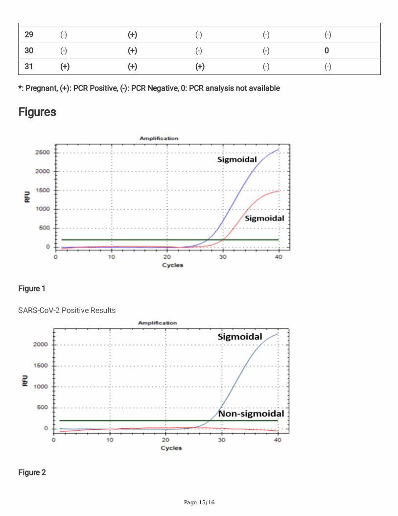
The diagnosis of patients with suspected COVID-19 presented to our hospital was con�rmed by the SARSCoV-2 PCR test. Viral RNAs extracted from the nasopharyngeal swab samples using vNAT solution wereanalyzed by Bio-Speedy SARS-CoV-2 RT-qPCR kits (single-step reverse transcription (RT) and real-time PCR(qPCR) kit targeting ORF1ab and N gene fragments [RT-qPCR]) and Bio-Rad CFX96 TouchTM device. Thecervicovaginal samples collected from the patients were appropriately transferred into tubes containingBio-Speedy viral nucleic acid buffer (vNAT) and then delivered to the microbiology laboratory as soon aspossible. Viral RNAs extracted using vNAT solution were analyzed using Bio-speedy SARS-CoV-2 RT-qPCRkits and Bio-Rad CFX96 TouchTM device. RT-qPCR procedure targeting the ORF1ab and N gene fragmentswas performed. The oligo-nucleotide set targeting the human RNase P gene (internal control) wasexamined for controlling sampling, nucleic acid extraction and inhibition. The internal control (IC) (RNaseP) curve was marked in blue, while the SARS-CoV-2 (N) and SARS-CoV-2 (ORF1ab) curves were marked inred. When the internal control curve was a sigmoidal curve, this indicated that there was no problemregarding the sampling and isolation stages. Moreover, when the SARS-CoV-2 curve was a sigmoidal curve,

Page 5/16
the sample was reported as POSITIVE, and when the curve was not sigmoidal, the sample was reported asNEGATIVE (Figures 1 and 2).
Statistical analysis
IBM SPSS 22.0 software (SPSS Inc., Chicago, IL, USA) was used for statistical analysis. Chi-square testwas used for categorical variables, and Student’s t test was used for numerical variables. The results wereexpressed as Mean (± SD) and N (%).
Funding
Bioeksen R&D Technologies LLC, the institution that provides SARS CoV-2 PCR tests to our hospitalthrough the Ministry of Health, donated 100 test kits to be used in this study.
ResultsA total of 31 female patients aged 20–64 years were included in the study. The mean age of the patientswas 46.097 (±13.207), Gravida was 2.677 (±1,720), and parity was 2.387 (±1.801). Note that 12 (38.7%) ofthe patients had regular menstruation, 16 (51.7%) were in menopause, and 3 (9.6%) were pregnant. Themost common accompanying chronic diseases were hypertension (n = 9, 29.0%), diabetes (n = 4, 12.9%)and chronic pulmonary disease (n = 4, 12.9%). Only 11 (35.4%) patients had COVID-19 positive spouses(Table 1). Moreover, 96.7% (n, 30) of the patients had muscle and joint pain, 93.5% (n, 29) had weakness,54.8% (n, 17) had cough, 29% (n, 9) had loss of taste, 29% (n, 9) had loss of smell, 25.8% (n, 8) hadshortness of breath, and 16.1% (n, 5) had fever. Note that 27 (87.1%) patients whose overall condition wasgood were followed-up in the ward, and 4 (12.9%) patients whose overall condition was moderate werefollowed up in the intensive care unit. There were no patients with a poor overall condition to requireintubation. Hydroxychloroquine + azithromycin + enoxaparin treatment was administered to 51.6% (n, 16)of patients, Hydroxychloroquine + favipiravir + enoxaparin treatment was administered to 38.7% (n, 12) ofthe patients, and lopinavir + ritonavir + enoxaparin treatment was administered to 9.7% (n, 3) of patients(Table 2). While 77.4% (n, 24) of patients had lung involvement on CT consistent with COVID-19, 12.9% (n,4) had no lung involvement on CT. CT was not performed for three of the patients as they were pregnant.Mean laboratory results were as follows: WBC: 5.7 (2.0-14.3), CRP: 2.2 (0.2-10.9), Ferritin: 86.7 (5.0-577.0),Troponin I: 2.3 (0.2-9.3), D-Dimer: 0.52 (0.34), and Fibrinogen: 387.3 (255.0-624.0) (Table-3). The patientswere hospitalized for an average of 14 days for treatment and follow-up purposes. No death because ofCOVID-19 or any other cause was observed in the patients included in our study. Four patients in theintensive care unit were discharged with recovery after their treatment. One of the three pregnant womengave birth to a preterm baby by normal vaginal route at 34 weeks of gestation; however, the other twopregnant women gave birth to term babies by cesarean section. SARS-COV-2 RNA results was negative innasopharyngeal swab samples of babies after birth. The preterm baby born at 34 weeks of gestation wasfollowed up in the neonatal intensive care for ~ 1week because of prematurity. No other problem wasobserved afterwards. There was no evidence of COVID-19 in the one-month follow-up of babies. The �rstnasopharyngeal swab sample was SARS-COV-2 RNA positive in all patients (n, 31). The control

Page 6/16
nasopharyngeal swab samples after 14 days was SARS-COV-2 RNA negative in 87.1% (n, 27) of thepatients and positive in 12.9% (n, 4) of the patients. The �rst and second cervical swab samples werecollected from 22 patients 14 days apart. The �rst cervical swab was collected from nine patients; however,the second swab could not be collected after 14 days. This was because they were menstruating or missedthe appropriate time for sampling. SARS-COV-2 RNA result was negative in 100% of a total of 53cervicovaginal swab samples collected (Table 4).
DiscussionCoronaviruses are a single-stranded RNA virus from the Coronaviridae family of the Nidovirales virusgroup. The name coronavirus comes from crown-like spikes on the outer surface of virus. Coronavirusesare small 65–125-nm-sized samples. The subgroups of the coronavirus family are alpha (α), beta (β),gamma (γ) and delta (δ) coronavirus [16]. 2019-nCoV (SARS-CoV-2), a beta coronavirus, is the seventhmember of the coronavirus family that infects humans after MERS-nCoV and SARS-nCoV [17]. It binds toangiotensin converting enzyme (ACE) 2 via the surface spike protein, which infects the target cell andcauses severe damage similar to SARS-CoV [18, 19]. Although the lung is believed to be the target organ of2019-nCoV, multiple non-respiratory symptoms have been reported, suggesting the involvement of otherorgans during the disease. ACE2 RNA expression is reported in all reproductive tissues of women (vagina,ovaries, fallopian tubes, endometrium, and cervix) and men (ductus deferens, testis, epididymis, seminalvesicle, and prostate) [13]. The study by Li et al. demonstrated that the presence of SARS-CoV-2 may bedetected in semen during the active phase of the disease [20]. This suggests that SARS-CoV-2 can besexually transmitted. Another study has reported higher ACE2 expression in male reproductive organs,particularly in the testicles; however, ACE2 expression observed in female reproductive tissues has beenreported to be at lower levels. Moreover, it has the potential to infect female reproductive tissues despitelow susceptibility to SARS-CoV-2 infection [13]. The �rst study investigating the presence of SARS-COV-2RNA in cervicovaginal �uid was conducted by Qiu et al. This study was conducted in ten postmenopausalwomen aged 52–80 years hospitalized with the diagnosis of severe COVID-19. Vaginal swab samples werecollected from patients 17–40 days after the onset of SARS-CoV-2 infection, and SARS-COV-2 RNA wasanalyzed. The presence of SARS-CoV-2 was investigated in the vaginal �uid samples of the patients.SARS-CoV-2 result was negative in all RT-PCR tests. Note that SARS-CoV-2 may have transmissionmechanisms similar to SARS-CoV and that there was no report that clearly indicated that SARS-CoVinvaded the female reproductive system or no reports on SARS-CoV detected in vaginal �uid. Moreover, theauthors noted that the absence of SARS-CoV-2 in vaginal �uid was proof that there was no sexualtransmission or no vertical transmission from mother to baby [21]. In one of the �rst studies on thissubject, Cui et al. investigated SARS-COV-2 RNA in the cervicovaginal and anal swab samples of 35patients diagnosed with COVID-19. Of all the samples, they detected SARS-COV-2 RNA in only one analswab sample. All other samples were negative for SARS-COV-2 RNA. RT-PCR positivity could not beobserved in the vaginal environment because of the absence of ACE2 expression, which is the SARS-CoV-2receptor, in the tissues of the vagina and cervix. The authors noted that there was no evidence that SARS-CoV-2 was transmitted from a woman to her partner via vaginal intercourse [22]. Aslan et al. investigatedthe presence of SARS-COV-2 RNA in the vaginal �uid of pregnant women with COVID-19. Twelve pregnant
Page 7/16
women with mild symptoms of con�rmed COVID-19 were included in the study, and the presence of SARS-COV-2 RNA was investigated in vaginal swab samples. All samples tested negative for SARS-COV-2 RNA[23]. However, in a systematic review including 156 newborns, the vertical transmission rate was 3.91%[24]. Despite the increasing number of published studies on COVID-19 in pregnancy, there is insu�cientdata to draw unbiased conclusions about the complications of COVID-19, vertical transmission, andperinatal complications in pregnant women. In this study, a total of 53 cervicovaginal �uid samples werecollected from 31 patients between the ages of 18-65 who were diagnosed with COVID-19 and had mildand moderate clinical symptoms. Samples were collected twice from 22 patients with an interval of 14days, and only one time from 9 patients. SARS-COV-2 RNA was analyzed using RT-PCR method, and allsamples were reported to be negative. In this study, the �rst samples were collected from the patientsduring the active disease period. The second samples were collected after 14 days. Nevertheless, allsamples were negative for SARS-COV-2 RNA, which was consistent with the literature. When the perinatalresults of three pregnant women in the study were evaluated, SARS-COV-2 RNA result was negative basedon the nasopharyngeal swabs of the three newborns. In terms of health status, there were no resultssuggestive of COVID-19. The main difference of this study from other studies was that sequential sampleswere collected during the active disease period. Moreover, the number of patients, age range and inclusionof pregnant patients increased the observation power of the study.
Study Limitations
Although the number of patients in this study is su�cient compared to similar studies, it is still one of theprimarily limitations. Another limitation is that the study was conducted in a single center. Studies onCOVID-19 are rapidly continuing, and there is a requirement for comprehensive multi-center studies thatinclude more cases.
ConclusionNegative SARS-COV-2 RNA results in cervicovaginal swabs suggest that there is no sexual transmission ofCOVID-19 and no vertical transmission during pregnancy. However, the number of studies conducted onthis subject and the sample size examined are still not su�cient. There is a requirement for comprehensivestudies on this subject with more cases, and SARS-CoV-2 may be investigated in genital tissues usingmore invasive methods.
DeclarationsThe authors have no �nancial disclosures to declare.
The authors have no con�ict of interest.
Ethics committee approval: Ethics committee approval was obtained from the Ahi Evran University EthicsCommittee (Date: June 10, 2020 Number: 2020-08/53).
Consent to participate (Informed consent): The participants provided their signed informed consent.
Page 8/16
Funding: This study was supported by Bioeksen R&D Technologies. 100 test kits were donated.
Financial Disclosure: The authors declared that this study has received no �nancial support.
Authors’ Contributions:
Barış Çıplak: Planning, Designing, Data Collection, Literature Review, Data Analysis, Writing
Yavuz Şimşek: Planning, Designing, Literature Review, Writing, Critical Review
Mustafa Kara: Planning, Designing, Supervision, Data Analysis, Statistical Analysis, Writing
Rukiye AKYOL: Designing, Data Processing, Writing Methods, Critical Review
Lokman HIZMALI: Designing, Data Collection, Critical Review
Veysel AKÇA: Data Collection
Işıl SAYHAN: Data Collection
Fikriye MİLLETLİ SEZGİN: Data Collection and Processing, Writing Methods
Yahya ŞAHİN: Supervision, Critical Review
Sercan EROĞLU: Supervision, Critical Review
Selda SONGUR DAĞLI: Supervision, Critical Review
Fatma ÇELİK: Data Collection, Critical Review
Zeynep KÖYLÜ: Data Collection
References1. Wiersinga WJ, Rhodes A, Cheng AC, Peacock SJ, Prescott HC (2020) Pathophysiology, transmission,
diagnosis, and treatment of coronavirus disease 2019 (COVID-19): a review. JAMA 324:782-793.
2. Zhu N, Zhang D, Wang W, Li X, Yang B, Song J, et al. (2020) A novel coronavirus from patients withpneumonia in China, 2019. New Eng J Med. 20;382(8):727-733
3. Liu Y, Gayle AA, Wilder-Smith A, Rocklov J (2020) The reproductive number of COVID-19 is highercompared to SARS coronavirus. J Travel Med. 13;27(2):taaa021.
4. Velavan TP,Meyer CG (2020) The COVID‐19 epidemic. Trop Med Intl Health 25:278.
5. WHO. (2020) Coronavirus disease 2019 (COVID-19): situation report, 72.

Page 9/16
�. WHO. (2020) Modes of transmission of virus causing COVID-19: implications for IPC precautionrecommendations: scienti�c brief, 27 March 2020
7. Chan JFW, Yuan S, Kok KH, To KK, Chu H, Yang J, et al (2020) A familial cluster of pneumoniaassociated with the 2019 novel coronavirus indicating person-to-person transmission: a study of afamily cluster. The Lancet 395: 514-523.
�. Huang C, Wang Y, Ren L, Zhao J, Hu Y, Zhang L, et al. (2020) Clinical features of patients infected with2019 novel coronavirus in Wuhan, China. The Lancet 395: 497-506.
9. Wang D, Hu B, Hu C, Zhu F, Liu X, ZHang J et al. (2020) Clinical characteristics of 138 hospitalizedpatients with 2019 novel coronavirus–infected pneumonia in Wuhan, China. JAMA 323: 1061-1069.
10. Zhang C, Shi L, Wang FS. (2020) Liver injury in COVID-19: management and challenges. LancetGestroenterol Hepatol 5: 428-430.
11. Chen N, Zhou M, Dong X, Qu J, Gong F, Han Y, et al. (2020) Epidemiological and clinical characteristicsof 99 cases of 2019 novel coronavirus pneumonia in Wuhan, China: a descriptive study. The Lancet395: 507-513.
12. Mao L, Jin H, Wang M, Hu Y, Chen S, He Q, et al. (2020) Neurologic manifestations of hospitalizedpatients with coronavirus disease 2019 in Wuhan, China. JAMA Neurol. 77(6):683-690.
13. Zupin L, Pascolo L, Zito G, Ricci G, Crovella S. (2020) SARS-CoV-2 and the next generations: whichimpact on reproductive tissues? J Assist Reprod Genet 37: 2399-2403.
14. Varga Z, Flammer AJ, Steiger P, Haberecker M, Andermatt R, Zinkernagel AS, et al. (2020). Endothelialcell infection and endotheliitis in COVID-19. The Lancet 395: 1417-1418.
15. Ackermann M, Verleden SE, Kuehnel M, Haverich A, Welte T, Laenger F, et al. (2020) Pulmonaryvascular endothelialitis, thrombosis, and angiogenesis in Covid-19. New Eng J Med. 383:120-128
1�. Shereen MA, Khan S, Kazmi A, Bashir N, Siddique R. (2020) COVID-19 infection: Origin, transmission,and characteristics of human coronaviruses. J Adv Res 24: 91-98.
17. Cheng ZJ, Shan J. (2020). 2019 Novel coronavirus: where we are and what we know. Infection 48:155-163.
1�. Lu R, Zhao X, Li J, Niu P, Yang B, Wu H, et al. (2020) Genomic characterisation and epidemiology of2019 novel coronavirus: implications for virus origins and receptor binding. Lancet 395: 565-574.
19. Zhuang MW, Cheng Y, Zhang J, Jiang XM, Wang L, Deng J, Wang PH. (2020) Increasing host cellularreceptor-angiotensin-converting enzyme 2 expression by coronavirus may facilitate 2019-nCoV (orSARS-CoV-2) infection. J Med Virol. 92(11):2693-2701. doi: 10.1002/jmv.26139.
20. Li D, Jin M, Bao P, Zhao W, Zhang S. (2020) Clinical characteristics and results of semen tests amongmen with coronavirus disease 2019. JAMA Netw Open 3: e208292.
21. Qiu L, Liu X, Xiao M, Xie J, Cao W, Liu Z, et al. (2020) SARS-CoV-2 is not detectable in the vaginal �uidof women with severe COVID-19 infection. Clin Infect Dis 71: 813-817.
22. Cui P, Zhen C, Wang T, Dai J, Zhang J, Ding T, et al. (2020) Clinical features and sexual transmissionpotential of SARS-CoV-2 infected female patients: a descriptive study in Wuhan, China. medRxiv; DOI:10.1101/2020.02.26.20028225.

Page 10/16
23. Aslan MM, Uslu Yuvaci H, Kose O, Toptan H, Akdemir N, Koroglu M, et al. (2020) SARS-CoV-2 is notpresent in the vaginal �uid of pregnant women with COVID-19. J Matern-Fetal Neonatal Med 16; 1-3.
24. Chi J, Gong W, Gao Q. (2020) Clinical characteristics and outcomes of pregnant women with COVID-19and the risk of vertical transmission: a systematic review. Arch Gynecol Obstet 303(2):337-345
TablesTable 1: Demographic characteristics

Page 11/16
Age
Mean (∓SD) 46,097(∓13,207)
Age Range 20-64
<45 (n, %) 15(48%)
≥45 (n, %) 16(52%)
Gravida 2,677(∓1,720)
Parity 2,387(∓1,801)
Menstruation N(%)
Regular Menstruation 12 (%38,7)
Menopausal 16 (%51,7)
Pregnant 3 (%9,6)
Chronic Diseases N(%)
None 13(%41,9)
Hypertension 9(%29,0)
Diabetes 4(%12,9)
Pulmonary Disease 4(%12,9)
Other 4(%12,9)
Employment N(%)
Employed 10(%32,3)
Not Employed 21(%67,7)
COVID-19 Positivity in the Family N(%)
No 9(%29,0)
Spouse 11(%35,4)
Other family members 17(%54,8)
Table 2: Clinical �ndings
Page 12/16
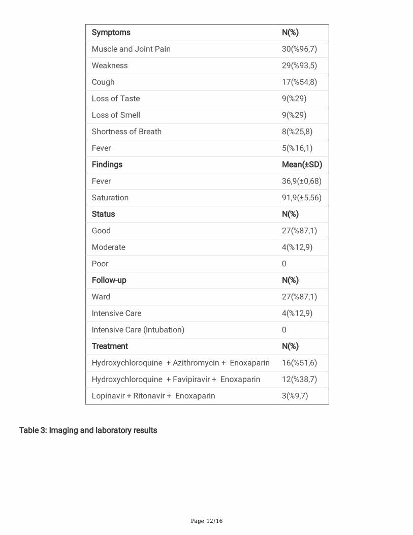
Symptoms N(%)
Muscle and Joint Pain 30(%96,7)
Weakness 29(%93,5)
Cough 17(%54,8)
Loss of Taste 9(%29)
Loss of Smell 9(%29)
Shortness of Breath 8(%25,8)
Fever 5(%16,1)
Findings Mean(±SD)
Fever 36,9(±0,68)
Saturation 91,9(±5,56)
Status N(%)
Good 27(%87,1)
Moderate 4(%12,9)
Poor 0
Follow-up N(%)
Ward 27(%87,1)
Intensive Care 4(%12,9)
Intensive Care (Intubation) 0
Treatment N(%)
Hydroxychloroquine + Azithromycin + Enoxaparin 16(%51,6)
Hydroxychloroquine + Favipiravir + Enoxaparin 12(%38,7)
Lopinavir + Ritonavir + Enoxaparin 3(%9,7)
Table 3: Imaging and laboratory results
Page 13/16
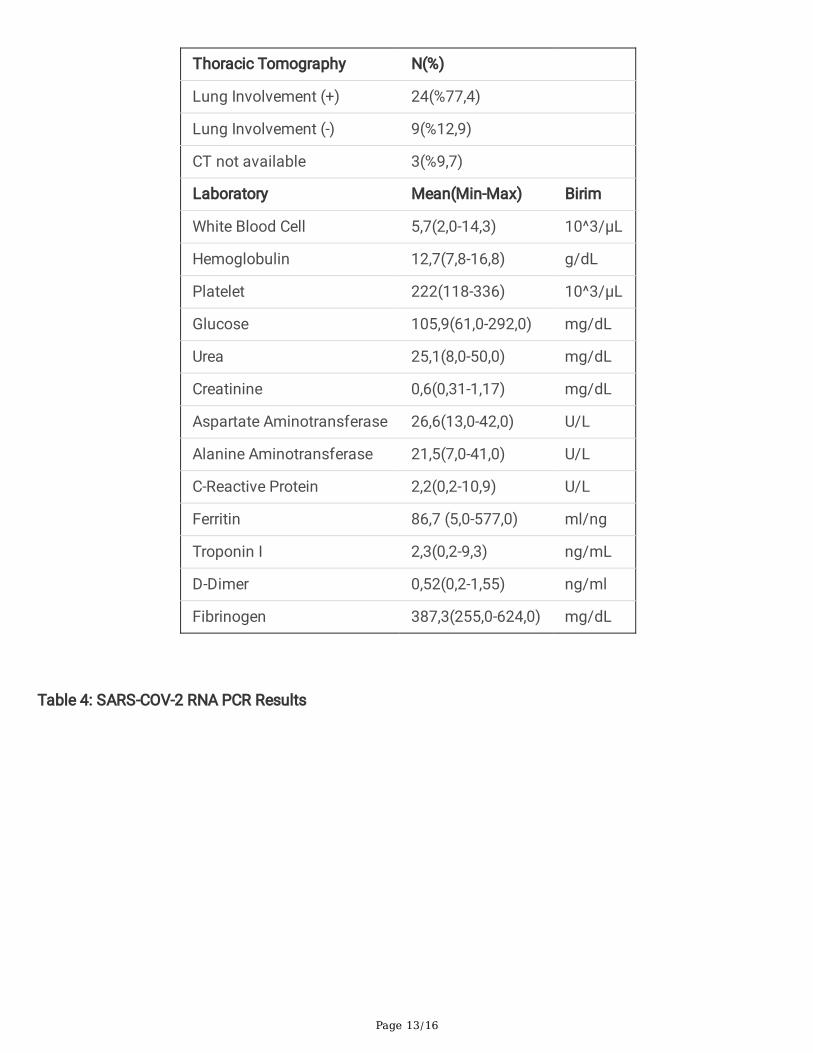
Thoracic Tomography N(%)
Lung Involvement (+) 24(%77,4)
Lung Involvement (-) 9(%12,9)
CT not available 3(%9,7)
Laboratory Mean(Min-Max) Birim
White Blood Cell 5,7(2,0-14,3) 10^3/µL
Hemoglobulin 12,7(7,8-16,8) g/dL
Platelet 222(118-336) 10^3/µL
Glucose 105,9(61,0-292,0) mg/dL
Urea 25,1(8,0-50,0) mg/dL
Creatinine 0,6(0,31-1,17) mg/dL
Aspartate Aminotransferase 26,6(13,0-42,0) U/L
Alanine Aminotransferase 21,5(7,0-41,0) U/L
C-Reactive Protein 2,2(0,2-10,9) U/L
Ferritin 86,7 (5,0-577,0) ml/ng
Troponin I 2,3(0,2-9,3) ng/mL
D-Dimer 0,52(0,2-1,55) ng/ml
Fibrinogen 387,3(255,0-624,0) mg/dL
Table 4: SARS-COV-2 RNA PCR Results

Page 14/16
PatientNo
Nasopharyngeal
PCR (Spouse's)
Nasopharyngeal
PCR (Day 1)
Nasopharyngeal
PCR (Day 14)
CervicovaginalPCR (Day 1)
CervicovaginalPCR (Day 14)
1* (+) (+) (-) (-) (-)
2 (+) (+) (-) (-) (-)
3 (+) (+) (-) (-) (-)
4 (+) (+) (-) (-) 0
5 (-) (+) (-) (-) 0
6 (+) (+) (-) (-) (-)
7 (+) (+) (-) (-) 0
8 (-) (+) (-) (-) (-)
9* (+) (+) (-) (-) (-)
10* (-) (+) (-) (-) (-)
11 (+) (+) (-) (-) (-)
12 (-) (+) (-) (-) (-)
13 (+) (+) (-) (-) (-)
14 (-) (+) (-) (-) 0
15 (-) (+) (-) (-) 0
16 (-) (+) (-) (-) 0
17 (-) (+) (-) (-) (-)
18 (-) (+) (-) (-) 0
19 (-) (+) (-) (-) (-)
20 (-) (+) (-) (-) (-)
21 (+) (+) (-) (-) (-)
22 (-) (+) (-) (-) (-)
23 (-) (+) (-) (-) 0
24 (-) (+) (-) (-) (-)
25 (-) (+) (+) (-) (-)
26 (-) (+) (+) (-) (-)
27 (-) (+) (+) (-) (-)
28 (-) (+) (-) (-) (-)
Page 15/16
29 (-) (+) (-) (-) (-)
30 (-) (+) (-) (-) 0
31 (+) (+) (+) (-) (-)
*: Pregnant, (+): PCR Positive, (-): PCR Negative, 0: PCR analysis not available
Figures
Figure 1
SARS-CoV-2 Positive Results
Figure 2

Page 16/16
SARS-CoV-2 Negative Results