cervical melanosis- an under-recognised melanocytic lesion ... · cervical melanosis- an...
TRANSCRIPT
Central Medical Journal of Obstetrics and Gynecology
Cite this article: Agarwal S, Pandey P, Durgapal P, Maurya G, Singh SK (2015) Cervical Melanosis- An Under-Recognised Melanocytic Lesion of Cervix: Report of Three Cases. Med J Obstet Gynecol 3(4): 1066.
*Corresponding authorsPinki Pandey, Type 5, B 301, New Campus, UPRIMS and R, Saifai, Etawah UP, India, Tel: +919458547252; Email:
Submitted: 15 July 2015
Accepted: 28 July 2015
Published: 30 July 2015
ISSN: 2333-6439
Copyright© 2015 Pandey et al.
OPEN ACCESS
Case Report
Cervical Melanosis- An Under-Recognised Melanocytic Lesion of Cervix: Report of Three CasesSavita Agarwal, Pinki Pandey*, Prashant Durgapal, Geeta Maurya, Sanjeev Kumar SinghDepartments of Pathology, U P Rural Institute of Medical Sciences and Research, India
Keywords•Cervix•Melanosis•Pigmentation
INTRODUCTIONMelanosis of uterine cervix is a rare pigmented lesion
characterized by melanocytic hyperpigmentation in basal layer of the squamous mucosa with or without melanocytic proliferation [1]. Though uncommon, these lesions carry great clinical and pathologic significance. There are multiple causes of cervical pigmentation ranging from reactive to neoplastic processes and hence careful evaluation of each case is mandatory in order to clearly separate clinically innocuous lesions like cervical melanosis from more grave lesions like cervical melanoma. Thus biopsy and meticulous microscopic examination in each case of cervical pigmentation should be undertaken to overcome the clinical dilemma. We report three cases of cervical melanosis which is an extremely rare entity characterised by melanocytic pigmentation in basal layer of uterine cervix.
CASE SUMMARY
Case 1
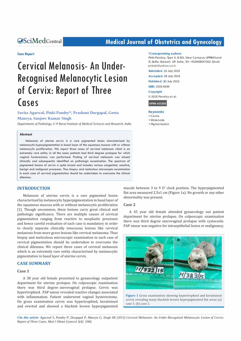
A 38 year old female presented to gynaecology outpatient department for uterine prolapse. On colposcopic examination there was third degree uterovaginal prolapse. Cervix was hypertrophied. PAP smear revealed reactive changes associated with inflammation. Patient underwent vaginal hysterectomy. On gross examination cervix was hypertrophied, keratinised and everted and showed a blackish brown hyperpigmented
macule between 3 to 9 O’ clock position. The hyperpigmented flat area measured 2.5x1 cm (Figure 1a). No growth or any other abnormality was present.
Case 2
A 45 year old female attended gynaecology out patient department for uterine prolapse. On colposcopic examination there was third degree uterovaginal prolapse with cystocoele. PAP smear was negative for intraepithelial lesion or malignancy.
Abstract
Melanosis of uterine cervix is a rare pigmented lesion characterized by melanocytic hyperpigmentation in basal layer of the squamous mucosa with or without melanocytic proliferation. We report three cases of cervical melanosis which is an extremely rare entity. In all the cases patients had third degree prolapse for which vaginal hysterectomy was performed. Finding of cervical melanosis was missed clinically and subsequently identified on pathologic examination. The spectrum of pigmented lesions of cervix is quite broad and includes various congenital, reactive, benign and malignant processes. Thus biopsy and meticulous microscopic examination in each case of cervical pigmentation should be undertaken to overcome the clinical dilemma.
A) B)
Figure 1 Gross examination showing hypertrophied and keratinised cervix revealing many blackish brown hyperpigmented flat areas (a) case 1; (b) case 2.

Central
Pandey et al. (2015)Email:
Med J Obstet Gynecol 3(4): 1066 (2015) 2/3
Patient underwent vaginal hysterectomy. On gross examination cervix was hypertrophied and revealed three black- brown hyperpigmented flat areas. Lesion extended from 6- 7 O’ clock position, 10 to 11 O’ clock position and 1- 3 O’ clock position. Size of these lesions ranged from 2.5x1 cm to 2x1 cm (Figure 1b). No growth or any other abnormality was present.
Case 3
A 52 year old female came to the gynaecology out patient department for complaints of something coming out of the vagina for past one year. On colposcopic examination there was third degree uterovaginal prolapse with cystocoele and rectocoel. PAP smear was reported as negative for intraepithelial lesion or malignancy. Patient underwent vaginal hysterectomy. On gross examination cervix was hypertrophied and revealed single black- brown hyperpigmented flat area measuring 1x 0.5 cm and extending from 2- 3 O’ clock position. No growth or any other abnormality was present.
Microscopic examination of the cervical lesion in all three cases showed stratified squamous epithelium without atypia, deposition of melanin pigments in the basal layer resulting in hyperpigmentation and no significant increase in melanocytes (Figures 2 and 3). Subepithelial tissue was unremarkable. Masson Fontana stain for melanin was positive. On immunohistochemistry S100 positive cells were seen. Considering these findings
diagnosis of cervical melanosis was made. There was no evidence of dysplasia or malignancy or any other hyperpigmented lesion elsewhere in the body. Follow up status in all the three cases till date was uneventful.
DISCUSSIONHyperpigmented lesions of the cervix are rare and of these
cervical melanosis is even rarer. Other relatively common hyperpigmented lesions of cervix are blue nevi and melanoma [2]. Melanocytic lesions are of importance primarily because of malignant melanoma, which is the single most common potentially lethal neoplasm of the skin. Although less common than the familiar basal and squamous cell tumours of the skin, they are much more frequently fatal, due to their intrinsic tendency to lymphatic and haematogenic metastasis. The incidence of melanoma has risen dramatically over the last several decades. However, the mortality has risen less dramatically than the incidence, likely due to earlier diagnosis. Melanoma is one of the most important cancers when considered as a cause of loss of life as it is commonly diagnosed in relatively young people, and can be fatal if untreated.
Melanosis of the uterine cervix is encountered very rarely in routine clinical practice and deserves meticulous examination in order to exclude other hyperpigmented lesions carrying significant clinical, pathologic and therapeutic implications. Spectrum of these lesions is quite broad and includes various congenital, reactive, benign and malignant processes [3].
Recently Tran et al [3] studied various grossly visible pigmented lesions of the uterine cervix and concluded that both melanocytic and non melanocytic lesions can lead to cervical hyperpigmentation. Most common lesion of melanocytic nature in this series was blue nevi followed by melanotic macule. Various non melanocytic lesions encountered were focal granulomatous vasculitis, collection of hemosiderin laden macrophages at the biopsy site, hemorrhagic nabothian cyst, hemangioma and giant cell reaction to foreign material. All cases of blue nevi lesions were located in the stroma of the endocervix whereas cervical macules were present in the ectocervix. In this study the incidence of pigmented lesions of cervix was 1.6% and authors emphasized on the rarity of cervical hyperpigmentation and also mentioned that majority of them are of benign nature however each case mandates careful evaluation to exclude possibility of cervical melanoma.
All three cases in the present study were missed clinically to be identified only on pathologic examination of the specimen. In the study by Tran et al [3] all the cases of blue nevi were either missed or misinterpreted as reaction to previous biopsy on initial pathologic examination. Deb et al [4] also mentioned that pigmented lesions are seldom detected clinically or on colposcopy. These facts suggest that these lesions tend to be missed or misinterpreted frequently by both clinicians and pathologists and hence they need to be aware of these entities in order to diagnose them correctly.
Vezzani and Sola [5] summarized theories pertaining to the presence of melanocytes in the cervix which include migration of neural crest elements, migration of melanocytes from adjacent mucocutaneous junction and appearance of melanocytes in
Figure 2 Photomicrograph showing stratified squamous epithelium of the uterine cervix with hyperpigmentation of the basal layer (Hematoxylin & Eosin, X100).
Figure 3 Magnified image of the stratified squamous epithelium of cervix with hyperpigmentation of the basal layer cells without increase in melanocytes (Hematoxylin & Eosin, X400).

Central
Pandey et al. (2015)Email:
Med J Obstet Gynecol 3(4): 1066 (2015) 3/3
Agarwal S, Pandey P, Durgapal P, Maurya G, Singh SK (2015) Cervical Melanosis- An Under-Recognised Melanocytic Lesion of Cervix: Report of Three Cases. Med J Obstet Gynecol 3(4): 1066.
Cite this article
association with transformed cervical squamous epithelium which resembles epidermis representing a metaplastic change. The latter seems relevant in situations associated with chronic irritation or trauma such as uterine prolapse and cryotherapy [1,6,7]. In the present study all three cases presented with third degree uterine prolapse thus favoring metaplastic theory as etiologic possibility.
Very sparse literature is available on this benign entity possibly due to its rarity however, all published cases share marked similarity in their presentation, appearance and clinical behaviour. In various published case reports patients either had uterine prolapse [7] or it was an incidental finding [8] during work up for other reasons like infertility [9], menorrhagia [10], dysfunctional uterine bleeding [11] and in one case cervical melanosis appeared following cryotherapy for epithelial dysplasia [7].
It is worth noting that there are various causes of cervical pigmentation and to reliably distinguish these causes on gross examination alone is not possible hence all pigmented lesions of the cervix should be subjected to biopsy and microscopic examination in order to reliably distinguish innocuous lesions like melanosis from more ominous lesions like melanoma.
REFERENCES1. Yilmaz AG, Chandler P, Hahm GK, O’Toole RV, Niemann TH. Melanosis
of the uterine cervix: a report of two cases and discussion of pigmented cervical lesions. Int J Gynecol Pathol. 1999; 18: 73-76.
2. Chang D, Tiburzio GB. Melanosis of the uterine cervix: a case report and literature review. J Bras Patol Med Lab. 2013; 49: 222-224.
3. Tran TA, Niu G, Tomasello CA, Tran HV, Ross JS, Carlson JA. The spectrum of grossly visible pigmented lesions in the uterine cervix: a prospective study. Int J Gynecol Pathol. 2014; 33: 89-99.
4. Deb P, Swarup D, Bhopte AG. Blue nevus of the uterine cervix. MJAFI. 2000; 56: 342-343.
5. Vezzani M, Sola P. [Melanin pigmentation of the epithelium of the exocervix]. Pathologica. 1979; 71: 657-661.
6. Kapoor NK. Melanosis of uterine cervix: a report of two cases. Indian J Pathol Microbiol. 2003; 46: 102-103.
7. Hytiroglou P, Domingo J. Development of melanosis of uterine cervix after cryotherapy for epithelial dysplasia. A case report and brief review of the literature on pigmented lesions of the cervix. Am J Clin Pathol. 1990; 93: 802-805.
8. Deppisch LM. Cervical melanosis. Obstet Gynecol. 1983; 62: 525-526.
9. Saikia UN, Dey P, Saikia B, Gupta I. Melanin containing cells of the uterine cervix and a possible histogenesis--a case report. Indian J Pathol Microbiol. 2004; 47: 22-23.
10. Kaplan PA, Griffo MD, Diaz-Arias AA. Melanosis of the uterine cervix: a case report. J Reprod Med. 2005; 50: 867-870.
11. Balamurugan S, Rameshrao K, Vijayshree R, Nayar S. Blue nevus of cervix – a case report. Internet J Gynecol Obstet. 2012; 16.