cervical emg profile differences between patients of neck...
TRANSCRIPT
RESEARCH PAPER
Cervical EMG profile differences between patients of neck pain andcontrol
SHRAWAN KUMAR1 & NARASIMHA PRASAD2
1University of North Texas Health Science Center, Physical Medicine Institute Department of Osteopathic Manipulative
Medicine, Center for BioHealth, Fort Worth, Texas 76107, USA and 2University of Alberta, Mathematical and Statistical
Sciences, Central Academic Building, Edmonton, Alberta, Canada
Accepted March 2010
AbstractPurpose. The objective of this study was to investigate EMG signals of cervical muscles in five directional efforts fromchronic neck pain patients and compare them with those of the healthy controls to discern differences between patients andcontrols with respect to strength and EMG characteristics.Method. Seventeen male and 17 female idiopathic and non-specific chronic neck pain patients without any diagnosedpathologies or prior surgery in the age group 18–65 years were recruited into the study. The controls consisted of 30 maleand 33 female subjects with no history of neck pain in the past 12 months. Both patients and controls performed theexperimental activities of flexion, left anterolateral flexion, left lateral flexion, left posterolateral extension and extension. Thepatients exerted to their 20% maximum voluntary contraction (MVC), pain threshold and pain tolerance levels in threeseparate contractions. Similarly, the control subjects exerted to their 20% MVC, 60% MVC and MVC in random order. Thedescriptive statistics for strength, normalised peak EMG, median frequency (MF), 10% frequency bands and their powerwere calculated. Eight levels of wavelet decomposition and their coefficients were calculated and subjected to principalcomponent analysis. These variables were subjected to analysis of variance and regression analysis to distinguish betweenpatients and controls. The full wave rectified linear envelope detected EMG of patients and controls were plotted againsttime to reveal pattern differences.Results. There was a lack of significant difference in the MF of the two samples indicating that the muscle conductionvelocity was not disturbed by the pain. Significant differences were also found in 10 percentile frequency bands betweenpatients and controls (p5 0.05). The wavelet decomposition with principal component analysis revealed that patients andcontrols could be identified as such 100% of the time at 20% MVC; and, patients and controls could be identified correctly100% and 90% of the time respectively at pain threshold/60% MVC.Conclusion. Thus, a combination of EMG spectral frequency banding and wavelet decomposition with regression can beused to distinguish chronic pain patients from controls.
Keywords: Chronic neck pain, classification, identification
Introduction
Neck pain is a common occurrence in our society
and constitutes significant social and economic
burden. In a cross-sectional study [1], it was
reported that the life-time occurrence of problems
of neck pain was 66.7% and the point prevalence was
22.22% in Saskatchewan adults. Overall 58.8% of
women and 47.2% of men had experienced neck
pain in the previous 6 months. In another prospec-
tive cohort study after involvement in traffic acci-
dents, 58.8% of sample was reported to be work
disabled [2]. In a cross-sectional study reported a
life-time prevalence of neck pain 78% in military
office workers, whereas the point prevalence was
59% [3]. On the other hand, among sedentary
workers 6-month neck pain prevalence was reported
to be 23.5% [4]. Among helicopter pilots, 3-month
prevalence of neck pain was reported being 57% [5].
Thus, the prevalence of neck pain may vary with
occupation and exposure to risk factors, but its
occurrence in society is common. From epidemio-
logical data of population survey and accounting the
insurance claims, it would appear that it is the minor
Correspondence: Shrawan Kumar, University of North Texas Health Science Center, Physical Medicine Institute Department of Osteopathic Manipulative
Medicine, 3500 Camp Bowie Boulevard, Center for BioHealth, Fort Worth, TX 76107, USA. E-mail: [email protected]
Disability and Rehabilitation, 2010; Early Online, 1–10
ISSN 0963-8288 print/ISSN 1464-5165 online ª 2010 Informa UK Ltd.
DOI: 10.3109/09638288.2010.481029
Dis
abil
Reh
abil
Dow
nloa
ded
from
info
rmah
ealth
care
.com
by
Uni
vers
ity o
f A
lber
ta o
n 08
/26/
10Fo
r pe
rson
al u
se o
nly.

accidents and events that are responsible for the large
majority of these cases. The injuries are most likely to
be located in musculoligamentous structure of the
neck [6,7]. The latter group of authors demonstrated
that in a simulated rear-end impact a speed change
or 5 kph or less could cause 179% of maximal
voluntary contraction in the sternocleidomastoid
muscle. This rate of progression of muscle stress is
likely to cause injury to these tissues resulting in pain.
A similar finding for trapezius muscle has also been
shown [8].
In a voluntary cervical motion the neuromotor
control exercised by the central nervous system
determines the time, intensity and a nature of
excitation of agonists and antagonist muscles [9].
The neuro-motor control is responsible for carrying
out two functions: (a) produce torque about a given a
spinal joint to carry out the task at hand, and (b)
develop appropriate forces required to stabilise it. In
symptomatic subjects due to pain, the pattern of the
neuro-motor control is disrupted altering the rela-
tionship between the muscle excitation, regional
muscle balance and resultant mechanical output
[10–12]. Therefore, the global objective of this study
was to determine differences in EMG characteristics
of muscles in patients with neck pain in comparison
to those of normal controls. The hypothesis of the
study was that the power spectrum profile time
domain features identified by wavelet decomposition
of the EMG of cervical muscles will deviate in pain
patients from those off the normal subjects.
Methods
Sample
The study was approved by the University of Ethics
Review Committee prior to initiation of the project.
17 male and 17 female patients of idiopathic and
non-specific chronic neck pain lasting 3 months or
more in the age group of 18–60 years were recruited
for the study. The exclusion criterion was any
diagnosed pathology, trauma or surgery. The inclu-
sion/exclusion criteria were established by taking
history, imaging and EMG studies. The mean body
mass index (BMI) of male patients was 29.8 and that
of females was 25.8. There were 30 male controls
with a mean BMI of 24.2 and 33 female control
subjects with a mean BMI of 24.2.
Subject preparation
After suitable skin preparation Delsys bipolar active,
knife edge, fixed inter-electrode distance (10 mm)
surface electrodes were applied to upper trapezius
(level with C4), splenius capitis and the sternoclei-
domastoids bilaterally to both patients and control
subjects. These subjects were required to exert
individual muscle specific activities to discern cross-
talk; if any crosstalk was present the electrodes were
replaced to an ideal location to minimise it. Such
prepared subjects were seated in the experimental set
up for cervical testing.
Testing device
The testing device consisted of an adjustable chair,
sliding platform and floor mounted strength measur-
ing device (Figure 1). The chair consisted of a
molded plastic seat mounted on an iron platform
with telescopic metal legs fixed to the base plate. The
back-rest and seat and were fitted with a Velcro four-
point restraint system to stabilise torso and both
shoulders. The chair with its base plate was pivoted
in the center and had casters on the periphery for
circular motion with stability. The circular plate was
graduated in intervals of 58 and holed through which
bolts could be placed to match the holes in the
sliding board of the platform. Two bolts were placed
at opposite ends and tightened for a rigid fixation of
the chair in the desired position of a subject. The
resistance device consisted of a vertical telescopic
15 cm wide rectangular metal tube welded to a thick
iron plate rigidly bolted in the floor. The 12-cm wide
tube could be raised or lowered and securely locked
into place. On top of the inner tube ball bearing was
mounted to which another hollow tube was attached
allowing it to rotate freely. Attached perpendicular to
this tubing was an adjustable arm that was uphol-
stered at the farther end for head contact and force
application. At the lower end of the tubing, a
counterweight was attached with an adjustable length
rod to compensate for variable positioning of the
horizontal resistance of arm. 14 cm below the pivot
point a horizontal metal rod was built at right angles
to the tubing which could be attached to a fixed
object with an intervening load cell (I-250). Thus,
any force exerted on the upholstered horizontal arm
was registered on the load cell, which was fixed in its
mechanical path. The directions of force exertions
were chosen from a scheme shown in Figure 2.
Tasks
The subjects were informed about the objectives and
procedure of the experiment. After signing the
informed consent form, the prepared subjects were
seated in the experimental set up for cervical testing.
The subjects were asked to exert in isometric flexion,
extension, left lateral flexion, left antero-lateral
2 S. Kumar & N. Prasad
Dis
abil
Reh
abil
Dow
nloa
ded
from
info
rmah
ealth
care
.com
by
Uni
vers
ity o
f A
lber
ta o
n 08
/26/
10Fo
r pe
rson
al u
se o
nly.

flexion and left postero-lateral extension according to
the scheme shown (Figure 2). The sequences of
these exertions were fully randomised. The chair was
rotated in the desired position for the testing
condition. Using the adjustability, the pivot point
of the resistance of arm was set at the same height as
the horizontal upholstered bar. Prior to the start of a
trial, the subjects were informed to exert their
appropriate effort concentrating on using their neck
only. The subjects where instructed to bring their
contraction to the desired level in the first 2 s and
hold it for another three. At this time the trial was
terminated. The cervical muscle contraction force
magnitude was measured in Newtons (N). The
reliability and accuracy of this equipment and
procedure was previously established [13]. The
patients were asked to exert to their estimated 20%
maximum voluntary contraction (MVC), pain
threshold and pain tolerance levels. The control
subjects were also asked to exert to their 20%
maximum voluntary contraction (MVC), 60%
MVC and MVC. All exertions were required to be
5 s long. While the subjects exerted force, the
strength of exertion and EMG of all six channels
were sampled at 2 kHz.
Data analysis
Force/strength. The force of contraction recorded in
cervical isometric flexion, extension left lateral flex-
ion, left antero-lateral flexion and left postero-lateral
extension. These percentage data were quantitatively
compared between the patients and normal control at
contraction levels of 20%, 60% (pain threshold for
patients) and MVC for both, controls and patients.
EMG. The raw EMG signals were band pass
filtered with low cutoff frequency of 20 Hz and high
cutoff frequency of 450 Hz. The signals in this
frequency band were pre-amplified at the site by a
factor of 10 and differentially amplified again with a
gain of 100. The time constant used was 25 ms and
the amplification system had a common mode
Figure 2. Directional scheme for cervical exertion.
Figure 1. Cervical strength testing device.
EMG signature of pain 3
Dis
abil
Reh
abil
Dow
nloa
ded
from
info
rmah
ealth
care
.com
by
Uni
vers
ity o
f A
lber
ta o
n 08
/26/
10Fo
r pe
rson
al u
se o
nly.

rejection ratio of 92 dB. Such recorded signals were
full wave rectified and linear envelope detected over
the 5-s recording duration. The peak amplitude of
EMG was then measured and recorded. The peak
EMG was normalised according to the following
scheme. The sternocleidomastoid EMG in various
activities was normalised against the peak EMG of
the sternocleidomastoid in flexion. The EMG scores
of splenius capitis were normalised against the peak
EMG of left splenius capitis in left lateral flexion.
Similarly, the EMG scores of the trapezii were
normalised against the peak EMG of trapezii in
extension. The pattern of EMG was observed by
plotting the normalised EMG in time with one
standard deviation confidence interval.
For the frequency domain analysis the raw EMG
signals were subjected to DC removal. Subsequently,
the sections of data that were in a stable force and
EMG activities were marked and selected for Fast
Fourier Transform analysis. Those activities were
isometric and the data segments were stationary
when chosen for such analysis. However, a test of
the stationarity of the data was carried out before
proceeding with the rest of the analysis. The spectral
data analysis of each muscle in each of the 10
activities was done separately for patients of neck
pain and control subjects. From the spectral analysis
the lower and upper 3 dB frequencies and the
bandwidths were extracted for patients and controls.
All these three parameters were calculated for all
three contractions (20% MVC, 60% MVC and
MVC.) The median frequency of each of the cervical
muscle in each of the 10 activities of both patients
and controls were also extracted.
The descriptive statistics of the variables of
strength, EMG magnitude, peak power normalised
EMG, median frequency, mean power frequency,
total power, peak power, frequency at peak power,
time to onset and time to peak EMG was calculated.
Each of these variables was also subjected to one-way
analysis of variance to examine the differences
between ‘control subjects’ and ‘neck-pain patients’
with respect to these features.
Time domain analysis. These EMG signals were first
rectified and subjected to wavelet analysis to obtain
eight levels of decomposition of signals for each subject
under each condition and for each channel. Daube-
chies wavelet transformation was used for this purpose.
The eight levels or scales, of the raw signals decom-
position, were confined to following frequency bands:
0–7.08125, 7.8125–15.625, 15.625–31.25, 31.25–
62.5, 62.5–125, 125–250, 250–500 and 500–1000 Hz.
At each of the abovementioned eight scales, the
following were computed as frequency features.
. Eight RMS values (RMS1, RMS2 and RMS8)
. Mean (m), dispersion (d), skewness (s) and
kurtosis (k).
For time domain features, rectified EMG signals
were subjected to fourth-order autoregressive analysis
Table I. Cervical muscle strength in cervical pain patients (n¼34) and normal controls (n¼ 63) (N).
Gender Subject type
Exertion levels
20% MVC Threshold/60% MVC Tolerance/MVC
Peak force (N)
Mean Std Deviation Mean Std Deviation Mean Std Deviation
Male Patient Flexion 14.63 3.68 38.70 22.28 46.70 23.52
Left antero 10.78 3.38 30.45 17.86 45.37 22.14
Left lateral 9.80 1.41 32.60 22.33 48.38 28.74
Left postero 9.73 2.85 34.48 19.15 47.76 27.10
Extension 14.83 4.01 47.52 30.97 63.12 29.84
Control Flexion 13.72 17.56 26.31 9.61 47.08 30.23
Left antero 9.46 5.08 19.45 5.50 40.21 26.35
Left lateral 10.44 5.61 22.34 9.60 41.56 22.46
Left postero 12.96 7.13 27.56 14.15 47.31 25.25
Extension 12.15 5.07 31.07 14.56 46.40 20.45
Female Patient Flexion 15.94 3.35 37.34 24.06 51.89 27.38
Left antero 17.84 3.94 38.15 19.48 48.20 24.82
Left lateral 18.76 4.81 37.90 21.56 53.45 28.65
Left postero 16.72 6.50 40.16 21.90 52.12 21.77
Extension 23.29 5.21 48.38 29.42 56.11 22.24
Control Flexion 14.80 6.20 32.72 6.57 55.84 16.99
Left antero 14.18 4.80 50.17 40.67 59.58 24.42
Left lateral 13.43 6.59 35.35 16.61 61.87 29.23
Left postero 15.74 5.53 36.83 11.23 68.39 23.23
Extension 19.06 6.59 44.43 15.61 76.10 27.94
4 S. Kumar & N. Prasad
Dis
abil
Reh
abil
Dow
nloa
ded
from
info
rmah
ealth
care
.com
by
Uni
vers
ity o
f A
lber
ta o
n 08
/26/
10Fo
r pe
rson
al u
se o
nly.

using the ‘arfit.m’ in Matlab. From this fourth-order
autoregressive model, autocorrelations (rxx1, rxx2,
rxx3, rxx4) and auto-regression coefficients up (phi1,
phi2, phi3, phi4) to order 4 were extracted to
describe time-domain features, namely time depen-
dency of the signals. All these signals were also
subjected to wavelet analysis to obtain frequency
domain features and time series analysis to obtain
time domain features on all 97 subjects.
Logistic regression analysis was applied to identify
best frequency-domain and time-domain features in
classifying subjects to ‘control’ and ‘neck-pain group’
by treating group as a binary response variable and
all above-mentioned frequency domain and time-
domain measures as predictors. Separate logistic
models were fitted for three exertion levels. After
identifying the ‘best model’ with minimum number of
predictors, classification procedure was formed based
on the best model by classifying a subject to neck-pain
group if predicted probability from the model was
greater than or equal to 0.5, otherwise the subject was
classified to control group. On the basis of one-fold
(delete one) and cross-validation approach, mis-
classification errors were computed to evaluate the
performance of the proposed classification scheme.
We evaluated the accuracy of this classification
method and miss-classification errors were calcu-
lated using re-substitution accuracy measures (the
accuracy that is evaluated when the classifier is
evaluated on the same sample that was used to
construct the classification rule.) However, this
accuracy measure is ‘optimistically biased’. To
address this criticism, we also used one-fold (delete
one) cross-validation approach to estimate miss-
classification errors.
In identifying best predictors under step-wise
selection scheme the standard rule was used: a
variable was entered into the model if the probability
of its score statistic was less than the 0.05 and is
removed if the probability is greater than the 0.10.
With the selected features from the logistic
regression analysis we also performed linear discri-
minate classification to examine the discrimination
power. Figures 3 and 4 display two-sigma bars on
linear discriminate scores, separately for control and
neck pain groups and hence demonstrates the
appropriateness of the proposed feature selections
to classify subjects to these two groups.
Results
Strength
The means and standard deviations of all strength of
patient and control samples for 20% MVC, 60%
MVC (control)/pain threshold (patients) and MVC
(control) and pain tolerance (patients) for flexion,
left antero-lateral flexion, left lateral flexion, left
postero-lateral extension and extension are presented
in Table I. Neck pain patient’s pain threshold
strength values turned out to be between 67 and
82% of their MVC for males and between 48 and
67% for females. Pain tolerance values of the patients
were significantly different from those of the max-
imum voluntary contraction efforts of the control
subjects (p5 0.05). The ANOVA revealed that there
was no significant difference between the body
weight normalised strength due to gender. However,
there were significant difference between patients
and controls for pain tolerance/MVC contraction
Figure 3. Classification of patients of cervical pain and normal
control subjects through linear discriminant score at 20%
maximum voluntary contraction in flexion with 95% confidence
interval.
Figure 4. Classification of patients of cervical pain and normal
control subjects through linear discriminant score at pain
threshold/60% maximum voluntary contraction in flexion with
95% confidence interval.
EMG signature of pain 5
Dis
abil
Reh
abil
Dow
nloa
ded
from
info
rmah
ealth
care
.com
by
Uni
vers
ity o
f A
lber
ta o
n 08
/26/
10Fo
r pe
rson
al u
se o
nly.

efforts in flexion (p5 0.05), left antero-lateral flexion
(p5 0.05), left lateral flexion (p5 0.05), left pos-
tero-lateral extension (p5 0.05) and extension
(p5 0.05).
EMG
The sample plots of EMG of cervical muscles during
20% MVC, pain threshold/60% MVC and MVC
contractions in flexion are shown in Figures 5–7.
Clearly, there is significantly greater EMG activity in
the cervical muscles of patients in flexion (p5 0.05).
A similar pattern has been found in all five exertions
(p5 0.05). In pain threshold contractions patient’s
EMG was significantly greater than those of controls
in 60% MVC contractions for all six muscles
(p5 0.05). The ANOVA revealed that the left and
right sternocleidomastoid in both males and females
were found to be significantly different in patients as
compared to the controls in all five contractions
(p5 0.05). The splenius capitis muscle was signifi-
cantly different between patients and controls in
flexion, antero-lateral flexion and left lateral flexion
both in males and females (p5 0.05). The trapezius
muscle showed a significant difference between
patients and controls except in extension (p5 0.05).
The median frequency of the cervical muscles
ranged between 80 and 130 Hz for both males and
females in patients and controls. The median
frequency of individual muscles also varied slightly
between the five different activities. The ANOVA
revealed no consistent and significant differences
between patients and controls in the median
frequency. When the total EMG bandwidth was
divided into 10% intervals and compared between
patients and controls activity specific differences
emerged. In flexion and left antero-lateral pain
tolerance/MVC contractions the right sternocleido-
mastoids were significantly different between pain
patients and controls in each of the 10 percentile
intervals (p5 0.05). In left postero-lateral extension
Figure 5. Sample plot of the EMG for RSCM, LSCM, RSPL, LSPL, RTRP and LTRP for patients and controls for pain 20% MVC in
flexion.
6 S. Kumar & N. Prasad
Dis
abil
Reh
abil
Dow
nloa
ded
from
info
rmah
ealth
care
.com
by
Uni
vers
ity o
f A
lber
ta o
n 08
/26/
10Fo
r pe
rson
al u
se o
nly.
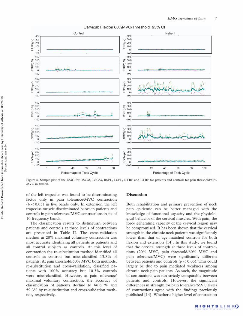
of the left trapezius was found to be discriminating
factor only in pain tolerance/MVC contraction
(p5 0.05) in five bands only. In extension the left
trapezius muscle discriminated between patients and
controls in pain tolerance/MVC contractions in six of
10 frequency bands.
The classification results to distinguish between
patients and controls at three levels of contractions
are presented in Table II. The cross-validation
method at 20% maximal voluntary contraction was
most accurate identifying all patients as patients and
all control subjects as controls. At this level of
contraction the re-substitution method identified all
controls as controls but miss-classified 13.8% of
patients. At pain threshold/60% MVC both methods,
re-substitution and cross-validation, classified pa-
tients with 100% accuracy but 10.3% controls
were miss-classified. However, at pain tolerance/
maximal voluntary contraction, the accuracy of
classification of patients decline to 66.6 % and
59.3% by re-substitution and cross-validation meth-
ods, respectively.
Discussion
Both rehabilitation and primary prevention of neck
pain epidemic can be better managed with the
knowledge of functional capacity and the physiolo-
gical behavior of the cervical muscles. With pain, the
force generating capacity of the cervical region may
be compromised. It has been shown that the cervical
strength in the chronic neck patients was significantly
lower than that of age matched controls for both
flexion and extension [14]. In this study, we found
that the cervical strength at three levels of contrac-
tions (20% MVC, pain threshold/60% MVC and
pain tolerance/MVC) were significantly different
between patients and controls (p5 0.05). This could
largely be due to pain mediated weakness among
chronic neck pain patients. As such, the magnitude
of contractions was not strictly comparable between
patients and controls. However, the significant
differences in strength for pain tolerance/MVC levels
of contractions agree with the findings previously
published [14]. Whether a higher level of contraction
Figure 6. Sample plot of the EMG for RSCM, LSCM, RSPL, LSPL, RTRP and LTRP for patients and controls for pain threshold/60%
MVC in flexion.
EMG signature of pain 7
Dis
abil
Reh
abil
Dow
nloa
ded
from
info
rmah
ealth
care
.com
by
Uni
vers
ity o
f A
lber
ta o
n 08
/26/
10Fo
r pe
rson
al u
se o
nly.

required to engender pain sensation at pain threshold
level was due to chronicity of the pain cannot be
established by this study. It is, however, noteworthy
that in levels of effort exerted by patients and
controls at 20% MVC and pain tolerance/MVC
were comparable and hence the results obtained are
also comparable.
The cervical spine is a mechanical structure which
has both, strength and flexibility. It has been
suggested that the biomechanics of the spinal move-
ment is affected by abnormal patterns of muscle
activity which could result in mechanically induced
pain. Abnormal patterns of muscle activity during
spinal movement may also predispose the spine to an
unstable state and therefore result in abnormal
loading causing neuromuscular dysfunction resulting
in pain [15,16]. Asymmetry of muscle activity has
also been assigned as a cause of pain development.
Asymmetry of muscle activity can occur and thereby
it may decrease or increase activity compared to
normal symmetrical response. The decreased activity
is attributed to the reflex inhibition and the increased
activity has been attributed to muscle spasm which
would prevent painful movement [17].
The sample plots of EMG of the six cervical
muscles for flexion shown in Figures 5–7 clearly
demonstrate a significantly different pattern and
magnitude of activity between patients and controls.
In this patient sample high EMG amplitude indicates
to the fact that there may be hypersensitivity [17].
This has been assigned as muscle spasm which
would prevent painful movements [17,18]. A pre-
sence of such a pattern in 20% MVC, pain threshold/
60% MVC and in MVC/pain tolerance contractions
remained unchanged indicating a possible presence
of spasm.
Significant differences in normalised peak EMG
were reported in 20% MVC, pain threshold/60%
MVC and pain tolerance/MVC contractions. How-
ever, there were more muscles which were showing
significant differences in the 20% MVC and pain
threshold/60% MVC contraction. In these, the left
and right sternocleidomastoids in both males and
females were significantly different in patients as
Figure 7. Sample plot of the EMG for RSCM, LSCM, RSPL, LSPL, RTRP and LTRP for patients and controls for pain tolerance/MVC in
extension.
8 S. Kumar & N. Prasad
Dis
abil
Reh
abil
Dow
nloa
ded
from
info
rmah
ealth
care
.com
by
Uni
vers
ity o
f A
lber
ta o
n 08
/26/
10Fo
r pe
rson
al u
se o
nly.

compared to controls for all five contractions
(flexion, left antero-lateral flexion, left lateral flexion,
left postero-lateral extension and extension). The
splenius capitis muscle was found to be significantly
different for flexion, left antero-lateral flexion and left
lateral flexion. It would clearly appear that the role of
the sternocleidomastoids is a dominant one. In all
flexion activities, symmetrical or asymmetrical,
sternocleidomastoids were the major flexor muscles
and they had to balance the mechanics of the neck as
agonists, synergists or antagonists. In extensor
activities, as one would expect, that the trapezius
muscle were significantly different in patients as
compared to the controls. It would, therefore, appear
that when pain threshold contractions are compared
with the sub-maximal 60% MVC contraction of
normal controls, there is a significant difference and
can be used as a useful classifier. This observation is
also supported by the findings of Sohn et al. [19]
who reported a significant increase in the twitch
amplitude as a result of experimental pain induced
by injection of 0.2 ml Capsaicin (p5 0.05) without
changes to half relaxation time and contraction time.
They also reported no significant changes to single
motor unit twitch properties. They suggested twitch
force may be a passive compensation mechanism to
maintain constant force output in painful muscles.
Others [20] have demonstrated that in repeated
contractions of shoulder muscles there was a shift in
the mean frequency of the surface EMG that was
greater in patients than in controls. However, no
repetitive contractions were studied in this study.
No significant difference between the median
frequency of the cervical muscles between patients
and controls indicates that they had insignificant
changes in the conduction velocity of the muscles.
However, when Levene’s test for equality of variances
was conducted for spectral power at 10% of band-
width of the muscles the sternocleidomastoids
had significantly different power in left and right
sternocleidomatoids for flexion and left antero-lateral
flexion. The left trapezius muscle had significantly
different power between patients and controls for left
postero-lateral extension and extension. This clearly
indicates that the prime mover muscles demonstrated
clear power differences between patients and controls
in activities specific to their functions.
An accurate classification of patients and controls
with cross-validation method at 20% MVC, and with
both methods (re-substitution and cross-validation)
at pain threshold/60% MVC contractions suggests
that at lower levels of contraction there is a greater
discriminatability between these groups using the
technique employed. It is conceivable that at the
initiation of contraction and at lower magnitudes of
contraction the decomposed components of wavelets
show a higher level of differentiation, perhaps due to
the spasmodic nature of the muscles in patients. As
the level of contraction increases the subtle differ-
ences between the signals of muscles of patients and
controls become smaller in proportion to the overall
magnitude of the signals or get submerged and hence
less discriminatory. However, a high level of dis-
crimination between patients and controls at two
levels of contraction is valuable and potentially
clinically useful. Such techniques could assist in
localizing pain by identifying muscles with altered
characteristics. Also, a future study can explore if
relief in pain begins to reverse these EMG changes.
Such approach can be used to determine efficacy of
treatments. Additionally, it is also possible that this
methodology may be able to differentiate between
psychogenic and physical pain, and thereby assist in
strategy for treatment selection.
Limitations
The limitations of the study are that the findings
apply only to patients with non-specific idiopathic
Table II. Patient (n¼ 34) and control (n¼63) classification for cervical pain.
Effort level (MVC) Method of classification Subject type
Predicted group
membership (%)
TotalPatient Control
20% MVC Re-substitution Patient 86.2 13.8 100
Control 0.0 100.0 100
Cross-validation Patient 0.0 100.0 100
Control 0.0 100.0 100
Patient pain-threshold and
Control 60% MVC
Re-substitution Patient 100.0 0.0 100
Control 10.3 89.7 100
Cross-validation Patient 100.0 0.0 100
Control 10.3 89.7 100
MVC Re-substitution Patient 66.7 33.3 100
Control 9.1 90.9 100
Cross-validation Patient 59.3 40.7 100
Control 11.4 88.6 100
EMG signature of pain 9
Dis
abil
Reh
abil
Dow
nloa
ded
from
info
rmah
ealth
care
.com
by
Uni
vers
ity o
f A
lber
ta o
n 08
/26/
10Fo
r pe
rson
al u
se o
nly.

neck-pain. Thus, the effect of specific pathologies can
not be inferred from the data presented. The data
relates only to the muscles identified and activities
studied. Also, these findings relate only to patients
with chronic neck pain lasting for 3 months or more.
Conclusions
The patients of neck pain demonstrate lower
muscle strength than normal controls. The EMG
responses in patients are pronounced in some
muscles than those of controls. The median
frequency of EMG does not vary between patients
and controls indicating no difference in conduction
velocity of muscles. A frequency banding of EMG at
10% bandwidth to determine differential power of
the prime muscles in appropriate motion frequently
differentiated between patients and controls by
demonstrating significant differences between these
groups. The time series data obtained from wavelet
analysis decomposition demonstrated a perfect dif-
ferentiation between patients and controls at 20%
MVC when analysed by cross-validation method.
The patients were also identified as such at pain
threshold contraction using either method though
controls were miss-classified as patients 10% of the
time. The classification of patients and controls at
pain tolerance/maximum voluntary contraction de-
teriorated significantly leading to a correct identifica-
tion of patients between 59.3% and 67.7% of times.
Thus using described methodology for analysis of
EMG signals at 20% MVC using cross validation
method can be used to differentiate between patients
and controls.
Acknowledgements
Technical assistance of Mr. Yogesh Narayan in data
collection and data analysis is gratefully acknowl-
edged. Dr. Siddiqi’s help in patient recruitment are
gratefully acknowledged. This study was supported
in its entirety by NSERC, Collaborative Health
Research Program, Canada.
References
1. Cote P, Cassidy JD, Caroll L. The Saskatchewan health and
back pain survey: the prevalence of neck pain and are related
to civility in Saskatchewan adults. Spine 1998;23:1689–1698.
2. Buitenhuis J, de Jong PJ, Jaspers JP, Groothoff JW. Work
disability after whiplash: a prospective cohort study. Spine
2009;34:262–267.
3. De Loose V, Burnotte F, Cagnie B, Stevens V, Van Tiggelen
D. Prevalence and risk factors of neck pain in military office
workers. Mil Med 2008;173:474–479.
4. Tsauo JY, Jang Y, Du CL, Liang HW. Incidence and risk
factors of neck discomfort: a 6-month sedentary-worker
cohort study. J Occup Rehabil 2007;17:171–179.
5. Ang B, Harms-Ringdahl K. Neck pain and related disability in
helicopter pilots: a survey of prevalence and risk factors. Aviat
Space Environ Med 2006;77:713–179.
6. Kumar S, Narayan Y, Amell T. Role of awareness in head
neck acceleration in low velocity rearend impacts. Acc Anal
Prev 2000;32:233–241.
7. Kumar S, Narayan Y, Amell T. An electromyographic study
low velocity rearend impacts. Spine 2002;27:1044–1055.
8. Mork PJ, Westgaard RH. The association between nocturnal
trapezius muscle activity and shoulder neck pain. Eur J Appl
Physiol 2004;92:18–25.
9. Falla D, Jull G, Edwards S, Koh K, Rainoldi A. Neuromus-
cular efficiency of the sternocleidomastoid and anterior
scalene muscles in patients with chronic neck pain. Disabil
Rehabil 2004;26:712–117.
10. Falla D, Bilenkij G, Jull G. Patients with chronic neck pain
demonstrate altered patterns of muscle activation during
performance of a functional upper limb task. Spine 2004;29:
1436–1440.
11. Johnston V, Jull G, Souvlis T, Jimmieson NL. Neck move-
ment and muscle activity characteristics in female office
workers with neck pain. Spine 2008;33:555–563.
12. O’Leary S, Falla D, Elliott JM, Jull G. Muscle dysfunction in
cervical spine pain: implications for assessment and manage-
ment. J Orthop Sports Phys Ther 2009;39:324–333.
13. Kumar S, Narayan Y, Amell T. Cervical strength of young
adults in sagittal, coronal, and intermediate planes. Clin
Biomech 2001;16:280–288.
14. Jordan A, Mehelsen J, Bulow PM. Maximal isometric and
strength of cervical vasculature in 100 healthy volunteers.
Spine 1999;24:1343–1348.
15. Panjabi MM. The stabilizing system of the spine. Part I.
function, dysfunction, adaptation, and enhancement. J Spinal
Disord 1992;5:383–389.
16. Cholewicki J, McGill SM. Mechanical or stability of the
in vivo lumbar spine: implications for injury and chronic low
back pain. Clin Biomech 1996;11:1–15.
17. Falla D, Bilenkij G, Jull G. Patients with chronic neck pain
demonstrate altered patterns of muscle activation during
performance of a functional upper limb task. Spine 2004;29:
1436–1440.
18. Price JP, Clare MH, Ewehardt FH. The studies in low
backache with persistent muscle spasm. Arch Phys Med
1948;29:703–709.
19. Sohn MK, Graven-Nielsen T, Arendt-Nielsen L, Svenson P.
Effects of experimental muscle pain on mechanical properties
of single motor units in human masseter. Clin Neurophysiol
2004;115:76–84.
20. Lundblad I, Elert J, Gerdle B. Worsening of the neck and
shoulder complaints in humans are correlated with frequency
parameters of electromyogram recorded one year-earlier. Eur
J Appl Physiol 1998;79:7–16.
10 S. Kumar & N. Prasad
Dis
abil
Reh
abil
Dow
nloa
ded
from
info
rmah
ealth
care
.com
by
Uni
vers
ity o
f A
lber
ta o
n 08
/26/
10Fo
r pe
rson
al u
se o
nly.