cervical disorders in the performance...
TRANSCRIPT
1
Melinda R Story, DVM
Diplomate, ACVS
Diplomate, ACVSMR
Certified, Medical Acupuncture for Veterinarians
Certified, International Veterinary Chiropractic Association
Colorado State University
Equine Sports Medicine and Rehabilitation
CERVICAL DISORDERS IN THE
PERFORMANCE HORSE
ACKNOWLEDGMENTS
• Dr. Kevin Haussler
• Dr. Yvette Nout-Lomas
• Dr. Tawfik Aboellail
• Dr. Myra Barrett
• Dr. Kurt Selberg
BACKGROUND - EQUINE
• Cervical vertebral stenotic myelopathy is the leading cause of spinal ataxia
• Reed et al, 2008
• Osteophyte formation of the articular facet joint is the most common cause of spinal cord
compression in older horses.
• Levine et al, 2007
• Growing interest in equine cervical spinal anatomy, function and dysfunction

2
BACKGROUND - EQUINE
• Enlargement of C5-6 is common in older horses and there is no correlation to breed, sex,
discipline or clinical signs.
• Down SS, Henson FMD; 2009 EVJ
• OA of the cervical facet joints has been found to be most severe at C3-4 and C5-T1. Severity
increases with size and age of horse
• Rombach, Stubbs, Clayton: 2014 EVJ
• Periarticular proliferation invading the intervertebral foramina can cause cervical nerve root
compression, neck pain, forelimb lameness
• Ricardi and Dyson: 1993 EVJ, Marks: 1999 J Eq Vet Sci
• Spinal stability in the horse under closer consideration
• Intervertebral disc disease
• Foss et al: 1993 Can V J, Jansson 2001 JAVMA
• Deep perivertebral musculature
• Rombach, Stubbs, Clayton: 2014 AJVR
BACKGROUND - HUMAN
• Spinal stability: the spine’s ability to maintain its alignment and to provide protection to the
neural structures it encloses during physiologic loading
• Kim, Perry, Garfin: 2005 Semin. Musculosk. Radiol
• “Injured facet joints do not a priori dictate that the spine is mechanically unstable. However,
proprioceptive and nociceptive nerve endings in the facet joint can respond to overload,
damage or injury to alter the musculature feedback and control for providing support to the
spinal column.”
• Injured nerves can become nonresponsive to loading or motion
• Leads to abnormal sensory feedback for the central nervous system’s coordination of spinal
tissues and paraspinal muscles that may lead to mechanical instability
• Panjabi; 2003 J Electromyogr Kinesiol
BACKGROUND - HUMAN
• Human literature
• Whiplash injury is a major focus of research (3240)
• Peripheral structures (cervical facet joints) are frequent sources of nociception
• Hyperalgesia occurs over the cervical spine, and remotely at unaffected body regions
• Globally reduced pain thresholds and pain tolerance seems to be a clinical
manifestation
• Scott, Jull, Sterling: 2005 Clin J Pain

3
BACKGROUND - HUMAN
• Chronic persistent neck pain has been reported in almost 50% of individuals who have
had neck pain at some point in their lives
• 14% report grade II-IV neck pain with high pain intensity and disability
• Neck pain is well recognized as a source of disability in the working population
• Falco FJE, Sukdeb D, Et al: 2012, Pain Physician
• Cervical facet joints have been implicated as a source of pain in the neck, head and upper
extremities in 36-60% of patients
• 32% of patients presenting for chronic pain remained undiagnosed
• Yin W, Bogduk: 2008, Pain Med
• Medical imaging in vivo may fail to identify lesions found at post-mortem
• Bogduk, N: 2011, Spine
CERVICAL FACET JOINT ANESTHESIA - HUMAN
• Good evidence to support intra-articular anesthesia for the diagnosis of cervical facet joint
pain
• Criterion standard of 75% pain relief and the ability to perform multiple maneuvers which
were painful prior to block
• Complications are “exceedingly rare”
• Dural puncture, spinal cord trauma, subdural injection, neural trauma, injection into
the intervertebral foramen and arteries, infectious complications
• Falco FJ, Datta S; 2012 Pain physician
• Cervical facet anesthesia is also reported in the equine literature
• Dyson S; 2011 Vet Clin North Am Eq Pract
ANATOMY
• 7 cervical vertebrae
• 25 Total joints
• Articular process joints
• Large joint capsule
• Many pain fibers
• Joints at 45 degree angle
• 8 Spinal nerves

4
CERVICAL FACET JOINT – ANATOMY AND
PHYSIOLOGY• 3D anatomy has been described in the
horse
• Claridge, Piercy et al; 2010 EVJ
CERVICAL FACET JOINT – BIOMECHANICS
• Joint capsule
• When the capsule is stretched, nerve afferents that innervate it are also stretched
• Capsule contains afferents that respond to firing at both low and high thresholds of
strain (10% and 47%)
• Afferents responding to both types of strain exhibit persistent generation of after-
discharge for up to 5 minutes after the strain stops
• This could potentially have long-term affects to the CNS
• Jaumard, Welch and Winkelstein; 2011 J Biomech Eng
• In rats it has been shown that distraction at C6-7 results in a 3 -fold increase in
behavioral hypersensitivity
• Lee, Davis et al; 2004 Stapp Car Crash J

5
EQUINE CERVICAL PAIN AND DYSFUNCTION
EQUINE CERVICAL PAIN AND DYSFUNCTION• Clinical presentation
• Neck pain
• Neck stiffness
• Unwilling to work on the bit
• Gait abnormalities, not necessarily ataxia
• General decreased performance
• Forelimb lameness
• Abnormal head/neck posture
• Abnormal/changed behavior, over-reactive, angry
• Abnormal sweat patterns
EQUINE CERVICAL PAIN AND DYSFUNCTION
• Possible diagnoses
• CVSM
• Fractures
• Nuchal ligament desmopathy/enthesopathy
• Intervertebral disk disease
• Costal osteoarthritis
• Cervical facet joint arthropathy
• Osteoarthritis
• Osteochondrosis
Ricardi et al 1993, Mattoon et al. 2004

6
FRACTURES
• May present for acute trauma
• Radiography
• Supportive care
• Anti-inflammatory medications, limit motion
• Surgery rarely indicated
• Sequestra
• Not uncommon to present for neck stiffness and poor performance
• Physical exam
• Radiography
FRACTURES
FRACTURES

7
NUCHAL LIGAMENT DESMOPATHY
• Nuchal ligament
• Bilobed, fans at the insertion
• New bone formation may be incidental
• 85% of 302 warmbloods, 5% of thoroughbreds
Dyson SJ; 2010
NUCHAL LIGAMENT DESMOPATHY
• History
• May have history of trauma or excessive amount of lunging in side reins
• Clinical signs
• Unwilling to stay straight in the bride
• Will not flex at the poll
• Rearing/shaking the head
• Hindlimb impulsion is usually normal
• Not necessarily painful to palpation at the poll
• Evaluation
• Muscle symmetry, radiographs and ultrasound
• Local anesthesia has been described
• Caution with proximity to epidural space
NUCHAL LIGAMENT DESMOPATHY
• 13 year old Dutch warmblood mare
• Prix St George
• Resistant to flex cranial cervical spine to
the right
• “Head shy”

8
NUCHAL LIGAMENT DESMOPATHY
NUCHAL LIGAMENT DESMOPATHY
• Acupuncture
• Manual therapy
• Massage
• Shockwave
• Wide crown piece with padding
• Cut back crown piece
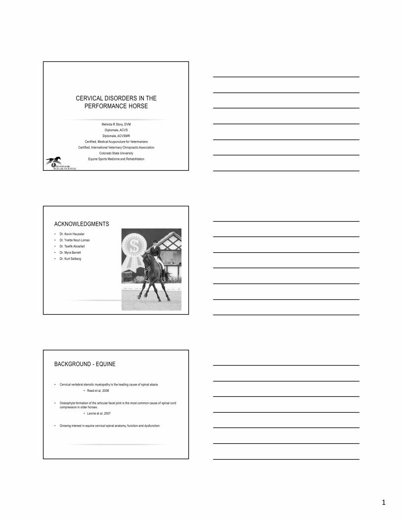
INTERVERTEBRAL DISK DISEASE
• Asymptomatic disk disease is reported in humans and horses
• 103 vertebral columns evaluated from horses birth to 23 years without signs of ataxia
• There appears to be an age related degenerative change of the disk in horses
• The changes noted worsened from cranial to caudal
• Bollwein, Hanichen; 1989 Tierarztl. Prax
9
EQUINE INTERVERTEBRAL DISK DISEASE
Speltz, Olson, et al; 2006 J Eq Vet S
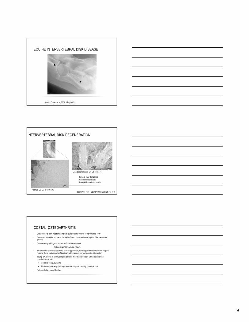
INTERVERTEBRAL DISK DEGENERATION
Disk degeneration: C4-C5 (843474)
Normal: C6-C7 (F1501595)
Severe fiber disruption
Chondrocyte clones
Basophilic acellular matrix
Speltz MC, et al. J Equine Vet Sci 2006;26:413-419.
COSTAL OSTEOARTHRITIS
• Costovertebral joint: head of the rib with superolateral surface of the vertebral body
• Costotransverse joint: connects the angle of the rib to anterolateral aspect of the transverse
process
• Cadaver study: 48% gross evidence of costovertebral OA
• Nathan et al; 1964 Arthritis Rheum
• T4 syndrome: paresthesias of one or both upper limbs, refered pain into the neck and scapular
regions. Case study reports of treatment with manipulation and exercise intervention
• Young, BA, Gill HE in 2008: joint pain patterns in normal volunteers with injection of the
costotransverse joint
• Ipsilateral, deep, dull ache
• T2 showed referred pain 2 segments cranially and caudally to the injection
• Not reported in equine literature

10
COSTAL OSTEOARTHRITIS
Right side Left side
CERVICAL FACET JOINT ARTHROPATHY
CERVICAL ARTICULAR FACETS
C3-C4 - 843209
11
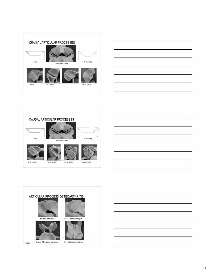
CRANIAL ARTICULAR PROCESSES
C2- - 843945 C2-C3 - 844079C3-C4 -
Dorsocaudal ViewNormal Remodelling
CAUDAL ARTICULAR PROCESSES
C3-C4 - 838905 C4-C5 - 843209 C6-C7 - 843209 C5-C6 - 838905
Dorsocaudal ViewNormal Remodelling
ARTICULAR PROCESS OSTEOARTHRITIS
843209 Caudal C6 articular processes
C6-C7 intervertebral joint
C6 joint capsule entheses
NNormal articulation
12
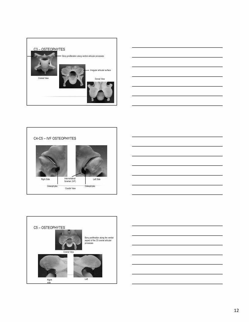
C3 – OSTEOPHYTES
Cranial View Dorsal View
Bony proliferation along ventral articular processes
Irregular articular surface
C4-C5 – IVF OSTEOPHYTES
Left SideRight Side
Caudal View
Intervertebral
foramen (IVF)
OsteophytesOsteophytes
C5 – OSTEOPHYTES
Rightt
side
Left
Bony proliferation along the ventral
aspect of the C5 cranial articular
processes
Cranial View

13
C5-C6-C7 – ARTICULAR DEFORMATION
C6-C7C5-C6
Left Cranioventral ViewMild Deformation Moderate Deformation
FACET JOINT ARTHROPATHY• Physical exam
• Abnormal posture
• Muscle symmetry
• Body condition
• Conformation
• Sweat patterns
• DAPE
• Motion exam
• Lameness exam
• Diagnostic imaging
CLINICAL EXAM
• Motion exam
• Lateral bending, ventral
and dorsal mobility
• Lameness exam
• Diagnostic imaging
14
DIAGNOSTIC IMAGING
• Lateral-lateral radiographs are often acquired as initial diagnostic:
• Neck pain
• Myelopathies
• Postural abnormalities
• Forelimb lameness
• Rider complaint of poor performance
Hudson et al. 2005, Birmingham 2010, Ricardi et
al. 1993
DIAGNOSTIC IMAGING
• The symmetrical anatomy causes superimposition.
• Potential to obscure lesions
• Little correlation of lateral radiographic findings with gross pathology in equids
• Oblique radiographic projections have been described to better visualize facet joints
individually
Unt et al. 2008, Withers et al., 2009,
Dimock et al 2010
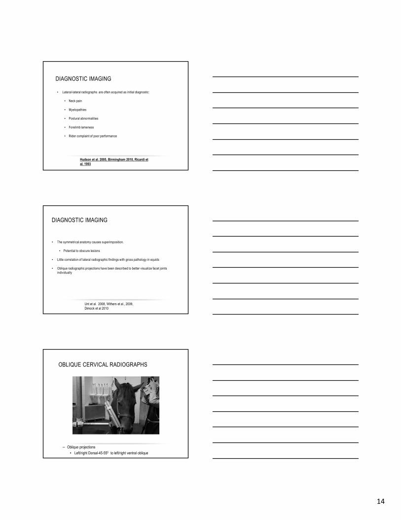
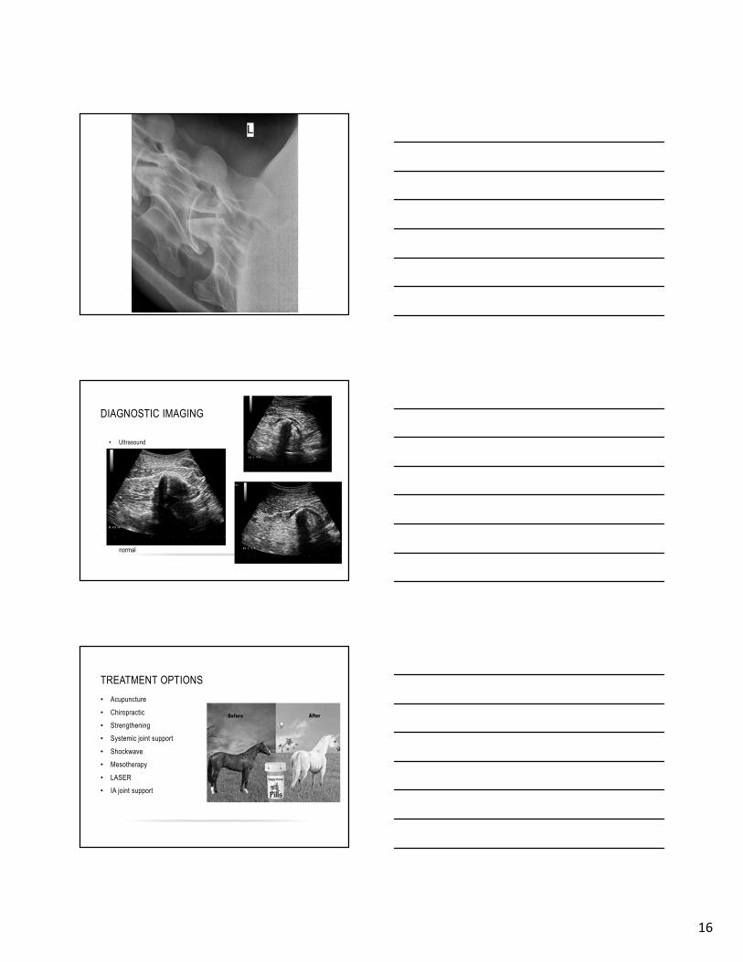
OBLIQUE CERVICAL RADIOGRAPHS
– Oblique projections
• Left/right Dorsal-45-550 to left/right ventral oblique
15
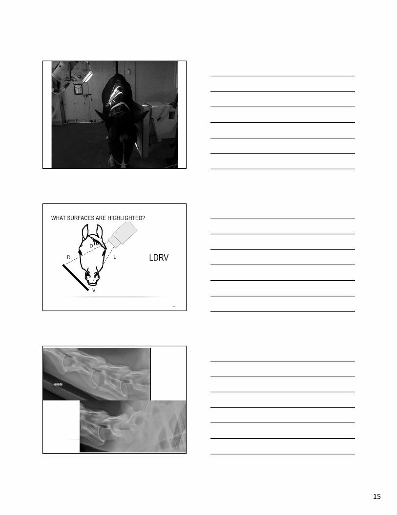
WHAT SURFACES ARE HIGHLIGHTED?
44
LDRV
16
DIAGNOSTIC IMAGING
• Ultrasound
normal
TREATMENT OPTIONS
• Acupuncture
• Chiropractic
• Strengthening
• Systemic joint support
• Shockwave
• Mesotherapy
• LASER
• IA joint support

17
CERVICAL FACET JOINT CORTICOSTEROID
INJECTIONS
• Human literature:
• Facet joint injection is effective at reducing pain in
patients with cervical radiculopathy secondary to disk
herniation
• Compared to transforaminal injection: much safer
• Bureau, Moser; 2014 Spine
• Not uncommon in the horse
• Ultrasound guided
• May be used as a diagnostic modality as well as
therapeutic
CALUSAR
• 16 year old Holsteiner gelding
• Used for Dressage
• Left Coxo-femoral joint has been treated in the past
• Recently fell when being lunged and was acutely lame RF
BASELINE LAMENESS

18
INITIAL EXAMINATION
• Mild shortening of cranial phase of stride in the RF at the
walk
• No pain, heat or swelling present in the distal limb.
• Firm palpation of right shoulder and lower neck elicits
consistent painful response.
• Flexion of the right shoulder elicits significant discomfort.
• 2/5 lame in the RF when trotted in a straight line over hard
ground, and 2+/5 when circled to the right. Grade 1 of 5
left hind lameness
PLAN
• Blocking
• Abaxial nerve block performed to rule out foot pain,
although clinical exam provided strong indication
that source of lameness was in the proximal
forelimb
• Radiographs
• Ultrasound
RIGHT SHOULDER

19
POST CERVICAL BLOCK
TREATMENT AND REHABILITATION
• No articular medication was administered at this time due to the recent history of
acute trauma and local anesthesia.
• Restricted to stall/run rest with 57mg oral firocoxib SID and topical Surpass
application for the next 2 weeks.
• After initial inflammation has subsided, re-evaluation and medication of the right
articular facet of C6-7 with corticosteroids will be performed if deemed necessary.
• Weekly acupuncture treatments
• Cervical stretches and individual mobilization of each cervical joint are also
indicated to improve mobility and reduce muscle spasm.
• Soft tissue injury in this region may also be amenable to laser therapy or ESWT
FLORYAN - HISTORY
• 8 year old castrated male Hanoverian
• Presented for back pain, worse in the thoracolumbar area
• 7 year history of neurological signs, progressively worse in the past year
• "Shivers" like behavior: strange behavior when backing and picking up his feet
for the farrier
• Muscle biopsy - normal muscle with mild vasculitis
• Owner notes that he drags his toes, but doesn't have problems going over
cavalletti rails
• No other medical problems noted
• Bloodwork within the past year, was WNL
• Bruxism under saddle: no change with Gastroguard

20
FLORYAN – NEUROLOGIC EXAM
• Generalized poor muscling
• No ataxia noted
• Moderate weakness in the hindlimbs (3/5) bilaterally symmetric
• Knuckling both hind limbs walking downhill
• Occasionally hitting himself or curb with front limbs
• Cranial nn WNL
• Sway: moderate paresis bilaterally (standing and walking)
FLORYAN – NEUROLOGIC EXAM
• Summary:
• Symmetrical moderate hind end paresis
• Symmetric mild forelimb paresis
• Shiver's like signs while backing, but not while having his feet picked up.
• The hindlimbs are more severely affected than the forelimbs
• Shivers can partially explain Floryan's neurological signs, it does not explain
all of them.
FLORYAN - MUSCULOSKELETAL
• 2+ of 5 left hindlimb lameness
• Moderately positive to stifle flexion
• Mild positive to phalangeal flexion
• No change with tarsal flexion.
• Mild effusion of the femoropatellar joint on the left hindlimb.
• Significant back pain elicited with palpation and motion
• Normal lateral bending of the cervical spine

21
FLORYAN – NUCLEAR SCINTIGRAPHY
• Moderate right sacroiliac
• Mild to moderate diffuse left and right caudal thoracic spine dorsal articulations
• Spinous processes of the caudal thorax and to a lesser extent the mid thorax
• Mild, diffuse, distal right tarsus
• Mild hind left hind fetlocks
• Mild left fore fetlock
• Mild, medial aspect of mid MC3/MC2, bilateral
• Mild medial palmar process left front distal phalanx.
FLORYAN – ULTRASOUND: T/L AND PELVIS
• Conclusions
1. Moderate right sacroiliac osteoarthrosis.
2. Mild osseous remodeling of the left sacroiliac articulation.
3. Lumbosacral disc fibrosis versus mineralization and mild L6 end plate
remodeling, of questionable clinical significance.
4. Mild arthrosis of the articular process joints at T16-17, T17-18 and very mild at
T18-L1
FLORYAN - CERVICAL
• Ultrasound: mild OA left C6-7
• Radiology: Mild enlargement of C6-7. Mild peri-articular osteophytosis and
sclerosis of the cranial articular facets of C7 on the oblique views. Sagittal ratios
51-53%

22
FLORYAN
FLORYAN
FLORYAN

23
FLORYAN - TREATMENT
• 7/8/15 The left and right sacroiliac regions were injected from cranial and caudal
approaches with methylprednisolone and Amikacin
• 7/30/15: left and right T16-17, T17-18 and T18-L1 articular facet joints treated with
Methylprednisolone and Amikacin
• The rDVM treated with one dose of OsPhos

24
T 16: DRG
Shrunken neurons and
Hypercellular foci
Normal DRG

25
Sacral DRG with Hypercellularity, neuronal vaculation
and periganglionic hemorrhage
FLORYAN - HISTOLOGY
• 1. Dorsal root ganglia (DRG), T9-T18: ganglionic neuronal degeneration and
necrosis, multifocal loss with minimal lymphohistiocytic ganglionitis.
• 2. Lumbosacral DRG and dorsal spinal nerve roots: ganglionic neuronal
degeneration and necrosis, multifocal loss with epi- and perineural hemorrhage.
• 3. C4-C6 dorsal root ganglia: neuronal vacuolation, necrosis and minimal
lymphohistiocytic ganglionitis.
SUMMARY
• The grumpy horse is likely trying to tell you something
• Cervical disease must be considered in cases of decreasing performance
• Assessing for cervical pain critical
• Evaluate for proper function
• Diagnostic imaging is important but does not necessarily answer all the questions
• Complex in the human model, increased level of difficulty in the horse because of
diagnostic limitations
• Integrative modalities as diagnostic and therapeutic options extremely valuable

26