cerebral localization localization...functional localization of cerebral cortex ‐‐‐history...
TRANSCRIPT
CEREBRAL LOCALIZATIONCEREBRAL LOCALIZATION
PRESENTED BY:HARSHIT MISHRA

Definition 1. The diagnosis of the location in the cerebrum of a
b i l i d ith f th i d
Definition
brain lesion, made either from the signs and symptoms manifested by the patient or from an investigation modality investigation modality.
2. The mapping of the cerebral cortex into areas, and the correlation of these areas with cerebral functionthe correlation of these areas with cerebral function.

Functional Localization of Cerebral Cortex ‐‐‐HISTORYFunctional Localization of Cerebral Cortex ‐‐‐HISTORY
PhrenologyPhrenology ofof GallGall ((17811781)) andand SpurzheimSpurzheim
Phrenology: Analysis of the shapes and lumps of the skull would reveal a person’s personality and intellect.Identified 27 basic faculties like imitation, spirituality
Paul Paul BrocaBroca (1861): Convincing evidence of speech (1861): Convincing evidence of speech lateralitylaterality
“Tan” : Aphasic patient
Carl Carl WernickeWernicke (1874): (1874):
TTemporal lesion disturbs comprehension.
Connectionism model of languagePredicated conduction aphasia

Experimental evidencesExperimental evidencesFritschFritsch andand HitzigHitzig ((18701870)) ‐‐‐‐‐‐motormotor cortexcortex
vonvon GuddenGudden ((18701870)) ‐‐‐‐‐‐‐‐ visualvisual cortexcortex
FerrierFerrier ((18731873)) ‐‐‐‐‐‐‐‐ auditoryauditory cortexcortex
Korbinian Brodmann (1868-1918):
BASED ON CYTOARCHITECTONIC STUDIESBASED ON CYTOARCHITECTONIC STUDIES
Established the basis for comparativecytoarchitectonics of the mammaliancortex.
47 areas47 areasmost popular
Vogt and Vogt (1919) Vogt and Vogt (1919) -- over 200 areasover 200 areas
von von EconomoEconomo (1929) (1929) ---- 109 areas109 areas

HARVEY CUSHING:HARVEY CUSHING:-- Mapped the human Mapped the human cerebral cortex with faradic electrical stimulationcerebral cortex with faradic electrical stimulationcerebral cortex with faradic electrical stimulation cerebral cortex with faradic electrical stimulation in the conscious patient.in the conscious patient.
PENFIELD & RASMUSSEN:- Outlined the motor & sensory Homunculus.y
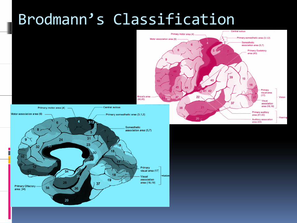
Brodmann’s Classification
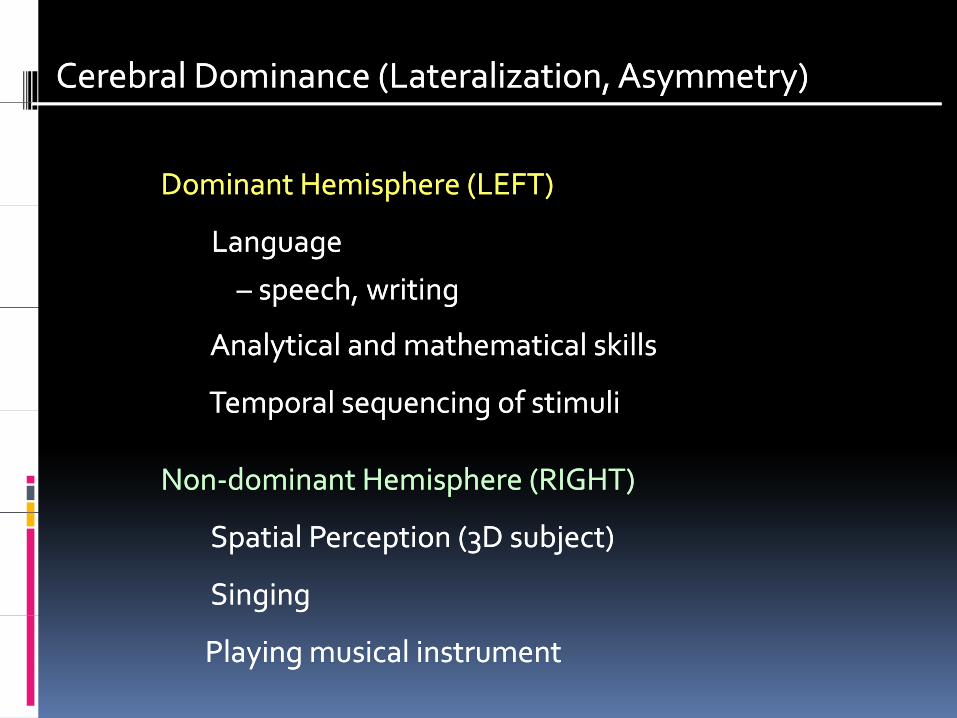
Cerebral Dominance (Lateralization, Asymmetry)Cerebral Dominance (Lateralization, Asymmetry)
Dominant Dominant Hemisphere (LEFT)Hemisphere (LEFT)
LanguageLanguage
–– speech, writingspeech, writing
Analytical and mathematical skillsAnalytical and mathematical skills
Temporal sequencing of stimuliTemporal sequencing of stimuliTemporal sequencing of stimuliTemporal sequencing of stimuli
NonNon‐‐dominant dominant Hemisphere (RIGHT)Hemisphere (RIGHT)
Spatial Perception (3D subject)Spatial Perception (3D subject)
SingingSinging
Playing musical instrumentPlaying musical instrument

METHODS
I. CLINICAL
II. ELECTROPHYIOLOGICAL
III. RADIOLOGICAL
IV INTRA OPERATIVEIV. INTRA‐OPERATIVE
V. EXPERIMENTAL
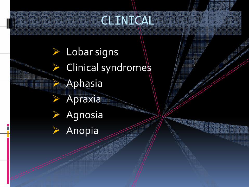
CLINICAL
Lobar signsLobar signsClinical syndromesA h i Aphasia ApraxiaAgnosiaAnopiaAnopia
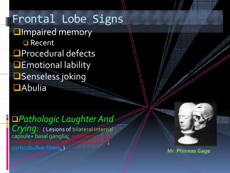
Frontal Lobe SignsImpaired memory
Recent
g
Procedural defectsEmotional labilityS l j kiSenseless jokingAbulia
Pathologic Laughter And g gCrying: ( Lesions of bilateral internal capsule+ basal ganglia; substantia nigra, cerebral peduncles, and hypothalamus; corticobulbar fibers )corticobulbar fibers, ) Mr. PhineasPhineas GageGage

Frontal Lobe Signs
Alien Hand Syndrome
g
Alien Hand Syndrome− Hand contra lateral to lesion performs purposeful movements
against will of patient− Lesion in Dominant Frontal Lobe (SMA anterior cingulate gyrus and medial − Lesion in Dominant Frontal Lobe (SMA, anterior cingulate gyrus and medial
prefrontal cortex
Magnetic Gait − Mesial Frontal lesionMesial Frontal lesion
Salutatory Seizure − Origin in SMA
kAkinetic Mutism− B/L Mesial Frontal Lesion
ParatoniaPrimitive Reflexes

Frontal Lobe Signs
Pseudobulbar Palsy
g
Pseudobulbar Palsy − Opercular Syndrome
Broca’s AphasiaBroca s Aphasia− Lesions in Left frontoparietal opercular− region− Speech and writing are impaired p g p− Telegraphic speech
Pure Agraphiag p− Affection of the posterior part of the − Left second frontal gyrus− (Exner's area)

Frontal Lobe Signs
• Executive Function Loss
g
I. Orbitofrontal syndromeDisinhibitedImpulsive Impulsive Poor judgment and insight
II. Frontal convexity syndromeA th ti Apathetic Aggressive Poor word list generation
fIII. Medial frontal syndromeAkineticIncontinent

Parietal Lobe
Elemental SomatosensoryDisturbances
P d h l i d • Pseudothalamic sensory syndrome • Lesion of parietal operculum, posterior
insula• Impairment of elementary sensation
i l d• Cortical sensory syndrome• Astereognosis, • Graphesthesia, position sense impairedGraphesthesia, position sense impaired

Parietal LobeDisturbances of Body Schema� and Spatial Relationshipsp p
Common with Right Hemisphere Common with Right Hemisphere Lesions Lesions AnosognosiaAnosognosiaPhantom limbConstructional apraxiaGeographical apraxiaDressing apraxiaH i l tHemineglect
Dressing apraxia

Disturbances of SensorimotorIntegration and Movement Execution
• Ideomotor apraxia• Failure to perform a pantomime• Most severe with lesions in the
region of Left intraparietalsulcus
•• Left frontal lesions
• buccofacial apraxia, right p , ghemiparesis, and left limb apraxia
•• Left parietal lesionsLeft parietal lesions
• buccofacial apraxia and bilateral limb apraxia

Temporal lobe
Hearing loss• Auditory agnosia
p
• Auditory agnosiao Hearing intacto Sounds not recognizedo Temporal lobe damage U/L or B/Lo Temporal lobe damage U/L or B/L
• Pure word deafnessB/L T l C ti l L i o B/L Temporal Cortical Lesion
• Left Hemispheric Damage Impaired Discrimination of Words lyrics Discrimination of Words, lyrics
• Right Hemispheric Damage Impaired Discrimination of Musical sounds

Temporal lobe
Complex hallucinationsComplex hallucinations− Otoscopic phenomena
p
Otoscopic phenomena− Illusory phenomena (micropsia,
metamorphopsia)Uncinate fitsUncinate fits− Olfactory hallucinationsGustatory Hallucinations
Temporo Parietal Seizures− Temporo ‐ Parietal Seizures
Déjà vu, jamais vu, Déjà vecu, jamais vecuj− Neocortex of temporal lobeCPS

Aphasias
Sensory Language Sensory Language AreaArea((Wernike'sWernike's areaarea) ) 22 39 022 39 0((Wernike'sWernike's areaarea) ) ‐‐‐‐‐‐‐‐22, 39, 4022, 39, 40
ReceptiveReceptive AphasiaAphasia ‐‐ area 22area 22•• defect defect in in comprehensioncomprehension•• defect defect in in comprehensioncomprehension•• good spontaneous speechgood spontaneous speech
AnomicAnomic AphasiaAphasia ‐‐ area 38 20 21area 38 20 21 22
39,40
AnomicAnomic AphasiaAphasia ‐‐ area 38, 20, 21area 38, 20, 21•• word finding difficultyword finding difficulty
JargonJargon aphasiaaphasia
3820,21
gg pp••fluent, but unintelligible fluent, but unintelligible jargon jargon •• 39 39 ((supramarginalsupramarginal gyrusgyrus),), 40 40 ((angular angular gyrusgyrus))

Aphasias
Superior Longitudinal FasciculusSuperior Longitudinal Fasciculusgg
•• Conduction AphasiaConduction Aphasia•• good comprehension,good comprehension,
good spontaneousgood spontaneousg pg pspeechspeech
•• poor repetition, poorpoor repetition, poorresponseresponse
44,4544,45
Motor Language Area (Motor Language Area (Broca’sBroca’s area) area) ‐‐‐‐‐‐44, 4544, 45
Motor AphasiaMotor AphasiaMotor AphasiaMotor Aphasia•• good comprehension,good comprehension,
no speechno speech

Occipital Lobe
Simple HallucinationsI fI f M di l O i it l DiM di l O i it l Di
p
− InferoInfero Medial Occipital DiseaseMedial Occipital Disease− Migraine (fortification)− Seizures (multicolored)Seizures (multicolored)
Hemianopia with/without Macular Sparing− Congruent Congruent
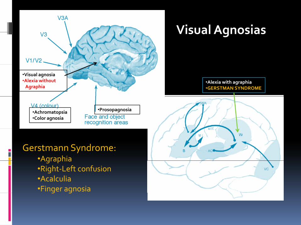
Visual AgnosiasVisual Agnosias
•Visual agnosia•Alexia without
Agraphia•Alexia with agraphia•GERSTMAN SYNDROME
•Achromatopsia•Color agnosia
•Prosopagnosia
Gerstmann Syndrome:•AgraphiaAgraphia•Right‐Left confusion•Acalculia•Finger agnosiag g
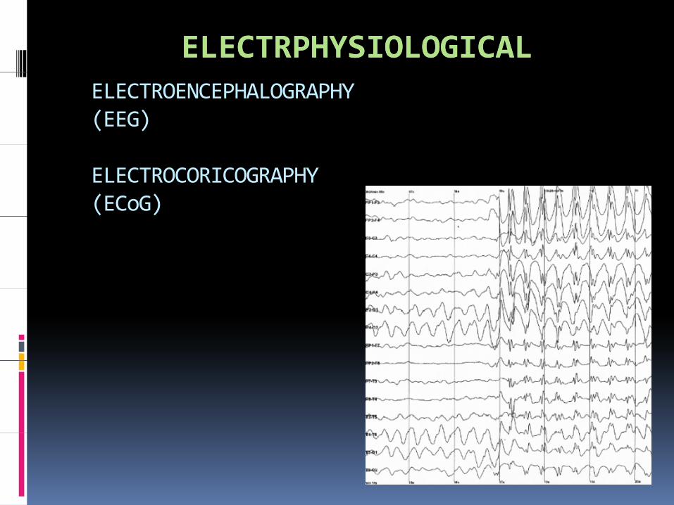
ELECTRPHYSIOLOGICALELECTRPHYSIOLOGICALELECTROENCEPHALOGRAPHY(EEG)
ELECTROCORICOGRAPHY (ECoG)
RADIOLOGICAL
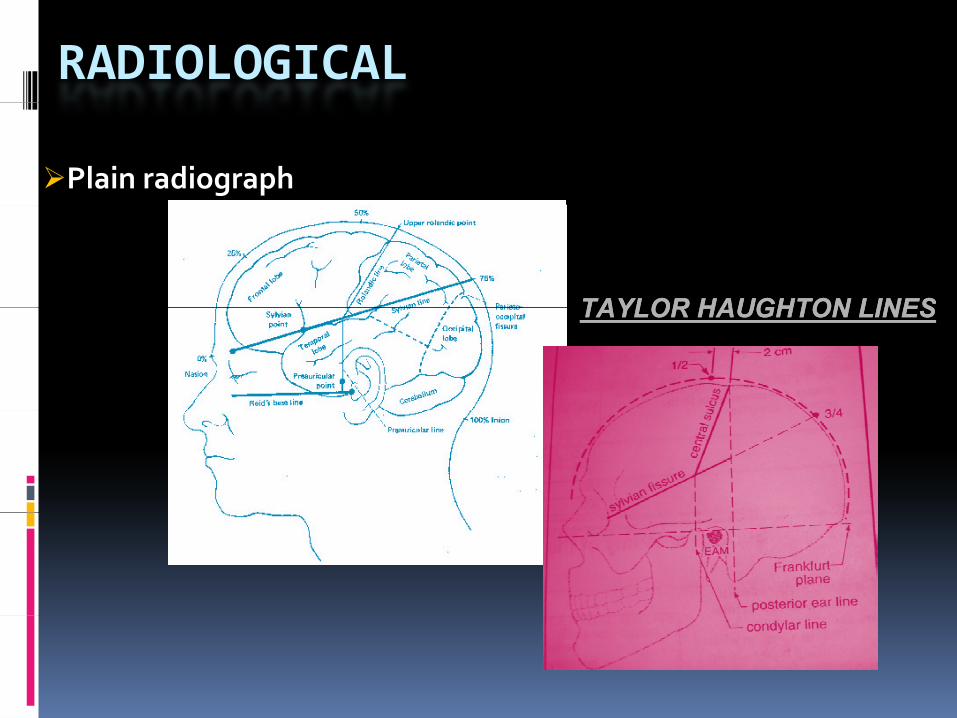
Plain radiograph
TAYLOR HAUGHTON LINESTAYLOR HAUGHTON LINESTAYLOR HAUGHTON LINESTAYLOR HAUGHTON LINES
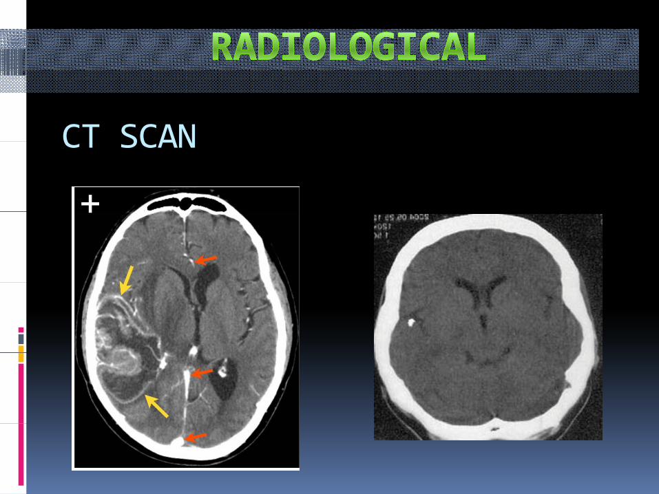
CT SCAN CT SCAN

MRI MRI

Positron Emission Tomography (PET)
H215O PETHemodynamic changes
FDG PETCerebral MetabolismCerebral Metabolism
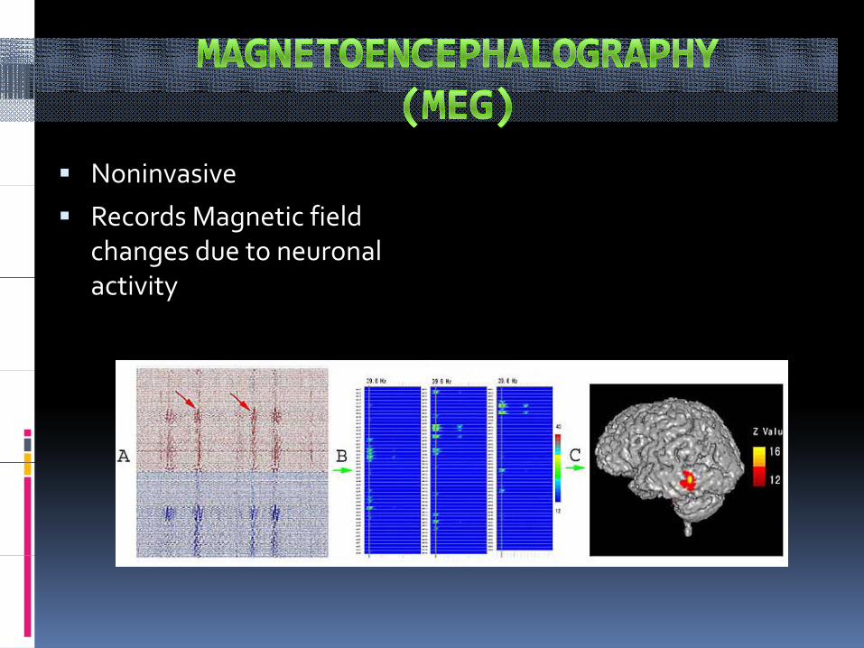
Noninvasive
Records Magnetic field changes due to neuronal
ti it activity

Functional MAGNETIC RESONANCE (f )IMAGING (fMRI)
Based on the concept of Blood Oxygenation p ygLevel-dependent Contrast (BOLD)
- Oxyhemoglobin is diamagnetic (like biological tissue). Magnetic fieldbiological tissue).
− Deoxyhemoglobin (dHb) is paramagneticinduce susceptibility effect around dHb
φr
θag et c e d
vessel φr

Anisotropic Diffusion Tensor i ( h )Imaging (Tractography)
Direction of maximum diffusivity of diffusivity of water corresponds to
i f Whit axis of White Matter tracts
•Displacement
•Edema
I filt ti•Infiltration
•Destruction
INTRAOPERATIVE INTRAOPERATIVE LOCALIZATION INLOCALIZATION INLOCALIZATION INLOCALIZATION INNEUROSURGERYNEUROSURGERY

INTRAOPERATIVE ULTRASOUND (IOUS)

SonographicallySonographically Guided Procedures in the BrainGuided Procedures in the BrainIntraoperative Doppler Ultrasound3‐D Transcranial UltrasoundContrast Enhanced Transcranial Ultrasonography
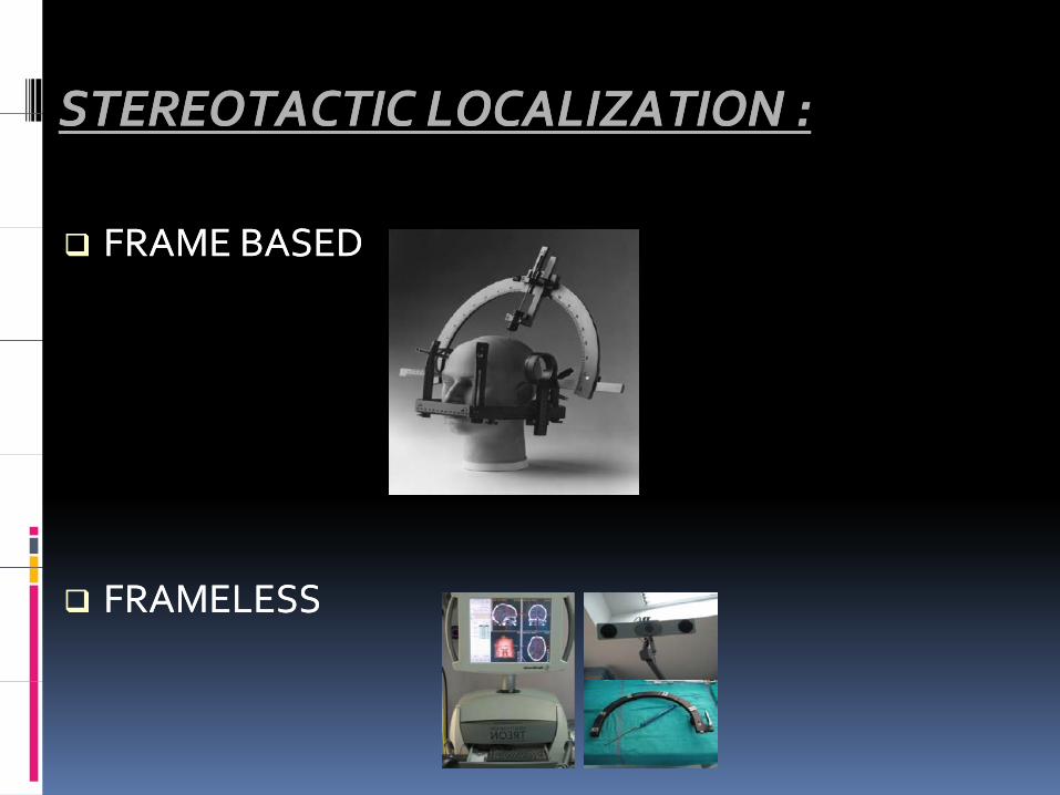
STEREOTACTIC LOCALIZATION :STEREOTACTIC LOCALIZATION :STEREOTACTIC LOCALIZATION :STEREOTACTIC LOCALIZATION :
FRAME BASEDFRAME BASED
FRAMELESS FRAMELESS

IntraoperativeIntraoperative / Mobile Ct Scan :/ Mobile Ct Scan :

Intraoperative MRI :
Also k /a “BRAINSUITE”Also k /a BRAINSUITE

CORTICAL MAPPING :

WADA ProcedureWADA Procedure

EXPERIMENTAL Single Unit Recording
› Animal studies› Advantage :great spatial and temporal resolution› Disadvantage : sampling only a very small fraction of a functional neural systemof a functional neural system
Transcranial Magnetic Stimulation› Coil placed over target brain
region› Lesion : strong fieldg› Excitation : mild field › Cognitive failures recorded
Optical imaging

Split-brainC ll t− Corpus callosotomy

Utility of Cerebral Localization
P ti Pl i
Utility of Cerebral Localization
1. Pre‐ operative Planning2. Create a Road Map of Brain Depicting
El t “N G ” ll Eloquent “No‐Go” areas as well as potential functional targetsI i i i f ti 3. Increasing precision of resection
4. Development of Minimally invasive techniq estechniques
5. Recognition of concept of plasticity of brainbrain
