cerebral hemodynamics tension induced by...
TRANSCRIPT
CEREBRALHEMODYNAMICSDURINGCONTROLLEDHYPO-TENSION INDUCEDBY THE CONTINUOUSINFUSION OF
GANGLIONIC BLOCKINGAGENTS(HEXAMETHONIUM,PENDIOMIDEANDARFONAD)1
By JOHNH. MOYERANDGEORGEMORRISWITH THE TECHNICAL ASSISTANCE OF
C. POLKSMITH(From the Departments of Pharmacology, Surgery and Medicine, Baylor University College of
Medicine, Houston, Texas, and the Cardiac Clinic of Jefferson Davis Hospital)
(Submitted for publication December 7, 1953; accepted April 17, 1954)
Although the acute cerebral hemodynamic re-sponse to blood pressure elevation has been stud-ied in normal man (1), there is very little informa-tion regarding the effect upon cerebral hemodynam-ics of reduction in blood pressure to hypotensiveranges. A moderate reduction in the systemicblood pressure appears to have very little effecton the cerebral circulation (2). However, whenthe blood pressure is markedly reduced one wouldanticipate that compensatory vasodilation of thecerebral vessels may be exceeded. Accordingly,the primary purpose of the current study was anestimation of the effect on the cerebral circulationof reduction in blood pressure produced by gang-lionic blocking agents. An attempt was made todetermine the critical level of reduction in arterialblood pressure beyond which further depressionmight produce cerebral ischemia and cerebral hy-poxia. An estimation of this type is especiallyindicated because of the current use of controlledhypotension for some surgical procedures. Theobservations on the cerebral circulation in the cur-rent study were made on unanesthetized subjectsin whom the blood pressure was reduced by con-tinuous infusion of hexamethonium,2 Pendiomide,8or Arfonad,4 all of which are ganglionic blockingagents.
1 This work was supported in part by grants from theMedical Research and Development Board, Office of theSurgeon General, Department of the Army under con-tract number DA-49-007-MD-314, and the Houston HeartAssociation.
2 Supplied through the courtesy of Burroughs Well-come & Co. as Hexameton, and Warner-Chilcott as Meth-ium.
8 Pentamethyl-diethyl-3-aza-pentane-1, 5 diammoniumdibromide, supplied through the courtesy of Ciba Pharma-ceutical Products as Pendiomide.
4D-3 4(1', 3"-dibenzyl-2' keto-immdizalido)-1,2,-tri-methylene thiophanium d-camphor sulfonate, suppliedthrough the courtesy of Hoffman-LaRoche as Arfonad.
METHODS
Observations on cerebral hemodynamics were made on19 normal individuals using the nitrous oxide technique(3, 4) for estimating cerebral blood flow. The patientswere divided into three groups. There were eight pa-tients in group A who received hexamethonium, five pa-tients in group B who received Pendiomide and six pa-tients in group C who received Arfonad. The subjectswere unanesthetized in order that obvious disturbancesin cerebration could be detected should they occur whenthe blood pressure was reduced. All but one of the pa-tients (No. 9) had normal arterial blood pressure at thetime the studies were performed. The exception was apatient with labile hypertension whose blood pressureincreased under stress, but was normal most of the time.Several additional patients showed a slight increase insystolic pressure at the time of the study, probably a re-sult of the apprehension associated with the carrying outof the procedure. After suitable control observations(supine position) the blood pressure was reduced by ad-ministering one of the ganglionic blocking agents (hexa-methonium,2 Pendiomides or Arfonad4). The cerebralblood flow studies were repeated after the desired reduc-tion in arterial blood pressure had been obtained and theblood pressure had remained stable for 30 to 60 minutes.In patients who were particularly sensitive to any of theagents, an effort was made not to reduce the mean ar-terial blood pressure below 50 mm. Hg because of pos-sible deleterious effects on the patient. The blood pres-sure was determined by simultaneous direct intra-arterialmanometry and by auscultation and a mercury sphygmo-manometer. The drugs were administered by continu-ous intravenous infusion using a concentration of hexa-methonium and Pendiomide of 0.5 to 2 mg. per cc. of solu-tion and of Arfonad of 4 mg. per cc. of solution in 5 percent glucose in distilled water. The hexamethonium wasgiven at the rate of 2 to 8 mg. per minute depending on thedegree of reduction in arterial blood pressure that was at-tained. One patient who was relatively unresponsive tohexamethonium was given the drug at a rate of 8 mg. perminute for a total of 750 mg. Pendiomide was given atthe rate of 3 to 6 mg. per minute. Usually a "floor" inthe blood pressure was reached with both hexamethoniumand Pendiomide and it was difficult to lower the pressuremuch farther. However, if the infusion was entirely dis-continued the blood pressure usually rose 10 nun. Hg or
1081
1082 JOHN H. MOYERAND GEORGEMORRIS
TABLE I
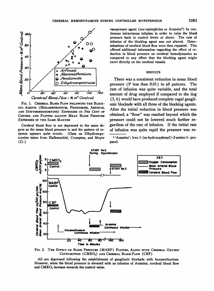
Cerebral hemodynamic response to blood pressure reduction with hexamethonium, Pendiomide or Arfonad *
Auscultatory blood pressureMean blood Cerebral Cerebrovascular Cerebral 02 Total
Control Drug pressure blood flow resistance uptake Dose dosetmg./ milli-
Patient Syst. Diast. Syst. Diast. C D DT C D DT C D DT C D DT min. grams
Group A. Response to hexamethonium1 147 85 80 50 101 53 63 54 34 36 1.9 1.6 1.8 3.6 2.9 2.5 4 2752 120 80 68 S0 96 54 61 56 40 40 1.7 1.4 1.5 2.8 3.1 2.3 4 3003 100 60 70 46 76 56 57 52 47 50 1.5 1.2 1.1 2.5 2.7 2.4 2 904 142 85 100 78 100 80 - 69 58 - 1.5 1.4 - 3.0 2.6 - 8 7505 128 90 73 55 104 60 - 75 36 - 1.4 1.7 - 4.1 3.3 - 2 606 140 80 96 56 98 66 - 50 38 - 2.0 1.7 - 2.6 2.4 - 4 2457 140 100 100 60 111 66 - 51 36 - 2.2 1.8 - 2.9 2.7 - 3 2758 145 90 90 54 108 61 - 52 44 - 2.1 1.4 - 4.1 3.7 - 3 80
Mean 133 84 85 56 99 62 60 57 42 42 1.8 1.5 1.5 3.2 2.9 2.4P Value <0.01 <0.01 <0.05 <0.3Mean per centl
of control 64 68 63 67 73 78 87 85 93 82
Group B. Response to Pendiomide9 178 100 70 48 122 54 63 46 42 46 2.7 1.3 1.4 2.8 3.2 3.4 3 100
10 116 70 80 50 94 54 58 51 45 60 1.8 1.2 1.0 2.3 2.4 3.0 5 7001 1 130 66 98 56 90 65 72 57 60 62 1.6 1.1 1.2 2.5 2.9 3.3 6 50012 120 70 90 66 84 72 81 S0 38 40 1.7 1.9 2.0 3.3 3.1 3.2 6 35013 136 90 84 54 111 60 - 49 51 - 2.3 1.2 - 2.9 3.2 - 5 500
Mean 136 79 84 55 100 61 69 51 47 52 2.0 1.3 1.4 2.8 3.0 3.2P Value <0.01 <0.4 <0.05 <0.4Mean per cent$
of control 64 72 63 73 93 102 70 75 108 120
Group C. Response to Arfonad14 116 70 60 40 92 48 46 51 34 30 1.8 1.4 1.5 3.5 2.4 2.5 5 30015 120 78 70 46 94 44 42 41 25 27 2.3 1.8 1.6 2.9 2.6 2.0 9 80016 140 92 78 50 110 58 47 48 35 48 2.3 1.7 1.0 2.5 2.7 2.4 9 65017 112 80 92 60 96 75 - 58 38 - 1.7 2.0 - 2.8 2.9 - 21 1,50018 130 86 92 64 106 66 - 73 56 - 1.5 1.2 - 4.1 4.1 - 6 50019 132 90 70 52 96 52 - 40 25 - 2.4 2.1 - 2.6 2.0 - 4 225
Mean 125 83 77 52 99 57 45 52 36 35 2.0 1.7 1.4 3.1 2.8 2.3P Value <0.01 <0.05 <0.2 <0.50Mean per centt
of control 62 63 58 46 68 75 86 65 91 79
Mean Value,Group A, B,and C 131 82 82 54 99 60 S9 54 41 441 1.9 1.5 1.4 3.0 2.9 2.7
Mean per centof control,$Groups A, B, C(19 patients) 63 67 61 63 77 87 82 75 96 96
P Value (19 patients,Groups A, B, and C) <0.01 <0.01 I <0.01 <0.3
* Mean blood pressure (mm. Hg) = direct arterial manometry; Cerebral blood flow = cc./100 Gm. brain/minute; Cerebral vascular resist-ance - mean blood pressure/cerebral blood flow; Cerebral 02 Uptake = cc./100 Gm. brain/minute; C, D, and DT = See Table II for key toabbreviations.
Includes all patients who received ganglionic blocking agents (hexamethonium, Pendiomide, or Arfonad).Mean value for per cent of control observations of individual studies.Increase in cerebral blood flow due to head down tilt was not statistically significant, P < 0.2.
more within a few minutes. When the infusion was thenstarted again the pressure could be depressed nearlyto the previous levels, indicating that the rate of infu-sion exerted some effect (although small) on the degreeof reduction in blood pressure after ganglionic blockadewas fairly well established. Since the hypotensive re-sponse to Arfonad was more marked and the duration ofaction relatively short as compared to hexamethoniumand Pendiomide, the rate of infusion was more closelyrelated to the hypotensive response. Somewhat largerdoses of this drug were used than in the cases of hexa-methonium and Pendiomide. The rate of infusion ofArfonad varied from 4 to 21 mg. per minute. After maxi-mumblood pressure reduction was attained with theseagents the concentration of nitrous oxide in the jugularvenous blood failed to approximate the concentration of
nitrous oxide in the arterial blood within the usual 10minutes. Therefore, it was necessary to continue the in-halation of nitrous oxide and sampling procedures for 20minutes in order to obtain dependable determinations.
Following the observations in the horizontally supineposition the infusion of the blocking agent was continuedin some of the patients in each group and they wereplaced in a 30 degree head down tilted supine position.The manometer was adjusted to the level of the carotidartery, and was connected in such a way that it auto-matically corrected for the difference in elevation betweenthe femoral artery and the carotid artery. After the ar-terial blood pressure had stabilized in this position, de-terminations of cerebral blood flow were again made.Seven of the patients who were not tilted were given a
CEREBRALHEMODYNAMICSDURING CONTROLLEDHYPOTENSION
4
*V.3 ac
01 &
4C
A*XA
)
)
PI
0// 000 /
*0
I -
I -
0.(o 4A'Fnaof,[ ~ o o OiAqdoeqcrw
/ */> // i
I/ I . I I I I0 20 40 60 80 /00 (2o
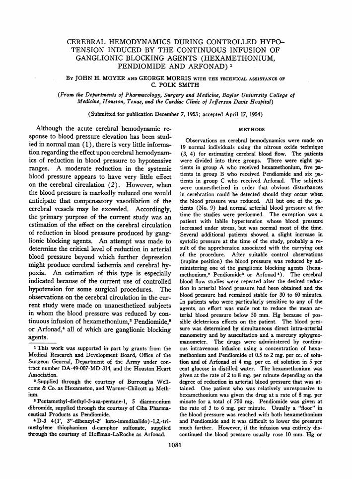
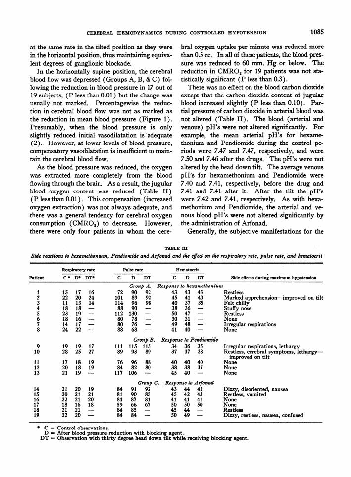
CG4e&z18&0odj'low - 9 af4ControtFIG. 1. CEREBRALBLooD FLOw FOLLOWINGTHE BLOCK-
ING AGENTS (HEXAMETHONIUM, PENDIOMIDE, ARFONAD,AND DIHYDROERGOCORNINE)EXPRESSEDIN PER CENT OFCONTROLAND PLOTrED AGAINST MEANBLOOD PRESSUREEXPRESSEDIN THE SAMEMANNER
Cerebral blood flow is not depressed to the same de-gree as the mean blood pressure is and the pattern of re-sponse appears quite erratic. (Data on Dihydroergo-cornine taken from Hafkenschiel, Crumpton, and Moyer(2).)
vasopressor agent (nor-epinephrine or Aramine') by con-tinuous intravenous infusion in order to raise the bloodpressure back to control levels or above. The rate ofinfusion of the blocking agent was not altered. Deter-minations of cerebral blood flow were then repeated. Thisoffered additional information regarding the effect of re-duction in blood pressure on cerebral hemodynamics ascompared to any effect that the blocking agent mightexert directly on the cerebral vessels.
RESULTS
There was a consistent reduction in mean bloodpressure (P less than 0.01) in all patients. Therate of infusion was quite variable, and the totalamount of drug employed if compared to the dog(5, 6) would have produced complete vagal gangli-onic blockade with all three of the blocking agents.After the initial reduction in blood pressure wasobtained, a "floor" was reached beyond which thepressure could not be lowered much farther re-gardless of the rate of infusion. If the initial rateof infusion was quite rapid the pressure was re-
5 'Aramine'; levo 1-(m-hydroxyphenyl)-2-amino-i- pro-panol.
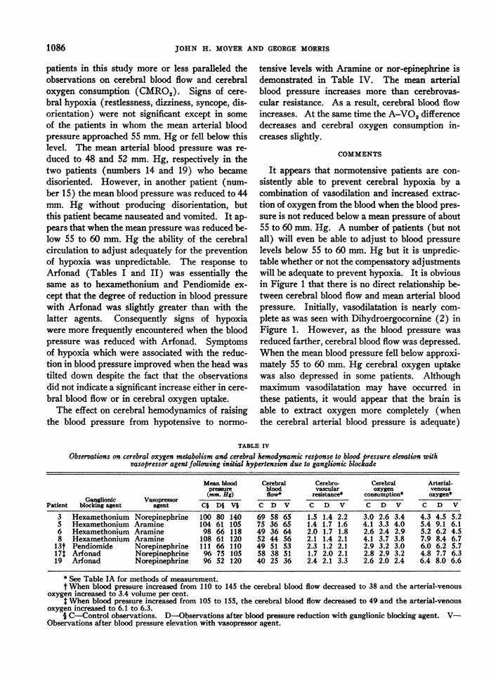
STUDY No2During Hypotenslon
/Xsxr>' tSTUDI
KEYESO55SyOgen Consumpion
oeon Arterial BloodPressureCerebral Blood Flow
,I
Cotro
-CBF \ l
Control \
I t Aromine~~~~~Continuous lnfuslon--a
HexamehonumContnuu nuon
20 40 60 s0" 140 160Time InMh tes
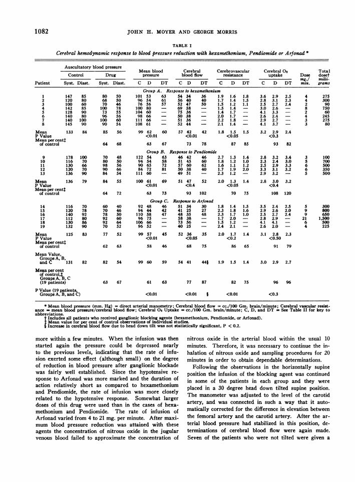
FIG. 2. THE EFFECT ON BLOODPREssuRE (MABP) PLOTTED, ALONGWITH CEREBRALOXYGENCONSUMPTION(CMRO2) ANDCEREBRALBLooD-FLOW (CBF)
All are depressed following the establishment of ganglionic blockade with hexamethonium.However, when the blood pressure is elevated with an infusion of Aramine, cerebral blood flowand CMRO,increase towards the control value.
1205?
4' oo
it
80
lb60
4
il20
1083
-
JOHN H. MOYERAND GEORGEMORRIS
TABLE II
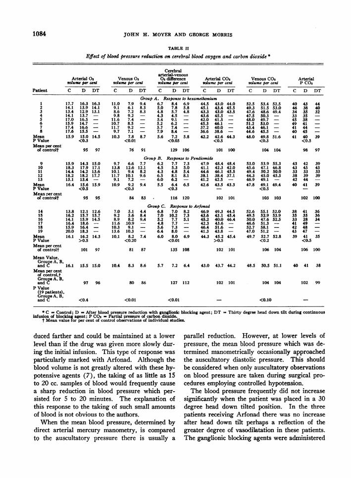
Effect of blood pressure reduction on cerebral blood oxygen and carbon dioxide *
Cerebralarterial-venous
Arterial 02 Venous 02 02 difference Arterial C02 Venous C02 Arterialvolume per cent voume per cent volume per cent volume per cent volume per cent P Co2
Patient C D DT C D DT C D DT C D DT C D DT C D DT
Group A. Response to hexamethoxium1 17.7 16.3 16.3 11.0 7.9 9.4 6.7 8.4 6.9 44.5 43.0 44.0 52.5 53.4 52.5 40 43 442 14.1 13.9 14.1 9.1 6.1 8.3 5.0 7.8 5.8 45.1 43.4 45.5 49.3 51.5 53.0 46 38 403 13.4 12.9 13.1 8.6 7.2 8.3 4.8 5.7 4.8 43.3 43.0 43.5 47.6 48.6 49.4 34 35 324 14.1 13.7 - 9.8 9.2 - 4.3 4.5 - 43.6 45.5 - 47.5 50.3 - 33 35 -5 17.0 16.5 - 11.6 7.4 - 5.4 9.1 - 42.0 41.5 - 48.0 49.7 - 45 38 -6 15.9 14.7 - 10.7 8.5 - 5.2 6.2 - 45.3 46.1 - 51.2 53.0 - 49 41 -7 17.4 16.6 - 11.7 9.2 - 5.7 7.4 - 37.2 40.0 - 43.4 46.1 - 41 44 -8 17.6 15.5 - 9.7 7.1 - 7.9 8.4 - 36.6 38.6 - 44.6 45.5 - 40 45 -
Mean 15.9 15.0 14.5 10.3 7.8 8.7 5.6 7.2 5.8 42.2 42.6 44.3 48.0 49.8 51.6 41 40 39P Value <0.3 <0.01 <0.05 <0.5 <0.3 <0.5Mean per cent
of controlt 95 97 76 91 129 106 101 100 104 104 98 97
Group B. Response to Pendiomide9 15.9 14.3 15.0 9.7 6.6 7.7 6.2 7.7 7.3 47.9 48.4 48.4 53.0 53.9 S5.3 43 42 39
10 18.3 17.9 17.1 13.8 12.6 12.1 4.5 5.3 5.0 41.1 42.3 42.0 45.6 47.1 46.8 43 45 4511 14.4 14.2 13.6 10.1 9.4 8.2 4.3 4.8 5.4 44.6 46.1 45.5 49.4 50.2 50.0 33 33 3312 18.2 18.2 17.7 11.7 10.1 9.6 6.5 8.1 8.1 38.1 38.4 37.1 44.3 45.0 45.5 38 39 3913 15.1 13.4 - 9.1 7.2 - 6.0 6.2 - 41.2 42.5 - 46.9 49.1 - 42 44 -
Mean 16.4 15.6 15.9 10.9 9.2 9.4 5.5 6.4 6.5 42.6 43.5 43.3 47.8 49.1 49.4 40 41 39P Value <0.5 <0.3 <0.3 - <0.5 -
Mean per centof controlt 95 95 84 83 , 116 120 102 101 103 103 102 100
Group C. Response to Arfonad14 13.8 12.1 12.6 7.0 5.1 4.4 6.8 7.0 8.2 46.9 49.3 44.5 52.6 55.1 52.0 38 41 3615 16.2 1S.7 15.7 9.2 5.6 8.4 7.0 10.2 7.3 43.6 43.1 45.4 49.5 S2.9 53.9 3S 3S 3616 14.1 15.9 14.5 8.9 8.2 9.4 5.2 7.7 5.1 45.2 40.0 46.4 50.0 47.6 52.5 33 28 3417 16.4 18.6 - 11.6 10.9 - 4.8 7.7 - 42.3 43.6 - 46.6 51.3 - 41 49 -18 15.9 16.4 - 10.3 9.1 - 5.6 7.3 - 46.4 51.6 - 52.7 58.1 - 42 48 -19 20.0 18.3 - 13.6 10.3 - 6.4 8.0 - 41.3 43.8 - 47.0 51.2 - 43 47 -
Mean 16.1 16.2 14.3 10.1 8.2 7.4 6.0 8.0 6.9 44.3 45.2 45.4 49.7 S2.7 52.8 39 41 35P Value >0.5 <0.20 <0.01 >0.5 <0.2 <0.5Mean per cent
of controlt 101 97 81 87 135 108 102 101 106 104 106 100
Mean Value,Groups A, B.and C 16.1 15.S 15.0 10.4 8.3 8.6 5.7 7.2 6.4 43.0 43.7 44.2 48.5 50.5 51.1 40 41 38
Mean per centof control,tGrou Ps A, B.andC 97 96 80 86 127 112 102 101 104 104 102 99
P Value(19 patients),Groups A, B,and C <0.4 <0.01 <0.01 - <0.10
* C - Control; D - After blood pressure reduction with ganglionic blocking agent; DT Thirty degree head down tilt during continuousinfusion of blocking agent; P C02 - Partial pressure of carbon dioxide.
t Mean value for per cent of control observations of individual studies.
duced farther and could be maintained at a lowerlevel than if the drug was given more slowly dur-ing the initial infusion. This type of response wasparticularly marked with Arfonad. Although theblood volume is not greatly altered with these hy-potensive agents (7), the taking of as little as 15to 20 cc. samples of blood would frequently causea sharp reduction in blood pressure which per-sisted for 5 to 20 minutes. The explanation ofthis response to the taking of such small amountsof blood is not obvious to the authors.
When the mean blood pressure, determined bydirect arterial mercury manometry, is comparedto the auscultatory pressure there is usually a
parallel reduction. However, at lower levels ofpressure, the mean blood pressure which was de-termined manometrically occasionally approachedthe auscultatory diastolic pressure. This shouldbe considered when only auscultatory observationson blood pressure are taken during surgical pro-cedures employing controlled hypotension.
The blood pressure frequently did not increasesignificantly when the patient was placed in a 30degree head down tilted position. In the threepatients receiving Arfonad there was no increaseafter head down tilt perhaps a reflection of thegreater degree of vasodilatation in these patients.The ganglionic blocking agents were administered
1084
CEREBRALHEMODYNAMICSDURING CONTROLLEDHYPOTENSION
at the same rate in the tilted position as they were
in the horizontal position, thus maintaining equiva-lent degrees of ganglionic blockade.
In the horizontally supine position, the cerebralblood flow was depressed (Groups A, B, & C) fol-lowing the reduction in blood pressure in 17 out of19 subjects, (P less than 0.01) but the change was
usually not marked. Percentagewise the reduc-tion in cerebral blood flow was not as marked as
the reduction in mean blood pressure (Figure 1).Presumably, when the blood pressure is onlyslightly reduced initial vasodilatation is adequate(2). However, at lower levels of blood pressure,
compensatory vasodilatation is insufficient to main-tain the cerebral blood flow.
As the blood pressure was reduced, the oxygen
was extracted more completely from the bloodflowing through the brain. As a result, the jugularblood oxygen content was reduced (Table II)(P less than 0.01). This compensation (increasedoxygen extraction) was not always adequate, andthere was a general tendency for cerebral oxygen
consumption (CMRO2) to decrease. However,there were only four patients in whom the cere-
bral oxygen uptake per minute was reduced morethan 0.5 cc. In all of these patients, the blood pres-sure was reduced to 60 mm. Hg or below. Thereduction in CMRO2for 19 patients was not sta-tistically significant (P less than 0.3).
There was no effect on the blood carbon dioxideexcept that the carbon dioxide content of jugularblood increased slightly (P less than 0.10). Par-tial pressure of carbon dioxide in arterial blood was
not altered (Table II). The blood (arterial andvenous) pH's were not altered significantly. Forexample, the mean arterial pH's for hexame-thonium and Pendiomide during the control pe-
riods were 7.47 and 7.47, respectively, and were
7.50 and 7.46 after the drugs. The pH's were notaltered by the head down tilt. The average venous
pH's for hexamethonium and Pendiomide were
7.40 and 7.41, respectively, before the drug and7.41 and 7.41 after it. After the tilt the pH'swere 7.42 and 7.41, respectively. As with hexa-methonium and Pendiomide, the arterial and ve-
nous blood pH's were not altered significantly bythe administration of Arfonad.
Generally, the subjective manifestations for the
TABLE III
Side reactions to hexamethonium, Pendiomide and Arfonad and the effect on the respiratory rate, pulse rate, and hematocrit
Respiratory rate Pulse rate Hematocrit
Patient C * D* DT* C D DT C D DT Side effects during maximum hypotension
Group A. Response to hexamethonium1 15 17 16 72 90 92 43 43 43 Restless2 22 20 24 101 89 92 45 41 40 Marked apprehension-improved on tilt3 11 13 14 114 96 98 40 37 35 Felt chilly4 18 18 - 88 90 - 38 36 Stuffy nose5 23 19 - 112 130 - 50 47 - Restless6 18 16 80 78 - 30 31 - None7 14 17 - 80 76 - 49 48 Irregular respirations8 24 22 - 88 68 - 41 40 None
Group B. Response to Pendiomide9 19 19 17 111 115 115 34 36 35 Irregular respirations, lethargy
10 28 25 27 89 93 89 37 37 38 Restless, cerebral symptoms, lethargy-improved on tilt
11 17 18 19 76 96 88 40 40 40 None12 20 18 19 84 82 80 38 38 37 None13 21 19 117 106 - 45 40 None
Group C. Response to Arfonad14 21 20 19 84 91 92 43 44 42 Dizzy, disoriented, nausea15 20 21 21 81 90 85 45 42 43 Restless, vomited16 22 21 20 84 87 81 41 41 41 None17 18 16 18 59 66 67 50 50 50 None18 21 21 84 85 - 45 44 Restless19 22 20 84 84 - 50 49 Dizzy, restless, nausea, confused
* C = Control observations.D = After blood pressure reduction with blocking agent.
DT = Observation with thirty degree head down tilt while receiving blocking agent.
1085
JOHN H. MOYERAND GEORGEMORRIS
patients in this study more or less paralleled theobservations on cerebral blood flow and cerebraloxygen consumption (CMRO2). Signs of cere-bral hypoxia (restlessness, dizziness, syncope, dis-orientation) were not significant except in someof the patients in whom the mean arterial bloodpressure approached 55 mm. Hg or fell below thislevel. The mean arterial blood pressure was re-duced to 48 and 52 mm. Hg, respectively in thetwo patients (numbers 14 and 19) who becamedisoriented. However, in another patient (num-ber 15) the mean blood pressure was reduced to 44mm. Hg without producing disorientation, butthis patient became nauseated and vomited. It ap-pears that when the mean pressure was reduced be-low 55 to 60 mm. Hg the ability of the cerebralcirculation to adjust adequately for the preventionof hypoxia was unpredictable. The response toArfonad (Tables I and II) was essentially thesame as to hexamethonium and Pendiomide ex-cept that the degree of reduction in blood pressurewith Arfonad was slightly greater than with thelatter agents. Consequently signs of hypoxiawere more frequently encountered when the bloodpressure was reduced with Arfonad. Symptomsof hypoxia which were associated with the reduc-tion in blood pressure improved when the head wastilted down despite the fact that the observationsdid not indicate a significant increase either in cere-bral blood flow or in cerebral oxygen uptake.
The effect on cerebral hemodynamics of raisingthe blood pressure from hypotensive to normo-
tensive levels with Aramine or nor-epinephrine isdemonstrated in Table IV. The mean arterialblood pressure increases more than cerebrovas-cular resistance. As a result, cerebral blood flowincreases. At the same time the A-VO2 differencedecreases and cerebral oxygen consumption in-creases slightly.
COMMENTS
It appears that normotensive patients are con-sistently able to prevent cerebral hypoxia by acombination of vasodilation and increased extrac-tion of oxygen from the blood when the blood pres-sure is not reduced below a mean pressure of about55 to 60 mm. Hg. A number of patients (but notall) will even be able to adjust to blood pressurelevels below 55 to 60 mm. Hg but it is unpredic-table whether or not the compensatory adjustmentswill be adequate to prevent hypoxia. It is obviousin Figure 1 that there is no direct relationship be-tween cerebral blood flow and mean arterial bloodpressure. Initially, vasodilatation is nearly com-plete as was seen with Dihydroergocornine (2) inFigure 1. However, as the blood pressure wasreduced farther, cerebral blood flow was depressed.When the mean blood pressure fell below approxi-mately 55 to 60 mm. Hg cerebral oxygen uptakewas also depressed in some patients. Althoughmaximum vasodilatation may have occurred inthese patients, it would appear that the brain isable to extract oxygen more completely (whenthe cerebral arterial blood pressure is adequate)
TABLE IV
Observations on cerebral oxygen metabolism and cerebral hemodynamic response to blood pressure elevation withvasopressor agent following initial hypertension due to ganglion c blockade
Patient
3568
13t17t19
Ganglionic Vasopressorblocking agent agent
Hexamethonium NorepinephrineHexamethonium AramineHexamethonium AramineHexamethonium AraminePendiomide NorepinephrineArfonad NorepinephrineArfonad Norepinephrine
Mean bloodpressure
(mm. Hg)
Ci Di V§100 80 140104 61 10598 66 118
108 61 120111 66 110
96 75 10596 52 120
Cerebralbloodflow*
C D V
69 58 6575 36 6549 36 6452 44 5649 51 5358 38 5140 25 36
Cerebro-vascular
resistance*
C D V
1.5 1.4 2.21.4 1.7 1.62.0 1.7 1.82.1 1.4 2.12.3 1.2 2.11.7 2.0 2.12.4 2.1 3.3
Cerebraloxygen
consumption*C D V
3.0 2.6 3.44.1 3.3 4.02.6 2.4 2.94.1 3.7 3.82.9 3.2 3.02.8 2.9 3.22.6 2.0 2.4
Arterial-venous
oxygen*C D V
4.3 4.5 5.25.4 9.1 6.15.2 6.2 4.57.9 8.4 6.76.0 6.2 5.74.8 7.7 6.36.4 8.0 6.6
* See Table IA for methods of measurement.t When blood pressure increased from 110 to 145 the cerebral blood flow decreased to 38 and the arterial-venous
oxygen increased to 3.4 volume per cent.$ When blood pressure increased from 105 to 155, the cerebral blood flow decreased to 49 and the arterial-venous
oxygen increased to 6.1 to 6.3.§ C-Control observations. D-Observations after blood pressure reduction with ganglionic blocking agent. V-
Observations after blood pressure elevation with vasopressor agent.
1086
CEREBRALHEMODYNAMICSDURING CONTROLLEDHYPOTENSION
from the blood flowing through it, as has beenpreviously demonstrated under conditions of hy-perventilation (8) and after the administration ofAminophylline (9). The failure of more completeextraction of oxygen in the current study may bedue in part to inadequate perfusion of plasma andoxygen through the capillary walls at these re-duced blood pressure levels.
The reduction in cerebral blood flow appears tobe related to the degree of blood pressure reduc-tion rather than to the blocking agent employed.Since Arfonad is the most potent of the ganglionicblocking agents used one can expect to approach orpass below this critical level more frequently withthis agent than with hexamethonium or Pendio-mide if maximum blood pressure reduction is em-ployed. Henry, Gauer, Kety, and Kramer (10)concluded that under conditions of gravitationalstress, unconsciousness was not lost until the meanarterial blood pressure approached 25 mm. Hg.This is probably related to their method for lower-ing the blood pressure (positive acceleration) andthe conclusions are hardly applicable to the cur-rent study. Scheinberg and Stead (11) observedthat when the mean cerebral arterial pressure wasreduced from 84 to 55 mm. Hg by changing fromthe supine to the upright position, there was a sig-nificant reduction in cerebral blood flow. Thisis in essential agreement with the current obser-vations.
Cerebral vasoconstrictor tone is probably notaffected directly by blockade of the sympatheticganglia because procaine blockade of the superiorcervical ganglia (12, 13) fails to increase cerebralblood flow. Since cerebral blood flow is mostlikely regulated by autonomous regulatory mecha-nisms at the local tissue level, the depression incerebral blood flow which occurs during hypo-tension is merely a reflection of inadequate hydro-static pressure. In the current study, when a vaso-constrictor agent was administered and the bloodpressure raised from hypotensive levels to normo-tensive ones, the cerebral blood flow increased.The increase in cerebral blood flow which occursunder these conditions is in direct contrast to thecerebrovascular response to these same pressoragents (14) when administered to normotensiveindividuals with a subsequent increase in bloodpressure to hypertensive levels since under the
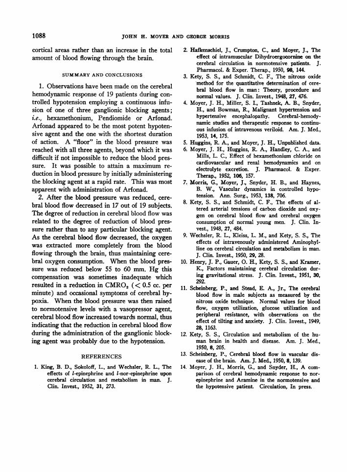
110
90
80
70
60
.
000
0 0
'V
00
O50 -
40 - ,"J/30 -
- I~~~~0P.,ndiomideI10 -" Af.
10 20 30 40 50 6070 80 90 100 110Cerebrol Blood Flow - Percent of Control -
FIG. 3. CEREBRAL BLOoD FLow EXPRESSED AS PERCENT OF CONTROLPLOTTED AGAINST OXYGENCONSUMP-TION EXPRESSEDIN THE SAMEMANNER
Cerebral blood flow is depressed in excess of cerebraloxygen consumption due to increased extraction of oxy-gen from the cerebral blood.
latter circumstances cerebral blood flow is re-duced. If the reduction in cerebral blood flowwhich also occurs following the administration ofganglionic blocking agents were not a result ofthe hypotension, then raising the pressure withan adrenergic drug would not be expected to in-crease cerebral blood flow, but might actually re-duce it (14). If such a pressure-flow relation-ship is accepted, then the value of vasopressoragents in the treatrnent of normal volemic hypo-tensive states seems well substantiated in so faras cerebral blood flow is concerned. These obser-vations would tend to invalidate the suggestion(1) that the use of vasoconstrictor agents suchas norepinephrine might depress cerebral bloodflow when used for the treatment of severe hypo-tensive states.
The failure of head down tilt to increase cere-bral blood flow was a completely unexpected ob-servation, and one for which the authors have noexplanation. Some of the patients who demon-strated mental disturbances during hypotensionwere improved when the head was tilted down.This observation is not necessarily incompatiblewith the observations of a continued reduction inaverage cerebral blood flow per unit volume ofbrain since the improvement in cerebral functionmay merely represent improved circulation to the
1087
JOHN H. MOYERAND GEORGEMORRIS
cortical areas rather than an increase in the totalamount of blood flowing through the brain.
SUMMARYANDCONCLUSIONS
1. Observations have been made on the cerebralhemodynamic response of 19 patients during con-
trolled hypotension employing a continuous infu-sion of one of three ganglionic blocking agents;i.e., hexamethonium, Pendiomide or Arfonad.Arfonad appeared to be the most potent hypoten-sive agent and the one with the shortest durationof action. A "floor" in the blood pressure was
reached with all three agents, beyond which it was
difficult if not impossible to reduce the blood pres-
sure. It was possible to attain a maximum re-
duction in blood pressure by initially administeringthe blocking agent at a rapid rate. This was mostapparent with administration of Arfonad.
2. After the blood pressure was reduced, cere-
bral blood flow decreased in 17 out of 19 subjects.The degree of reduction in cerebral blood flow was
related to the degree of reduction of blood pres-
sure rather than to any particular blocking agent.As the cerebral blood flow decreased, the oxygen
was extracted more completely from the bloodflowing through the brain, thus maintaining cere-
bral oxygen consumption. When the blood pres-
sure was reduced below 55 to 60 mm. Hg thiscompensation was sometimes inadequate whichresulted in a reduction in CMRO2(< 0.5 cc. per
minute) and occasional symptoms of cerebral hy-poxia. When the blood pressure was then raisedto normotensive levels with a vasopressor agent,cerebral blood flow increased towards normal, thusindicating that the reduction in cerebral blood flowduring the administration of the ganglionic block-ing agent was probably due to the hypotension.
REFERENCES
1. King, B. D., Sokoloff, L., and Wechsler, R. L., Theeffects of l-epinephrine and l-nor-epinephrine uponcerebral circulation and metabolism in man. J.Clin. Invest., 1952, 31, 273.
2. Hafkenschiel, J., Crumpton, C., and Moyer,- J., Theeffect of intramuscular Dihydroergocormne on thecerebral circulation in normotensive patients. J.Pharmacol. & Exper. Therap., 1950, 98, 144.
3. Kety, S. S., and Schmidt, C. F., The nitrous oxidemethod for the-quantitative determination of cere-bral blood flow in man: Theory, procedure andnormal values. J. Clin. Invest., 1948, 27, 476.
4. Moyer, J. H., Miller, S. I., Tashnek, A. B., Snyder,H., and Bowman, R., Malignant hypertension andhypertensive encephalopathy. Cerebral-hemody-namic studies and therapeutic response to continu-ous infusion of intravenous veriloid. Am. J. Med.,1953, 14, 175.
5. Huggins, R. A., and Moyer, J. H.,. Unpublished data.6. Moyer, J. H., Huggins, R. A., Handley, C. A., and
Mills, L. C., Effect of hexamethonium chloride oncardiovascular and renal hemodynamics and onelectrolyte excretion. J. Pharmacol. & Exper.Therap., 1952, 106, 157.
7. Morris, G., Moyer, J., Snyder, H. B., and Haynes,B. W., Vascular dynamics in controlled hypo-tension. Ann. Surg., 1953, 138, 706.
8. Kety, S. S., and Schmidt, C. F., The effects of al-tered arterial tensions of carbon dioxide and oxy-gen on cerebral blood flow and cerebral oxygenconsumption of normal young men. J. Clin. In-vest., 1948, 27, 484.
9. Wechsler, R. L., Kleiss, L. M., and Kety, S. S., Theeffects of intravenously administered Aminophyl-line on cerebral circuilation and metabolism in man.J. Clin. Invest., 1950, 29, 28.
10. Henry, J. P., Gauer, 0. H., Kety, S. S., and Kramer,K., Factors maintaining cerebral circulation dur-ing gravitational stress. J. Clin. Invest., 1951, 30,292.
11. Scheinberg, P., and Stead, E. A., Jr., The cerebralblood flow in male subjects as measured by thenitrous oxide technique. Normal values for bloodflow, oxygen; utilization, glucose utilization andperipheral resistance, with observations on theeffect of tilting and anxiety. J. Clin. Invest., 1949,28, 1163.
12. Kety, S. S., Circulation and metabolism of the hu-man brain in health and disease. Am. J. Med.,1950, 8, 205.
13. Scheinberg, P., Cerebral blood flow in vascular dis-ease of the brain. Am. J. Med., 1950, 8, 139.
14. Moyer, J. H., Morris, G., and Snyder, H., A com-parison of cerebral hemodynamic response to nor-epinephrine and Aramine in the normotensive andthe hypotensive patient. Circulation, In press.
1088