cephlometric history, evolution & landmarks
TRANSCRIPT
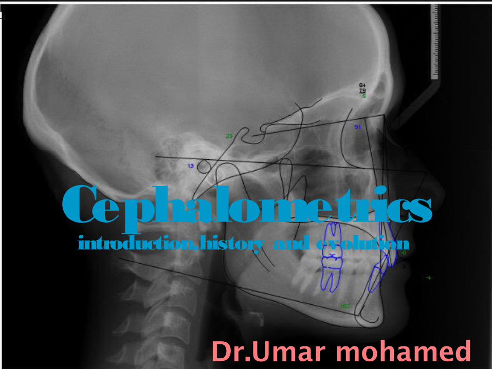
Cephalometricsintroduction,history and evolution
Dr.Umar mohamed
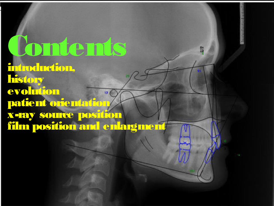
Contentsintroduction,history evolutionpatient orientationx-ray source positionfilm position and enlargment

Introduction
Cephalometric radiography was introduced in to orthodontics during the 1930s.
Orthodontists, in their attempts to change facio-oro-dental deviations from accepted norms, have adopted cephalometric measurement, a method employed in physical anthropology.
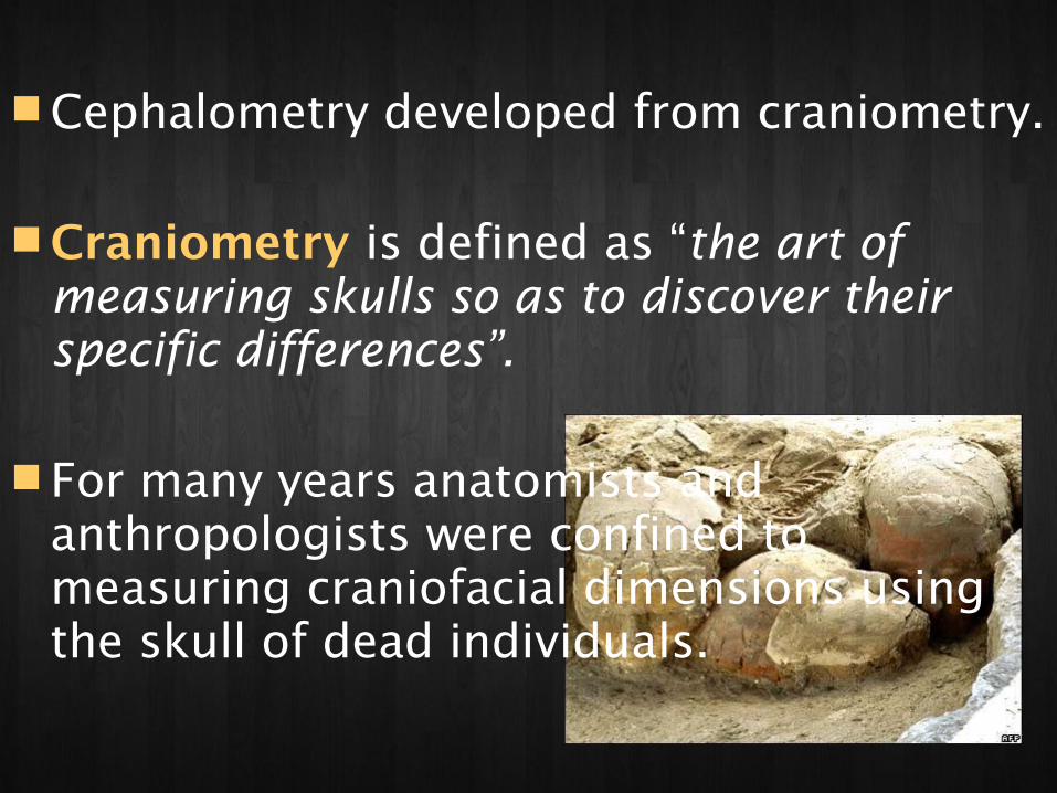
Cephalometry developed from craniometry. Craniometry is defined as “the art of
measuring skulls so as to discover their specific differences”.
For many years anatomists and anthropologists were confined to measuring craniofacial dimensions using the skull of dead individuals.
Cephalometry is concerned with measuring the head inclusive of soft tissues, be it living or dead.
Craniometry – measuring skull.Cephalometry – measuring head.
However Cephalometry had its limitations due to the inaccuracies that resulted from varying thickness of soft tissues.
With the discovery of X rays by Roentgen in 1895, Radiographic Cephalometry came in to being.
Defined as the measurement of head from bony and soft tissue land marks on the radiographic image
(Krogman & Sassouni 1957).

This approach combines the advantages of Craniometry and anthropometry.
The disadvantage is that it produces two dimensional image of a three dimensional structure.

History
History prior to the advent of radiography begins with the attempts of the scientists to classify the human physiques.
Basically it stems from the history of
Anthropometry.

In 500 BC, Hippocrates, Father of medicine, designated two physical types 1.Habitus phithicus – long thin body subject to tuberculosis,
& 2.Habitus apoplecticus -- short thick individual
susceptible to vascular diseases & apoplexy.
Kretschmer (1921) described : pyknic (compact), asthenic (without strength), & athletic.
Sheldon continued the work & refined it.
He classified physique in to three types. 1.Endomorphy is centered on the
abdomen, and the whole digestive system.
2.Mesomorphy is focused on the muscles and the circulatory system.
3.Ectomorphy is related to the brain and the nervous system.
Early History of measurements and proportions
Portrayal of human form demands not only artistic talent & technical ability but a disciplined & consistent style.
The ancient Egyptians developed an intricate quantitative system that defined the proportions of the human body. It became known as the Canon of proportions.

Initially the canons were enclosed in a grid system of equalized squares with 18 horizontal lines, line 18 drawn through hairline.
Later it was included in a grid system of 22 horizontal lines, line 21 drawn through the upper eyelid.

Period of Renaissance
Leonardo da vinci was probably the earliest to apply the theory of head measurement.
He used lines related to specific structures in the head in his study of human form.


His drawings included a study of facial proportions in natural head position. The profile was divided in to seven parts by eight horizontal lines.
The joining of the lower lip and chin and the tip of the jaw and the upper tip of the ear with the temple forms a perfect square; and each face is half of the head.

Albrecht Durer, published a study in 1528 on cranial measurements which comprised the “Vier Bucher von menschliche Proportion”.
Using geometrical methods he provided a analysis of the leptoprosopic face & euryprosopic face in coordinate system, where the horizontal and the vertical lines were drawn through the same land marks or facial features.


IN 18TH CENTURYThe Dutchman Petrus Camper was credited with the introduction of facial angle.
He oriented crania in a space on a horizontal from the middle of the porus acusticus to a point below the nose. Camper’s horizontal became the reference line for angular measurements used to characterize evolutionary trends in anthropological studies.

The facial angle as he described was formed by the intersection of a facial line and a horizontal plane.
The facial line was a line tangential to the most prominent part of the frontal bone and to the slight convexity anterior to the upper teeth.

The horizontal plane passes through the lower part of the nasal aperture, backwards along the line of the zygomatic arch, and through the centre of the external auditory meatus.
Camper’s facial angle was readily accepted as a standard measurement in craniology.

The terms prognathic and orthognathic introduced by Retzius are tied to Camper’s illustrations of facial form.
As a result the facial angle became a time honored anthropological method to determine the facial type.
Prognathism refers to the prominence of jaws, relative to forehead, &
a straight facial profile -- as orthognathous.

He is also credited with the introduction of cephalic index, the ratio of breadth to length of the skull expressed as a percentage.

History of Cephalometric Radiography
In 1895, Prof. Wilhelm Conrad Roentgen made a remarkable contribution to science with the discovery of x-rays.
Mrs. Röntgen's hand, the first X-ray
picture of the human body ever taken. photos courtesy of NASA

On December 28, 1895 he submitted a paper “On A New Kind of Rays, A Preliminary Communication” to the Wurzburg Physical Medical Society.
Prof. Wilhem Koening & Dr. Otto Walkhoff
simultaneously made the first dental radiograph in 1896.
It was clear that the use of x-rays provided the means of obtaining a different perspective on the arrangement and relation of bones thus expanding the horizons of craniometry & cephalometry .

Van Loon -- first to introduce cephalometrics to orthodontics. He applied anthropometric procedures in analyzing facial growth by making plaster casts of face in to which he inserted oriented casts of the dentition.
Hellman in 1920s used cephalometric techniques and described their value.

The first x- ray pictures of skull in the standard lateral view were taken by A.J.Pacini & Carrera in 1922.
Pacini received a research award from the American Roentgen Ray Society for a thesis entitled “Roentgen Ray Anthropometry of the Skull”.
Pacini introduced a teleroentgenographic technique for standardized lateral head radiography which proved to be of tremendous use in cephalometry, as well as in measuring growth and devlopment of face.
His method, which was rather primitive, involved a large fixed distance from the x ray source to the cassette.

He identified the following landmarks : gonion, pogonion, nasion, and anterior nasal spine. He also located the sella turcica & external auditory meatus.

In 1931 Cephalometric radiography came to full function when B. Holly Broadbent in USA published methods to obtain standardized head radiographs in the Angle Orthodontist (A new X ray tech & its application to orthodontia).
H. Hofrath simultaneously published the same in Fortschritte der Orthodontie in Germany.

Broadbent’s contribution
Broadbent’s interest in craniofacial growth began with his orthodontic education under E.H. Angle in 1920.
He continued to pursue that intrest along with his orthodontic practice, working with a leading anatomist J. Wingate Todd

The patient’s head was centered in the cephalostat with the superior borders of the external auditory meatus resting on the upper parts the two ear rods.

The lowest point on the inferior bony border of the left orbit, indicated by the orbital marker, was at the level of the upper parts of the ear rods.
Nose clamp was fixed at the root of the nose to support the upper face.
The focus film distance was set at 5 feet (152.4 cm) and the subject film distance could be measured to calculate image magnification.
With the two X ray tubes at right angles to each other in the same horizontal plane, two images (lateral & PA) could be simultaneously produced.

Hofrath’s technique differed from Broadbent’s in that the path of the central ray was not fixed in relation to the head and no plan was suggested for super positioning subsequent x-rays.

Cephalometric Analysis

The major use of radiographic cephalometry is in characterizing the patient’s dental and skeletal relationships.
This led to the development of a number of cephalometric analyses to compare a patient to the population standards.
William. B. Downs in 1948 developed the first cephalometric analysis.
Its significance was that it presented an objective method of portraying many factors underlying malocclusion.
Followed by other analyses by Steiner (1953), Tweed (1953), Ricketts (1958), Sassouni (1969), Enlow (1969), Jaraback (1970), Jacobson (1975) etc.

Evolution of Cephalometrics
The thoroughness of Broadbent’s design of the cephalometric method is evident from the fact that the basic technique has survived unchanged for over 70 years.
The instrumentation had evolved to a more suitable form for the individual practitioner through the pioneering efforts of Margolis, Higley & others.

Cephalometric Landmarks
-- are readily recognizable points on a cephalometric radiograph or tracing, representing certain hard or soft tissue anatomical structures (anatomical landmarks) & (constructed landmarks).

Requirements
Should be easily seen on the roentgenogram, Be uniform in out line, and easily reproducible. Should have significant relationship to the
vectors of growth. Should permit valid quantitative measurements
of lines and angles projected from them. Measurements should be amenable to
statistical analyses.

Landmarks in Lat.Ceph


Hard tissue landmarks A-point (Point A, Subspinale, SS) : the
most posterior midline point on the concavity between the ANS and prosthion.
Anterior nasal spine (ANS): the anterior tip of the sharp bony process of maxilla at he lower margin anterior nasal opening


Articulare (Ar) a point at the junction of the posterior border of ramus of mandible and inferior border of posterior cranial base (occipital bone).
B-point (Point B, Supramentale, sm): the most posterior midline point in the concavity of the mandible between the most superior point on the alveolar bone overlying the mandibular incisors (infradentale) and Pog.

Basion (Ba): the lowest point on the anterior rim of the foramen magnum.
Bolton (Bo) : the intersection of the outline of the occiptal condyle and the formen magnum at the highest point on the notch posterior to the occiptal condyle.

Gonion (Go): the most posterior inferior point on the outline of the angle of the mandible.
Gnathion (Gn) : the most anterior inferior point on the bony chin in the midsagittal plane.

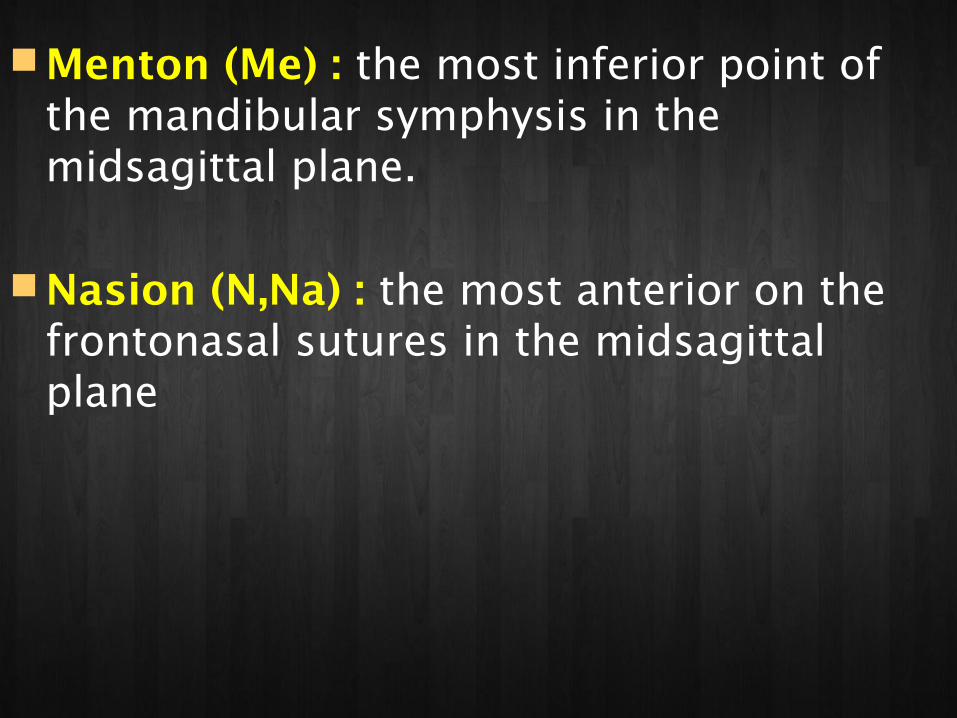
Menton (Me) : the most inferior point of the mandibular symphysis in the midsagittal plane.
Nasion (N,Na) : the most anterior on the frontonasal sutures in the midsagittal plane

Orbitale (Or) : the lowest point on the inferior margin of the orbit.
Pogonion (pog) : the most anterior point on the contour of the bony chin in the midsagittal plane
Porion (Po): the most superior point on the outline of the external auditory meatus (anatomic). The superior most point of the ear rods (machine porion) sometimes is used.

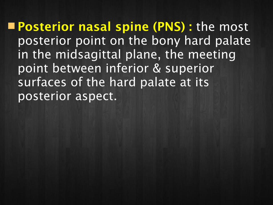
Posterior nasal spine (PNS) : the most posterior point on the bony hard palate in the midsagittal plane, the meeting point between inferior & superior surfaces of the hard palate at its posterior aspect.

Pterygomaxillary fissure (PTM,) : bilateral inverted tear drop shaped radiolucency whose anterior border represents the posterior surfaces of the tuberosities of the maxilla.
Sella (S) : the geometric centre of the pituitary fossa (sella turcica), determined by inspection – a constructed point in the midsagittal plane.

Conclusion
Broadbent’s gave us a three dimensional analysis, but orthodontics has remained preoccupied with the lateral view. The lateral view is easy to work with and the patient is also much more recognizable than in the frontal (P-A) view, especially with soft tissue enhancement.

Clinical orthodontics is yet to fully utilize Broadbent’s contribution.
We treat in three dimensions and the width dimension that are visualized on the frontal view are crucial in many cases.
