central ecmo - amazon s3€¦ · flow from central ecmo is directly from the outflow cannula into...
TRANSCRIPT
CENTRAL ECMO
WHEN AND HOW?
RANJIT JOHN, MDUNIVERSITY OF MINESOTA
Background How to do Case reports When to do Managing complications Post operative management strategies

46 year old male presented to outside hospital ER with severe shortness of breath and hypotension
Intubated, rapidly requiring pressorsCardiac arrest, requiring 15 minutes CPRPupils reportedly dilated, fixedOn arrival, SBP 60-70s, HR 140sExtremities cold, cyanotic, mottled WBC 18, Creatinine 3.4, INR 6, AST/ALT 5000s, Lactate 18Drips – Dopamine, Epinephrine, Neosynephrine
Bedside Echo – Severe biventricular dysfunction, EF <10%
CASE PRESENTATION

Treatment Options
1. IABP, Continue aggressive medical management
2. Take patient immediately to OR:Revascularization (CABG)
orTemporary mechanical support (temporary LVAD/RVAD – Centrimag
vs ECMO)or
Continuous flow ventricular device +/- RVAD
3. Take patient to Cath lab – percutaneous VAD support (Impella, Tandem heart, ECMO), treat underlying CAD
4. Do nothing, allow patient to die
Mechanical circulatory support has evolved markedly over recent years.
ECMO (extra corporeal membrane oxygenation) in particular has become more reliable with improving equipment, and increased experience, which is reflected in improving results.

What is the ideal device?
Uni- or biventricular support?
When to institute temporary support?
Where to institute it ? – ER/OR/Cath lab
For how long?
Ongoing Questions
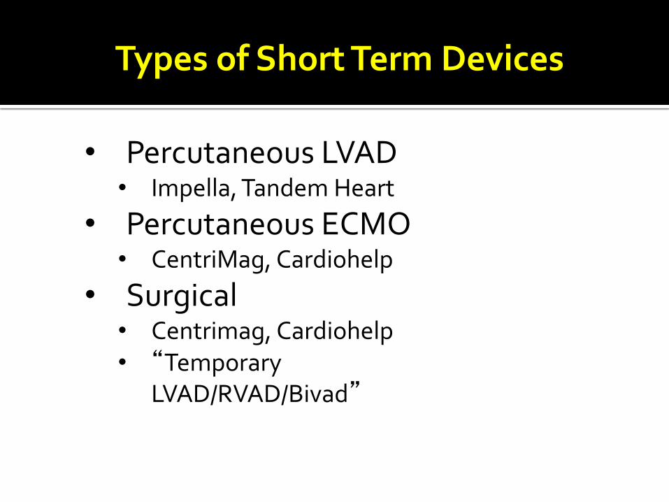
• Percutaneous LVAD• Impella, Tandem Heart
• Percutaneous ECMO• CentriMag, Cardiohelp
• Surgical • Centrimag, Cardiohelp• “Temporary
LVAD/RVAD/Bivad”
Types of Short Term Devices

Immediate circulatory support Maximum drainage without complicationsof venous obstruction Unobstructed inflow without distalIschemia Lowest risk of infection Mobilization of the patient when possible

Several considerations must be weighed: Likelihood of organ recovery.: only appropriate if disease
process is reversible with therapy and rest on ECMO Cardiac recovery: to either wait for further cardiac recovery
to allow implant of device (LVAD) or to list for transplantation.
Disseminated malignancy Advanced age Known severe brain injury Unwitnessed cardiac arrest or cardiac arrest of prolonged
duration. Technical contraindications to consider: aortic dissection or
aortic incompetence
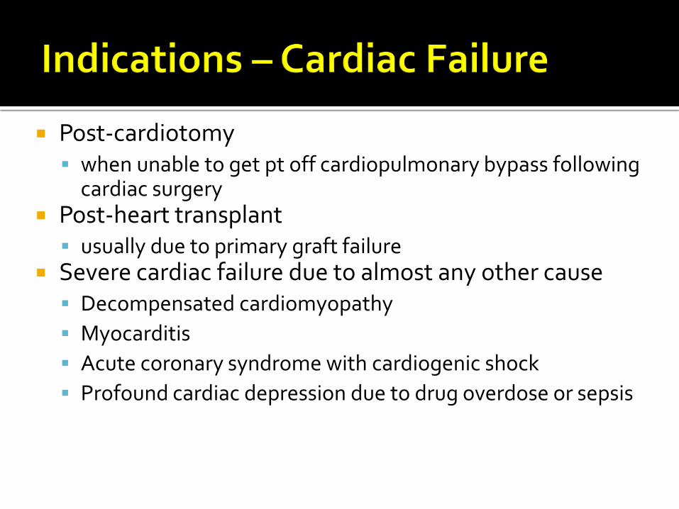
Post-cardiotomy when unable to get pt off cardiopulmonary bypass following
cardiac surgery Post-heart transplant usually due to primary graft failure
Severe cardiac failure due to almost any other cause Decompensated cardiomyopathy Myocarditis Acute coronary syndrome with cardiogenic shock Profound cardiac depression due to drug overdose or sepsis

ARTERIAL OPTIONS
Femoral artery– Perc / Retrograde, Leg ischemia, Infection Axillary artery
– Good flow, less CVA / Graft, Arm overcirculation
Aorta– Great flow / Sternotomy, infection
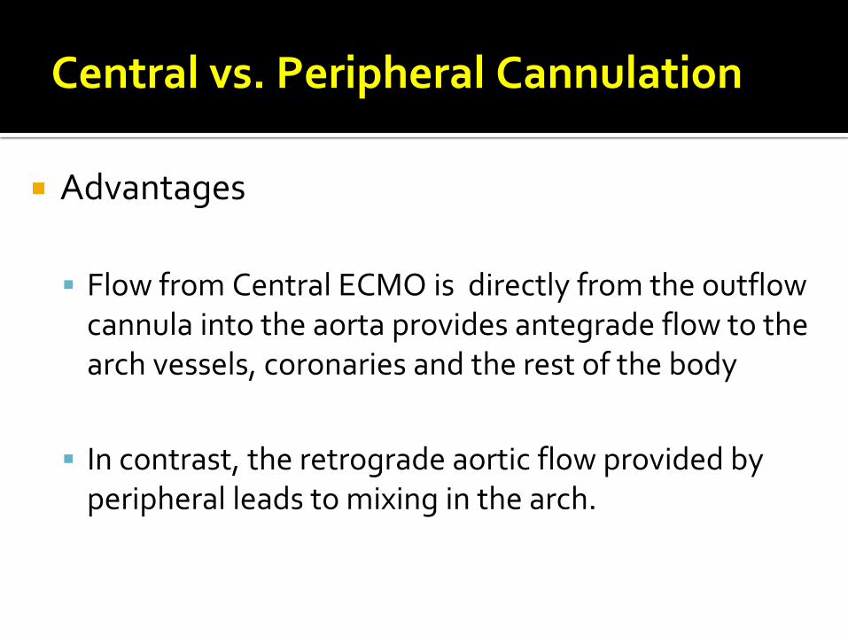
Central vs. Peripheral Cannulation
Advantages
Flow from Central ECMO is directly from the outflow cannula into the aorta provides antegrade flow to the arch vessels, coronaries and the rest of the body
In contrast, the retrograde aortic flow provided by peripheral leads to mixing in the arch.

Central vs. Peripheral Cannulation
Disadvantages
Previously insertion of central ECMO required leaving chest open to allow the cannulae to exit.▪ Increased the risk of bleeding and infection ▪ Newer cannulae are designed to be tunneled through the
subcostal abdominal wall allowing the chest to be completely closed.
Central cannula are costly (no longer??)
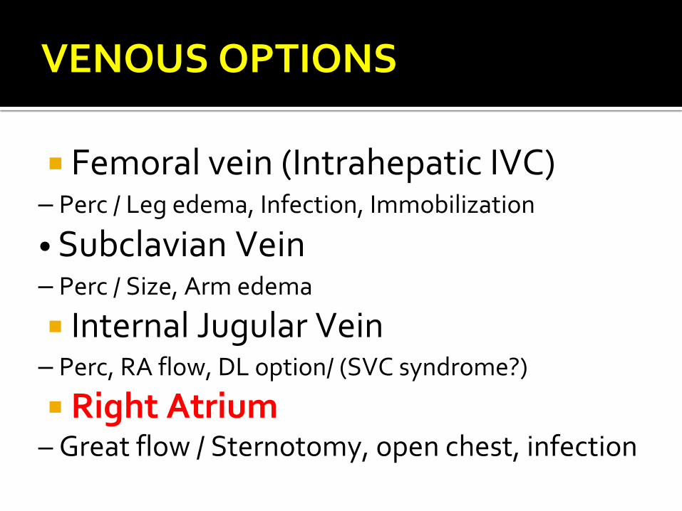
VENOUS OPTIONS
Femoral vein (Intrahepatic IVC)– Perc / Leg edema, Infection, Immobilization
• Subclavian Vein– Perc / Size, Arm edema
Internal Jugular Vein– Perc, RA flow, DL option/ (SVC syndrome?)
Right Atrium– Great flow / Sternotomy, open chest, infection
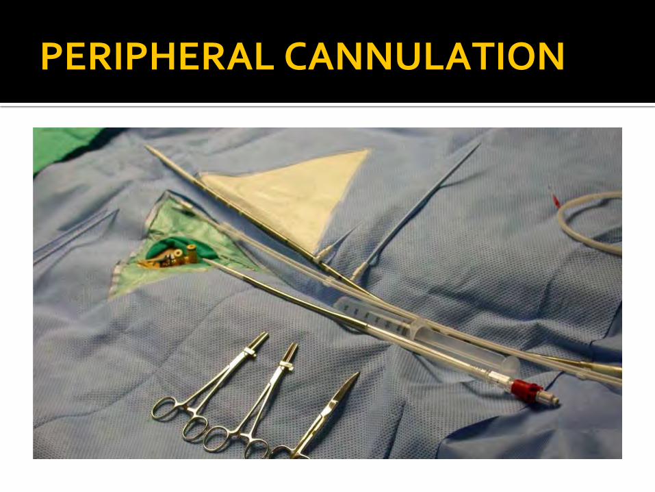
PERIPHERAL CANNULATION
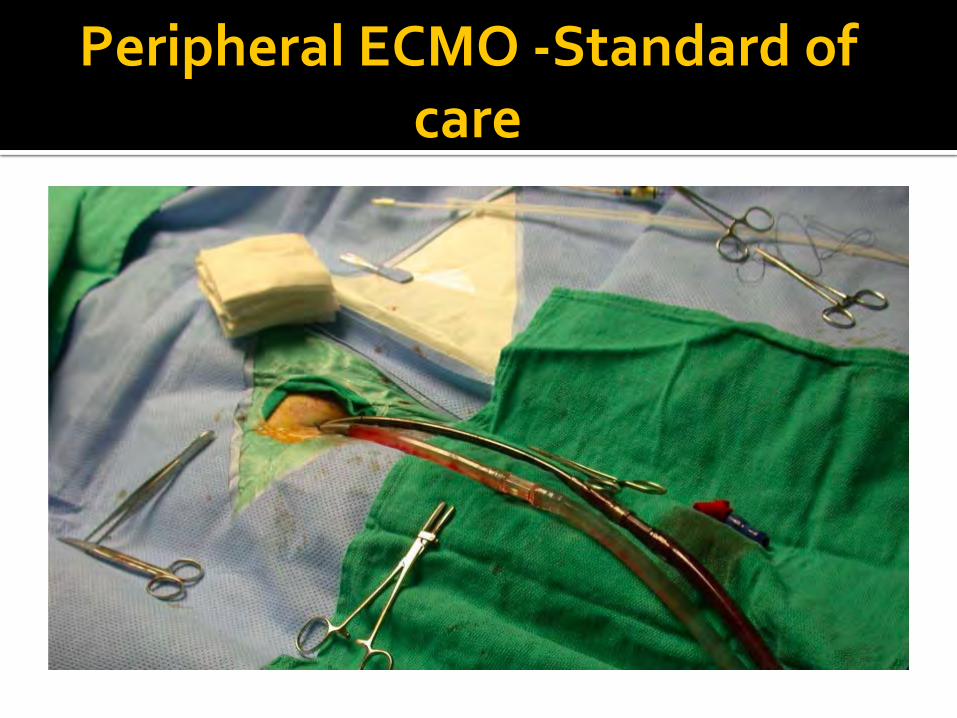
Peripheral ECMO -Standard of care

Central ECMO Cannulation
CASE REPORT # 1
56 year old female with history of dilated cardiomyopathy admitted with recurrent VT/VF
Hemodynamically unstable, refractory to medical therapy
Placement of peripheral ECMO via fem fem cannulation with distal perfusion cannula
Despite this, ischemic leg in 12 hours Ultrasound of contralateral LE vessels - PVD

Central ECMO placed with median sternotomy and aortic and right atrial cannulation
Concomitant removal of peripheral cannula, repair and embolectomy of right femoral artery
Eventually bridged to permanent LVAD.

CASE REPORT # 2
32 year old male transferred with cardiogenic shock with hemodynamic collapse
Peripheral ECMO placed via femoral vessels 8 hours later, ECHO shows LV severely
distended, pulmonary edema Plan for Impella 2.5; echo at time of Impella
placement shows clot in aortic root Options ?

Right mini thoracotomy and placement of LV vent through right superior pulmonary vein
Echo showed moderate LV decompression and stable ECMO flows
24 hours later, severe hemolysis likely from LV vent Median sternotomy, with Centrimag LVAD and RVAD
placement, with oxygenator in RVAD circuit –supported for 2 weeks
Subsequently bridged to permanent LVAD LV recovery in 6 months, with LVAD explant Currently doing well, normal EF
CASE REPORT # 3
58 year old male transferred with cardiogenic shock on multiple pressors
Hypotensive, intubated Severe aortic regurgitation secondary to
aortic valve endocarditis with enterococcus Options ?
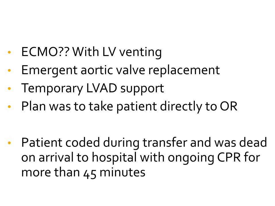
• ECMO?? With LV venting• Emergent aortic valve replacement• Temporary LVAD support• Plan was to take patient directly to OR
• Patient coded during transfer and was dead on arrival to hospital with ongoing CPR for more than 45 minutes

58 year old male transferred with cardiogenic shock on multiple pressors post emergent CABG
Hypotensive, intubated, open chest Acute renal and hepatic failure Echo – severe biventricular function Options ?

• Central ECMO via aortic and right atrial cannulation
• Wash out chest• Inspect grafts; Inspect all surgical sites• Chest closed in 3 days• Prolonged recovery and discharged to TCU

CASE REPORT # 5
Uneventful heart transplantation with primary graft failure immediately post tx
Usual maneuvers – nitric oxide, optimizing drips, AV pacing, IABP – still significant graft dysfunction with significant RV failure
Temporary RVAD, now LV failure Central ECMO Recovery in 5 days with subsequent
discharge

When to do CENTRAL ECMO Post- cardiotomy shock – biventricular failure
and/or hypoxia with respiratory failure Post-heart transplant primary graft failure Cardio-respiratory failure in patients with
severe peripheral vascular disease Ongoing peripheral ECMO with Limb ischemia Refractory groin bleeding/hematoma Rising lactate? – inadequate flows/ cannula size Retroperitoneal hematoma Need for LV venting ?
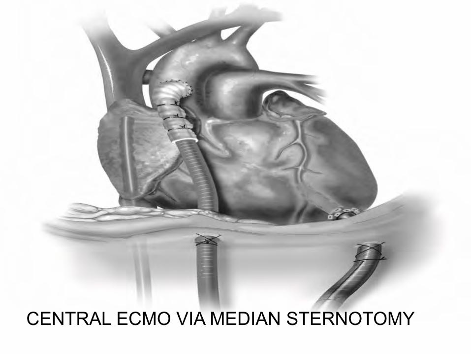
CENTRAL ECMO VIA MEDIAN STERNOTOMY

CENTRAL ECMO VIA Right MINI THORACOTOMY
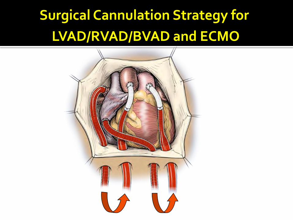
Surgical Cannulation Strategy for LVAD/RVAD/BVAD and ECMO

Surgical Cannulation Strategy for LVAD/RVAD/BVAD and ECMO
ECMO (CENTRAL)Outflow – ascending aorta
▪ ? LV apical cannula placed into aorta distal to aortic valve
Inflow – right atrium▪ Right atrial appendage▪ Body of right atrium
Surgical Cannulation Strategy for LVAD/RVAD/BVAD and ECMO
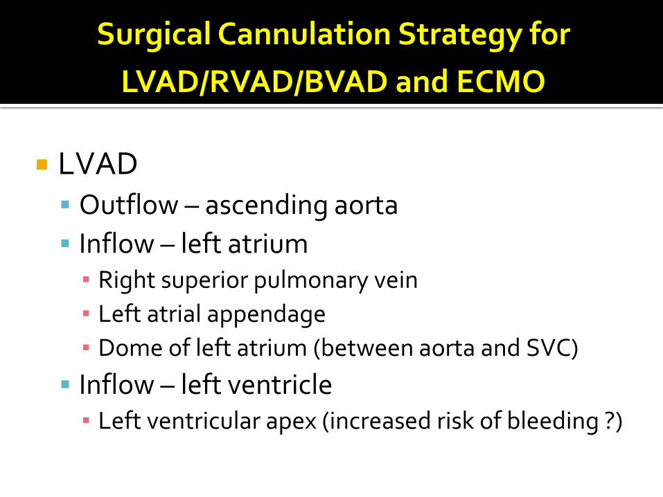
LVAD Outflow – ascending aorta Inflow – left atrium
▪ Right superior pulmonary vein▪ Left atrial appendage▪ Dome of left atrium (between aorta and SVC) Inflow – left ventricle
▪ Left ventricular apex (increased risk of bleeding ?)
Surgical Cannulation Strategy forLVAD/RVAD/BVAD and ECMO
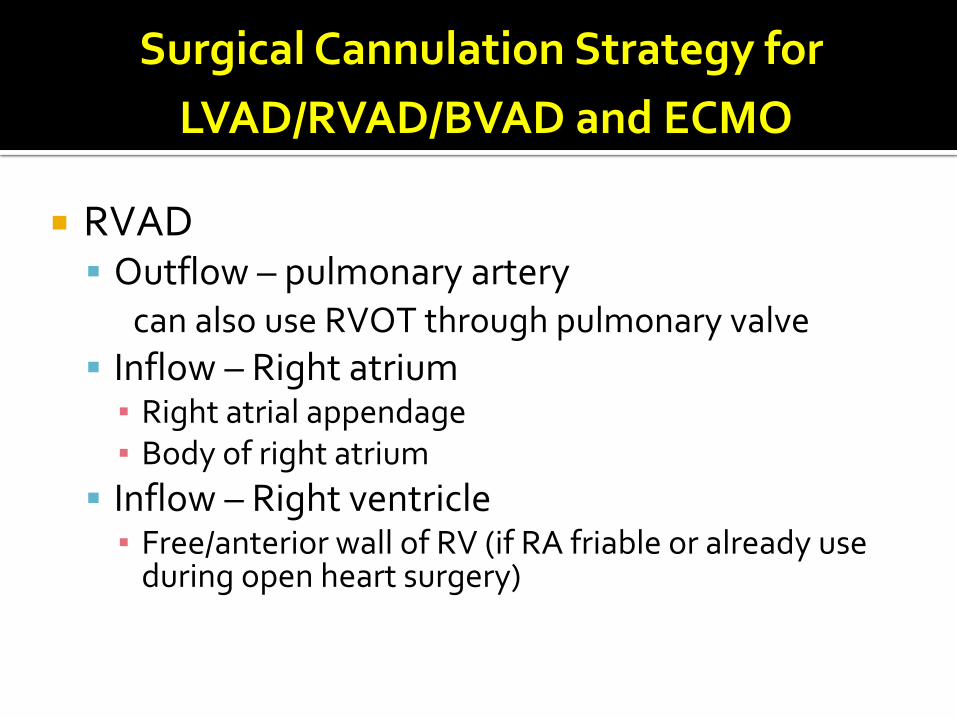
RVAD Outflow – pulmonary artery
can also use RVOT through pulmonary valve Inflow – Right atrium
▪ Right atrial appendage▪ Body of right atrium
Inflow – Right ventricle▪ Free/anterior wall of RV (if RA friable or already use
during open heart surgery)
Surgical Cannulation Strategy for LVAD/RVAD/BVAD And ECMO
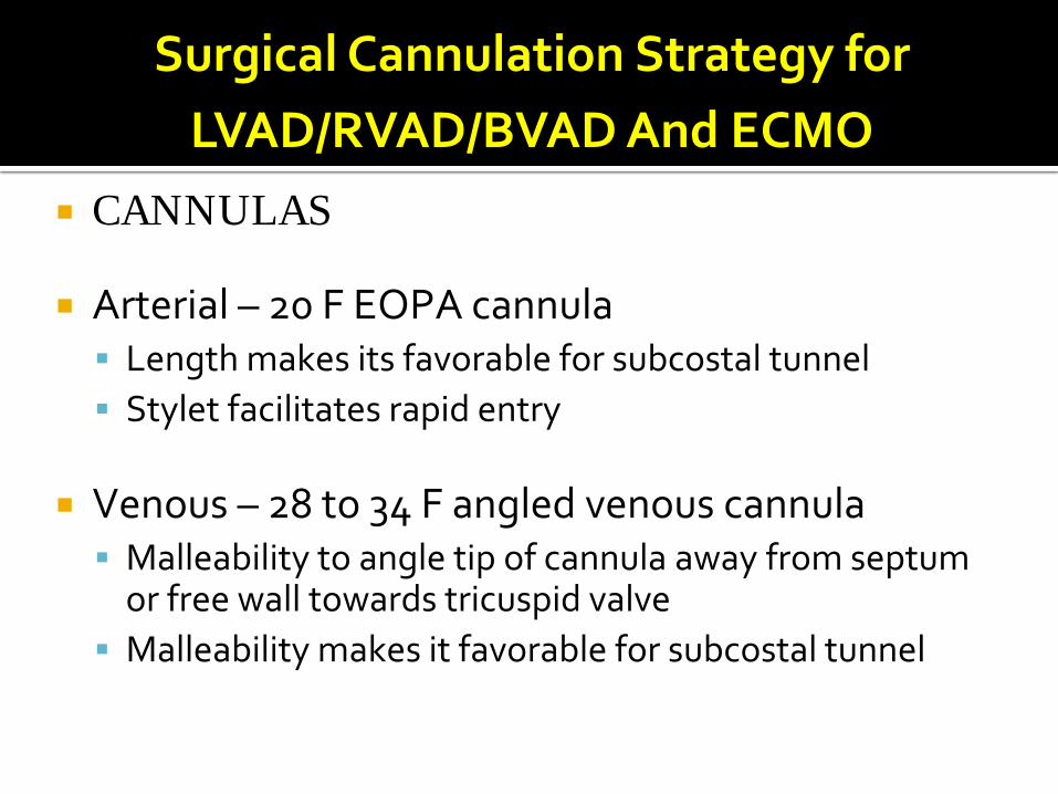
CANNULAS
Arterial – 20 F EOPA cannula Length makes its favorable for subcostal tunnel Stylet facilitates rapid entry
Venous – 28 to 34 F angled venous cannula Malleability to angle tip of cannula away from septum
or free wall towards tricuspid valve Malleability makes it favorable for subcostal tunnel
Surgical Cannulation Strategy for LVAD/RVAD/BVAD And ECMO
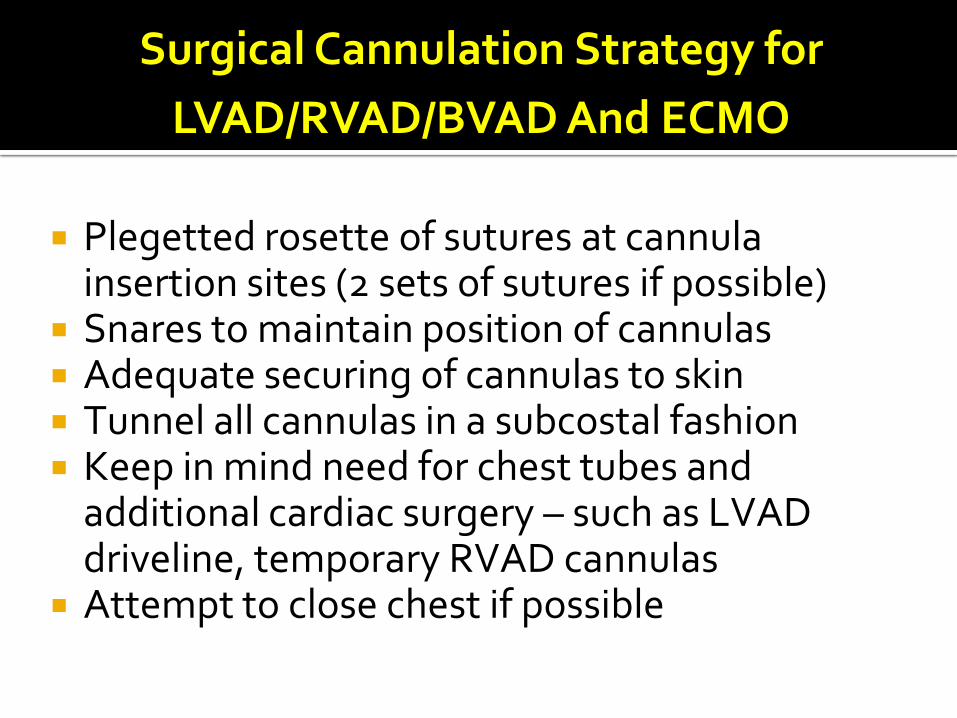
Plegetted rosette of sutures at cannula insertion sites (2 sets of sutures if possible)
Snares to maintain position of cannulas Adequate securing of cannulas to skin Tunnel all cannulas in a subcostal fashion Keep in mind need for chest tubes and
additional cardiac surgery – such as LVAD driveline, temporary RVAD cannulas
Attempt to close chest if possible
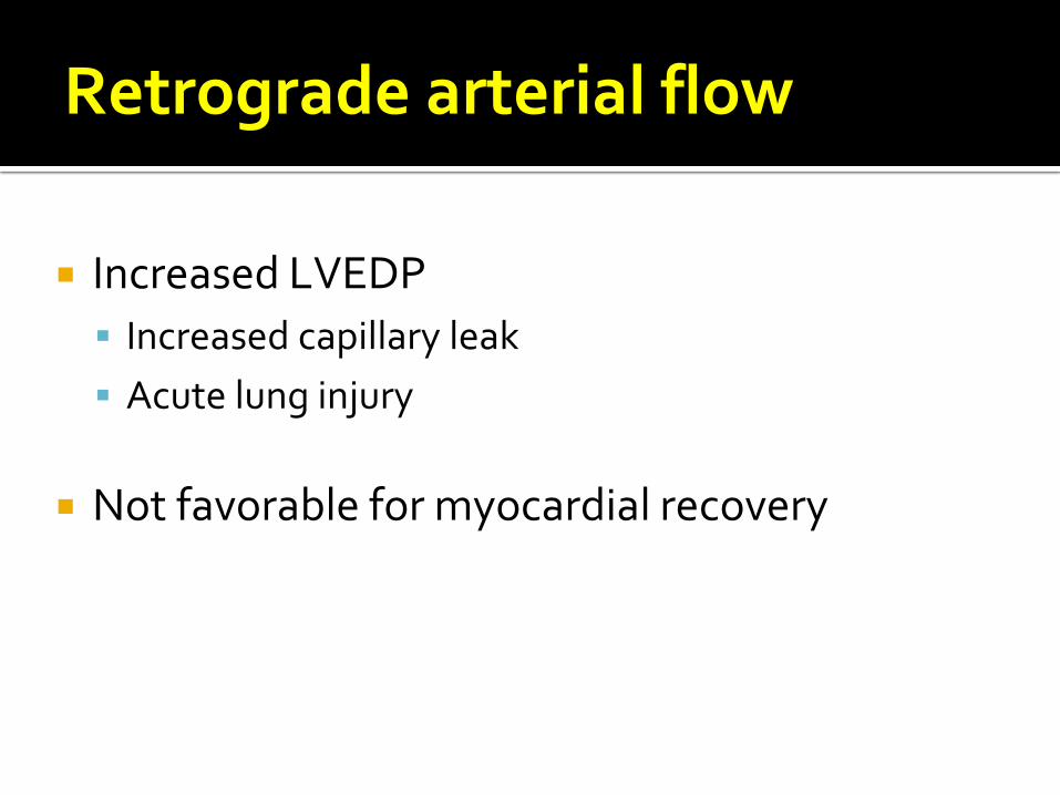
Retrograde arterial flow
Increased LVEDP Increased capillary leak Acute lung injury
Not favorable for myocardial recovery

Pulmonary Edema on ECMO
Central cannulation does not prevent it LA/LV venting – minithoracotomy LV vent Do Echo 6-24 hours after initiation of ECMO to
look for LV DISTENTION Pulmonary edema on CXR is an early clue Attempt inotropes to facilitate LV ejection
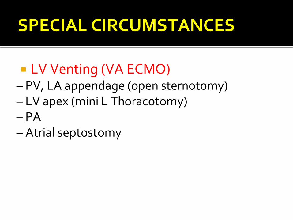
SPECIAL CIRCUMSTANCES
LV Venting (VA ECMO)– PV, LA appendage (open sternotomy)– LV apex (mini L Thoracotomy)– PA– Atrial septostomy
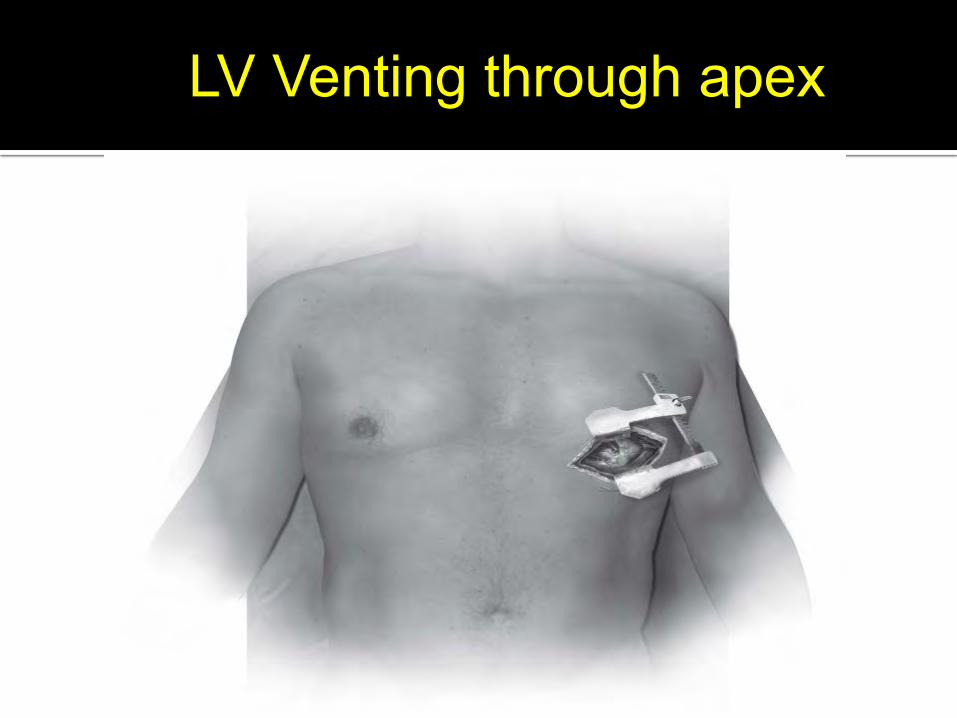
LV Venting through apex
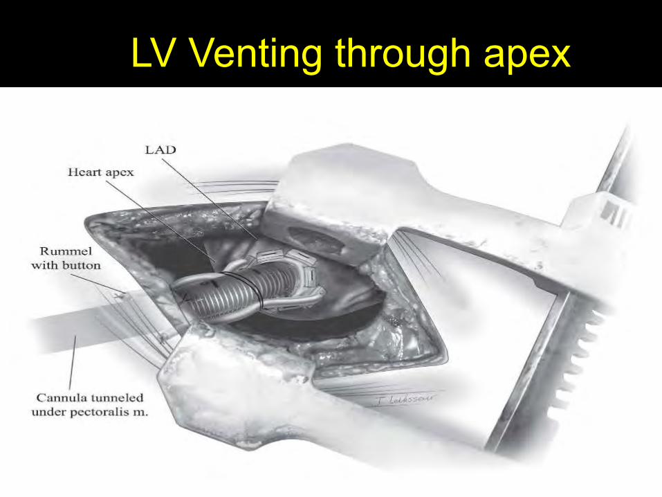
LV Venting through apex
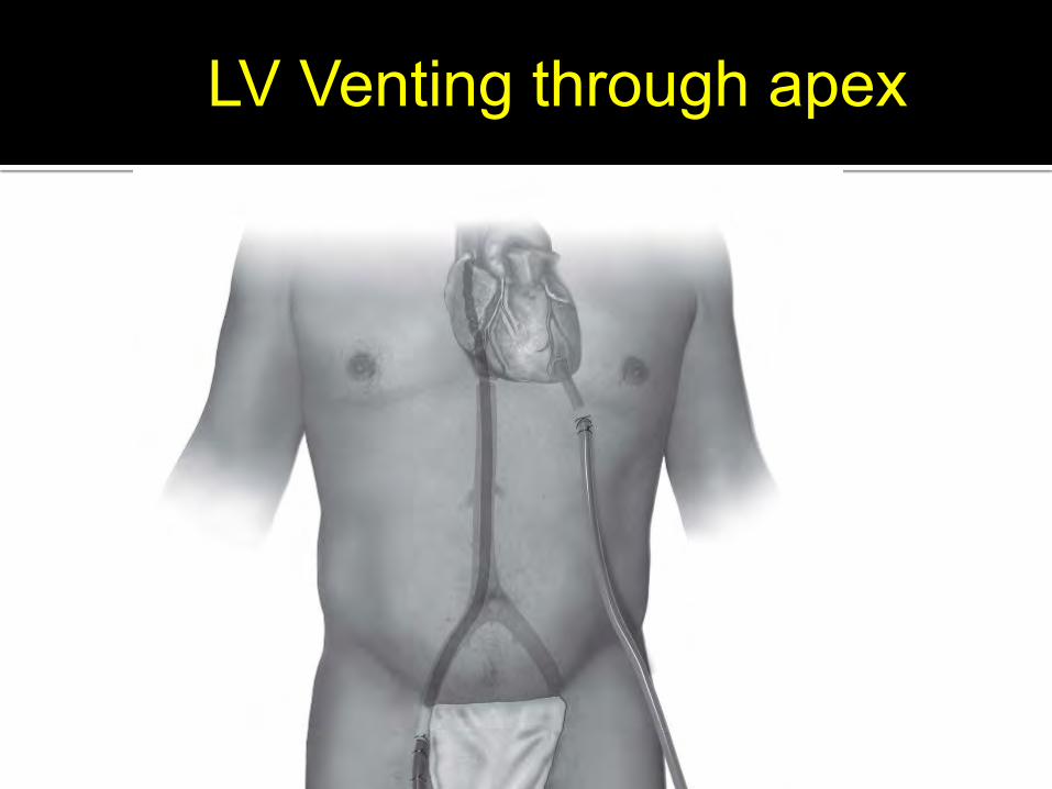
LV Venting through apex
Central ECMO Complications
1) Bleeding 2) Infection3) Neurologic sequelae
BLEEDING
Bleeding/Hemolysis Out of proportion to the degree of coagulopathy and patient
platelet count
Coagulopathy Continuous activation of contact and fibrinolytic
systems by the circuit Consumption and dilution of factors within minutes of
initiation of ECMO
BLEEDING – Central ECMO
Chest re-exploration Packing Inspection of cannulation sites with
reinforcement Common culprit after 24-72 hours is right
atrial cannula site – use of an additional suture tie around cannula including right atrial cuff
Aggressive replacement of FFP, platelets
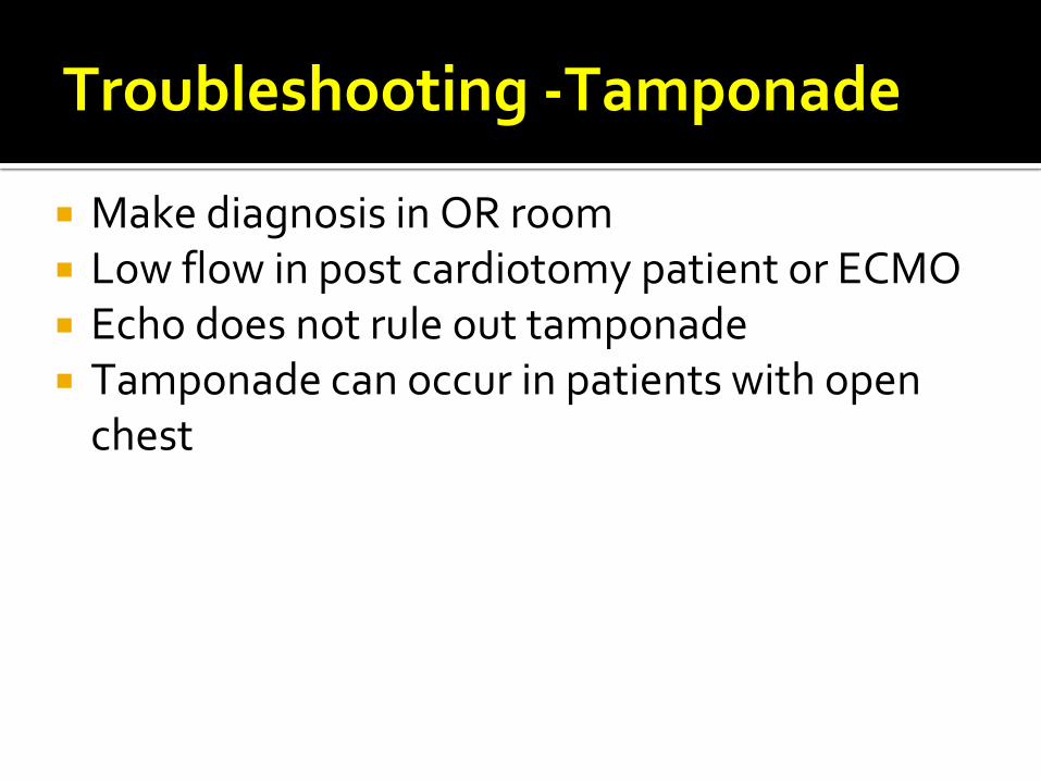
Troubleshooting -Tamponade
Make diagnosis in OR room Low flow in post cardiotomy patient or ECMO Echo does not rule out tamponade Tamponade can occur in patients with open
chest
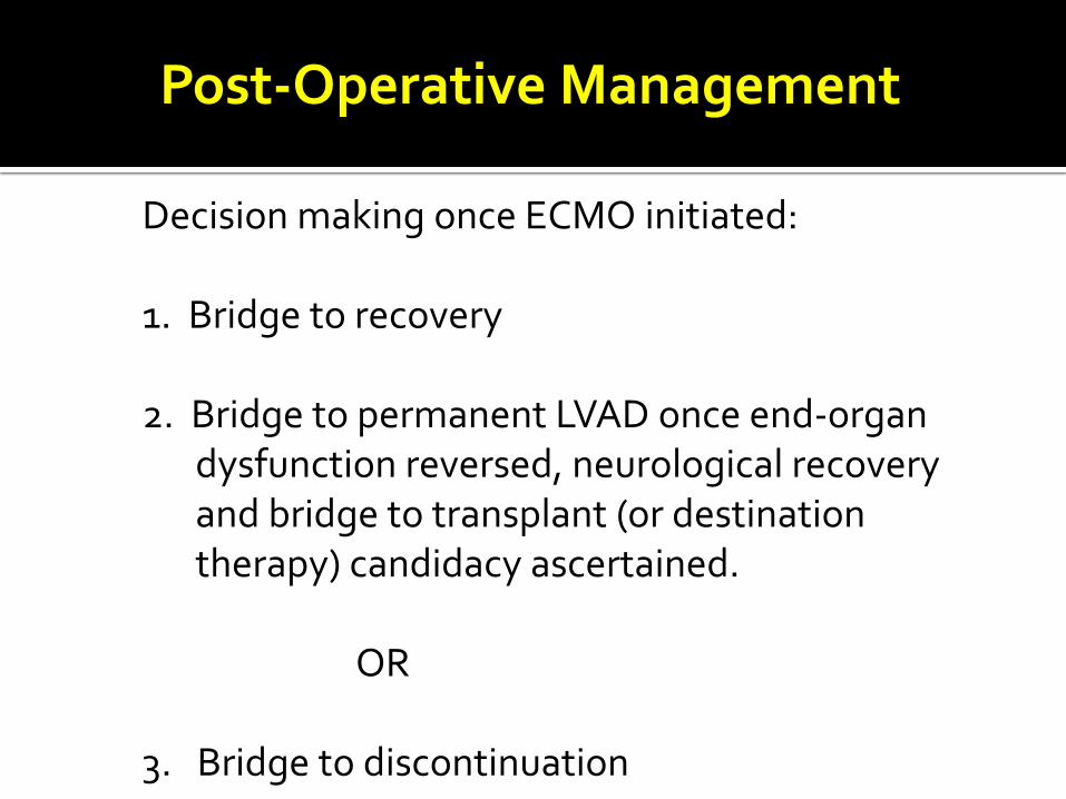
Post-Operative Management
Decision making once ECMO initiated:
1. Bridge to recovery
2. Bridge to permanent LVAD once end-organ dysfunction reversed, neurological recovery and bridge to transplant (or destinationtherapy) candidacy ascertained.
OR
3. Bridge to discontinuation
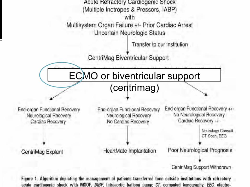
ECMO or biventricular support (centrimag)

Post-Operative Management
• Wean off pressors and inotropes as tolerated
• Maintain flows to keep MAPs 65-90mmHg, UOP >30cc/hr, lactate down trending, Sats > 90%
• Transfusion HgB> 8gm/dL, Platelets > 80
• CI >2.2 -2.4; decompress but maintain or encourage contractility

Post-Operative Management
•Heparin to maintain ACT 160 to 200 sec only when no bleeding issues exist (can hold up to 48 hours)
•Liberal use of diuretics or CVVHD
•Prophylactic antibiotics
•Early and aggressive nutritional support
•Extensive and frequent family discussions
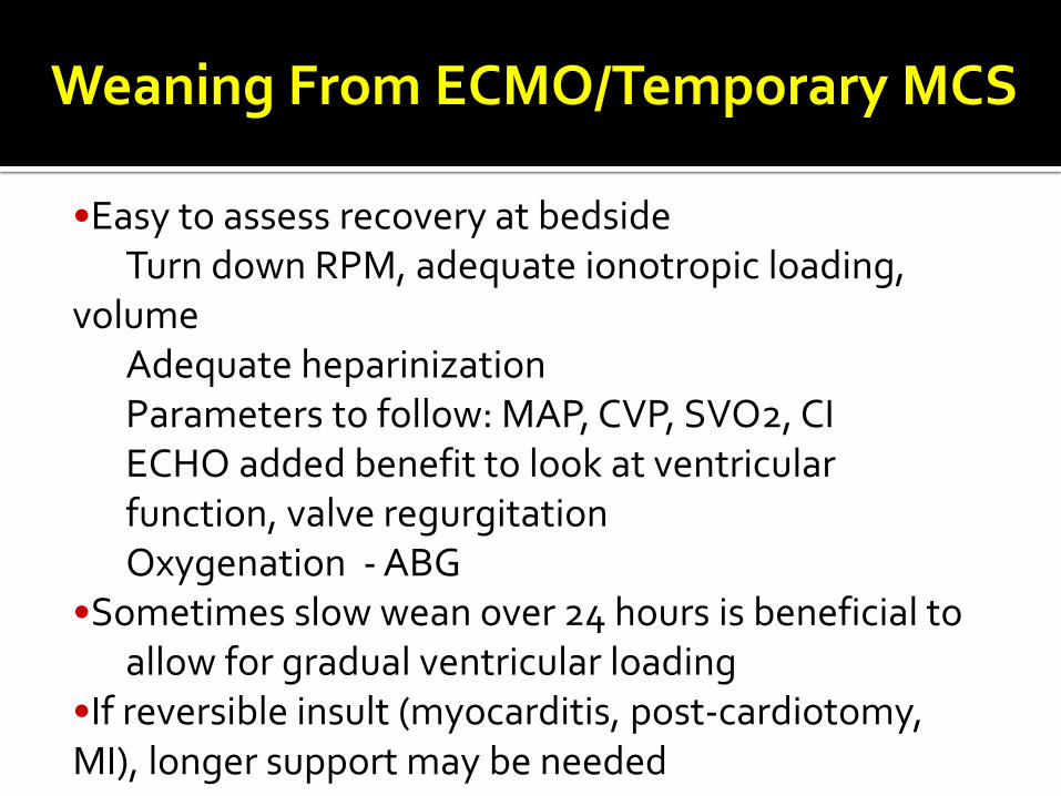
•Easy to assess recovery at bedsideTurn down RPM, adequate ionotropic loading,
volumeAdequate heparinizationParameters to follow: MAP, CVP, SVO2, CIECHO added benefit to look at ventricular function, valve regurgitationOxygenation - ABG
•Sometimes slow wean over 24 hours is beneficial to allow for gradual ventricular loading
•If reversible insult (myocarditis, post-cardiotomy, MI), longer support may be needed with
l i i ) d h ld i l
Weaning From ECMO/Temporary MCS
•Usually OR, one final TEE assessment•Heparinize, remove cannulas and clamp vessels•Allow for back bleeding from aortic and atrial cannulation sites, as clots can form around cannulas•Pursestring or more commonly, direct repair •Thorough irrigation of chest with antibiotic solutions•Consider IABP support, ionotropes and volume
Central ECMO - Decannulation
•Well defined indications for Central ECMO in current era•Can be done safely with easily reproducible surgical techniques•Should be done expeditiously in the post-cardiotomy situation •Critically ill patients can be transferred safely with central ECMO•Central ECMO is not recommended for all patients doing poorly on peripheral ECMO
Central ECMO - CONCLUSIONS