cellular/molecular achievinghigh ... · pdf filecellular/molecular...
TRANSCRIPT

Cellular/Molecular
Achieving High-Frequency Optical Control of SynapticTransmission
Skyler L. Jackman, Brandon M. Beneduce, Iain R. Drew, and Wade G. RegehrDepartment of Neurobiology, Harvard Medical School, Boston Massachusetts 02115
The optogenetic tool channelrhodopsin-2 (ChR2) is widely used to excite neurons to study neural circuits. Previous optogenetic studiesof synapses suggest that light-evoked synaptic responses often exhibit artificial synaptic depression, which has been attributed to eitherthe inability of ChR2 to reliably fire presynaptic axons or to ChR2 elevating the probability of release by depolarizing presynaptic boutons.Here, we compare light-evoked and electrically evoked synaptic responses for high-frequency stimulation at three synapses in the mousebrain. At synapses from Purkinje cells to deep cerebellar nuclei neurons (PC3DCN), light- and electrically evoked synaptic currents wereremarkably similar for ChR2 expressed transgenically or with adeno-associated virus (AAV) expression vectors. For hippocampalCA33CA1 synapses, AAV expression vectors of serotype 1, 5, and 8 led to light-evoked synaptic currents that depressed much more thanelectrically evoked currents, even though ChR2 could fire axons reliably at up to 50 Hz. The disparity between optical and electricalstimulation was eliminated when ChR2 was expressed transgenically or with AAV9. For cerebellar granule cell to stellate cell (grc3SC)synapses, AAV1 also led to artificial synaptic depression and AAV9 provided superior performance. Artificial synaptic depression alsooccurred when stimulating over presynaptic boutons, rather than axons, at CA33CA1 synapses, but not at PC3DCN synapses. Thesefindings indicate that ChR2 expression methods and light stimulation techniques influence synaptic responses in a neuron-specificmanner. They also identify pitfalls associated with using ChR2 to study synapses and suggest an approach that allows optogenetics to beapplied in a manner that helps to avoid potential complications.
Key words: AAV; channelrhodopsin; optogenetics; short-term plasticity; synapse
IntroductionThe genetically encoded light-gated cation channel channel-rhodopsin-2 (ChR2) is widely used both in vitro and in vivo toinvestigate neural circuits and to characterize synapses. ChR2 canbe expressed in specific neuronal populations by the targetedinjection of viral vectors and by using Cre-expressing driver lines(Fenno et al., 2011; Madisen et al., 2012). Light can then be usedto control neural activity with high temporal and spatial precision(Boyden et al., 2005). In this way, ChR2 can be used to activatedefined presynaptic inputs selectively, enabling characterizationof synapses that are not amenable to study by conventional ap-proaches (Petreanu et al., 2007; Xia et al., 2011; Mathews et al.,2012) because, for many synapses, paired recording or selectiveelectrical stimulation of specific presynaptic inputs is impractical.
The use of ChR2 for characterization of synapses, however,has been limited by a perception that ChR2 does not allow reli-
able synaptic activation at the high firing frequencies that occurphysiologically. Optically evoked synaptic currents have beenshown to depress more than those evoked electrically (Zhang andOertner, 2007; Cruikshank et al., 2010; Olsen et al., 2012), but thereason for this disparity is not known. It has been proposed thatChR2 desensitization may prevent reliable action potential gen-eration during high-frequency stimulation (Olsen et al., 2012).Alternatively, it is thought that the slow kinetics of ChR2 couldbroaden action potentials and increase the initial probability ofrelease (Zhang and Oertner, 2007), particularly when the presyn-aptic boutons are directly activated with light.
For many optogenetic approaches, it is vital to identify andovercome limitations of ChR2 to activate synapses at physiolog-ical frequencies. One approach to overcoming limitations in lightactivation has been to engineer new variants of ChR2 with lessdesensitization and faster kinetics (Nagel et al., 2005; Lin et al.,2009; Gunaydin et al., 2010). Here, we evaluate the ability ofChR2 to activate synaptic inputs at high frequencies and deter-mined whether widely available ChR2 variants can be used toactivate synapses at physiological frequencies. We comparedelectrically and optically evoked responses at three central syn-apses and found that the performance of ChR2 was synapsedependent, but there were important general principles. Stimu-lating axons with ChR2 reliably triggered presynaptic action po-tentials, yet light-evoked synaptic responses showed artificialdepression in some cases when certain AAV serotypes were usedto drive expression. When ChR2 was expressed transgenically or
Received Nov. 5, 2013; revised April 25, 2014; accepted April 30, 2014.Author contributions: S.L.J. and W.G.R. designed research; S.L.J., B.M.B., and I.R.D. performed research; S.L.J.
analyzed data; S.L.J. and W.G.R. wrote the paper.This work was supported by the National Institutes of Health Grant R01 NS032405 to W.G.R., Training Grant NS007484
to S.L.J, and a Leonard and Isabelle Goldenson Fellowship to S.L.J. We thank Kimberly McDaniels for help with mousebreeding; Justin Brousseau, Kaytee Flick, and Hannah Goulart for stereotaxic injections; Jasmine Vazquez for figure illustra-tions; and the University of Pennsylvania Vector Core, Arpiar Saunders, and Bernardo Sabatini for providing AAV vectors.
The authors declare no competing financial interests.Correspondence should be addressed to Wade G. Regehr, Department of Neurobiology, Harvard Medical School,
220 Longwood Avenue, Boston MA 02115. E-mail: [email protected]:10.1523/JNEUROSCI.4694-13.2014
Copyright © 2014 the authors 0270-6474/14/347704-11$15.00/0
7704 • The Journal of Neuroscience, May 28, 2014 • 34(22):7704 –7714
with AAV serotype 9, light-evoked responses more closely resem-bled responses evoked by conventional electrical stimulation. Al-though we have identified potential complications associatedwith using ChR2, our findings suggest a strategy to avoid suchproblems when using optogenetics to study synapses.
Materials and MethodsChR2 expression. All animals were handled in accordance with federalguidelines and protocols approved by Harvard University. Mice of eithersex of the following strains were used: wild-type (WT) C57BL/6N(Charles River Laboratories), as well as transgenic lines (Jackson Labo-ratories) Thy1-ChR2-YFP (B6.Cg-Tg(Thy1-COP4/EYFP)18Gfng/J),flox-hChR2 (B6;129S-Gt(ROSA)26Sor tm32(CAG-COP4*H134R/EYFP)Hze/J),PCP2-Cre (B6.Cg-Tg(Pcp2-cre)3555Jdhu/J), CaMKII-Cre (B6.Cg-Tg(Camk2a-cre)T29-1Stl/J), and the cerebellar granule-cell-specific �6-Cre line (Aller et al., 2003). The following AAV vectors were obtainedfrom the University of Pennsylvania Vector Core: AAV1-CAG.ChR2-Venus.WPRE.SV40, AAV5-CAG.ChR2-Venus.WPRE.SV40 and AAV9-CAG.ChR2-Venus.WPRE.SV40, AAV1-hSyn.hChR2(H134R)-EYFP.WPRE.hGH,AAV1-CMV.PI.Cre.rBG,AAV1-CB7.CI.TurboRFP.WPRE.rBG. AAV8.EF1a.FAS.ChR2(H134R)-mCherry was provided byBernardo Sabatini (Saunders et al., 2012).
Mice at postnatal day 16 (P16) to P30 were anesthetized by intraperi-toneal injection of a mixture of ketamine/xylazine/acepromazine (100/10/3 mg/kg) supplemented with 1– 4% isoflurane at 0.6 –1.4 L/min. Totarget expression to lobule V of the cerebellar cortex, a small craniotomywas made 1.35 mm caudal from lambda at the midline. Using a stereo-taxic instrument (Kopf Instruments), a fine glass capillary needle waslowered 0.8 mm below the surface of the brain and then slowly retractedto 0.7 mm after 3 min. To target expression to the hippocampal CA3region, injection coordinates were 2.69 mm rostral and 3 mm lateral(left) from lambda; the needle was lowered 2.9 mm and then retracted to2.8 mm. Viruses were diluted with PBS and 0.2 �l (cerebellum) or 1 �l(hippocampus) of virus suspension was delivered at a rate of 0.1 �l/minusing a microliter syringe (Hamilton) and a microsyringe pump con-troller (WPI). Five to 10 min after bolus injection, the needle wasslowly retracted. The scalp incision was sutured and postinjectionanalgesics (buprenophrine, 0.05 mg/kg) were administered subcuta-neously for 48 h.
ChR2 expression levels were sufficient to optically evoke synaptic re-sponses from PCs and CA3 cells within 1 week of injection. Due to thedifficulty of recording from the deep cerebellar nuclei (DCN) in olderanimals, all recordings were performed 10 –14 d after injection. Hip-pocampal recordings could be made in older animals, so we tested alarger range of wait times (1–7 weeks after injection), but found that theproperties of optically evoked transmission did not vary significantlywith wait time. ChR2 expression was lower in cerebellar granule cells,for which we found that �3 weeks were required to optically evokesynaptic responses; therefore, we performed experiments 3– 4 weeksafter injection.
Acute slice preparation. For hippocampal and stellate cell recordings,animals were deeply anesthetized using isoflurane and decapitated.Brains were removed and placed in an ice-cold dissecting solution con-taining the following (in mM): 82.7 NaCl, 65 sucrose, 23.8 NaHCO3, 23.7glucose, 6.8 MgCl2, 2.4 KCl, 1.4 NaH2PO4, and 0.5 CaCl2. Then, 300-�m-thick transverse hippocampal slices or 250-�m-thick transverse cer-ebellar slices were made in ice-cold dissecting solution. In hippocampalslices, a cut was made in stratum radiatum between CA3 and CA1 toprevent recurrent excitation. Slices were incubated at 32°C for 30 min inartificial CSF (ACSF) containing the following (in mM): 125 NaCl, 26NaHCO3, 25 glucose, 2.5 KCl, 2 CaCl2, 1.25 NaH2PO4, and 1 MgCl2,adjusted to 315 mOsm. For DCN recordings, animals were anesthetizedby intraperitoneal injection of ketamine/xylazine/acepromazine, intrac-ardially perfused with 32°C ACSF, and then 300-�m-thick sagittal cere-bellar slices were made in warm ACSF. Slices were incubated at 32°C for30 min in ACSF with 1.5 mM CaCl2 to facilitate comparison with previousstudies (Person and Raman, 2012) and allowed to equilibrate to roomtemperature for �30 min. Experiments were performed at 33 � 1°C with
flow rates of 4 ml/min. After recording, slices were imaged with an AxioImager 2 fluorescent microscope (Zeiss) to assess the extent of AAV-mediated expression and images were analyzed using ImageJ.
Electrophysiology. Data were acquired using a Multiclamp 700B ampli-fier (Molecular Devices) digitized at 20 kHz with an ITC-18 (Instrutech).Voltage-clamp recordings were low-pass filtered at 2 kHz. Acquisitionwas controlled by custom software written in IgorPro (generously pro-vided by Matthew Xu-Friedman, SUNY Buffalo). Whole-cell recordingswere obtained using patch pipettes (3– 6 M�) pulled from borosilicatecapillary glass (WPI) with a Sutter P-97 horizontal puller. To recordEPSCs in hippocampal CA1 and stellate cells, inhibition was blockedwith picrotoxin (20 �M), cells were voltage clamped at �60 mV, andpatch pipettes were filled with an internal recording solution containingthe following (in mM): 110 CsCl, 35 CsF, 10 EGTA, 10 HEPES, and 2QX-314, pH 7.2, 315–320 mOsm. For current-clamp recordings, theinternal solution contained the following (in mM): 150 K-gluconate, 3KCl, 10 HEPES, 0.5 EGTA, 3 MgATP, 0.5 GTP, 5 phosphocreatine-tris2,and 5 phosphocreatine-Na2, with the pH adjusted to 7.2 with NaOH. Torecord synaptically driven firing in CA1 neurons, inhibition was blockedwith picrotoxin (20 �M); to record ChR2-elicited spikes in CA3 neurons,excitation was also blocked with NBQX (5 �M) and CPP (2.5 �M). IPSCswere recorded in DCN neurons using the same K-gluconate internalsolution, cells were voltage clamped at 0 mV, and NBQX (5 �M), CPP(2.5 �M), and strychnine (2 �M) were included in the bath solution. Seriesresistance was monitored in voltage-clamp recordings with a 2 mV hy-perpolarizing pulse. For both electrical and optical stimuli, four to 10trials were typically conducted for each interstimulus interval andvoltage-clamp recordings were averaged over trials. For extracellularstimulation, glass monopolar electrodes (0.5–1 M�) were filled withACSF and controlled with a stimulus isolation unit (A360; WPI). Elec-trical stimulus artifacts were deleted for display. Averaged paired-pulseand train EPSC data are displayed with double exponential or polyno-mial curves fit in IgorPro.
ChR2 stimulation. Slices expressing ChR2 were stored in low light. Tostimulate ChR2, a 100 mW DPSS analog-controllable 473 nm blue laser(OptoEngine) was coupled through the excitation pathway of a BX51WIupright microscope (Olympus). Laser light was focused onto slicesthrough a 60� water-immersion objective to produce an 80 �m maxi-mum diameter spot. The spot size was adjusted using a dual-slit lightmodulator (Till Photonics). For hippocampal EPSC recordings, the areaof the laser illumination and the intensity of the electrical stimulationwere adjusted to produce initial EPSCs of �200 pA. To assess the laserintensity at the sample, an optical power meter (Ophir; Vega) was used tomeasure the absolute steady-state intensity below the objective. A pho-todiode was then used to measure the relative intensity during steady-state illumination and during a 0.5 ms laser pulse. The output of thephotodiode was used to determine the absolute intensity during 0.5 mspulses.
ResultsAssessing short-term synaptic plasticity using virallyexpressed ChR2To determine the utility of ChR2 for studying short-term synap-tic plasticity, we compared optically and electrically evoked syn-aptic transmission at three types of synapses with diverseproperties. We studied the inhibitory synapse between cerebellarPurkinje cells and DCN neurons (the PC3DCN synapse) thatconveys the output of the cerebellar cortex. PCs are spontane-ously active cells capable of firing at high frequencies and containvoltage-gated potassium channels that rapidly repolarize afteraction potentials; PC axons are myelinated (Palay and Chan-Palay, 1974). We also studied the excitatory synapse between theunmyelinated axon of CA3 and CA1 pyramidal cells (theCA33CA1 synapse), which is arguably the most extensivelystudied synapse in the mammalian brain (Creager et al., 1980;Manabe et al., 1993; Oertner et al., 2002). Finally, we studiedexcitatory synapses between cerebellar granule cells and stellate
Jackman et al. • Rapid Optical Control of Synaptic Transmission J. Neurosci., May 28, 2014 • 34(22):7704 –7714 • 7705

cells (the grc3SC synapse). Granule cellsare not spontaneously active, but are ca-pable of firing rapidly and their axons,commonly known as parallel fibers, areunmyelinated in mice and rats (Palkovitset al., 1971; Palay and Chan-Palay, 1974;Wyatt et al., 2005).
We targeted a ChR2-Venus fusionprotein to the either the cerebellar cortexor the CA3 region of the hippocampus bystereotaxic injection of an AAV vector(AAV1-CAG.ChR2-Venus). After waitingfor expression of ChR2-Venus (see Mate-rials and Methods), slices were cut and ax-ons were activated with a spot of light�200 –500 �m from the recorded neuroneither electrically with an extracellularelectrode or optically with an 80-�m-diameter spot of 473 nm laser light fo-cused through the microscope objective(0.5 ms, 160 mW/mm 2; Fig. 1A–C, top)and synaptic responses were monitoredwith whole-cell recordings.
An important test of short-term plas-ticity is how synapses respond to trainsbecause synapses are often repetitively ac-tivated by multiple presynaptic action po-tentials in vivo. At the PC3DCN synapse,PSCs evoked by 20 Hz electrical stimula-tion depressed modestly (Fig. 1A), as de-scribed previously for this synapse(Telgkamp and Raman, 2002). Remark-ably, responses evoked by optical stimulation exhibited the sameshort-term plasticity and showed no signs of the artificial synap-tic depression reported at other synapses (Cruikshank et al., 2010;Olsen et al., 2012). In contrast, there was a pronounced mismatchbetween optically and electrically evoked responses at theCA33CA1 synapse and the grc3SC synapse. When Schaffercollaterals were stimulated at 20 Hz, electrically evoked PSCsfacilitated throughout the train, whereas optically evoked PSCsfacilitated transiently and then depressed (Fig. 1B). Similar re-sults were seen at the grc3SC synapse (Fig. 1C), although differ-ences between electrical and optical stimulation were somewhatless pronounced.
Although AAV expression vectors are perhaps the most pop-ular method for driving ChR2 expression, the growing availabil-ity of ChR2 transgenic lines could enable optogeneticcharacterization of synapses. However, expression levels in trans-genic animals are often lower than vector-driven expression lev-els. To assess the suitability of transgenic ChR2 expression foractivating synapses, we expressed ChR2 in PCs either with AAVor transgenically (Fig. 2A) and compared optically evoked synap-tic responses at the PC3DCN synapse. To express ChR2 in PCs,we crossed the PC-specific PCP2-Cre line with the Ai32 line(which we will refer to as “flox-hChR2”; Madisen et al., 2012) thatdrives Cre-dependent expression of ChR2(H134R)-EYFP. Wefound we could stimulate PC axons with trains of 10 and 50 Hzand optically evoked PSCs closely resembled electrically evokedPSCs (Fig. 2B). Therefore, in contrast to what has been reportedat other synapses, at the PC3DCN synapse, ChR2 appears to beremarkably well suited for characterizing synaptic properties.
To explore the different performance of ChR2 between thePC3DCN synapse and the hippocampus, we more thoroughly
characterized optically evoked responses at the CA33CA1 syn-apse. AAV1 injections into the hippocampus resulted in robustexpression of ChR2-Venus in the CA3 pyramidal neurons andtheir axons, similar to what we observed in cerebellar PCs (Fig.3A). However, with 10 Hz stimulation, electrically evoked re-sponses showed modest facilitation, whereas optically evoked re-sponses depressed strongly (Fig. 3B, left). We found thissurprising because ChR2 is capable of driving reliable firing at 10Hz (Arenkiel et al., 2007). With 50 Hz stimulation, electricallyevoked responses exhibited strong transient facilitation that wasfollowed by sustained but only modestly facilitated synaptic re-sponses, whereas optically evoked responses showed transientfacilitation followed by strong depression (Fig. 3B, right). Opti-cally evoked steady-state synaptic responses were more stronglyattenuated for high-frequency stimulation.
To assess optically evoked short-term plasticity, we also usedpaired-pulse stimulation, a method often used to determine theinitial release probability (p) of synapses (Zucker and Regehr,2002). Typically, low p synapses facilitate, whereas high p syn-apses depress. At the CA33CA1 synapse, electrical stimulationresults in paired-pulse facilitation that peaks at approximately atwofold enhancement and decays to baseline with a time constantof �90 ms (Fig. 3C). Optically stimulating Schaffer collateralaxons with pairs of flashes produced paired-pulse ratios that de-viated from the electrically evoked curve for short interstimulusintervals (ISIs) but facilitated normally for ISIs of 50 ms andgreater (Fig. 3C). Prominent depression was observed for an ISIof 10 ms. These findings suggest that using ChR2 to assess the pand paired-pulse plasticity of a synapse can be misleading (Zhangand Oertner, 2007; Varga et al., 2009; Cruikshank et al., 2010;Ellender et al., 2011; Ledri et al., 2012; Pinol et al., 2012).
Figure 1. Comparing optically and electrically evoked synaptic plasticity at three central synapses. ChR2 was expressed inpresynaptic cells using AAV1-ChR2. Axons were stimulated at 20 Hz 200 –500 �m from the postsynaptic cell, either electricallywith an extracellular electrode or optically with blue light. Postsynaptic currents (PSCs) were measured in voltage clamp for thePC3DCN (A), CA33 CA1 (B), and grc3 SC synapses (C). Shown are the experimental configurations (A–C, top), representativeelectrical and optical responses recorded in the same neurons (A–C, middle), and averaged normalized PSC amplitudes evokedelectrically and optically (A–C, bottom). The numbers of neurons contributing to the summaries (electrical, optical) are as follows:11, 6 for A, 25, 31 for B, and 10, 13 for C. Vertical scale bars, 100 pA. Data are expressed as average � SEM.
7706 • J. Neurosci., May 28, 2014 • 34(22):7704 –7714 Jackman et al. • Rapid Optical Control of Synaptic Transmission

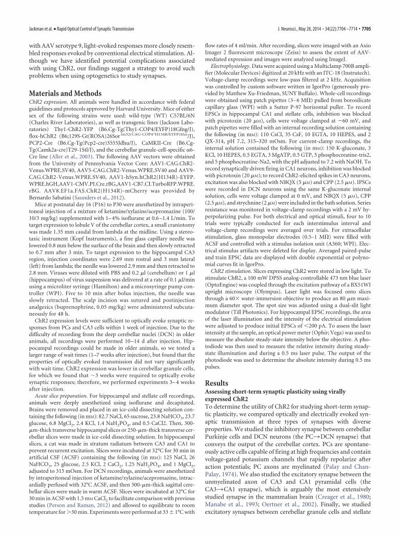
AAV expression vectors affect synaptic plasticityWe wondered whether deficits in optically evoked synaptic plas-ticity resulted from the inability of ChR2 to drive presynapticfiring. To determine the reliability of optically evoked firing, werecorded from CA3 pyramidal cells in current clamp while stim-ulating with a 0.5 ms flash focused either over the soma or overthe axon �500 �M away in the stratum radiatum (Fig. 4A). Op-tical stimulation over either the soma or axon could elicit spikes.As reported previously, somatically induced spikes were pre-ceded by a charging phase, whereas axonally induced spikesshowed little prior depolarization (Lewis et al., 2009; Petreanu etal., 2009; Madisen et al., 2012), confirming that spikes were ini-tiated far from the somatic recording pipette. Both stimulationmethods elicited spikes, although axonal stimulation requiredbrighter light (Fig. 4B). We worried that using bright light mightreduce the reliability of generating multiple spikes because ChR2inactivation increases with light intensity (Lin et al., 2009). How-ever, maximal intensity light (160 mW/mm 2) reliably evokedaxonal firing for ISIs of 20 –500 ms (Fig. 4C,D). Even an ISI of 10ms elicited pairs of spikes in most cells (11/13 cells) and CA3 axonscould be driven reliably at frequencies up to 50 Hz (Fig. 4E,F).
If unreliable firing does not account for deficits in opticallyevoked short-term plasticity, this raised the possibility that ourChR2 expression vector changed the properties of synapses. In-
terestingly, our observed deficits in optically evoked short-termplasticity closely resembled those of previous studies in whichvectors were also used to express ChR2 (Zhang and Oertner,2007; Cruikshank et al., 2010).
One possible way to reconcile our observations is if AAV alterssynaptic plasticity at the CA33CA1 synapse. If this is the case,then one might expect altered plasticity regardless of whetheraxons are stimulated optically or electrically. On the contrary, wefound that, on average, there was a slight reduction in the amountof paired-pulse plasticity between electrically evoked responses inuninjected and injected animals, but it was not statistically signif-icant (p � 0.1 for all ISIs, unpaired Student’s t test; Fig. 5E).However, this is not a fair test of the hypothesis that AAV alterssynaptic properties because optical and electrical stimulation ac-tivate different axons. Light activates only ChR2-expressing ax-ons that originate in the CA3 region near the injection site,whereas electrical stimulation activates both expressing and non-
Figure 2. Optically evoked plasticity is normal at the PC3DCN synapse. A, Fluorescenceimages in sagittal cerebellar slices of ChR2-Venus expression after AAV1-ChR2 injection (left)and hChR2-YFP expression from a PCP2-Cre X flox-hChR2 mouse (right). B, Top, RepresentativeIPSCs evoked by electrical and optical stimulation of presynaptic axons, for AAV1- andtransgenic-driven ChR2 expression. Scale bars, 1 nA for electrical and PCP2-Cre X flox-hChR2,100 pA for AAV1-ChR2. Bottom, Averaged normalized PSC amplitudes from 10 and 50 Hz trainsevoked electrically (n 11) and optically (AAV1-ChR2, n 6; PCP2-Cre � flox-hChR2, n 14). Data are expressed as average � SEM. Figure 3. Optically evoked responses exhibit abnormal depression at the hippocampal
CA33 CA1 synapse. A, Fluorescence image of ChR2-Venus expression in a transverse hip-pocampal slice after injection of AAV1-ChR2. B, Top, Representative EPSCs evoked by 10 and 50Hz trains recorded from the same CA1 pyramidal neuron for both electrical and optical stimula-tion of Schaffer collateral axons. Bottom, Averaged normalized EPSC amplitudes during trainsevoked electrically (n 25) and optically (n 31). C, Left, Representative EPSCs evokedoptically and electrically by pairs of stimuli at 10 –200 ms ISI recorded from the same neuron.Right, Averaged paired-pulse ratios evoked electrically (n 89) and optically (n 126).Vertical scale bars, 100 pA. Data are expressed as average � SEM.
Jackman et al. • Rapid Optical Control of Synaptic Transmission J. Neurosci., May 28, 2014 • 34(22):7704 –7714 • 7707
expressing axons that could arise at a distance from the injectionsite. Therefore, electrically evoked plasticity could be minimallyaffected if AAV changes plasticity in infected cells in a cell auton-omous manner or even if plasticity is altered in all cells near theinjection site. If AAV does alter synaptic properties, then theshort-term plasticity of electrically activated responses wouldonly be impaired if most of the electrically activated axons arefrom cells influenced by AAV.
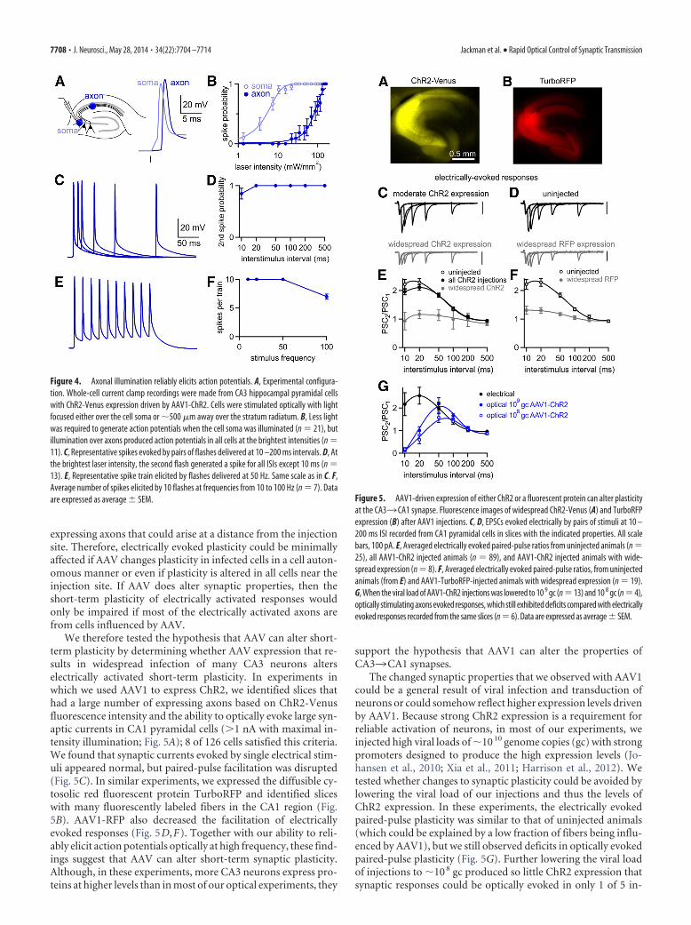
We therefore tested the hypothesis that AAV can alter short-term plasticity by determining whether AAV expression that re-sults in widespread infection of many CA3 neurons alterselectrically activated short-term plasticity. In experiments inwhich we used AAV1 to express ChR2, we identified slices thathad a large number of expressing axons based on ChR2-Venusfluorescence intensity and the ability to optically evoke large syn-aptic currents in CA1 pyramidal cells (�1 nA with maximal in-tensity illumination; Fig. 5A); 8 of 126 cells satisfied this criteria.We found that synaptic currents evoked by single electrical stim-uli appeared normal, but paired-pulse facilitation was disrupted(Fig. 5C). In similar experiments, we expressed the diffusible cy-tosolic red fluorescent protein TurboRFP and identified sliceswith many fluorescently labeled fibers in the CA1 region (Fig.5B). AAV1-RFP also decreased the facilitation of electricallyevoked responses (Fig. 5D,F). Together with our ability to reli-ably elicit action potentials optically at high frequency, these find-ings suggest that AAV can alter short-term synaptic plasticity.Although, in these experiments, more CA3 neurons express pro-teins at higher levels than in most of our optical experiments, they
support the hypothesis that AAV1 can alter the properties ofCA33CA1 synapses.
The changed synaptic properties that we observed with AAV1could be a general result of viral infection and transduction ofneurons or could somehow reflect higher expression levels drivenby AAV1. Because strong ChR2 expression is a requirement forreliable activation of neurons, in most of our experiments, weinjected high viral loads of �10 10 genome copies (gc) with strongpromoters designed to produce the high expression levels (Jo-hansen et al., 2010; Xia et al., 2011; Harrison et al., 2012). Wetested whether changes to synaptic plasticity could be avoided bylowering the viral load of our injections and thus the levels ofChR2 expression. In these experiments, the electrically evokedpaired-pulse plasticity was similar to that of uninjected animals(which could be explained by a low fraction of fibers being influ-enced by AAV1), but we still observed deficits in optically evokedpaired-pulse plasticity (Fig. 5G). Further lowering the viral loadof injections to �10 8 gc produced so little ChR2 expression thatsynaptic responses could be optically evoked in only 1 of 5 in-
Figure 4. Axonal illumination reliably elicits action potentials. A, Experimental configura-tion. Whole-cell current clamp recordings were made from CA3 hippocampal pyramidal cellswith ChR2-Venus expression driven by AAV1-ChR2. Cells were stimulated optically with lightfocused either over the cell soma or �500 �m away over the stratum radiatum. B, Less lightwas required to generate action potentials when the cell soma was illuminated (n 21), butillumination over axons produced action potentials in all cells at the brightest intensities (n 11). C, Representative spikes evoked by pairs of flashes delivered at 10 –200 ms intervals. D, Atthe brightest laser intensity, the second flash generated a spike for all ISIs except 10 ms (n 13). E, Representative spike train elicited by flashes delivered at 50 Hz. Same scale as in C. F,Average number of spikes elicited by 10 flashes at frequencies from 10 to 100 Hz (n 7). Dataare expressed as average � SEM. Figure 5. AAV1-driven expression of either ChR2 or a fluorescent protein can alter plasticity
at the CA33 CA1 synapse. Fluorescence images of widespread ChR2-Venus (A) and TurboRFPexpression (B) after AAV1 injections. C, D, EPSCs evoked electrically by pairs of stimuli at 10 –200 ms ISI recorded from CA1 pyramidal cells in slices with the indicated properties. All scalebars, 100 pA. E, Averaged electrically evoked paired-pulse ratios from uninjected animals (n 25), all AAV1-ChR2 injected animals (n 89), and AAV1-ChR2 injected animals with wide-spread expression (n 8). F, Averaged electrically evoked paired-pulse ratios, from uninjectedanimals (from E) and AAV1-TurboRFP-injected animals with widespread expression (n 19).G, When the viral load of AAV1-ChR2 injections was lowered to 10 9 gc (n 13) and 10 8 gc (n 4),optically stimulating axons evoked responses, which still exhibited deficits compared with electricallyevoked responses recorded from the same slices (n 6). Data are expressed as average � SEM.
7708 • J. Neurosci., May 28, 2014 • 34(22):7704 –7714 Jackman et al. • Rapid Optical Control of Synaptic Transmission
jected mice, but deficits in short-term plasticity were still evident(Fig. 5G). This suggests that if ChR2 is to be used to activateaxons, then AAV1-induced deficits cannot be avoided by reduc-ing expression levels.
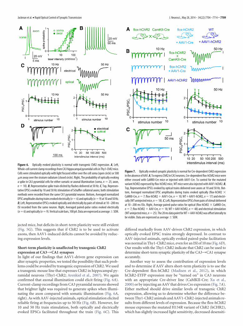
Short-term plasticity is unaffected by transgenic ChR2expression at CA33CA1 synapsesIn light of our findings that AAV1-driven gene expression canalter synaptic properties, we tested the possibility that such prob-lems could be avoided by transgenic expression of ChR2. We useda transgenic mouse line that expresses ChR2 in hippocampal py-ramidal neurons (Thy1-ChR2; Arenkiel et al., 2007). We againconfirmed that axonal illumination could elicit firing (Fig. 6A).Current-clamp recordings from CA3 pyramidal neurons showedthat brighter light was required to generate spikes when illumi-nating the axon compared with somatic illumination (Fig. 6A,right). As with AAV-injected animals, optical stimulation elicitedreliable firing at frequencies up to 50 Hz (Fig. 6B). However, for10 and 50 Hz train stimulation, both optically and electricallyevoked EPSCs facilitated throughout the train (Fig. 6C). This
differed markedly from AAV-driven ChR2 expression, in whichoptically evoked EPSC trains strongly depressed. In contrast toAAV-injected animals, optically evoked paired-pulse facilitationwas normal in Thy1-ChR2 mice, even for an ISI of 10 ms (Fig. 6D).Our results with the Thy1-ChR2 indicate that ChR2 can be used todetermine short-term synaptic plasticity of the CA33CA1 synapseaccurately.
Another way to assess the contribution of expression levelsand to determine if AAV alters short-term plasticity is to use theCre-dependent flox-hChR2 (Madisen et al., 2012), in whichhChR2-EYFP expression may be “turned on” in CA3 neuronswith an appropriate Cre-driver line (CaMKII-Cre; Xu et al.,2000) or by injecting an AAV that drives Cre expression (Fig. 7A).Either method should drive similar levels of transgenic ChR2expression, allowing us to determine whether the difference be-tween Thy1-ChR2 animals and AAV1-ChR2-injected animals re-sults from different levels of expression. Because the flox-hChR2mouse expresses the mutated H134R variant of ChR2 (hCHR2),which has slightly increased light sensitivity, decreased desensiti-
Figure 6. Optically evoked plasticity is normal with transgenic ChR2 expression. A, Left,Whole-cell current clamp recordings from CA3 hippocampal pyramidal cells in Thy1-ChR2 mice.Cells were stimulated optically with light focused either over the cell soma (open circle) or 500�m away over the stratum radiatum (closed circle). Right, The probability of optically evokinga spike in CA3 pyramidal cells for either somatic or axonal illumination (soma, n 25; axon,n 10). B, Representative spike train elicited by flashes delivered at 50 Hz. C, Top, Represen-tative EPSCs evoked by 10 and 50 Hz stimulation of Schaffer collateral axons; both stimulationmethods were recorded from the same CA1 pyramidal neuron. Bottom, Averaged normalizedEPSC amplitudes during trains evoked electrically (n 6) and optically (n 9) at 10 and 50 Hz.D, Left, Representative EPSCs evoked optically and electrically by pairs of stimuli at 10 –200 msISI recorded from the same neuron. Right, Averaged paired-pulse ratios evoked electrically(n 6) and optically (n 9). Vertical scale bars, 100 pA. Data are expressed as average�SEM.
Figure 7. Optically evoked synaptic plasticity is normal for Cre-dependent ChR2 expressionin the absence of AAV. A, To express ChR2 in CA3 neurons, Cre-dependent flox-hChR2 mice wereeither crossed with CaMKII-Cre mice or injected with AAV1-Cre. To control for the mutatedvariant hChR2 expressed by flox-hChR2 mice, WT mice were also injected with AAV1-hChR2. B,Top, Representative EPSCs evoked by optical trains delivered over axons at 10 and 50 Hz. Bot-tom, Averaged normalized EPSC amplitudes during trains evoked optically (flox-hChR2 �CaMKII-Cre, n 7; flox-hChR2 AAV1-Cre, n 10; WTAAV1-hChR2, n 17) and electri-cally (WT uninjected mice, n 18). C, Left, Representative EPSCs from pairs of stimuli deliveredat 10 –200 ms ISIs. Right, Average paired-pulse ratios for optical (flox-hChR2 � CaMKII-Cre,n 7; flox-hChR2 AAV-Cre, n 18; WTAAV-hChR2, n 48) and electrical stimulation(WT uninjected mice, n 25). The 20 ms data point for WTAAV-hChR2 was offset laterally tobe visible. Data are expressed as average � SEM.
Jackman et al. • Rapid Optical Control of Synaptic Transmission J. Neurosci., May 28, 2014 • 34(22):7704 –7714 • 7709

zation and slower channel closing (Nagel et al., 2005; Lin, 2011),we also injected AAV1-hChR2-EYFP in WT mice as a control.
Optically evoked EPSCs facilitated during train stimulation inflox-hChR2 � CaMKII-Cre mice, but strongly depressed for 50Hz stimulation in flox-hChR2 mice injected with AAV1-Cre andWT mice injected with AAV1-hChR2 (Fig. 7B). Optically evokedpaired-pulse ratios in flox-hChR2 � CaMKII-Cre mice weresimilar to electrically evoked paired-pulse ratios in uninjectedWT mice (Fig. 7C). In contrast, paired-pulse ratios were stronglydepressed in flox-hChR2 mice injected with AAV1-Cre. In WTanimals injected with AAV1-hChR2, paired-pulse ratios resem-bled those of WT animals injected with AAV1-ChR2 (Fig. 3),indicating that the different properties of the two variants did notaffect optically evoked synaptic plasticity significantly. These dataagain illustrate that transgenically expressed ChR2 can be used todetermine short-term plasticity, whereas AAV-driven expression ofeither Cre or hChR2 can alter synaptic properties.
Strategies for avoiding AAV-induced changes tosynaptic plasticityAAV vectors have become popular agents for gene delivery due totheir ability to drive strong, stable, nontoxic gene expression.There is little evidence that AAV vectors affect synaptic transmis-sion and AAV is often used to express ChR2 and other moleculesfor studies of synaptic transmission. However, it was reportedrecently that, in the mouse hippocampus, AAV-driven expres-sion of GFP can impair inhibitory transmission by reducingGABA levels in vesicles (Ortinski et al., 2010). It is unclear to whatextent AAV also affects excitatory synapses. Interestingly, AAV-induced changes to synaptic transmission depended on AAV se-rotype. AAV5 led to decreased synaptic inhibition, AAV9 did not,and AAV1 was not tested (Ortinski et al., 2010).
Thus far, our expression vectors were all serotype 1, which wechose for its strong transduction efficiency in hippocampal neu-rons (Royo et al., 2008). We wondered whether the changes weobserved in short-term plasticity might depend on AAV serotype.We therefore injected WT mice with vectors identical to theAAV1-ChR2 from Figures 1, 2, 3, 4, and 5, but with different viralserotypes (AAV5-CAG.ChR2-Venus and AAV9-CAG.ChR2-Venus) and with AAV8-EF1a.FAS.ChR2(H134R)-mCherry. Allthree vectors produced strong expression in the hippocampus(Fig. 8A).
When ChR2 expression was driven by AAV5 and AAV8, op-tically evoked EPSCs depressed strongly during train stimulation(Fig. 8B) and there were deficits in paired-pulse facilitation (Fig.8C). In marked contrast, when AAV9 was used to express ChR2,the responses to trains (Fig. 8B) and pairs of optical stimuli (Fig.8C) more closely resembled electrical stimulation. Therefore, itappears that AAV1, AAV5, and AAV8 alter the properties of theCA33CA1 synapse, but AAV9 does not.
We investigated whether using AAV9 to express ChR2 is alsoeffective at mitigating the synaptic depression observed with op-tically evoked trains at the grc3SC synapse (Fig. 9A). At thissynapse, electrical stimulation at 10 Hz produced only very mod-est synaptic facilitation and 50 Hz stimulation produced facilita-tion that was initially large, but modest by the tenth stimulus (Fig.9B). When AAV1-ChR2 was used, depression was apparent for10 Hz optical trains and more prominent for 50 Hz trains (Fig.9B). When AAV9-ChR2 was used, light-evoked synaptic plastic-ity was similar to electrically evoked synaptic plasticity for thefirst 5 stimuli (p � 0.1, unpaired Student’s t test), but by the tenthstimulus, the PSCs depressed, although not as much as whenAAV1 was used (Fig. 9B). These findings suggest that, for the
grc3SC synapse, as in the CA33CA1 synapse, superior perfor-mance of ChR2 is achieved when it is expressed with AAV9 ratherthan AAV1.
We unsuccessfully attempted to determine whether trans-genic ChR2 expression could also improve light-evoked re-sponses at the grc3SC synapse. We crossed flox-hChR2 micewith �6-Cre mice (Aller et al., 2003) to express hChR2-EYFPselectively in cerebellar granule cells. We found that hChR2-EYFP expressed, but at such low levels that we were unable toevoke PSCs with brief light pulses.
Over-bouton illuminationAlthough we found conditions that led to good agreement be-tween optically and electrically evoked short-term plasticity atboth PC3DCN and CA33CA1 synapses, our standard ap-proach was to stimulate axons far from the postsynaptic cell toavoid complications that may arise from directly stimulating pre-synaptic boutons. Previous studies suggest that over-boutonstimulation may alter synaptic plasticity either by changing thepresynaptic waveform or by allowing direct influx of Ca 2
through ChR2 (Zhang and Oertner, 2007; Olsen et al., 2012).Despite this potential problem, over-bouton stimulation can be
Figure 8. Effect of AAV serotype on optically evoked synaptic plasticity. A, ChR2 expressionafter injection of AAV5-ChR2, AAV8, or AAV9-ChR2 in WT mice. B, Top, Representative EPSCsevoked by optical trains delivered over axons at 10 and 50 Hz. Bottom, Averaged normalizedEPSC amplitudes during trains evoked optically (AAV5-ChR2, n 6; AAV8-ChR2, n 7; AAV9-ChR2, n 10). Curve fit to electrical stimulation in uninjected animals (Fig. 7) is also shown forcomparison. C, Left, Representative EPSCs from pairs of stimuli delivered at 10 –200 ms ISIs.Right, Average paired-pulse ratios for optical stimulation (AAV5-ChR2, n 6; AAV8-ChR2, n 7; AAV9-ChR2, n 14) and curve fit for electrical stimulation (black, WT uninjected mice, Fig.7). Vertical scale bars, 100 pA. Data are expressed as average � SEM.
7710 • J. Neurosci., May 28, 2014 • 34(22):7704 –7714 Jackman et al. • Rapid Optical Control of Synaptic Transmission

much more effective at evoking synaptic responses, so many op-togenetic studies stimulate synaptic transmission by illuminatingdirectly over presynaptic boutons. We therefore compared theproperties of optically evoked responses for stimulation over thepostsynaptic cell and over axons �200 �m from the postsynapticcell for the PC3DCN synapse and the CA33CA1 synapse (Fig.10A,B). In both cases, AAV9 was used to express ChR2 and anal-ysis was restricted to cells in which we recorded both over-axonand over-bouton stimulation.
At the PC3DCN synapse, short-term plasticity was remark-ably similar for over-bouton and over-axon stimulation with2–100 Hz stimulus trains. There was no sign that over-boutonstimulation caused artificial synaptic depression (Fig. 10C,E).The performance of over-bouton stimulation was very differentfor the CA33CA1 synapse. Over-bouton stimulation at 10 Hzresulted in modest depression, whereas over-bouton stimulationat 50 Hz resulted in strong depression (Fig. 10D). A comparisonof the frequency dependence of PSC10/PSC1 for electrical stim-ulation and optical stimulation over-axon and over-boutonrevealed the largest differences between over-bouton and over-axon stimulation occurred at frequencies of 20 Hz and above(Fig. 10F). Although these findings illustrate the extent to whichover-bouton stimulation can alter synaptic plasticity, such com-plications can be avoided simply by illuminating presynaptic fi-bers well away from the postsynaptic cell.
Summary of light-evoked responsesWe compared the average normalized responses to the secondand last stimulus of 50 Hz trains for electrically and opticallyevoked responses for each method of expressing ChR2 at all threesynapses. The amplitude of the second response (PSC2/PSC1) isoften used to assesses whether a synapse is high p (depresses) orlow p (facilitates), whereas the amplitude of the last response
(PSC10/PSC1) provides a measure of the steady-state PSC. At thePC3DCN synapse, PSC2/PSC1 and PSC10/PSC1 did not differsignificantly between electrically evoked PSCs and opticallyevoked PSCs for ChR2 expressed transgenically with AAV1 orwith AAV9 (Fig. 11A). At the CA33CA1 synapse, we used eightdifferent methods to express ChR2 in CA3 hippocampal pyrami-dal neurons (two transgenic, five viral, and one virally activatedtransgenic) with mixed effects on optically evoked synaptic plas-ticity (Fig. 11B). Purely transgenic approaches yielded opticallyevoked short-term plasticity similar to conventional electricalstimulation. In contrast, electrically and optically evoked synap-tic plasticities were significantly different when ChR2 was ex-pressed with AAV1, AAV5, and AAV8, but not with AAV9. At thegrc3SC synapse, both AAV1 and AAV9 resulted in opticallyevoked responses that facilitated normally in response to the sec-ond stimulus, but depressed significantly in response to the laststimulus relative to electrical stimulation (Fig. 11C). However,the response to the last stimulus depressed significantly less with
Figure 9. Effect of AAV serotype on optically evoked synaptic plasticity at the granulecell3stellate cell synapse. A, Experimental configuration. Parallel fiber axons from cerebellargranule cells were stimulated �500 �m from the recorded cell by laser illumination (bluecircle) or with extracellular electrode and synaptic responses were monitored in voltage-clampmode from stellate cells. ChR2 was expressed in granule cells by either AAV1-ChR2 or AAV9-ChR2. B, Top, Representative EPSCs evoked by trains of 10 stimuli delivered by extracellularelectrode or laser illumination at 10 Hz and 50 Hz. Bottom, Averaged normalized EPSC ampli-tudes during trains evoked electrically (n 10) and optically (AAV1-ChR2, n 13; AAV9-ChR2,n 12) at 10 Hz and 50 Hz. Vertical scale bars, 100 pA. Data are expressed as average � SEM.
Figure 10. The performance of over-bouton stimulation is synapse dependent. ChR2 wasexpressed using AAV9-ChR2 and over-bouton and over-axon optical stimulation were com-pared for the PC3DCN (A) and the CA33 CA1 synapse (B). C, D, Representative PSCs evokedby 10 and 50 Hz stimulation over-axon and over-bouton recorded from a single DCN (C) orhippocampal CA1 (D) neuron and averaged normalized amplitudes for all cells in which re-sponses to both stimuli were measured are shown for PC3DCN (B, n 5) and for CA33 CA1synapses (E, n 6). E, F, Average normalized amplitude of the tenth PSC during stimulus trainsof different frequencies, for electrical stimulation (black) and optical stimulation over-axon(red) and over-bouton (gray) for the PC3DCN (E) and CA33 CA1 synapse (F ). Scale bars: C,1000 pA; D, 100 pA. Data are expressed as average � SEM.
Jackman et al. • Rapid Optical Control of Synaptic Transmission J. Neurosci., May 28, 2014 • 34(22):7704 –7714 • 7711

AAV9 than with AAV1. Unfortunately,our attempts to drive transgenic ChR2 ex-pression at this synapse by crossing the�6-Cre and flox-hChR2 lines did not re-sult in adequate expression levels, so it isunclear whether the deficits we observedin optically evoked synaptic plasticity re-sulted from problems with virally drivenexpression or from ChR2 stimulationitself.
Method of ChR2 expression determinesthe reliability of synaptically drivencircuit activityTo date, ChR2 has been used sparingly tocharacterize synaptic physiology. Moreoften, ChR2 is used to drive defined pop-ulations of neurons to study neural cir-cuits (Gradinaru et al., 2009; Kravitz et al.,2010; Olsen et al., 2012). We investigatedwhether the synaptic depression we ob-served when ChR2 was expressed withAAV1 would impair the ability of opti-cally activated synaptic inputs to drivepostsynaptic activity at the CA33CA1synapse. We stimulated Shaffer collateralseither optically or electrically while re-cording from CA1 neurons in current-clamp conditions (Fig. 12). The electricalstimulus intensity and the size of the laserspot were adjusted to produce postsynaptic action potentials inresponse to �60% of single stimuli. For electrical stimulationwith trains of 10 and 50 Hz in uninjected WT animals, each of thelast nine stimuli usually evoked time-locked spikes. In contrast,optical stimulation in animals injected with AAV1-ChR2 evokedvery little firing late in the train. However, optical stimulation inslices from Thy1-ChR2 animals evoked postsynaptic firingthroughout the train, similar to electrical stimulation. The reli-able postsynaptic firing produced by electrical stimulation andoptical stimulation in Thy1-ChR2 animals is consistent with theobserved facilitation of synaptic currents (Fig. 6), whereas theunreliable optical stimulation with AAV1-expressed ChR2 isconsistent with the synaptic depression observed in these animals(Fig. 3). Therefore, the method of expressing ChR2 can stronglyinfluence the manner in which optical stimulation drives neuralcircuit activity.
DiscussionWe identified multiple difficulties in applying optogenetic ap-proaches to studies of synaptic transmission, but found that, un-der the right experimental conditions, widely available ChR2variants could be used to activate axons with physiologically rel-evant activity patterns and produce synaptic responses that weresimilar to electrically evoked responses. This was a surprise be-cause previous studies have questioned whether ChR2 could beused to reliably activate axons at high frequencies. Instead, wefound that deficits in optically evoked synaptic transmissioncould arise from either AAV expression vectors or stimulatingover synaptic boutons. When ChR2 was expressed transgenicallyor using AAV9, optically and electrically evoked responses weresimilar.
Figure 12. The method of ChR2 expression determines the reliability of synaptically drivencircuit activity. A, Current-clamp recordings from CA1 neurons. Action potentials were evokedby synaptic input from CA3 axons stimulated electrically (uninjected WT mice) and opticallywhen ChR2 was expressed with AAV1-ChR2 or in Thy1-ChR2 mice. The stimulus intensity wasadjusted to produce an action potential at the beginning of the train in �60% of trials. Spikeraster plots below the current-clamp traces show firing during six trials. Examples are shown for10 and 50 Hz stimulation. B, Average number of spikes elicited by each stimulus in the train isshown for 10 and 50 Hz stimulation (electrical, n 9; AAV1-ChR2, n 11, Thy1-ChR2; n 10). Data are expressed as average � SEM.
Figure 11. The method of ChR2 expression determines the properties of optically evoked short-term plasticity. A–C, Averagenormalized amplitude of the second response (top) and last response (bottom) to 50 Hz trains for all methods used to express ChR2at the PC3DCN (A), CA33 CA1 (B), and grc3 SC (C) synapses. Trains evoked by electrical stimulation were from uninjected WTanimals (B) or from slices expressing ChR2 (A, C). Statistical significance (**) was assessed by one-way ANOVA followed by Tukey’spost hoc test ( p � 0.01). Data are expressed as average � SEM.
7712 • J. Neurosci., May 28, 2014 • 34(22):7704 –7714 Jackman et al. • Rapid Optical Control of Synaptic Transmission
Some AAV expression vectors can alter the properties ofsome synapsesAAV serotypes influence synaptic responses in a highly synapse-dependent manner. AAV1 led to artificial synaptic depression atthe CA33CA1 and grc3SC synapses, but not at the PC3DCNsynapse. AAV5 and AAV8 also impaired synaptic transmission atthe CA33CA1 synapse, indicating that the problem was notspecific to the AAV1 serotype. AAV9 mitigated the deleteriouseffects of viruses on short-term plasticity at CA33CA1 synapses,and to a certain extent at grc3SC synapses.
We tested several hypotheses to explain the observed deficitsin optically evoked short-term plasticity when AAV1 was used toexpress ChR2. Initially, we assessed whether it was a result of theinherent inability of ChR2 to reliably stimulate axons, as had beenpreviously proposed (Wang et al., 2007; Cruikshank et al., 2010;Cruikshank et al., 2012). The observation that axonal illumina-tion reliably stimulated antidromic spikes in the soma of CA3pyramidal cells indicated that this was not the case. Instead, AAVwas implicated as the cause of impaired transmission becauseoptically evoked synaptic plasticity was altered when AAV wasused to either express ChR2 in WT animals or to express Cre inconditional ChR2 transgenic animals, whereas synaptic re-sponses were normal in transgenic mice that expressed ChR2without using AAV1. We also found that using AAV1 to producestrong and widespread expression of either Turbo RFP or ChR2led to strong alterations in electrically evoked short-term plastic-ity, suggesting that some aspect of AAV1 transfection could alterthe properties of short-term plasticity.
It is possible that some AAV serotypes induce reactive astro-cytosis, which in turn alters synaptic strength (Ortinski et al.,2010). In the hippocampus, AAV5 induces reactive astrocytosisthrough an unknown mechanism, but AAV9 does not (AAV1and AAV8 were not examined). Reactive astrocytosis reduced themagnitude of inhibition by downregulating the expression ofglutamine synthetase, which in turn reduced the availability ofGABA for synaptic release. This mechanism accounts for a reduc-tion of inhibitory transmission, but cannot account for changesin the short-term synaptic plasticity of excitatory synapses. It ispossible that reactive astrocytosis also affects the properties ofshort-term plasticity at excitatory synapses, but further experi-ments are needed to determine whether astrocytosis is the mech-anism by which some AAV serotypes modify short-termplasticity at excitatory synapses. Regardless of the mechanism bywhich AAV1, AAV5, and AAV8 alter short-term plasticity, ourfindings suggest that these serotypes may not be suitable for manyoptogenetic applications and the use of AAV9 is preferable.
Over-bouton stimulationFor many applications, the easiest way to optically evoke largesynaptic responses is to activate ChR2-labeled inputs with full-field, over-bouton illumination. However, we found that illumi-nating directly over CA3 synaptic boutons led to artificialsynaptic depression, whereas axonal stimulation at more distantsites produced more physiological synaptic responses. It is likelythat the slow kinetics of ChR2 results in a prolonged depolarizationof presynaptic boutons that affects endogenous voltage-gated chan-nels and changes the probability of neurotransmitter release. Illumi-nating over axons at a distance from synapses provides the mostreliable way of activating presynaptic inputs and axons can be stim-ulated with precise timing even at high frequencies.
In contrast, PC3DCN synaptic responses evoked by over-bouton light stimulation were indistinguishable from thoseevoked electrically. This illustrates that problems associated with
over-bouton stimulation are highly dependent on the synapsebeing studied. One potential explanation for the superior perfor-mance of over-bouton stimulation at PC3DCN synapses is thatPCs are spontaneously active at high frequencies and express fastpotassium channels at high density to rapidly repolarize actionpotentials. This may allow PC presynaptic boutons to overcomeChR2 conductances and rapidly repolarize after over-boutonstimulation.
Widely available ChR2 variants allow synaptic activation atphysiological frequenciesOptogenetic techniques have proliferated rapidly across neuro-science in recent years, but ChR2 has not been fully exploited toinvestigate synaptic transmission. This may reflect a perceptionthat ChR2 cannot reliably drive the physiological patterns of ac-tivity required for quantitative studies of synaptic short-termplasticity. Synaptic physiologists may have therefore approachedthe use of optogenetics cautiously. Efforts to characterize ChR2and other microbial opsins have highlighted shortcomings suchas inactivation and slow channel kinetics (Lin, 2011). To that end,great effort has gone into engineering ChR2 variants with greaterlight sensitivity, decreased desensitization and faster kinetics(Nagel et al., 2005; Lin et al., 2009; Gunaydin et al., 2010). Al-though improved ChR2 variants will doubtless be of great utility,we find that, with proper control of gene expression, ChR2 andhChR2 are already remarkable tools for the study of synapses.
General strategy for using ChR2 to characterize synapsesThe possibility that over-bouton stimulation and the use of cer-tain AAV serotypes can produce artificial synaptic depressionmust be taken into account for most but not all applications ofChR2. When determining basic synaptic properties, such aswhether a synapse exists between two cell types or the identity ofthe neurotransmitter released, artificial synaptic depression is notan issue and transgenic animals or any of the AAV serotypescould be used. However, if the goals are to characterize whethersynapses facilitate or depress, to determine responses to high-frequency stimulus trains, or to stimulate synapses in a realisticmanner in vivo, then the means of expressing ChR2 and the man-ner of stimulation matter a great deal.
Our findings suggest that it is preferable to express ChR2 inthe presynaptic cell of interest using transgenic animals eitherwith an appropriate BAC transgenic line or by crossing Crelines with flox-hChR2 mice, as we did for the CA33CA1 andPC3DCN synapses. Unfortunately, it may not always be pos-sible to obtain an appropriate Cre line or to achieve suffi-ciently high levels of ChR2 expression, as was the case forgrc3SC synapses.
If transgenic approaches are not an option, then AAV9 ap-pears to be the preferable method for expressing ChR2. With thisserotype, it was possible to obtain light-evoked responses thatclosely approximated electrically evoked responses at theCA33CA1 and PC3DCN synapses. At the grc3SC synapse,the performance of AAV9 was superior to AAV1, although sus-tained optical stimulation still resulted in depression. This sug-gests that, unless electrical stimulation can be used to verify theproperties of light-evoked responses, responses to sustained acti-vation are difficult to interpret. Our findings at three synapsessuggest that axonal stimulation of ChR2 expressed with AAV9can at least be used to assess paired-pulse plasticity and estimatethe release properties of a synapse.
In some cases, it is preferable to use over-bouton stimulationrather than axonal stimulation, which makes it easier to evoke
Jackman et al. • Rapid Optical Control of Synaptic Transmission J. Neurosci., May 28, 2014 • 34(22):7704 –7714 • 7713
synaptic responses. Over-bouton stimulation produced good re-sponses at the PC3DCN synapse, but not at the CA33CA1synapse. Therefore, a reasonable strategy to determine whetherover-bouton stimulation can be used might be to compareresponses evoked by over-bouton and over-axon stimulation.If there is good agreement in the short-term plasticity evokedby both methods of stimulation, then over-bouton stimula-tion can be used. However, if over-bouton stimulation pro-duces artificial depression, then it should be avoided duringsynaptic characterization.
ReferencesAller MI, Jones A, Merlo D, Paterlini M, Meyer AH, Amtmann U, Brickley S,
Jolin HE, McKenzie AN, Monyer H, Farrant M, Wisden W (2003) Cer-ebellar granule cell Cre recombinase expression. Genesis 36:97–103.CrossRef Medline
Arenkiel BR, Peca J, Davison IG, Feliciano C, Deisseroth K, Augustine GJ,Ehlers MD, Feng G (2007) In vivo light-induced activation of neuralcircuitry in transgenic mice expressing channelrhodopsin-2. Neuron 54:205–218. CrossRef Medline
Boyden ES, Zhang F, Bamberg E, Nagel G, Deisseroth K (2005) Millisecond-timescale, genetically targeted optical control of neural activity. Nat Neu-rosci 8:1263–1268. CrossRef Medline
Creager R, Dunwiddie T, Lynch G (1980) Paired-pulse and frequency facil-itation in the CA1 region of the in vitro rat hippocampus. J Physiol 299:409 – 424. Medline
Cruikshank SJ, Urabe H, Nurmikko AV, Connors BW (2010) Pathway-specificfeedforwardcircuitsbetweenthalamusandneocortexrevealedbyselectiveopticalstimulation of axons. Neuron 65:230–245. CrossRef Medline
Cruikshank SJ, Ahmed OJ, Stevens TR, Patrick SL, Gonzalez AN, Elmaleh M,Connors BW (2012) Thalamic control of layer 1 circuits in prefrontalcortex. J Neurosci 32:17813–17823. CrossRef Medline
Ellender TJ, Huerta-Ocampo I, Deisseroth K, Capogna M, Bolam JP (2011)Differential modulation of excitatory and inhibitory striatal synaptic trans-mission by histamine. J Neurosci 31:15340–15351. CrossRef Medline
Fenno L, Yizhar O, Deisseroth K (2011) The development and applicationof optogenetics. Annu Rev Neurosci 34:389 – 412. CrossRef Medline
Gradinaru V, Mogri M, Thompson KR, Henderson JM, Deisseroth K (2009)Optical deconstruction of parkinsonian neural circuitry. Science 324:354 –359. CrossRef Medline
Gunaydin LA, Yizhar O, Berndt A, Sohal VS, Deisseroth K, Hegemann P (2010)Ultrafast optogenetic control. Nat Neurosci 13:387–392. CrossRef Medline
Harrison TC, Ayling OG, Murphy TH (2012) Distinct cortical circuit mech-anisms for complex forelimb movement and motor map topography.Neuron 74:397– 409. CrossRef Medline
Johansen JP, Hamanaka H, Monfils MH, Behnia R, Deisseroth K, Blair HT,LeDoux JE (2010) Optical activation of lateral amygdala pyramidal cellsinstructs associative fear learning. Proc Natl Acad Sci U S A 107:12692–12697. CrossRef Medline
Kravitz AV, Freeze BS, Parker PR, Kay K, Thwin MT, Deisseroth K, Kreitzer AC(2010) Regulation of parkinsonian motor behaviours by optogenetic con-trol of basal ganglia circuitry. Nature 466:622–626. CrossRef Medline
Ledri M, Nikitidou L, Erdelyi F, Szabo G, Kirik D, Deisseroth K, Kokaia M(2012) Altered profile of basket cell afferent synapses in hyper-excitabledentate gyrus revealed by optogenetic and two-pathway stimulations. EurJ Neurosci 36:1971–1983. CrossRef Medline
Lewis TL Jr, Mao T, Svoboda K, Arnold DB (2009) Myosin-dependent tar-geting of transmembrane proteins to neuronal dendrites. Nat Neurosci12:568 –576. CrossRef Medline
Lin JY (2011) A user’s guide to channelrhodopsin variants: features, limita-tions and future developments. Exp Physiol 96:19 –25. CrossRef Medline
Lin JY, Lin MZ, Steinbach P, Tsien RY (2009) Characterization of engi-neered channelrhodopsin variants with improved properties and kinetics.Biophys J 96:1803–1814. CrossRef Medline
Madisen L, Mao T, Koch H, Zhuo JM, Berenyi A, Fujisawa S, Hsu YW, GarciaAJ 3rd, Gu X, Zanella S, Kidney J, Gu H, Mao Y, Hooks BM, Boyden ES,Buzsaki G, Ramirez JM, Jones AR, Svoboda K, Han X, Turner EE, Zeng H(2012) A toolbox of Cre-dependent optogenetic transgenic mice forlight-induced activation and silencing. Nat Neurosci 15:793– 802.CrossRef Medline
Manabe T, Wyllie DJ, Perkel DJ, Nicoll RA (1993) Modulation of synaptic
transmission and long-term potentiation: effects on paired pulse facilita-tion and EPSC variance in the CA1 region of the hippocampus. J Neuro-physiol 70:1451–1459. Medline
Mathews PJ, Lee KH, Peng Z, Houser CR, Otis TS (2012) Effects of climbingfiber driven inhibition on purkinje neuron spiking. J Neurosci 32:17988 –17997. CrossRef Medline
Nagel G, Brauner M, Liewald JF, Adeishvili N, Bamberg E, Gottschalk A(2005) Light activation of channelrhodopsin-2 in excitable cells of Cae-norhabditis elegans triggers rapid behavioral responses. Curr Biol 15:2279 –2284. CrossRef Medline
Oertner TG, Sabatini BL, Nimchinsky EA, Svoboda K (2002) Facilitation atsingle synapses probed with optical quantal analysis. Nat Neurosci 5:657–664. CrossRef Medline
Olsen SR, Bortone DS, Adesnik H, Scanziani M (2012) Gain control by layersix in cortical circuits of vision. Nature 483:47–52. CrossRef Medline
Ortinski PI, Dong J, Mungenast A, Yue C, Takano H, Watson DJ, Haydon PG,Coulter DA (2010) Selective induction of astrocytic gliosis generatesdeficits in neuronal inhibition. Nat Neurosci 13:584 –591. CrossRefMedline
Palay SL, Chan-Palay V (1974) The cerebellar cortex: cytology and organi-zation. New York: Springer.
Palkovits M, Magyar P, Szentagothai J (1971) Quantitative histologicalanalysis of the cerebellar cortex in the cat. 3. Structural organization of themolecular layer. Brain Res 34:1–18. CrossRef Medline
Person AL, Raman IM (2012) Purkinje neuron synchrony elicits time-locked spik-ing in the cerebellar nuclei. Nature 481:502–505. CrossRef Medline
Petreanu L, Huber D, Sobczyk A, Svoboda K (2007) Channelrhodopsin-2-assisted circuit mapping of long-range callosal projections. Nat Neurosci10:663– 668. CrossRef Medline
Petreanu L, Mao T, Sternson SM, Svoboda K (2009) The subcellular orga-nization of neocortical excitatory connections. Nature 457:1142–1145.CrossRef Medline
Pinol RA, Bateman R, Mendelowitz D (2012) Optogenetic approaches tocharacterize the long-range synaptic pathways from the hypothalamus tobrain stem autonomic nuclei. J Neurosci Methods 210:238 –246. CrossRefMedline
Royo NC, Vandenberghe LH, Ma JY, Hauspurg A, Yu L, Maronski M,Johnston J, Dichter MA, Wilson JM, Watson DJ (2008) Specific AAVserotypes stably transduce primary hippocampal and cortical cultureswith high efficiency and low toxicity. Brain Res 1190:15–22. CrossRefMedline
Saunders A, Johnson CA, Sabatini BL (2012) Novel recombinant adeno-associated viruses for Cre activated and inactivated transgene expressionin neurons. Front Neural Circuits 6:47. CrossRef Medline
Telgkamp P, Raman IM (2002) Depression of inhibitory synaptic transmis-sion between Purkinje cells and neurons of the cerebellar nuclei. J Neu-rosci 22:8447– 8457. Medline
Varga V, Losonczy A, Zemelman BV, Borhegyi Z, Nyiri G, Domonkos A, HangyaB, Holderith N, Magee JC, Freund TF (2009) Fast synaptic subcortical con-trol of hippocampal circuits. Science 326:449–453. CrossRef Medline
Wang H, Peca J, Matsuzaki M, Matsuzaki K, Noguchi J, Qiu L, Wang D,Zhang F, Boyden E, Deisseroth K, Kasai H, Hall WC, Feng G, AugustineGJ (2007) High-speed mapping of synaptic connectivity using photo-stimulation in Channelrhodopsin-2 transgenic mice. Proc Natl Acad SciU S A 104:8143– 8148. CrossRef Medline
Wyatt KD, Tanapat P, Wang SS (2005) Speed limits in the cerebellum: con-straints from myelinated and unmyelinated parallel fibers. Eur J Neurosci21:2285–2290. CrossRef Medline
Xia Y, Driscoll JR, Wilbrecht L, Margolis EB, Fields HL, Hjelmstad GO(2011) Nucleus accumbens medium spiny neurons target non-dopaminergic neurons in the ventral tegmental area. J Neurosci 31:7811–7816. CrossRef Medline
Xu B, Gottschalk W, Chow A, Wilson RI, Schnell E, Zang K, Wang D, NicollRA, Lu B, Reichardt LF (2000) The role of brain-derived neurotrophicfactor receptors in the mature hippocampus: modulation of long-termpotentiation through a presynaptic mechanism involving TrkB. J Neuro-sci 20:6888 – 6897. Medline
Zhang YP, Oertner TG (2007) Optical induction of synaptic plasticity usinga light-sensitive channel. Nat Methods 4:139 –141. CrossRef Medline
Zucker RS, Regehr WG (2002) Short-term synaptic plasticity. Annu RevPhysiol 64:355– 405. CrossRef Medline
7714 • J. Neurosci., May 28, 2014 • 34(22):7704 –7714 Jackman et al. • Rapid Optical Control of Synaptic Transmission