cellular uptake and toxicity of positively and negatively ... · cellular uptake and toxicity of...
TRANSCRIPT
Cellular uptake and toxicity of positively and negatively charged silica nanoparticles in
A549 human lung cells
Elisabeth Elje1,2,3, Julia Schölermann1, Mihaela Roxana Cimpan1*, Maria Dusinska2*
1) Biomaterials, Department of Clinical Dentistry, University of Bergen, Årstadveien 19, 5009 Bergen, Norway
2) NILU - Norwegian Institute for Air Research, Instituttveien 18, 2007 Kjeller, Norway
3) Department of Chemistry, University of Bergen, Allégaten 41, 5007 Bergen, Norway
*) Contributed equally
1
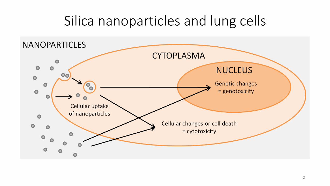
Silica nanoparticles and lung cells
2
Aims of the study
• To identify the effect of silica nanoparticle surface charge on the cytotoxic and genotoxic effects in A549 cells.
• To identify the effect of silica nanoparticle surface charge on the cellular uptake in A549 cells.
3
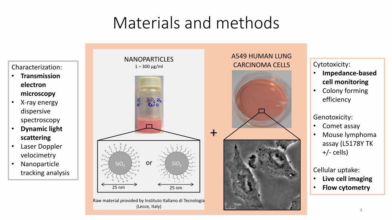
Characterization:• Transmission
electron microscopy
• X-ray energy dispersive spectroscopy
• Dynamic light scattering
• Laser Doppler velocimetry
• Nanoparticle tracking analysis
Cytotoxicity:• Impedance-based
cell monitoring• Colony forming
efficiency
Genotoxicity:• Comet assay• Mouse lymphoma
assay (L5178Y TK +/- cells)
Cellular uptake:• Live cell imaging• Flow cytometry
Materials and methods
SiO2or
+++++
++
++++
++++++
+++
++
++
SiO2
----
--
----
------
--
--
--
--
+
NANOPARTICLES A549 HUMAN LUNGCARCINOMA CELLS
25 nm 25 nm
1 – 300 µg/ml
Raw material provided by Instituto Italiano di Tecnologia(Lecce, Italy)
4
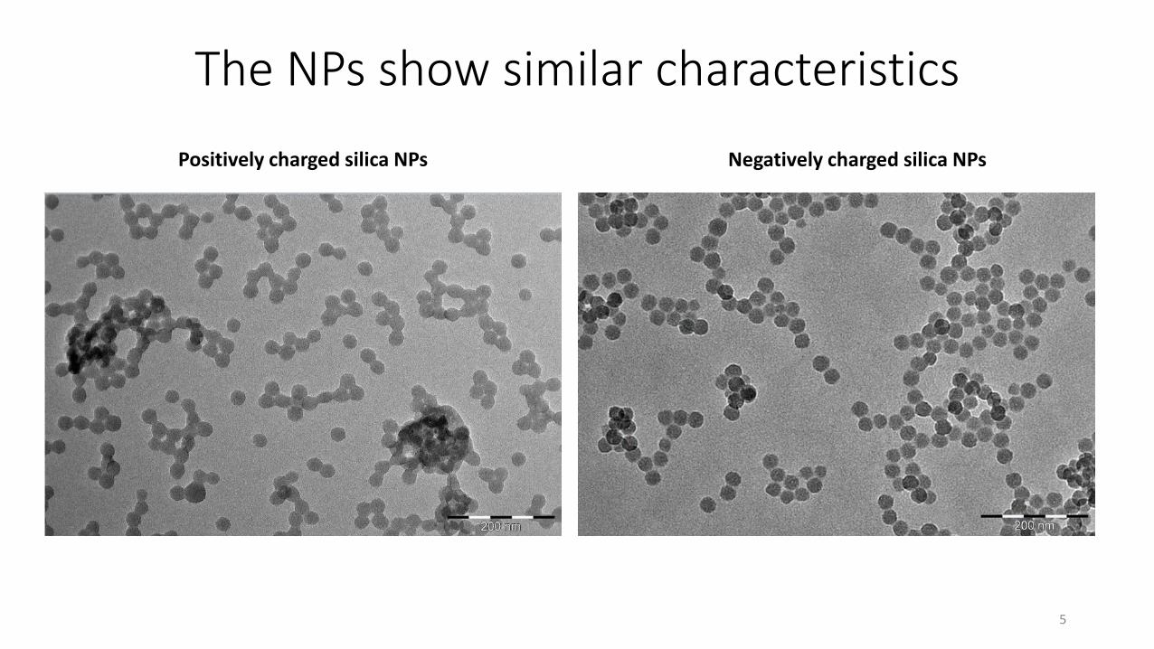
The NPs show similar characteristics
Positively charged silica NPs Negatively charged silica NPs
5
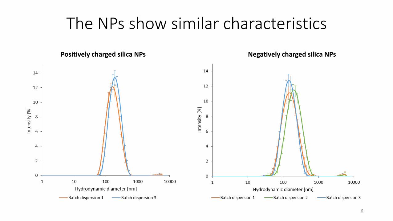
The NPs show similar characteristics
Positively charged silica NPs Negatively charged silica NPs
6
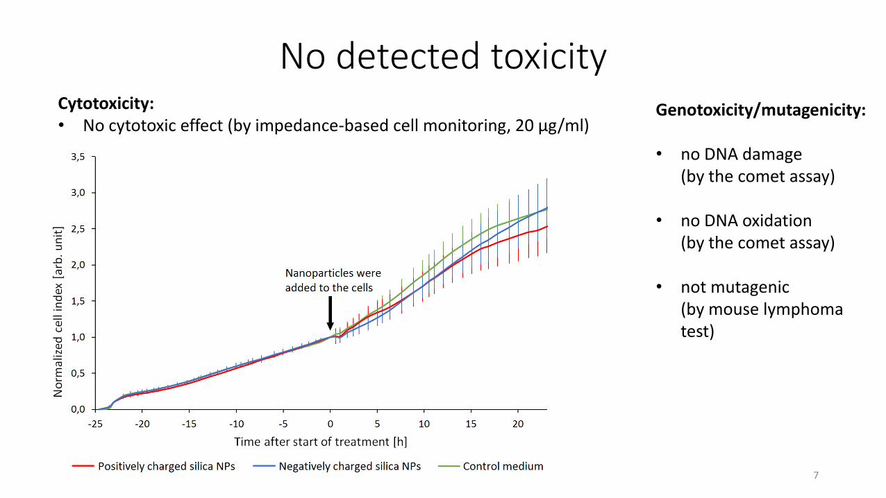
No detected toxicityGenotoxicity/mutagenicity:
• no DNA damage(by the comet assay)
• no DNA oxidation(by the comet assay)
• not mutagenic(by mouse lymphoma test)
Cytotoxicity:• No cytotoxic effect (by impedance-based cell monitoring, 20 µg/ml)
7
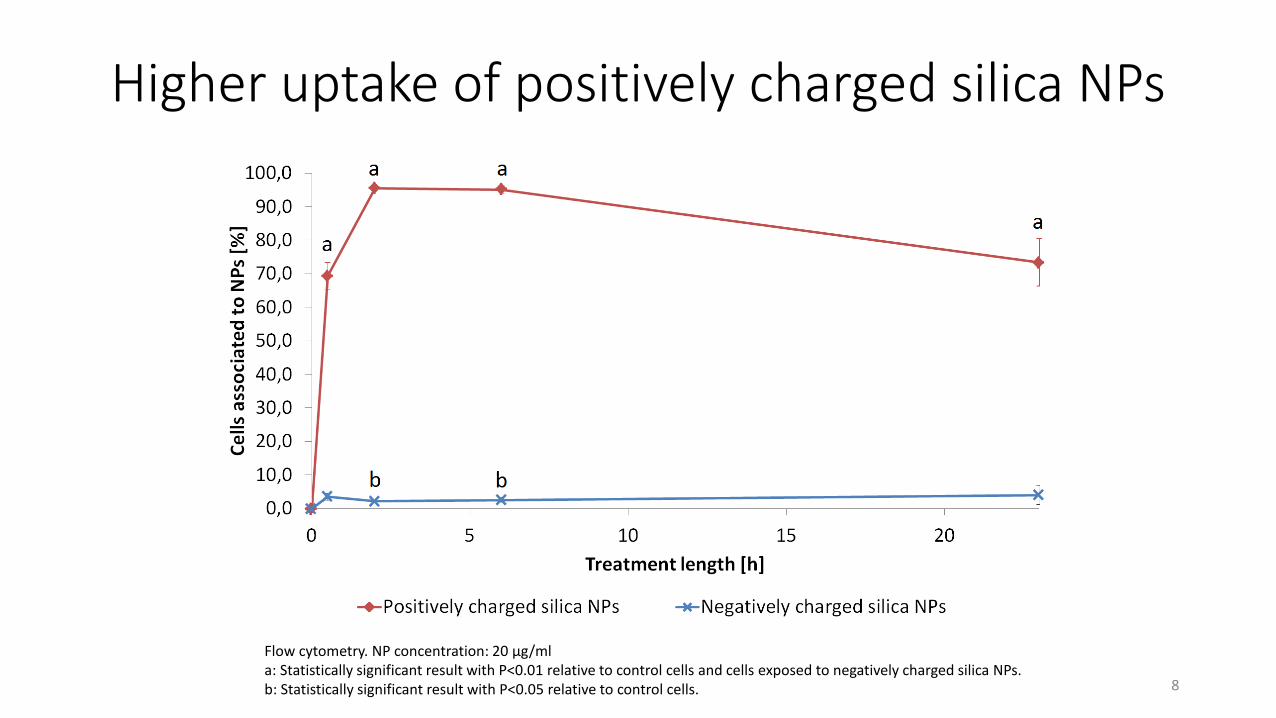
Higher uptake of positively charged silica NPs
Flow cytometry. NP concentration: 20 µg/mla: Statistically significant result with P<0.01 relative to control cells and cells exposed to negatively charged silica NPs.b: Statistically significant result with P<0.05 relative to control cells. 8
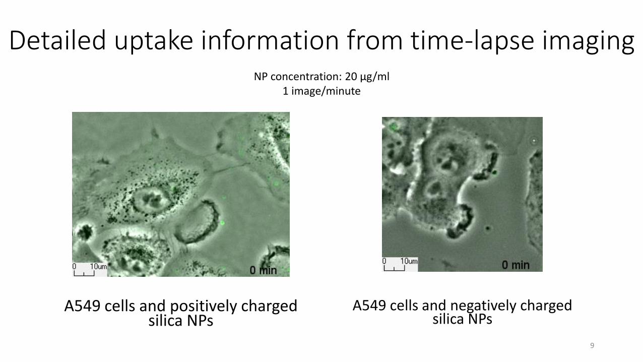
Detailed uptake information from time-lapse imagingNP concentration: 20 µg/ml
1 image/minute
A549 cells and positively charged silica NPs
A549 cells and negatively charged silica NPs
9
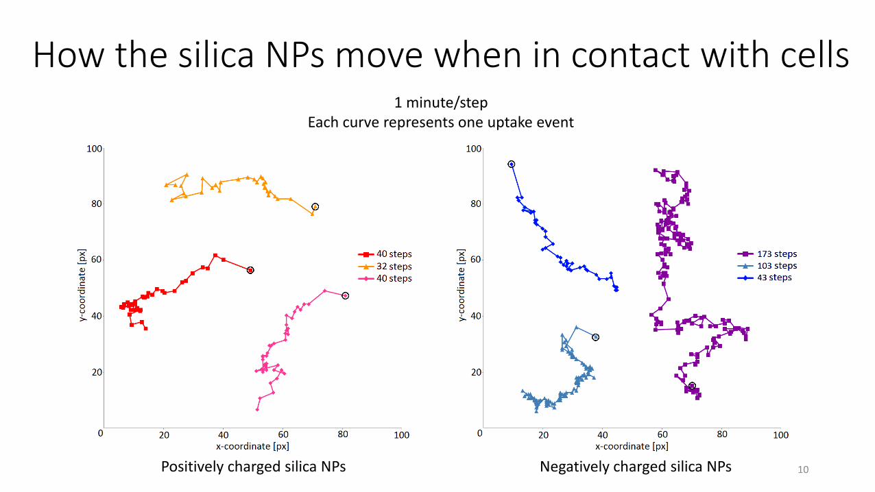
How the silica NPs move when in contact with cells
Positively charged silica NPs Negatively charged silica NPs
1 minute/stepEach curve represents one uptake event
10
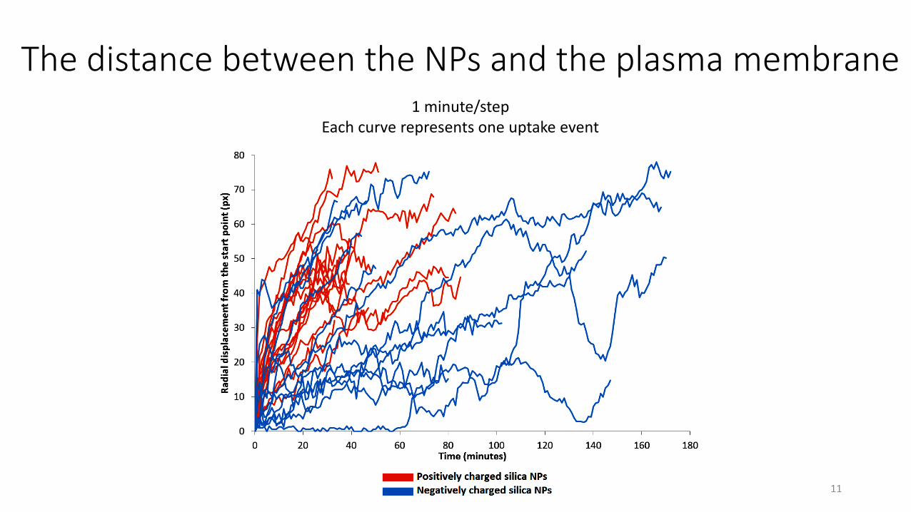
The distance between the NPs and the plasma membrane1 minute/step
Each curve represents one uptake event
11
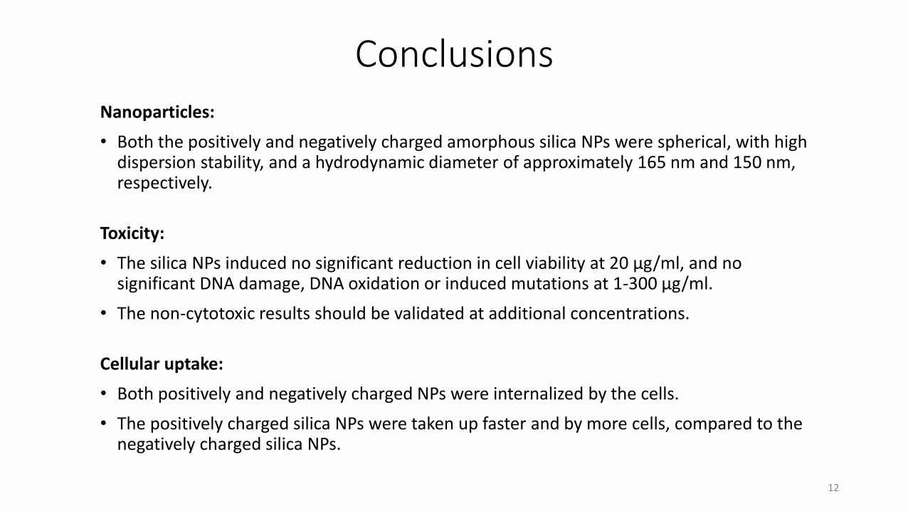
ConclusionsNanoparticles:
• Both the positively and negatively charged amorphous silica NPs were spherical, with high dispersion stability, and a hydrodynamic diameter of approximately 165 nm and 150 nm, respectively.
Toxicity:
• The silica NPs induced no significant reduction in cell viability at 20 µg/ml, and no significant DNA damage, DNA oxidation or induced mutations at 1-300 µg/ml.
• The non-cytotoxic results should be validated at additional concentrations.
Cellular uptake:
• Both positively and negatively charged NPs were internalized by the cells.
• The positively charged silica NPs were taken up faster and by more cells, compared to the negatively charged silica NPs.
12

Supplementary figures
13
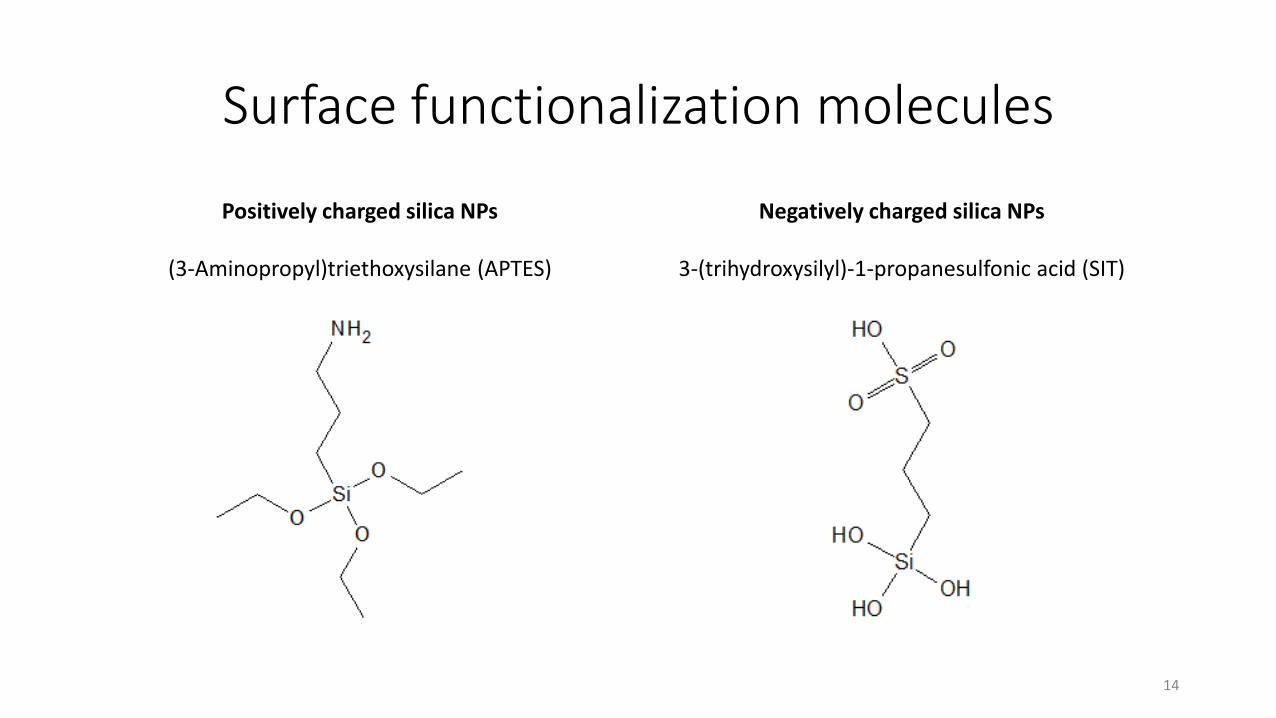
Surface functionalization molecules
14
Positively charged silica NPs
(3-Aminopropyl)triethoxysilane (APTES)
Negatively charged silica NPs
3-(trihydroxysilyl)-1-propanesulfonic acid (SIT)
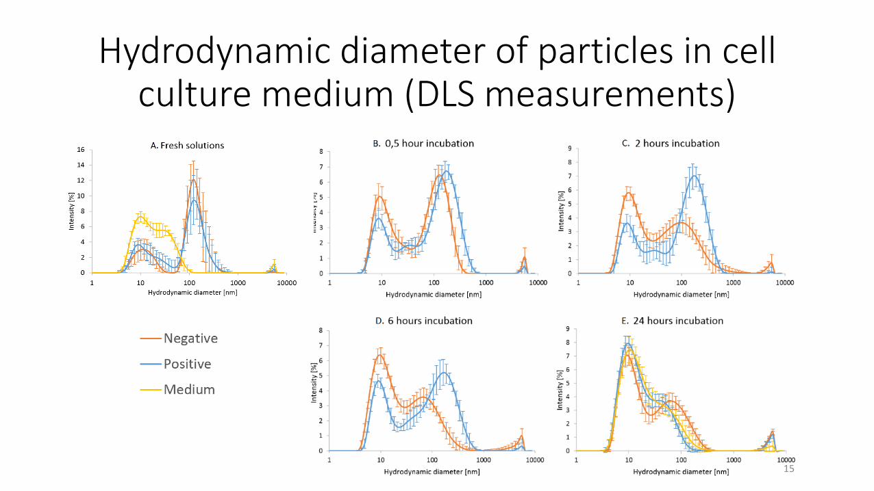
Hydrodynamic diameter of particles in cell culture medium (DLS measurements)
15