cellular mechanisms for information coding in auditory brain stem
TRANSCRIPT
Cellular Mechanisms for Information Coding in Auditory Brain Stem Nuclei.
Laurence Trussell Oregon Hearing Research Center and Vollum Institute, L-335A Oregon Health Sciences University 3181 SW Sam Jackson Park Rd Portland, OR 97201 Phone 503-494-3424 FAX 503-494-3403 Email [email protected]
Chapter 3 in Integrative Functions in the Mammalian Auditory Pathway, edited by D. Oertel, R.R. Fay and A.N Popper. New York: Springer 2002, pp. 72-98.
Introduction The brain stem auditory nuclei carry out a wide variety of transformations of the signals
carried by the auditory nerve. Although basic frequency and intensity information is first encoded in the cochlea, brain stem circuitry must perform further neural definitions and refinements of these parameters, as well as integrate the cues necessary for the localization of sounds in space. Each of these aspects is associated not just with certain cell types, morphologies, and synaptic connections, but with cells having characteristic electrical response profiles. Such response properties are an outcome of the complement of ion channels that the cells possess and of the dynamic properties of the synapses through which cells communicate.
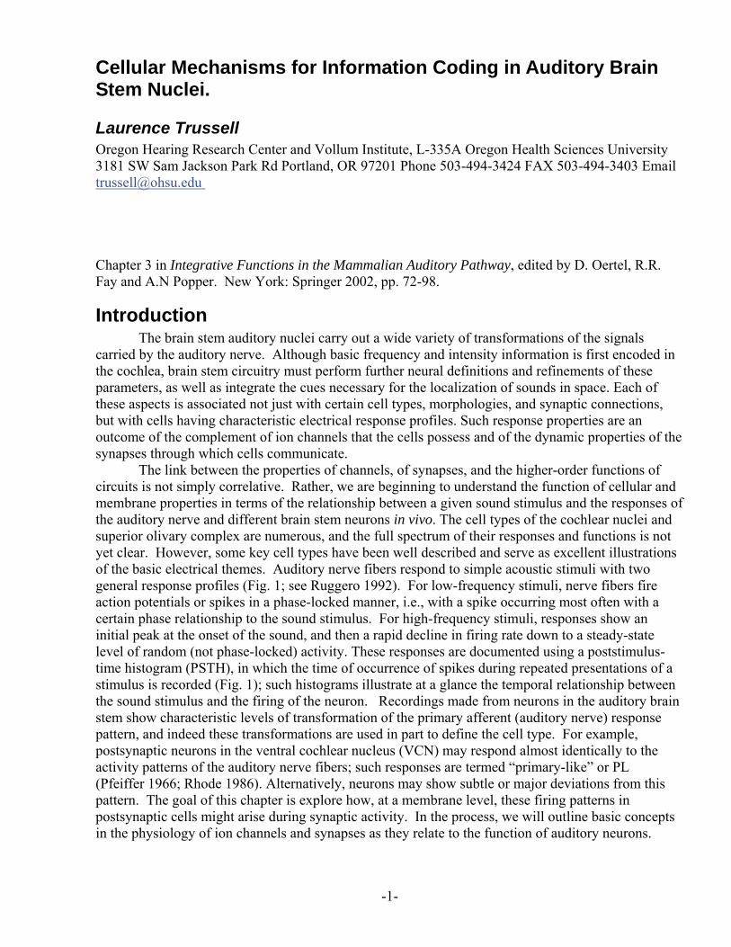
The link between the properties of channels, of synapses, and the higher-order functions of circuits is not simply correlative. Rather, we are beginning to understand the function of cellular and membrane properties in terms of the relationship between a given sound stimulus and the responses of the auditory nerve and different brain stem neurons in vivo. The cell types of the cochlear nuclei and superior olivary complex are numerous, and the full spectrum of their responses and functions is not yet clear. However, some key cell types have been well described and serve as excellent illustrations of the basic electrical themes. Auditory nerve fibers respond to simple acoustic stimuli with two general response profiles (Fig. 1; see Ruggero 1992). For low-frequency stimuli, nerve fibers fire action potentials or spikes in a phase-locked manner, i.e., with a spike occurring most often with a certain phase relationship to the sound stimulus. For high-frequency stimuli, responses show an initial peak at the onset of the sound, and then a rapid decline in firing rate down to a steady-state level of random (not phase-locked) activity. These responses are documented using a poststimulus-time histogram (PSTH), in which the time of occurrence of spikes during repeated presentations of a stimulus is recorded (Fig. 1); such histograms illustrate at a glance the temporal relationship between the sound stimulus and the firing of the neuron. Recordings made from neurons in the auditory brain stem show characteristic levels of transformation of the primary afferent (auditory nerve) response pattern, and indeed these transformations are used in part to define the cell type. For example, postsynaptic neurons in the ventral cochlear nucleus (VCN) may respond almost identically to the activity patterns of the auditory nerve fibers; such responses are termed “primary-like” or PL (Pfeiffer 1966; Rhode 1986). Alternatively, neurons may show subtle or major deviations from this pattern. The goal of this chapter is explore how, at a membrane level, these firing patterns in postsynaptic cells might arise during synaptic activity. In the process, we will outline basic concepts in the physiology of ion channels and synapses as they relate to the function of auditory neurons.
-1-
2. Techniques The earliest studies of single neurons in the brain stem used extracellular recording techniques,
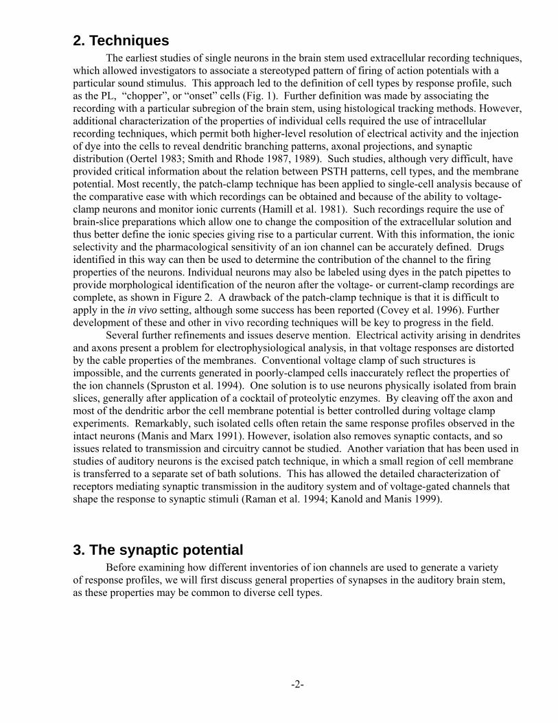
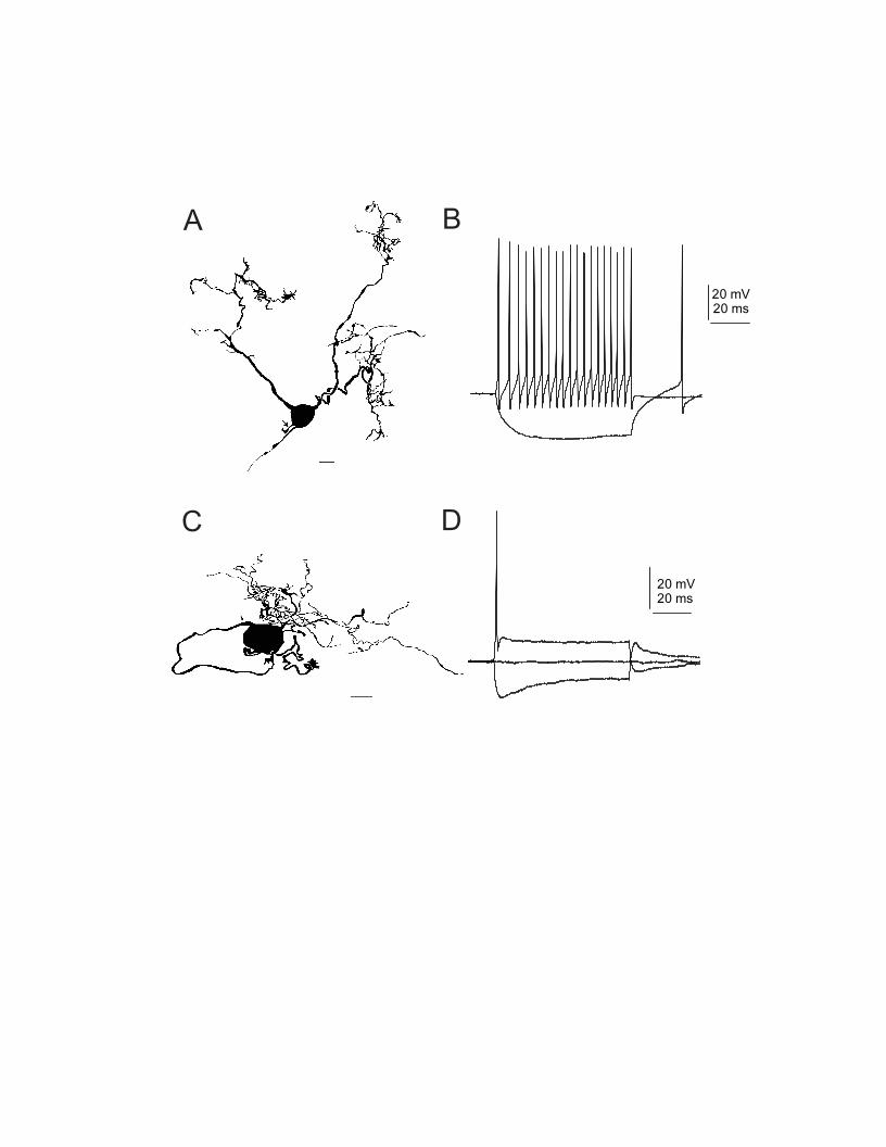
which allowed investigators to associate a stereotyped pattern of firing of action potentials with a particular sound stimulus. This approach led to the definition of cell types by response profile, such as the PL, “chopper”, or “onset” cells (Fig. 1). Further definition was made by associating the recording with a particular subregion of the brain stem, using histological tracking methods. However, additional characterization of the properties of individual cells required the use of intracellular recording techniques, which permit both higher-level resolution of electrical activity and the injection of dye into the cells to reveal dendritic branching patterns, axonal projections, and synaptic distribution (Oertel 1983; Smith and Rhode 1987, 1989). Such studies, although very difficult, have provided critical information about the relation between PSTH patterns, cell types, and the membrane potential. Most recently, the patch-clamp technique has been applied to single-cell analysis because of the comparative ease with which recordings can be obtained and because of the ability to voltage-clamp neurons and monitor ionic currents (Hamill et al. 1981). Such recordings require the use of brain-slice preparations which allow one to change the composition of the extracellular solution and thus better define the ionic species giving rise to a particular current. With this information, the ionic selectivity and the pharmacological sensitivity of an ion channel can be accurately defined. Drugs identified in this way can then be used to determine the contribution of the channel to the firing properties of the neurons. Individual neurons may also be labeled using dyes in the patch pipettes to provide morphological identification of the neuron after the voltage- or current-clamp recordings are complete, as shown in Figure 2. A drawback of the patch-clamp technique is that it is difficult to apply in the in vivo setting, although some success has been reported (Covey et al. 1996). Further development of these and other in vivo recording techniques will be key to progress in the field.
Several further refinements and issues deserve mention. Electrical activity arising in dendrites and axons present a problem for electrophysiological analysis, in that voltage responses are distorted by the cable properties of the membranes. Conventional voltage clamp of such structures is impossible, and the currents generated in poorly-clamped cells inaccurately reflect the properties of the ion channels (Spruston et al. 1994). One solution is to use neurons physically isolated from brain slices, generally after application of a cocktail of proteolytic enzymes. By cleaving off the axon and most of the dendritic arbor the cell membrane potential is better controlled during voltage clamp experiments. Remarkably, such isolated cells often retain the same response profiles observed in the intact neurons (Manis and Marx 1991). However, isolation also removes synaptic contacts, and so issues related to transmission and circuitry cannot be studied. Another variation that has been used in studies of auditory neurons is the excised patch technique, in which a small region of cell membrane is transferred to a separate set of bath solutions. This has allowed the detailed characterization of receptors mediating synaptic transmission in the auditory system and of voltage-gated channels that shape the response to synaptic stimuli (Raman et al. 1994; Kanold and Manis 1999).
3. The synaptic potential Before examining how different inventories of ion channels are used to generate a variety
of response profiles, we will first discuss general properties of synapses in the auditory brain stem, as these properties may be common to diverse cell types.
-2-
3.1 Excitatory transmission Rapid excitatory postsynaptic potentials (EPSPs) in the auditory pathway, such as those
mediating auditory nerve signaling, are generated by ionotropic glutamate receptors (reviewed by Parks 2000). Glutamate receptors fall into three major categories, the AMPA (alpha-amino-3-hydroxy-5-methyl-4-isoxazolepropionic acid ) receptor, the kainate receptor, and the NMDA (N-methyl-D-aspartate) receptor (reviewed by Hollmann 1999). While there is evidence for all three receptor subtypes in the cochlear nuclei and superior olivary complex, the weight of evidence indicates that the AMPA receptor plays the major role in fast transmission. For example, quinoxaline-derived antagonists, which act at AMPA and kainate receptors, completely block transmission mediated by auditory nerve fibers in the cochlear nuclei or by axons of bushy cells in the medial nucleus of the trapezoid body (MNTB) (Wu and Kelly 1992b; Zhang and Trussell 1994b; Isaacson and Walmsley 1995). The non-competitive antagonist of AMPA receptors GYKI-52466 also blocks auditory nerve transmission (T. Otis and L. Trussell, unpublished observations), and synaptic responses on bushy cells and MNTB neurons are modulated by cyclothiazide, a drug selective for the AMPA receptor (Trussell et al. 1993; Wu and Borst 1999). NMDA receptors are also activated by the excitatory neurotransmitter of auditory nerve fibers (Zhang and Trussell 1994a; Isaacson and Walmsley 1995; Ferragamo et al. 1998), but their role in auditory transmission is less clear, perhaps providing slow, stable depolarization or the mediation of long-term plasticity. In bushy cells and MNTB neurons, the expression of NMDA receptors subsides during development (Bellingham et al. 1998; Taschenberger and von Gersdorff 2000). It remains even less clear what the role of kainate receptors is in the auditory system (Petralia et al. 1996). These have been proposed to regulate transmitter release presynaptically in other brain regions (Rodriguez-Moreno et al. 1997), but such analyses have not yet been extended to auditory synapses. Regarding the identity of the transmitter itself, available evidence suggests that it is glutamate. Although both glutamate and aspartate are commonly found in nerve terminals, only glutamate is an effective agonist for both AMPA and NMDA receptors (Patneau and Mayer 1990). There is evidence however that a novel, as yet unidentified compound may be the excitatory transmitter, at least at the hair cell synapse (Sewell and Mroz 1990).
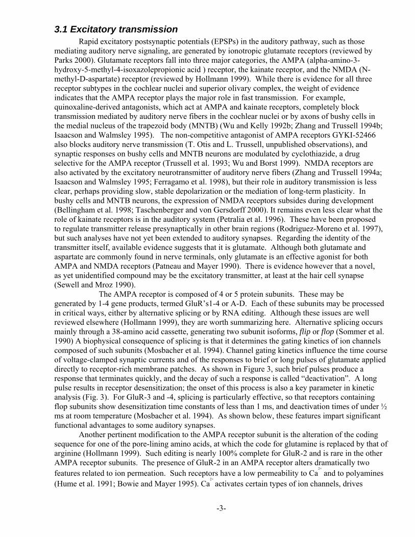
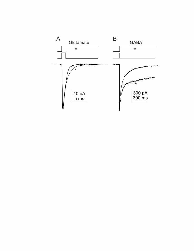
The AMPA receptor is composed of 4 or 5 protein subunits. These may be generated by 1-4 gene products, termed GluR’s1-4 or A-D. Each of these subunits may be processed in critical ways, either by alternative splicing or by RNA editing. Although these issues are well reviewed elsewhere (Hollmann 1999), they are worth summarizing here. Alternative splicing occurs mainly through a 38-amino acid cassette, generating two subunit isoforms, flip or flop (Sommer et al. 1990) A biophysical consequence of splicing is that it determines the gating kinetics of ion channels composed of such subunits (Mosbacher et al. 1994). Channel gating kinetics influence the time course of voltage-clamped synaptic currents and of the responses to brief or long pulses of glutamate applied directly to receptor-rich membrane patches. As shown in Figure 3, such brief pulses produce a response that terminates quickly, and the decay of such a response is called “deactivation”. A long pulse results in receptor desensitization; the onset of this process is also a key parameter in kinetic analysis (Fig. 3). For GluR-3 and -4, splicing is particularly effective, so that receptors containing flop subunits show desensitization time constants of less than 1 ms, and deactivation times of under ½ ms at room temperature (Mosbacher et al. 1994). As shown below, these features impart significant functional advantages to some auditory synapses.
Another pertinent modification to the AMPA receptor subunit is the alteration of the coding sequence for one of the pore-lining amino acids, at which the code for glutamine is replaced by that of arginine (Hollmann 1999). Such editing is nearly 100% complete for GluR-2 and is rare in the other AMPA receptor subunits. The presence of GluR-2 in an AMPA receptor alters dramatically two features related to ion permeation. Such receptors have a low permeability to Ca
2+
and to polyamines (Hume et al. 1991; Bowie and Mayer 1995). Ca
2+
activates certain types of ion channels, drives
-3-
cellular metabolic processes leading to long-term alterations in electrical responsiveness or the biochemical state of the neuron, and may also lead to cell death. How these reactions to the transmitter may play out in auditory function is not clear.
Molecular biological and immunohistochemical studies have revealed the presence of GluR- 3 and -4 in many neurons of the mammalian and avian auditory brain stem and inferior colliculus, particularly in cells which only weakly express GluR-1 and -2 (Rubio and Wenthold 1997; Wang et al. 1998; Caicedo and Eybalin 1999; Ravindranathan et al. 2000). Reverse transcriptase polymerase chain reaction (RT-PCR) indicated further that most subunits in the avian nucleus magnocellularis (NM), angularis (NA) and laminaris (NL), and the rat MNTB were of the flop isoform (Geiger et al. 1995; Ravindranathan et al. 2000). The functional implications of this expression pattern is well supported by physiological studies, as described below.
3.1.1 Time course of the synaptic response How do receptor properties work with other cellular parameters to shape the EPSP? The
synaptic potential is generated by the charging effect of the excitatory synaptic current (i.e, the EPSC) on the membrane capacitance. For synapses with very rapid EPSCs, the decay of the EPSP will be determined by the rate at which the membrane discharges, the membrane time constant. The time constant in turn is dependent on the membrane conductance imparted by the ion channels that are active near the resting potential of the cell. Thus, the duration of the AMPA receptor EPSP will be limited by intrinsic membrane properties. By contrast, the duration of NMDA EPSPs are limited by the sluggish NMDA EPSC, which is generally longer than the membrane time constant (Lester et al. 1990; see section 4).
The duration of the EPSP has important implications for how neurons integrate synaptic signals. Brief synaptic potentials summate only when several synapses fire very close in time; in this way, the output of the postsynaptic cell depends on synchronous activity of presynaptic fibers. For neurons in the timing pathways of acoustic processing, the timing of activity of the inputs fibers is phase-locked to the phase of the sound stimulus. Thus, convergence of weak, brief synaptic potentials provides a simple mechanism for ensuring that the timing information is well preserved through sequential synaptic levels (Joris et al. 1994). When synaptic inputs are large, such as in MNTB neurons, brevity is also critical. As the synapses fire at relatively high rates, EPSPs must be brief enough that sequential events do not overlap and distort the timing of action potential generation in the postsynaptic cell.
3.1.2 Synaptic activation of glutamate receptors The simplest picture for the molecular dynamics of a typical fast excitatory synaptic current
begins with the release of the contents of a glutamate-filled vesicle. The transmitter then binds and activates a limited number of postsynaptic receptors opposite the active zone, producing a so-called miniature excitatory postsynaptic current (mEPSC). Glutamate binding to receptors is weak, i.e. it binds with low affinity. The remaining transmitter that has not bound diffuses away quickly, probably within a few hundred microseconds. The short lifetime of the bound state and of available transmitter together ensure that the transmitter response is terminated quickly. In the brief period that glutamate is still bound, the receptor protein swings between channel open and closed conformations. The frequency with which such transitions occur also contributes to the speed of the overall synaptic response (Trussell 1998).
mEPSCs thus provide a convenient monitor of the overall effects of receptor affinity and
-4-
channel gating kinetics. Measurements of mEPSC decay times in different cell types reveal a striking trend: the fastest decays of any synaptic current in the brain is found in the central auditory neurons, with typical values of between 400-500 μsec at room temperature (Trussell 1998). Where examined, these synaptic currents also show high Ca
2+
permeability and high sensitivity to polyamine channel blockers (Otis et al. 1995; Zhou et al. 1995; Gardner et al. 1999). Together these observations are consistent with the expression of receptors composed largely of GluR-3flop or -4flop. Direct measurement of receptor deactivation and desensitization supports this conclusion. Deactivation time constants measured in patches are identical to those of the mEPSCs (Otis et al. 1996b; Gardner et al. 1999; Gardner et al. 2001). These, and the desensitization time constants, are identical to those of GluR-3flop and -4flop measured in heterologous expression systems (Gardner et al. 2001). Thus, synaptic transmission at many auditory synapses depends upon selective expression of one or two specialized receptor subunits.
Recent studies suggest that expression of the fast-gating subunits may be a consequence of cellular interactions during development of synaptic contacts. Auditory nerve fibers branch upon entering the brain and innervate a wide variety of cell types, at least two in birds and seven in mammals (Warchol and Dallos 1990; Cant 1992). In every case examined, the cells postsynaptic to auditory nerve fibers produce the fast-gating subunits and, in all but one case, express little GluR-2 (Raman et al. 1994; Rubio and Wenthold 1997; Wang et al. 1998; Ravindranathan et al. 2000; Gardner et al. 2001). Moreover, developmental analysis of the receptors indicates that channel gating in AMPA receptors is slow, and sensitivity to polyamines is low, at the time of innervation (Lawrence and Trussell 2000). Finally, isolation of neurons in cell culture prior to innervation prevents the change in channel kinetics (Lawrence and Trussell 2000). These results suggest that some molecular features of the auditory system arises through interactions between pre- and post-synaptic cells.
Besides receptor properties, additional factors also shape the EPSC. Because auditory synapses generally release a large number of vesicles, needed to ensure a suprathreshold postsynaptic response, the time period over which the vesicles fuse substantially broadens the EPSC. At end-bulb and calyceal synapses, release of vesicles requires several hundred microseconds after arrival of an action potential at the nerve terminal; this period, convolved with the duration of the mEPSC, results in an EPSC that lasts about 1 ms (Isaacson and Walmsley 1995; Borst and Sakmann 1996). Additional slower phases of the EPSC are attributed to the gradual clearance of low levels of residual transmitter (Otis et al 1996a).
3.2 Inhibition The major inhibitory neurotransmitters of the auditory system brainstem nuclei are GABA and
glycine (Sato et al. 2000). The GABAA and glycine receptors activate chloride-selective channels whose activation may have a variety of effects. When the reversal potential for chloride ions (ECl) is negative to the resting potential, the transmitter produces a hyperpolarizing inhibitory postsynaptic potential (IPSP). By shunting excitatory currents and bringing the membrane potential away from threshold, the IPSP curtails excitatory signaling. Examples in the auditory system include glycinergic transmission in the VCN, LSO, MSO and MNTB (Banks and Smith 1992; Grothe and Sanes 1993; Kotak et al. 1998; Wu and Fu 1998; Smith et al. 2000). In the cochlear nuclei, inhibition has been proposed to account for the refinement of the range of frequencies resulting in excitation or of the responsiveness to tones in the presence of background noise. Accordingly, frequency-response curves (tuning curves) may be modified in vivo by local application of antagonists of GABA and glycine receptors (Caspary et al. 1993; Evans and Zhao 1993; Caspary et al. 1994). However, the precise neural circuitry that modifies tuning characteristics has not been defined; potential sources of such
-5-
inhibition exist throughout the superior olivary complex and in the cochlear nuclei themselves (Ostapoff et al. 1990; Saint Marie et al. 1991; Wickesberg et al. 1991; Ferragamo et al. 1998).
Inhibitory mechanisms play a clearer role in the convergence of excitation and inhibition on neurons of the LSO. Here, the timing of convergence of the two signals is believed to be critical for encoding of interaural sound level differences, such that sounds louder at the contralateral ear will suppress activity of neurons excitated by weaker ipsilateral inputs (Tsuchitani and Boudreau 1967). Another key feature of this inhibition is that the IPSPs are comparatively brief, on the order of a few ms, which is presumably required for precise temporal convergence of ipsi- and contralateral signals (Wu and Kelly 1992a). Glycinergic signals are also seen in bushy cells of the ventral cochlear nucleus, and are thought to be mediated by tuberculoventral cells of the dorsal cochlear nucleus (Wu and Oertel 1986; Wickesberg et al. 1991). These inputs, in which the inhibitory neurons have a similar characteristic frequency as the bushy cell, have been proposed to play a role in the suppression of echos generated within the ear (Wickesberg and Oertel 1990). Again, rapid synaptic signaling and precise convergence would be essential to this mechanism.
However, this picture of inhibitory function as mediated by precise convergence of depolarizing and hyperpolarizing signals is not universal. Early in development of the LSO, the reversal potential for chloride ions is depolarizing, and the inhibitory terminals from the MNTB release GABA as well as glycine (Sanes and Friauf 2000). Due to their strong depolarizing action, these transmitters have excitatory actions in the developing brain whose functions may be related to the activation of voltage-activated Ca
2+
channels and Ca2+
dependent synapse stabilization (Sanes and Friauf 2000). In some regions, GABA and glycine are depolarizing even in the mature animal. In the dorsal cochlear nucleus, for example, glycinergic cartwheel cells provide depolarizing IPSPs onto other cartwheel cells and hyperpolarizing IPSPs onto fusiform neurons (Golding and Oertel 1996, 1997). The effect of the depolarizing IPSP is apparently complex, such that when the cartwheel cell is relatively quiescent, the IPSP will facilitate effects of excitatory signals, while during more intense periods of activity, the IPSP is inhibitory to action potential generation. Generally, inhibition produced by depolarizing IPSPs is interpreted in terms of the shunting effects of the glycinergic or GABAergic conductance, which drives the membrane potential below spike threshold. Developmentally stable depolarizing IPSPs are also evident in the avian bushy cells of NM, as described below.
Another intriguing variable in the IPSPs of auditory neurons is their time course. While glycinergic IPSPs are generally of short duration, GABAergic signals are slower to decay, often outlasting the EPSP by 10’s of ms, a result of the slow gating kinetics of GABA receptors (Fig. 3). In mammals, there are few direct demonstrations of GABAergic IPSPs in the cochlear nuclei and olivary complex (Ferragamo et al. 1998; Lim et al. 2000), but in birds GABA is the major inhibitory transmitter. Neurons of the avian NM and NL, which receive depolarizing GABAergic input from the superior olive (Hyson et al. 1995), respond to trains of stimuli delivered to the GABAergic fibers with depolarizing plateau potentials of about 10 mV; after the period of stimulation, IPSPs decay gradually back to baseline over 100 ms (Yang et al. 1999; Lu and Trussell 2000). Depolarizing inhibition shortens the membrane time constant, thus ensuring that only perfectly convergent EPSPs summate to threshold (Funabiki et al. 1998). The mechanism for generation of protracted inhibition appears to involve a combination of pre-and postsynaptic factors (Lu and Trussell 2000). Slow IPSPs, due to the slower gating kinetics of the GABA receptor, summate during high frequency activity to produce a relatively smooth plateau. Presynaptically, however, the timing of the response of the synapses to presynaptic action potentials appears to become disrupted, and GABA is released at random intervals during a train of regular stimuli. This desynchronization is due to Ca
2+
accumulation in the terminal, the dynamics of vesicle depletion and replenishment, and the facilitation of vesicle fusion probability. The slow decline of these processes after cessation of presynaptic action potential activity delays the termination of the IPSP (Lu and Trussell 2000).
-6-
Such late inhibition have been suggested to play computationally significant roles in sound localization in the inferior colliculus (Klug et al. 1999).
3.3 Presynaptic regulation of synaptic transmission Synaptic potentials vary in amplitude during repetitive synaptic stimulation. Some variation
is random from event-to-event, a consequence of the probabilistic nature of transmitter release (del Castillo and Katz 1954). Some, however, is use-dependent, resulting from simple forms of short-term synaptic plasticity, facilitation and depression (Zucker 1989). These are synapse-specific features whose significance for sensory processing are only just being appreciated as brain slice and voltage clamp methods are applied to auditory synapses. As noted above, facilitation and depression also play a key role in shaping inhibitory plateaus in NM. In large end-bulb and calyceal synapses, transmitter is released with relatively high probability, ensuring a rapid approach to action potential threshold. With continuous activity at high rates, however, these terminals undergo synaptic depression, with EPSPs and EPSCs falling to a smaller, steady-state level depending on stimulus frequency, temperature, and age of the animal (Wu and Oertel 1987; Brenowitz et al. 1998; Taschenberger and von Gersdorff 2000). A use-dependent reduction in release is understandable in terms of a simple model of the synapse. Each synaptic site has a limited number of vesicles available for release (the readily-releasable pool) and these are replenished at a finite rate, usually with a time course of at least tens of milliseconds (Dittman and Regehr 1998; Wang and Kaczmarek 1998). This model has also been invoked to account for adaptation of spike rate during tones in auditory nerve fibers (Geisler and Greenberg 1986; Moser and Beutner 2000). Besides depletion of vesicles, other pre- and postsynaptic factors may also contribute to lack of responsiveness of synapses (Otis et al. 1996a; Wu and Borst 1999; Waldeck et al. 2000). Nevertheless, despite massive reductions over time in the number of vesicles that are released per stimulus, mature terminals are still able to generate suprathreshold EPSPs due to the large number of release sites that remain active. For example at MNTB calyces, EPSPs remain suprathreshold for at least 8-10 stimuli at 600 Hz (Taschenberger and von Gersdorff 2000). For multiply-innervated bushy cells, depression at each synapse may be profound, but is compensated by the larger number of synapses contributing to the depolarization.
Release at calyceal and end-bulb terminals may also be regulated by activation of presynaptic receptors. For example, metabotropic glutamate, GABAB, and adenosine receptors inhibit release through inhibition of presynaptic Ca
2+
channels (Forsythe and Barnes-Davies 1995; Takahashi et al. 1996; Brenowitz et al. 1998). However, although these receptor systems are capable of dramatically altering the size of EPSCs elicited at low frequencies, their function in physiological contexts remains obscure. In chick bushy cells (NM), it has been suggested that GABAB receptors may act to reduce synaptic depression, as lowering transmitter release appears to lessen postsynaptic receptor desensitization . Still, it has not yet been shown that such receptors are activated in vivo at these synapses. Lim and Walmsley (2000) found that synaptic strength at glycinergic synapses could be enhanced by antagonists of GABAB receptors, suggesting that GABA, perhaps co-released with glycine, inhibits release of glycine. More recently, glycine receptors themselves have been demonstrated on MNTB calyces. Activation of these receptors appears to enhance EPSPs through an unusual mechanism, in which opening of glycine-gated chloride channels in the nerve terminal depolarizes the synapse, resulting in an elevation of presynaptic Ca
2+
and a facilitation of release (Turecek and Trussell 2001). These glycine receptors may be activated following stimulation of nearby glycinergic fibers, implying that glycine must effectively diffuse from glycinergic boutons to excitatory terminals.
-7-
4. Intrinsic properties of neurons As we have discussed, the response of a neuron to synaptic stimuli depends on the
characteristics of the synaptic receptors and of the ion channels they activate. The outcome of this change in membrane conductance, from the perspective of the output of the neuron, is related to the inventory of voltage-gated ion channels, the electrotonic structure of the neuron, the number and location of synapses, and the temporal structure of their activity. Gathering information on all of these factors in one experimental system borders on impossibility. Thus, to describe how these factors converge to generate a basic stereotypical response, we must draw upon both in vivo and in vitro experimental data and also computer simulations. When based upon real biological parameters, such simulations permit the testing of concepts that significantly enhance the interpretation of data and the subsequent design of experiments.
4.1 Choppers The response of neurons in the cochlear nucleus to sound stimuli is characterized by the
patterns observed in the PSTH, with additional information provided by other statistical analysis of the response, such as interspike intervals and latencies. One of the major groupings, the “chopper”, is characterized by the presence of a regular train of action potentials whose intervals are consistent (thus the term “regular spiking”) and are not related to the frequency of the sound stimulus (Rhode and Smith 1986; Rhode and Greenberg 1992). The chopping may persist during the stimulus or dampen out after a few ms, in which case the spiking becomes random from trial to trial. In response to a low-frequency sound, the choppers often show poor phase locking. In vivo studies have identified neurons showing sustained (CS) or transient (CT) chopping as stellate cells of the ventral cochlear nucleus (Rhode et al. 1983a). This identification of neurons is supported by in vitro studies using microelectrodes or patch pipettes. These show that neurons identified morphologically as stellate cells, respond to prolonged, square pulses of positive current with a train of regularly spaced action potentials, as shown in Figure 2A, B (Oertel 1983; Manis and Marx 1991; Isaacson and Walmsley 1995). Larger current steps yield a higher frequency of spikes, with a minimum-to-maximum frequency range of several fold. This profile of response, in its crudest form, can be mimicked with computational models that resemble the classical squid giant axon in channel composition: an inactivating Na
+
channel, a delayed rectifier K+ channel and a non-selective leak
current (Banks and Sachs 1991; Manis and Marx 1991; White et al. 1994). Although the squid axon does not behave exactly like a chopper, simple adjustments in the properties of the component ion channels, such as gating kinetics and thresholds for activation, apparently yield a model that shows many of the features of choppers observed experimentally. The intracellular studies support this assessment, as voltage clamp of enzymatically-isolated regular-spiking neurons exhibit a dominant outward potassium current, blocked by tetraethylammonium ions (TEA), a sodium current, and a background “leakage” current (Manis and Marx 1991). Based on the properties of these ionic currents, Manis and Marx (1991) were able to recontruct the basic regular-spiking response in a computer simulation. Depolarizing currents bring the cells to threshold, setting in motion the classical Hodgkin-Huxley cycle of activation, inactivation and deactivation of the Na
+ and K
+ current. When
the depolarizing current is sustained, the cycle can be repeated, primarily because the channels are able to reprime themselves sufficiently after the last action potential despite the persistent depolarization provided by the stimulus. Regularity of spike interval occurs because the degree to which the channels can reprime themselves is similar from spike to spike. These results imply that the stellate cell could produce a chopping response given a basic complement of ion channels, but specifically in response to a simple, prolong stimulus.
-8-
Beyond the voltage-clamp characterization described by Manis and Marx (1991), there is little information about the molecular identity of the voltage-gated channels of choppers. The key features required of the K
+
channels to facilitate chopping include 1) rapid channel kinetics, which allow channels to open and close in the time scale of an action potential, 2) a high (more depolarized) voltage “threshold” for the channels, allowing the cell to deactivate the K
+
current even during a persistent depolarizing stimulus, and 3) little or no channel inactivation, which would otherwise impede repolarization and repetitive firing later in trains of stimuli. One candidate is the Kv3.1 potassium channel subunit. When expressed in heterologous expression systems, Kv3.1 produces a K
+
current with a high threshold for activation and rapid kinetics. Repetitive spiking neocortical neurons express this channel (Martina et al. 1998; Erisir et al. 1999). However, in situ hybridization indicates that only a fraction of stellate-like cells express this channel (Perney and Kaczmarek 1997), and thus other channels with similar properties may be important in stellate neurons.
How can these cells produce a chopping response during a barrage of complex synaptic stimuli? This question has been approached both experimentally and through computational methods. Banks and Sachs (1991) developed a model of the stellate cell that incorporated the basic morphological features of the neuron, including several dendrites, an axon, and a cell body. The pattern of synaptic activity driving the cell was made to simulate the actual temporal features of auditory nerve responses to tones. Two major parameters that were examined were the effects of varying synapse location and of synapse number. When a small number (4) of synapses converged close to the cell body, the individual synaptic potentials were large and summated to variable degrees. As a result, the cell’s firing pattern was highly irregular. Synapses placed on electrotonically distant dendritic locations produced synaptic response that were heavily filtered (made smaller and slower) more severely by the resistance and capacitance of the dendrites, i.e. their cable properties. Increasing the number of synaptic inputs to ten could compensate for the reduction in amplitude, producing a smooth depolarizing response at the cell body. When the action potential was generated in or near the cell body, the output of the cell was now a regular train of spikes, reminiscent of the response of the model to a simple square pulse of current. In principle, differences among cells in the regularity of spiking could reflect differences in the location of synapses; morphological data indicates that stellate cells may indeed differ in the relative position of excitatory synapses (Smith and Rhode 1989). Similar conclusions were reached using more sophisticated models based on actual morphometric data (White et al. 1994). The weakness of phase locking then would be an outcome of the rigid response profile of the cell to a stimulus and the filtering properties of the dendrites, which together would blur the temporal information contained in auditory nerve firing.
More recent brain slice studies suggest that the number of inputs to VCN stellate cells of the mouse (5) are not as many as were required in the models described above to account for the regularity of spike output (Ferragamo et al. 1998). Moreover, it appears that the actual synaptic waveform itself was more complex than that employed in the model. Excitation was mediated by a fast, AMPA receptor-dependent EPSP, but also by an NMDA-receptor EPSP that lasted for over 100 ms after the synaptic stimulus was terminated. Blockade of NMDA receptors resulted a loss of firing during periods greater than a few ms after the stimulus. Late, fast EPSPs were also observed, suggesting that disynaptic pathways may enhance excitation of the stellate cell following auditory nerve stimulation. These data indicate the irregular chopping resulting from a small number of auditory nerve synaptic inputs might be alleviated by additional slow or delayed excitatory events. However, it should be noted that these effects occur over a period of 100’s of ms, while chopping per se is defined during a period of <50 ms. Thus, it remains unclear how multiple receptor subtypes and recurrent excitation may shape the earliest response to sound in choppers.
-9-
4.2 Primary-like (PL) responses and phase locking The main features of the PL response are the gradual adaptation and irregularity of firing to
high-frequency stimuli, and phase-locking to lower-frequency inputs. A related profile is primary-like with notch (PL-N), which shows a sharp transient drop in firing after an initial well-timed spike, and excellent phase locking at low frequencies. Onset-lock (OL) units are similar to PL-N, but have a lower rate of firing late in the response to a high-frequency tone (Rhode and Greenberg 1992). What cellular properties permit such a close mimicking of the properties of the synaptic input, which is clearly required for preservation of timing information?
PL and PL-N responses are most likely generated by AVCN spherical and globular bushy cells, respectively (Rhode et al. 1983a; Rhode and Smith 1986; Smith and Rhode 1987; Smith et al. 1993). The most basic features bushy cells must have in order to account for these responses are strong, brief synaptic inputs and ion channels that allow the membrane voltage to follow closely the changes in synaptic current and to recover quickly after each action potential. The latter have been well-studied in brain-slice preparations. Oertel (1983) first showed that mouse bushy cells respond to a prolonged, strong depolarizing current injection with a single action potential, a finding confirmed in bushy cells of rat and guinea pig AVCN and chick NM (Fig. 2C, D; Reyes et al. 1994; Zhang and Trussell 1994b; Isaacson and Walmsley 1995; Schwarz et al. 1998). Repetitive, brief stimuli, however, easily elicit action potentials, indicating that the cell membranes are designed for permitting a single suprathreshold response but a rapid recovery following the termination of the stimulus. Unlike choppers, even weak depolarizing currents cause a sharp decrease in membrane resistance (increase in conductance) in bushy cells, indicating that ion channels open with a very low threshold relative to the resting potential. Further studies using voltage clamp techniques support this inference, and defined in bushy cells two important K
+
currents (Manis and Marx 1991; Rathouz and Trussell 1998). A low-threshold current (LTC) is slightly active at the resting potential and becomes more completely activated when the cell is depolarized by 10-15 mV. The K
+ channel producing this
current is sensitive to 4-aminopyridine (4-AP) and dendrotoxin. A high-threshold current (HTC) similar to that of stellate cells activates quickly when during an action potential the membrane potential exceeds about –20 mV. Blockade of HTC by tetraethylammonium (TEA) ions results in a broadened action potential, but does not initiate repetitive firing. By contrast, blockade of LTC with 4-AP or dendrotoxin transforms the neurons into repetitively spiking cells, supporting the notion that LTC may be key to distinguishing the electrical properties of the stellate and bushy cell.
How do these ionic currents shape a single synaptic response in bushy cells? With simultaneous activation of a suitable number of inputs, the inward synaptic current depolarizes the neuron to threshold and an action potential is fired. The rapid initiation of the spike is due to the large number of synaptic vesicles released, and the nearness of the somatic synapses to the spike initiation zone on the axon. The cell then quickly repolarizes due the shut down of Na
+
and K+
channels, and the abrupt decay of the synaptic current, mediated by the fast-acting AMPA receptors. Thus, the cell is then ready to respond again. Several factors may limit the likelihood of a subsequent response. Voltage-gated channels must reprime, and stimuli at too close an interval will fall within the cell’s spike refractory period, i.e., when spike threshold is elevated. Synapses also have a refractoriness of their own (see section 3.3): presynaptic active zones, once they have released a vesicle, must prepare a new release-ready vesicle, and this may take at least several ms. Postsynaptic receptors may be briefly desensitized by the transmitter, and require time to recover. One factor that might aid in restoration of the synapse is that transmission is mediated by a large number of release sites, not all of which release in response to any one presynaptic action potential. Thus, a moderate release probability may aid in the ability of the synapse to transmit at high frequencies (Schneggenburger et al. 1999).
Phase-locking of auditory nerve signals is never perfect, and so the arrival of signals at the
-10-
spherical bushy cell occurs with variable latencies (jitter) (Young et al. 1988; Blackburn and Sachs 1989). Later in the response to a tone, such jitter is even more enhanced, particularly during high-frequency tones in which phase locking is lost. The synaptic delay, the interval between the arrival of the presynaptic spike and the onset of the postsynaptic response, is also a stochastic process contributing to transmission jitter (Barrett and Stevens 1972). Spherical bushy cells receive input from only a few auditory nerve axons, unlike globular bushy cells (Sento and Ryugo 1989; Liberman 1991), and one would expect that strength of their synapses to be correspondingly larger. When a few, strong EPSPs occur with some jitter, the one with the shortest latency will initiate a postsynaptic action potential. However, when a larger number of smaller EPSPs occur, as with globular bushy cells, they must summate until threshold is reached. Two factors ensure that such summation does not destroy the preservation of timing, but may actually enhance it (Joris et al. 1994). First, each EPSP is very brief, and so they can only summate effectively if they occur with a similar latency. Second, if the jitter is such that the summation occurs over a longer period of time, activation of the LTC will reduce the effectiveness of the EPSPs and threshold cannot be reached. As a result, postsynaptic spikes will only occur when driven by summation of well-timed subthreshold EPSPs. Such a mechanism accounts for the differences in firing rate of PL and O-L cells late in the response to a high-frequency tone (Rhode and Smith 1986; Young et al. 1988). The “notch” on the PL-N PSTH arises apparently from the fact that synaptic convergence effectively drives the globular bushy cell to fire with little jitter; the ensuing refractory period of the bushy cell must therefore also occur with uniform latency. Rothman et al. (1993) have explored these aspects of bushy cells activity with an excellent computer model which accounts for many aspects of the membrane physiology.
4.3 The MNTB mirrors bushy cell activity Principal cells of the MNTB show membrane properties remarkably similar to those of bushy
cells, in terms of expression of voltage- and glutamate-evoked currents as well as the presence of a somatic glutamatergic synapse. The MNTB receives only a single, large calyceal terminal which reliably transmits signals from the globular bushy cell (Guinan and Li 1990; Smith et al. 1998). Thus, the MNTB neurons show a PL-N PSTH in response to tones, not because of convergence (as discussed above) but rather because of the neurons’ ability reproduce the activity of the globular bushy cell so faithfully.
The MNTB has received much attention regarding the molecular identity of the K+
channels responsible for the high precision in the temporal responsiveness synaptic input. As with bushy cells, dendrotoxin or 4-AP induces repetitive firing to long current steps (Banks and Smith 1992; Brew and Forsythe 1995). Under voltage clamp, these neurons exhibit a prominent LTC (Brew and Forsythe 1995). Several features of this current are very similar to those of currents produced by the Kv1.1 and/or 1.2 K
+
channels, which are also strongly expressed in MNTB as well as in VCN (Grigg et al. 2000). The HTC is also quite prominent in MNTB and, as with bushy cells, is sensitive to TEA. Wang et al. (1998) contrasted the properties of the Kv3.1 channel expressed in cell lines with the HTC of MNTB, finding a remarkable degree of similarity in their pharmacological and voltage sensitivity, and gating kinetics. Computer simulations incorporating the LTC and HTC suggested that the HTC is necessary to permit firing at high rates by rapidly terminating the action potential (see section 4.1). Accordingly, Kv3.1 is strongly expressed in the MNTB, as well as in bushy cells (Perney et al. 1992; Perney and Kaczmarek 1997; Wang et al. 1998).
-11-
4.4 Octopus cells and detection of coherent activity Octopus cells of the posterior ventral cochlear nucleus respond to high-frequency tones with a
strong O-L response, that is, an extremely well-timed initial response followed by a low level of random activity (Rouiller and Ryugo 1984; Rhode and Smith 1986). Octopus cells phase lock, and even entrain, extremely well to intense low-frequency sounds (≤ 1000 kHz). This behavior represents an outcome of several morphological and electrophysiological features of these neurons. Octopus cells send their stout dendrites across bands of auditory nerve fibers, sampling a broad spectrum of acoustic best frequencies (Osen 1969; Kane 1973). These fibers (and, rarely, other octopus cells) make excitatory synapses along the dendrites; it is estimated that at least 60 auditory nerve fibers innervate each octopus cell (Golding et al. 1995). The neurons also show striking levels of expression of two types of channel: the dendrotoxin-sensitive LTC channels and the hyperpolarization-activated non-selective cation channel, IH (Golding et al. 1995; Ferragamo and Oertel 1998; Bal and Oertel 2000; Grigg et al. 2000). As a result, octopus cells exhibit a very low input resistance and extremely short membrane time constant (200 μsec) near the resting potential. By contrast, bushy and MNTB neurons require depolarization to attain such a short time constant. As noted above, clicks or loud tones will activate a broad spectrum of auditory nerve fibers and evoke a well-timed spike in the octopus cell. However, during the later phases of high-frequency auditory nerve activity the input activity is random and summation cannot occur. Thus, octopus cells are well suited to respond to broad-band phase-locked responses of the auditory nerve, leading to the suggestion that they may serve to detect common inter-spike intervals in auditory nerve fibers, necessary to encoding pitch (Golding et al. 1995).
The leakiness of the membrane at rest, conferred by IH, and the sharp onset of LTC may work together to allow this computation in the face of very complex activity in fibers of diverse best frequency (Bal and Oertel 2000; Cai et al. 2000). For example, such a high degree of synaptic convergence would produce significant depolarizations in other neurons, simply through spontaneous fiber activity alone. Leakiness of the resting cell membrane shunts these synaptic currents and minimizes the chances of random simultaneous EPSPs reaching threshold. When a group of fibers are active nearly simultaneously, the LTC acts to assure that the response only occurs for the very best-timed inputs and that the latency to the response is short, in a way similar to that discussed above for bushy cells. As the membrane depolarizes, LTC activates, increasing the amount of current needed to drive the cell to threshold. Thus unless the cell can be depolarized quickly, LTC prevents excitation. In this way, octopus cells perform a rate-of-rise discrimination on synaptic signals, filtering out activity that is not sufficiently coherent (Ferragamo and Oertel 1998).
4.5 Other coincidence detection mechanisms Neurons of the medial superior olive and the avian nucleus laminaris are key elements in the
neural mechanism of sound localization in the horizontal plane (Carr and Konishi 1990; Yin and Chan 1990; Carr 1993). MSO and NL neurons have intrinsic membrane properties similar to those of bushy cells, and use these to detect the coincident arrival of signals from ipsi- and contralateral spherical bushy cells (see Yin, Chapter 4). A high degree of bushy cell convergence and the high sensitivity to the timing of signals is likely to produce the O-L responses and strong phase locking to monaural sound measure in MSO, using the principles outlined above (Rothman et al. 1993). However, studies in the avian NL suggest that dendrites may play a unique role in enhancing the ability of the neurons to respond particularly well to the coincidence of ipsi- and contralateral signals (Agmon-Snir et al. 1998).
Chicken NL comprises a sheet of neurons with clusters of dendrites at the dorsal/ventral poles of each cell body (Jhaveri and Morest 1982). Neurons receive input from ipsilateral bushy cells (from
-12-
NM) on their dorsal dendrites and input from contralateral bushy cells on their ventral dendrites. Both innervation and morphology of NL is tonotopic, in that cells receiving input from high best-frequency NM have short dendrites, while cells innervated by low BF NM have long dendrites (Rubel and Parks 1975). Agmon-Snir and colleagues explored the role of this remarkable dendritic layout using computer simulations (Agmon-Snir et al. 1998). When a given number of synapses are active in nearby membrane, the depolarization they produce reduces the driving force for current through the synaptic channels. This results in a reduction in the efficacy of that set of inputs in bringing the cell body to spike threshold. When inputs are separated at different dendritic locations, this effect of nonlinear summation is reduced, and that same number of inputs are more effective. The authors found that coincidence of ipsi- and contralateral inputs was apparently improved, and convergence from same-dendrite inputs diminished, by this mechanism. Moreover they found that the usefulness of the dendrites falls off with high frequency input, and noted that this may account for the comparative lack of dendrites in high best-frequency NL neurons.
4.6 Fine tuning of response properties in fusiform cells Fusiform (pyramidal) cells of the dorsal cochlear nucleus participate in circuits that integrate
somatosensory inputs with auditory signals and may play an important role in sound localization based on monaural spectral cues (see Young, chapter 5). In vivo recordings indicate that fusiform cells can exhibit a variety of PSTH profiles, including chopper, onset and build-up patterns (Pfeiffer 1966; Godfrey et al. 1975; Rhode et al. 1983b). Rhode and colleagues suggested that such patterns may shift with membrane potential (Rhode et al. 1983b). In view of the variety of voltage-sensitive ionic conductances in fusiform cells, it would not be surprising if resting potential altered the relative balance of these channels available for shaping the response to excitation. Manis (1990) tested this explicitly using brain slice recordings, finding that the pattern of spiking could be markedly altered by small shifts in the membrane potential before a depolarizing current step was delivered. With moderate resting potentials, cells showed a response profile similar to that of stellate cells, consistent with the chopping mode recorded in vivo. Preceding the excitation with a hyperpolarizing step led to a single spike, followed by a pause of over 70 ms before the chopping pattern re-emerged. When the excitation was slightly weaker, no initial spike was seen. These latter two profiles are reminiscent of the pauser and build-up PSTH patterns, respectively (Fig. 1).
Kanold and Manis (1999) examined the ionic conductances of fusiform cells in greater detail in order to determine what channels account for the shifting response profile described above. They found that fusiform cells express two types of transient outward currents, one of which, termed IKIF, showed very rapid activation and inactivation and recovery from inactivation. IKIF was highly voltage sensitive, such that half the channels inactivated at –85 mV. A detailed comparison of the gating kinetics of this channel with the timing of the pauses and buildup pattern seen under current-clamp recordings, led to the following scenario for how IKIF bestows such plasticity in the response to acoustic stimuli. At normal resting potentials, IKIF is mostly inactivated, so that depolarization results immediately in the generation of repetitive action potentials, probably using a mechanism similar to that of the stellate cells (section 4.1). However, if a fusiform neuron has been hyperpolarized long enough to reprime the IKIF channels, roughly 20-30 ms, subsequent depolarization activates the IKIF channel, thus opposing spike generation. Repetitive spiking begins as IKIF gradually inactivates. Hyperpolarizations needed to restore IKIF might, in principal, result from the activity of inhibitory synapses or from strong afterhyperpolarization following trains of spikes (Hirsch and Oertel 1988; Golding and Oertel 1996).
-13-
5. Summary Individual neurons in auditory brain stem nuclei respond to tones with stereotypical patterns of
action potentials. These patterns, which can mimic or transform the activity pattern of the synaptic inputs, result for the characteristic morphology and physiology of pre- and postsynaptic elements. The number and location of synapses and their transmitter release dynamics may determine the shape and size of synaptic potentials. Moreover, the biophysical properties of the transmitter receptors and of voltage-gated channels also determines how the cell will respond. Among the latter, K
+
channels figure most prominently. Key issues for future studies include 1) the molecular composition of the key ion channels, especially of K
+ channels, 2) the developmental events that determine which
channels are expressed, 3) the detailed circuitry of synaptic connections within the brain stem, especially contacts between excitatory cells and inhibitory cells, 4) how such circuitry is activated in response to sound.
Acknowledgements I thank Achim Klug and Dan Padgett for comments on the manuscript. Su Zhang and Josh
Lawrence kindly provided data in Figures 2 and 3. My work is supported by NIH grants NS28901 and DC04078.
References Agmon-Snir H, Carr CE, Rinzel J (1998) The role of dendrites in auditory coincidence
detection. Nature 393:268-272. Bal R, Oertel D (2000) Hyperpolarization-activated, mixed-cation current (I(h)) in octopus cells of
the mammalian cochlear nucleus. J Neurophysiol 84:806-817. Banks MI, Sachs MB (1991) Regularity analysis in a compartmental model of chopper units in
the anteroventral cochlear nucleus. J Neurophysiol 65:606-629. Banks MI, Smith PH (1992) Intracellular recordings from neurobiotin-labeled cells in brain slices
of the rat medial nucleus of the trapezoid body. J Neurosci 12:2819-2837. Cai Y, McGee J, Walsh EJ (2000) Contributions of ion conductances to the onset responses of
octopus cells in the ventral cochlear nucleus: simulation results. J Neurophysiol 83:301-314. Carr CE (1993) Processing of temporal information in the brain. Annu Rev Neurosci 16:223-243. Carr CE, Konishi M (1990) A circuit for detection of interaural time differences in the brain stem
of the barn owl. J Neurosci 10:3227-3246. Caspary DM, Backoff PM, Finlayson PG, Palombi PS (1994) Inhibitory inputs modulate discharge
rate within frequency receptive fields of anteroventral cochlear nucleus neurons. J Neurophysiol 72:2124-2133.
Caspary DM, Palombi PS, Backoff PM, Helfert RH, Finlayson PG (1993) GABA and glycine inputs control discharge rate within the excitatory response area of primary-like and phase-locked AVCN neurons. In: Merchan MA, Jiuz JM, Godfrey DA, Mugnaini E (eds) The mammalian cochlear nuclei. Organization and function. New York: Plenum, pp 239-252.
Covey E, Kauer JA, Casseday JH (1996) Whole-cell patch-clamp recording reveals subthreshold sound-evoked postsynaptic currents in the inferior colliculus of awake bats. J Neurosci
-14-
16:3009-3018. del Castillo J, Katz B (1954) Quantal components of the end-plate potential. J Physiol (Lond)
124:560-573. Dittman JS, Regehr WG (1998) Calcium dependence and recovery kinetics of presynaptic
depression at the climbing fiber to Purkinje cell synapse. J Neurosci 18:61476162. Erisir A, Lau D, Rudy B, Leonard CS (1999) Function of specific K(+) channels in sustained high-
frequency firing of fast-spiking neocortical interneurons. J Neurophysiol 82:2476-2489. Evans EF, Zhao W (1993) Neuropharmacological and neurophysiological dissection of inhibition in
the mammalian cochlear nuclei. In: Merchan MA, Jiuz JM, Godfrey DA, Mugnaini E (eds) The mammalian cochlear nuclei. Organization and function. New York: Plenum, pp 253-266.
Ferragamo MJ, Oertel D (1998) Shaping of synaptic responses and action potentials in octopus cells. Assoc Res Oto 21:96.
Ferragamo MJ, Golding NL, Oertel D (1998) Synaptic inputs to stellate cells in the ventral cochlear nucleus. J Neurophysiol 79:51-63.
Forsythe ID, Barnes-Davies M (1993) The binaural auditory pathway: excitatory amino acid receptors mediate dual timecourse excitatory postsynaptic currents in the rat medial nucleus of the trapezoid body. Proc R Soc Lond B Biol Sci 251:151-157.
Funabiki K, Koyano K, Ohmori H (1998) The role of GABAergic inputs for coincidence detection in the neurones of nucleus laminaris of the chick. J Physiol (Lond) 508:851-869.
Gardner SM, Trussell LO, Oertel D (1999) Time course and permeation of synaptic AMPA receptors in cochlear nuclear neurons correlate with input. J Neurosci 19:8721-8729.
Gardner SM, Trussell LO, Oertel D (2001) Comparison of AMPA receptors associated with different inputs in the cochlear nuclei. Soc. Neurosci. Abstr. 26: 908.
Geiger JR, Melcher T, Koh DS, Sakmann B, Seeburg PH, Jonas P, Monyer H (1995) Relative abundance of subunit mRNAs determines gating and Ca2+ permeability of AMPA receptors in principal neurons and interneurons in rat CNS. Neuron 15:193-204.
Geisler CD, Greenberg S (1986) A two-stage nonlinear cochlear model possesses automatic gain control. J Acoust Soc Am 80:1359-1363.
Godfrey DA, Kiang NY, Norris BE (1975) Single unit activity in the posteroventral cochlear nucleus of the cat. J Comp Neurol 162:247-268.
Golding NL, Oertel D (1996) Context-dependent synaptic action of glycinergic and GABAergic inputs in the dorsal cochlear nucleus. J Neurosci 16:2208-2219.
Golding NL, Oertel D (1997) Physiological identification of the targets of cartwheel cells in the dorsal cochlear nucleus. J Neurophysiol 78:248-260.
Golding NL, Robertson D, Oertel D (1995) Recordings from slices indicate that octopus cells of the cochlear nucleus detect coincident firing of auditory nerve fibers with temporal precision. J Neurosci 15:3138-3153.
Grigg JJ, Brew HM, Tempel BL (2000) Differential expression of voltage-gated potassium channel genes in auditory nuclei of the mouse brainstem. Hear Res 140:77-90.
Grothe B, Sanes DH (1993) Bilateral inhibition by glycinergic afferents in the medial superior olive. J Neurophysiol 69:1192-1196.
Guinan JJ, Jr., Li RY (1990) Signal processing in brainstem auditory neurons which receive giant endings (calyces of Held) in the medial nucleus of the trapezoid body of the cat. Hear Res 49:321-334.
Hamill OP, Marty A, Neher E, Sakmann B, Sigworth FJ (1981) Improved patch-clamp techniques for high-resolution current recording from cells and cell-free membrane patches. Pflugers Arch 391:85-100.
Hirsch JA, Oertel D (1988) Intrinsic properties of neurones in the dorsal cochlear nucleus of mice, in vitro. J Physiol (Lond) 396:535-548.
-15-
Hollmann M (1999) Structure of ionotropic glutamate receptors. In: Jonas P, Monyer H (eds) Ionotropic glutamate receptors in the CNS Berlin: Springer-Verlag, pp 1-78.
Hume RI, Dingledine R, Heinemann SF (1991) Identification of a site in glutamate receptor subunits that controls calcium permeability. Science 253:1028-1031.
Hyson RL, Reyes AD, Rubel EW (1995) A depolarizing inhibitory response to GABA in brainstem auditory neurons of the chick. Brain Res 677:117-126.
Isaacson JS, Walmsley B (1995) Receptors underlying excitatory synaptic transmission in slices of the rat anteroventral cochlear nucleus. J Neurophysiol 73:964-973.
Jhaveri S, Morest DK (1982) Neuronal architecture in nucleus magnocellularis of the chicken auditory system with observations on nucleus laminaris: a light and electron microscope study. Neuroscience 7:809-836.
Joris PX, Carney LH, Smith PH, Yin TC (1994) Enhancement of neural synchronization in the anteroventral cochlear nucleus. I. Responses to tones at the characteristic frequency. J Neurophysiol 71:1022-1036.
Kane EC (1973) Octopus cells in the cochlear nucleus of the cat: heterotypic synapses upon homeotypic neurons. Int J Neurosci 5:251-279.
Kanold PO, Manis PB (1999) Transient potassium currents regulate the discharge patterns of dorsal cochlear nucleus pyramidal cells. J Neurosci 19:2195-2208.
Klug A, Bauer EE, Pollak GD (1999) Multiple components of ipsilaterally evoked inhibition in the inferior colliculus. J Neurophysiol 82:593-610.
Kotak VC, Korada S, Schwartz IR, Sanes DH (1998) A developmental shift from GABAergic to glycinergic transmission in the central auditory system. J Neurosci 18:46464655.
Lawrence JJ, Trussell LO (2000) Long-term specification of AMPA receptor properties after synapse formation. J Neurosci 20:4864-4870.
Lester RA, Clements JD, Westbrook GL, Jahr CE (1990) Channel kinetics determine the time course of NMDA receptor-mediated synaptic currents. Nature 346:565-567.
Liberman MC (1991) Central projections of auditory-nerve fibers of differing spontaneous rate. I. Anteroventral cochlear nucleus. J Comp Neurol 313:240-258.
Lim R, Alvarez FJ, Walmsley B (2000) GABA mediates presynaptic inhibition at glycinergic synapses in a rat auditory brainstem nucleus. J Physiol 525 Pt 2:447-459.
Lu T, Trussell LO (2000) Inhibitory transmission mediated by asynchronous transmitter release [see comments]. Neuron 26:683-694.
Manis PB (1990) Membrane properties and discharge characteristics of guinea pig dorsal cochlear nucleus neurons studied in vitro. J Neurosci 10:2338-2351.
Manis PB, Marx SO (1991) Outward currents in isolated ventral cochlear nucleus neurons. J Neurosci 11:2865-2880.
Martina M, Schultz JH, Ehmke H, Monyer H, Jonas P (1998) Functional and molecular differences between voltage-gated K+ channels of fast-spiking interneurons and pyramidal neurons of rat hippocampus. J Neurosci 18:8111-8125.
Mosbacher J, Schoepfer R, Monyer H, Burnashev N, Seeburg PH, Ruppersberg JP (1994) A molecular determinant for submillisecond desensitization in glutamate receptors. Science 266:1059-1062.
Moser T, Beutner D (2000) Kinetics of exocytosis and endocytosis at the cochlear inner hair cell afferent synapse of the mouse. Proc Natl Acad Sci U S A 97:883-888.
Oertel D (1983) Synaptic responses and electrical properties of cells in brain slices of the mouse anteroventral cochlear nucleus. J Neurosci 3:2043-2053.
Osen KK (1969) Cytoarchitecture of the cochlear nuclei in the cat. J Comp Neurol 136:453-484. Ostapoff EM, Morest DK, Potashner SJ (1990) Uptake and retrograde transport of [3H]GABA
from the cochlear nucleus to the superior olive in the guinea pig. J Chem Neuroanat 3:285-
-16-
295. Otis T, Zhang S, Trussell LO (1996a) Direct measurement of AMPA receptor desensitization
induced by glutamatergic synaptic transmission. J Neurosci 16:7496-7504. Otis TS, Raman IM, Trussell LO (1995) AMPA receptors with high Ca2+ permeability mediate
synaptic transmission in the avian auditory pathway. J Physiol (Lond) 482:309-315. Otis TS, Wu YC, Trussell LO (1996b) Delayed clearance of transmitter and the role of glutamate
transporters at synapses with multiple release sites. J Neurosci 16:1634-1644. Parks TN (2000) The AMPA receptors of auditory neurons. Hear Res 147:77-91. Patneau DK, Mayer ML (1990) Structure-activity relationships for amino acid transmitter
candidates acting at N-methyl-D-aspartate and quisqualate receptors. J Neurosci 10:2385-2399.
Perney TM, Kaczmarek LK (1997) Localization of a high threshold potassium channel in the rat cochlear nucleus. J Comp Neurol 386:178-202.
Perney TM, Marshall J, Martin KA, Hockfield S, Kaczmarek LK (1992) Expression of the mRNAs for the Kv3.1 potassium channel gene in the adult and developing rat brain. J Neurophysiol 68:756-766.
Petralia RS, Wang YX, Zhao HM, Wenthold RJ (1996) Ionotropic and metabotropic glutamate receptors show unique postsynaptic, presynaptic, and glial localizations in the dorsal cochlear nucleus. J Comp Neurol 372:356-383.
Pfeiffer RR (1966) Classification of response patterns of spike discharges for units in the cochlear nucleus: tone-burst stimulation. Exp Brain Res 1:220-235.
Raman IM, Zhang S, Trussell LO (1994) Pathway-specific variants of AMPA receptors and their contribution to neuronal signaling. J Neurosci 14:4998-5010.
Rathouz M, Trussell L (1998) Characterization of outward currents in neurons of the avian nucleus magnocellularis. J Neurophysiol 80:2824-2835.
Ravindranathan A, Donevan SD, Sugden SG, Greig A, Rao MS, Parks TN (2000) Contrasting molecular composition and channel properties of AMPA receptors on chick auditory and brainstem motor neurons. J Physiol (Lond) 523 Pt 3:667-684.
Reyes AD, Rubel EW, Spain WJ (1994) Membrane properties underlying the firing of neurons in the avian cochlear nucleus. J Neurosci 14:5352-5364.
Rhode WS, Smith PH (1986) Encoding timing and intensity in the ventral cochlear nucleus of the cat. J Neurophysiol 56:261-286.
Rhode WS, Greenberg S (1992) Physiology of the cochlear nuclei. In: Popper AN, Fay RR (eds) The Mammalian Auditory Pathway: Neurophysiology. New York: Springer-Verlag, pp 94-152.
Rhode WS, Oertel D, Smith PH (1983a) Physiological response properties of cells labeled intracellularly with horseradish peroxidase in cat ventral cochlear nucleus. J Comp Neurol 213:448-463.
Rhode WS, Smith PH, Oertel D (1983b) Physiological response properties of cells labeled intracellularly with horseradish peroxidase in cat dorsal cochlear nucleus. J Comp Neurol 213:426-447.
Rodriguez-Moreno A, Herreras O, Lerma J (1997) Kainate receptors presynaptically downregulate GABAergic inhibition in the rat hippocampus. Neuron 19:893-901.
Rothman JS, Young ED, Manis PB (1993) Convergence of auditory nerve fibers onto bushy cells in the ventral cochlear nucleus: implications of a computational model. J Neurophysiol 70:2562-2583.
Rouiller EM, Ryugo DK (1984) Intracellular marking of physiologically characterized cells in the ventral cochlear nucleus of the cat. J Comp Neurol 225:167-186.
Rubel EW, Parks TN (1975) Organization and development of brain stem auditory nuclei of the
-17-
chicken: tonotopic organization of n. magnocellularis and n. laminaris. J Comp Neurol 164:411-433.
Rubio ME, Wenthold RJ (1997) Glutamate receptors are selectively targeted to postsynaptic sites in neurons. Neuron 18:939-950.
Ruggero MA (1992) Physiology of the auditory nerve. In: Popper AN, Fay RR (eds) The Mammalian Auditory Pathway: Neurophysiology. New York: Springer-Verlag, pp 3493.
Saint Marie RL, Benson CG, Ostapoff EM, Morest DK (1991) Glycine immunoreactive projections from the dorsal to the anteroventral cochlear nucleus. Hear Res 51:11-28.
Sanes DH, Friauf E (2000) Development and influence of inhibition in the lateral superior olivary nucleus. Hear Res 147:46-58.
Sato K, Shiraishi S, Nakagawa H, Kuriyama H, Altschuler RA (2000) Diversity and plasticity in amino acid receptor subunits in the rat auditory brain stem. Hear Res 147:137-144.
Schneggenburger R, Meyer AC, Neher E (1999) Released fraction and total size of a pool of immediately available transmitter quanta at a calyx synapse. Neuron 23:399-409.
Schwarz DW, Tennigkeit F, Adam T, Finlayson P, Puil E (1998) Membrane properties that shape the auditory code in three nuclei of the central nervous system. J Otolaryngol 27:311-317.
Sento S, Ryugo DK (1989) Endbulbs of held and spherical bushy cells in cats: morphological correlates with physiological properties. J Comp Neurol 280:553-562.
Sewell WF, Mroz EA (1990) Purification of a low-molecular-weight excitatory substance from the inner ears of goldfish. Hear Res 50:127-137.
Smith AJ, Owens S, Forsythe ID (2000) Characterisation of inhibitory and excitatory postsynaptic currents of the rat medial superior olive. J Physiol 529:681-698.
Smith PH, Rhode WS (1987) Characterization of HRP-labeled globular bushy cells in the cat anteroventral cochlear nucleus. J Comp Neurol 266:360-375.
Smith PH, Rhode WS (1989) Structural and functional properties distinguish two types of multipolar cells in the ventral cochlear nucleus. J Comp Neurol 282:595-616.
Smith PH, Joris PX, Yin TC (1993) Projections of physiologically characterized spherical bushy cell axons from the cochlear nucleus of the cat: evidence for delay lines to the medial superior olive. J Comp Neurol 331:245-260.
Smith PH, Joris PX, Yin TC (1998) Anatomy and physiology of principal cells of the medial nucleus of the trapezoid body (MNTB) of the cat. J Neurophysiol 79:3127-3142.
Sommer B, Keinanen K, Verdoorn TA, Wisden W, Burnashev N, Herb A, Kohler M, Takagi T, Sakmann B, Seeburg PH (1990) Flip and flop: a cell-specific functional switch in glutamate-operated channels of the CNS. Science 249:1580-1585.
Spruston N, Jaffe DB, Johnston D (1994) Dendritic attenuation of synaptic potentials and currents: the role of passive membrane properties. Trends Neurosci 17:161-166.
Takahashi T, Forsythe ID, Tsujimoto T, Barnes-Davies M, Onodera K (1996) Presynaptic calcium current modulation by a metabotropic glutamate receptor. Science 274:594-597.
Taschenberger H, von Gersdorff H (2000) Fine-tuning an auditory synapse for speed and fidelity: developmental changes in presynaptic waveform, EPSC kinetics, and synaptic plasticity. J Neurosci 20:9162-9173.
Trussell L (1998) Control of time course of glutamatergic synaptic currents. Prog Brain Res 116:59-69.
Trussell LO, Zhang S, Raman IM (1993) Desensitization of AMPA receptors upon multiquantal neurotransmitter release. Neuron 10:1185-1196.
Tsuchitani C, Boudreau JC (1967) Encoding of stimulus frequency and intensity by cat superior olive S- segment cells. J Acoust Soc Am 42:794-805.
Turecek R, Trussell LO (2001) Presynaptic glycine receptors enhance transmitter release in the
-18-
MNTB. Assoc. Res. Oto. 24: 196. Waldeck RF, Pereda A, Faber DS (2000) Properties and plasticity of paired-pulse depression at a
central synapse. J Neurosci 20:5312-5320. Wang LY, Kaczmarek LK (1998) High-frequency firing helps replenish the readily releasable pool
of synaptic vesicles. Nature 394:384-388. Wang LY, Gan L, Forsythe ID, Kaczmarek LK (1998) Contribution of the Kv3.1 potassium
channel to high-frequency firing in mouse auditory neurones. J Physiol (Lond) 509:183-194.
Wang YX, Wenthold RJ, Ottersen OP, Petralia RS (1998) Endbulb synapses in the anteroventral cochlear nucleus express a specific subset of AMPA-type glutamate receptor subunits. J Neurosci 18:1148-1160.
Warchol ME, Dallos P (1990) Neural coding in the chick cochlear nucleus. J Comp Physiol [A] 166:721-734.
White JA, Young ED, Manis PB (1994) The electrotonic structure of regular-spiking neurons in the ventral cochlear nucleus may determine their response properties. J Neurophysiol 71:1774-1786.
Wickesberg RE, Oertel D (1990) Delayed, frequency-specific inhibition in the cochlear nuclei of mice: a mechanism for monaural echo suppression. J Neurosci 10:17621768.
Wickesberg RE, Whitlon D, Oertel D (1991) Tuberculoventral neurons project to the multipolar cell area but not to the octopus cell area of the posteroventral cochlear nucleus. J Comp Neurol 313:457-468.
Wu LG, Borst JG (1999) The reduced release probability of releasable vesicles during recovery from short-term synaptic depression. Neuron 23:821-832.
Wu SH, Oertel D (1986) Inhibitory circuitry in the ventral cochlear nucleus is probably mediated by glycine. J Neurosci 6:2691-2706.
Wu SH, Oertel D (1987) Maturation of synapses and electrical properties of cells in the cochlear nuclei. Hear Res 30:99-110.
Wu SH, Kelly JB (1992a) Binaural interaction in the lateral superior olive: time difference sensitivity studied in mouse brain slice. J Neurophysiol 68:1151-1159.
Wu SH, Kelly JB (1992b) Synaptic pharmacology of the superior olivary complex studied in mouse brain slice. J Neurosci 12:3084-3097.
Wu SH, Fu XW (1998) Glutamate receptors underlying excitatory synaptic transmission in the rat's lateral superior olive studied in vitro. Hear Res 122:47-59.
Yang L, Monsivais P, Rubel EW (1999) The superior olivary nucleus and its influence on nucleus laminaris: a source of inhibitory feedback for coincidence detection in the avian auditory brainstem. J Neurosci 19:2313-2325.
Yin TC, Chan JC (1990) Interaural time sensitivity in medial superior olive of cat. J Neurophysiol 64:465-488.
Young ED, Robert JM, Shofner WP (1988) Regularity and latency of units in ventral cochlear nucleus: implications for unit classification and generation of response properties. J Neurophysiol 60:1-29.
Zhang S, Trussell LO (1994a) Voltage clamp analysis of excitatory synaptic transmission in the avian nucleus magnocellularis. J Physiol (Lond) 480:123-136.
Zhang S, Trussell LO (1994b) A characterization of excitatory postsynaptic potentials in the avian nucleus magnocellularis. J Neurophysiol 72:705-718.
Zhou N, Taylor DA, Parks TN (1995) Cobalt-permeable non-NMDA receptors in developing chick brainstem auditory nuclei. Neuroreport 6:2273-2276.
Zucker RS (1989) Short-term synaptic plasticity. Annu Rev Neurosci 12:13-31.
-19-
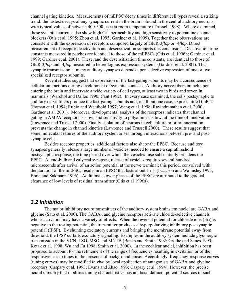
Figure legends Figure 1 Summary of poststimulus-time histograms discussed in this chapter. Panels adapted from Rhode and Greenberg (1992).
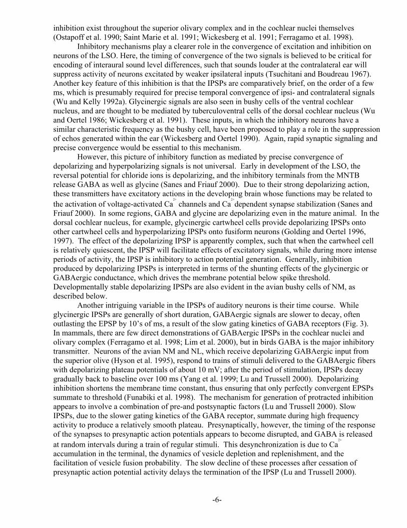
Figure 2 Stellate and bushy cells of the mouse ventral cochlear nucleus in a brain slice preparation. A, Camera lucida drawing of a stellate cell labeled using biocytin following patch-clamp recording. Arrow indicates the beginning of the axon. Calibration bar 10 μm. B, Response of a stellate cell to current steps of –200 and +200 pA. C, Camera lucida drawing of a biocytin-labeled bushy cell, with the axon marked by arrow, and calibration bar of 10 μm. D, response of a bushy cell to current steps of –400, 0 and +800 pA.
Figure 3 Response of short or long pulses of 1 mM glutamate (A) or 1 mM GABA (B) to an outside-out patch excised from a neuron in nucleus magnocellularis. The 2 upper traces indicate the time course of the transmitter application, while the lowest traces show the current responses. The asterisk marks the longer pulse and its response. In A, the response is mediated largely by AMPA receptors, whose deactivation and desensitization occur within milliseconds. Note that the desensitizing response (*) is slower than the deactivating response, reflecting different kinetic processes of the AMPA receptor. In B, GABA application activates GABA-A receptors, whose kinetics of deactivation and desensitization are far slower than those of the AMPA receptors. The slower time course of these responses accounts for the slower time course of the GABAergic IPSC.
-20-
0 10 20 30 40 500
15
30
45
60
75
0 10 20 30 40 500
25
50
75
100
125
0 10 20 30 40 500
30
60
90
120
150
0 40 80 120 160 2000
4
8
12
16
20
Primary-like
Onset, lock
Primary-like,notch
0 10 20 30 40 500
10
20
30
40
50
Chopper(sustained)
Pauser/buildup
msec
#o
fsp
ikes
0 10 20 30 40 500
50
100
150
200
250
Phase-lock(onset)
A B
C D
20 mV
20 ms
20 mV
20 ms
300 pA300 ms
40 pA5 ms
* *
*
*
Glutamate GABAA B