cellular forms: an artistic exploration of morphogenesis · cellular forms: an artistic exploration...
TRANSCRIPT

Cellular Forms: an Artistic Exploration ofMorphogenesis
Andy Lomas1
Abstract. Cellular Forms uses a simplified model of cellulargrowth to generate intricate sculptural shapes. Structures arecreated out of interconnected cells, with rules for the forcesbetween cells, as well as rules for how cells accumulate internalnutrients. When the nutrient level in a cell exceeds a giventhreshold the cell splits into two, with both the parent anddaughter cells reconnecting to their immediate neighbours. Manydifferent complex organic structures are seen to arise from subtlevariations of these rules, creating forms with strongreminiscences of plants, corals, internal organs and micro-organisms.1
The aim is to create structures emergently: exploring genericsimilarities between many different forms in nature rather thanrecreating any particular organism, and in the process exploringuniversal archetypal forms that can come from growth processesrather than top-down externally engineered design.
1 INTRODUCTION
The use of simulation methods in generative art can be seen as anatural extension of systems art [1], where the artist defines aprocess that can be run autonomously to create artefacts. With asufficiently rich simulation system there is an expectation that
1Website: www.andylomas.com, Email: [email protected]
surprising emergent results can be generated which would bedifficult, or potentially even impossible, to create without the aidof digital technology. The process can be seen as one ofexploration: both in defining the rules for the simulation systemsand exploring the range of results that can be achieved once asystem has been created.
In particular, simulation systems that are inspired bybiological processes, such as morphogenesis, can be used as apowerful means to explore the nature of organic form. Can theastonishingly complex forms that are seen in nature emerge fromsimple rules? This can be viewed as exploring the nature of thebasic fabric available to create structures when they aregenerated as a result of growth processes.
This paper describes Cellular Forms, an exploration of howrich evocative forms can be created using a simplified biologicalmodel of cellular growth. The model used is a deliberatelysimplified one, both to explore how a simple model can createrichly emergent results, and to be computationally sufficientlyefficient to allow the creation of structures with many millions ofcells in order to achieve a high level of complexity and detailwith the aim of evoking a powerful aesthetic result.
2 RELATED WORK
The relationship between growth and form has been the subjectof study for many years. Major influences behind the workdescribed here are Ernst Haeckel's studies of forms in nature [2]and D'Arcy Thompson's seminal “On Growth and Form” [3].
Alan Turing's paper “The Chemical Basis of Morphogenesis”[4] can be seen as the origin of using digital simulation methodsto examine potential mechanisms behind pattern generation andgrowth, performing biological experiments “in silico” rather than“in vitro” or “in vivo”. The reaction-diffusion equations thatTuring describes in his paper can create a surprisingly rich rangeof complex patterns, with remarkable resemblance to many ofthose seen in nature such as pigmentation patterns on the coats ofleopards, zebra and angelfish. Variations on reaction-diffusionequations, particularly following the work of Greg Turk [5], arecommonly used in computer graphics to create convincinglybiological textures for creatures.
Probably the most common method utilised to createconvincing biological structures is the use of L-Systems, asoriginally proposed by Aristid Lindenmayer [6]. Rules forbranching and the relative sizes of segments between branchesare expressed using a simple grammar with a generation rule thatcan be recursively applied to the form to create the next level. L-Systems are commonly used in computer graphics to producetree-like forms, and can be a very efficient way to createcomplex structures, but effects like branching are explicitly
Figure 1. Examples of Cellular Forms.

encoded into the system rather than arising emergently fromlower level processes.
Previous work by the author explored structures that can becreated by variations on diffusion-limited aggregation [7], usedas a simplified model of a growth system [8], [9]. This representsgrowth by repeated deposition: the structure is initialised with asingle seed particle. Successive new particles are allowed torandomly move in an external medium until they collide with thestructure generated so far. They then become attached to thestructure at that position, and the process is repeated. The resultsof this Aggregation series were a range of structures withreminiscences of dendritic plants and finely branched corals.
The previous works most related to the work described hereare Jaap Kaandorp's work on “Accretive Growth” [10] [11] andGeorge Hart's “Growth Forms” [12]. Both of these use a modelwhere cells are described by a surface of linked particles, withrules for when these cells split and how the topology of thesurface changes after cell division. In Kaandorp's work the aimis to mimic the growth of coral-like forms, with growth indifferent areas based on external nutrient gradients that mimicthe availability of water-borne food for marine organisms. InHart's work some particles are designated as 'buds' which areused to explicitly control and stimulate growth in local areas,causing effects such as branching. The results of Kaandorp andHaart's work are a variety of branched coral-like structures, andthe simulations described are run for a few thousand cellprimitives.
3 CELLULAR FORMS
In Cellular Forms the principal aim was to create a systemcapable of generating complex biologically evocative formsbased on the simulation of growth by cellular division.
Following the author's previous work, it was desired that themodel should be flexible enough that it should be capable of
producing results similar to those from the 'Aggregation' seriesas well as creating additional structures not achievable by thatsimulation framework. The aim was to be exploratory rather thancreate any specific target forms, but to be capable of producingstructures reminiscent of internal organs such as the foldedsurface of brains.
In order to create this greater range of possible forms it wasdecided to use a model based on a simplified version ofmorphogenesis through cellular division. The system should becapable of creating complex sheets of cells, with rules governingwhen cells divide, how the topology of the surface of cells isaffected by the newly created cells, and creating forces betweencells to induce the surface to fold into complex shapes.
By having a variety of different methods to induce growth itwas hoped to be able to create the extended range of structuresdesired. If growth was stimulated by external randomlytransported food particles which directly cause the first cell theyhit to divide, it should be capable of creating similar effects tothose seen in the Aggregation series. On the other hand, ifgrowth was based on concentrations of chemicals diffusingthrough the structure, potentially with all cells receiving thesame amount of nutrient, then it was hoped that structures morelike internal body organs could be produced.
It was also important that the simulation system should becapable of generating many millions of cell primitives. This wasdesired in order to achieve a compelling level of intricate detailin the structures, and had to be achievable within the limitationsof available conventional PC hardware.
4 MATHEMATICAL MODEL
The model used is based on a simplified version of cells. Aparticle system [13] representation is used, with each cellrepresented by one particle, and each particle linked to a numberof other particles that it is directly attached to.
Figure 2. Example image from the Aggregation series.
Figure 3. Initial ball of cells, with particles distributeduniformly on the surface of a sphere.
While the structures are three-dimensional, the topology ofthe connected particles is that of a two-dimensional surface. Inall the structures illustrated here, the system starts with a simpleball of cells with all the cells uniformly spaced on the surface ofa sphere.
Development of the form proceeds by a combination of forcesthat mediate interactions between the cells, and cell divisionsthat change the topology of the structure. The simulation takesplace over time, with time incrementing in uniform clock cyclesteps.
Cells that are directly linked try to maintain a constantdistance from each other. Additional rules try to restore the sheetto a locally planar state if there is a fold in the surface, and tobulge the sheet out when links are in compression. The intentionis that these two influences will work in competition with eachother, with different strength factors for each tending to createsurfaces with a variety of characteristics.
The actual implementation of all these effects is achieved bycalculating a new target position for each of these influences,and offsetting the cell's position towards the new target positionafter multiplying by a restoring factor. Values for these factorsare parameters for the simulation system as a whole.
Consider a system with simulation parameterslinkRestLength, springFactor, planarFactor and bulgeFactor.Let a cell have position P which is linked to n particles withpositions Lr, and the unit length normal to the surface at thecurrent cell position is N. The target positions for three differentinfluencing effects are calculated by the following methods:
1. The tendency for linked cells to maintain a constantdistance from each other is implemented using a linear spring-like system. The target position for the springs is calculated bytaking the average of the rest positions that each link would pushthe particle to if it were the only influence:
springTarget=1 /n∑r =1
n
(Lr+ linkRestLength×(̂P−Lr))
2. The planar target position, that acts in a similar manner to atorsion spring, is simply calculated by taking the average of allthe positions of directly linked particles. This is designed to havethe effect of tending to reduce folds and bumps in the surface,restoring the surface to a local planar state:
planarTarget=1/n∑r=1
n
Lr
3. The bulge target position is determined by calculating thedistance that each link would have to push the particle outwardsalong the direction of the surface normal in order to restore thelink to its rest length. This is designed to have an effect oftending to bulge the surface outwards in the direction of thenormal when links are in compression. The bulge distance due toeach link is calculated by a simple application of the cosineformula for triangles, and the average taken for all the links tocreate the desired overall distance bulgeDist in the direction ofthe normal. The bulgeTarget vector is then taken by goingbulgeDist in the direction of the surface normal from P.
dotN r=(L r−P)⋅N
bulgeDist=1/n∑r =1
n
linkRestLength2−∣Lr∣
2dotN r
2dotN r
bulgeTarget=P+bulgeDist×N
The new position for the particle position P' is then calculatedby offsetting P in the direction of each of these target positionusing the three different simulation factor values.
P '=P+springFactor×(springTarget−P)+ planarFactor×(planarTarget−P)+bulgeFactor×(bulgeTarget−P)
One thing to notice about this is that there is no momentumterm: the new position is simply calculated by moving it afraction of the distance towards the target positions controlled bythe restoring factors. This can be justified if we consider the cellsto be growing in a medium that is highly viscous relative to thecell size, so there is a large damping effect on any velocities. Italso has the advantage of making the system less prone tounstable oscillations, particularly when we are in effect addingenergy to the system every time cells divide.
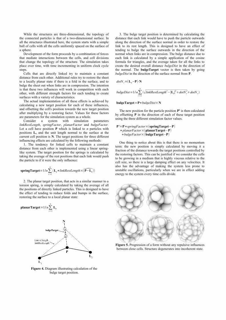
Figure 5. Progression of a form without any repulsive influencesbetween close cells. Structure degenerates into incoherent state.
Figure 4. Diagram illustrating calculation of thebulge target position.

Cells that aren't directly linked to each other but are in closeproximity experience a repulsive influence. Without this it wasfound that structures would degenerate into an incoherent stateas they started to generate folds. In effect the repulsive influencebetween cells imposes a constraint of structural coherence on theform. An analogy can be drawn with D'Arcy Thompson'sarguments for how physics imposes constraints on the possibleshapes for forms created by growth processes.
This repulsive influence is controlled by two other parametersfor the simulation system which define a radius of influence(roi) and repulsionStrength. The effect of the repulsion isapplied by calculating a collisionOffset vector to be added toeach particle's position where
collisionOffset=repulsionStrength∑r∈A
(roi 2
−|P−Pr|2
roi2 ×(̂P−Pr))
and A is the set of all particles within the radius of influence ofthe current particle that aren't directly linked to the currentparticle. Directly linked particles are excluded from the repulsioncalculations since they are considered directly attached to eachother, and the influences between them are already controlled bythe previously described other effects.
Each cell has an internal 'food level'. The food accumulates ina cell and when it exceeds a given threshold the cell is selectedfor splitting. Various different methods have been implementedto affect food levels, and therefore affect growth rates, including: Uniformly adding a random amount to each cell at
each time step. Using reaction-diffusion equations (RDEs) over the
surface [5] to create differential areas of growth. The RDEis calculated using the cells as the places where chemicalsare stored, and diffusing the chemicals along the direct linksbetween cells. One of the RDE chemicals is used as a'nutrient level', affecting how much the food level in thecells increments at each time step.
Using ray-tracing to simulate light coming in fromoutside the structure which stimulates nutrient creation inthe cells that the light hits. This nutrient is used to controlthe food increment in each cell, and can also diffuse fromone cell to another along the links between cells todistribute the nutrient through the structure.
When a cell has been selected for splitting, a number ofparameters are used to control how the split occurs. These areused to control how the topology of links between the cells ischanged by cell division. First, two links are chosen thatrepresent the plane of cleavage. All the links to one side of theplane of cleavage are left connected to the parent cell, while thelinks to the other side are disconnected from the parent andreplaced with links to the daughter cell. Along the plane ofcleavage links are made to both the parent and daughter cells. Anew link is also created directly between the parent anddaughter.
There are some key simplifications in this system comparedwith one that aims to accurately mimic biological processes. Inparticular: Apart from the potential for differential growth rates
as described above, there is no cell differentiation. Theparameters governing forces between cells and how celldivision takes place are constants for the whole system, anddon't change over time. The target link length is also thesame uniform constant value for all links between any cells.
Cells are represented by simple spheres with only aposition and radius. They don't have any principal axis ororientation.
There is no cell motility. The links that describeattachments between cells are formed immediatelyfollowing cell division.
As previously mentioned, the cells are alwaysconnected together in arrangements that are topologicallyequivalent to a simple closed surface rather than being asolid volume.
5 IMPLEMENTATION
Typical simulations are run for tens of thousands of iterationswith data sets growing to over 50 million particles.
Figure 6. Progression of a form with the same parameters butwith repulsive influence between close cells. Structure develops
coherent shape.
Figure 7. The process of cell division.
The software to run the simulations and render images fromthe data sets created is implemented in C++ and CUDA [14].
To make use of the general purpose parallel processingcapabilities of modern graphics hardware, all the calculationsinvolving the individual particles are executed using CUDA onthe GPU. This includes all the functions that simulate the effectsof forces between the particles, cast rays into the structure torender images or simulate light rays, and handle topologicalchanges to the surface that occur when cells divide. This meansthat all the data for the cellular structures can be kept purely inGPU memory, avoiding the need to transfer large amounts ofdata across the PCI bus between the host (CPU) memory and thedevice (GPU) memory. In the current implementation the onlyneed for transferring cell data between the CPU and GPU is if itis required to write out data to a file on disk, or to read data backfrom disk.
In particular, implementing using the GPU allowed a verysignificant speed improvement for the calculations of repulsiveinteractions between cells in close proximity but not directlylinked to each other. The previous implementation of this on theCPU was too slow to make it feasible to run simulations with therequired number of cells.
Class name Class description
BaseParticleGpu Base class defining particle data to be held on the GPU.
LinkedParticleGpu Derived from BaseParticleGpu. Extends the base class by adding a list of links between particles. Also implements supportfor reaction-diffusion equations using the links between particles.
ElasticSheetParticleGpu Derived from LinkedParticleGpu. Adds methods for dealing with the spring, planar and bulge effects, as well as repulsion effects between spatially close particles.
FoodSplitParticleGpu Derived from ElasticSheetParticleGPU. Adds methods to implement food levels, nutrient levels, and cell division.
Table 1. Class structure used for particle data.
The software is implemented as an extensible framework. Anobject oriented approach was used with a hierarchy of inheritingclasses representing the data for the structures. This starts with abase class for representing general particle data on the GPU,which is then specialised through a series of derived classes thatinherit from each other up until the final class used in all thesimulations shown here.
Rendering 2D images from the cellular simulation data isdone using ray-tracing techniques, with the cells treated asspheres with radii based on the average distances to linked cells.A number of different techniques are implemented, includingproducing solid surface renders with 'ambient occlusion' for theshading which represents the effects of a self-shadowing from auniform omnidirectional diffuse light, and “X-Ray” renders thattreat each sphere as a contributor to an accumulated densitycalculated by tracing a ray though the whole structure. All thekey rendering functions are implemented using CUDA kernelson the GPU, both for speed and so that all the particle data canbe kept solely on the GPU.
6 ARTISTIC ARTEFACTS
Simulations are first run with a maximum of a million cells tocreate quick initial sample tests of simulation parameter valueswhich may create interesting results. These tests typically takebetween 1 and 5 minutes to run for each sample. From these,candidates are selected to run full length simulations and high-resolution renders. These final simulations are executed to createthe most detailed structures possible, which typically meansbetween 52 million and 56 million cells before the memorylimits of the current hardware (NVIDIA GTX Titan with 6GBRAM) are reached. These full length simulations typically takebetween 1 and 4 hours each.
Rendered images are created directly from the simulations,which are used to make the artistic artefacts from the series.These artefacts can take various forms: High resolution prints of the final structures. Currently
these are rendered at 8600 by 8600 pixels, to allow veryfine detail when printing at large sizes.
High definition animations taken by rendering imagesat equally spaced time intervals, to show how the structuresdevelop incrementally over time.
3D stereo views taken by rendering pairs of imagesusing cameras with an interocular separation.
Currently, two main rendering styles are used for the finalartefacts:
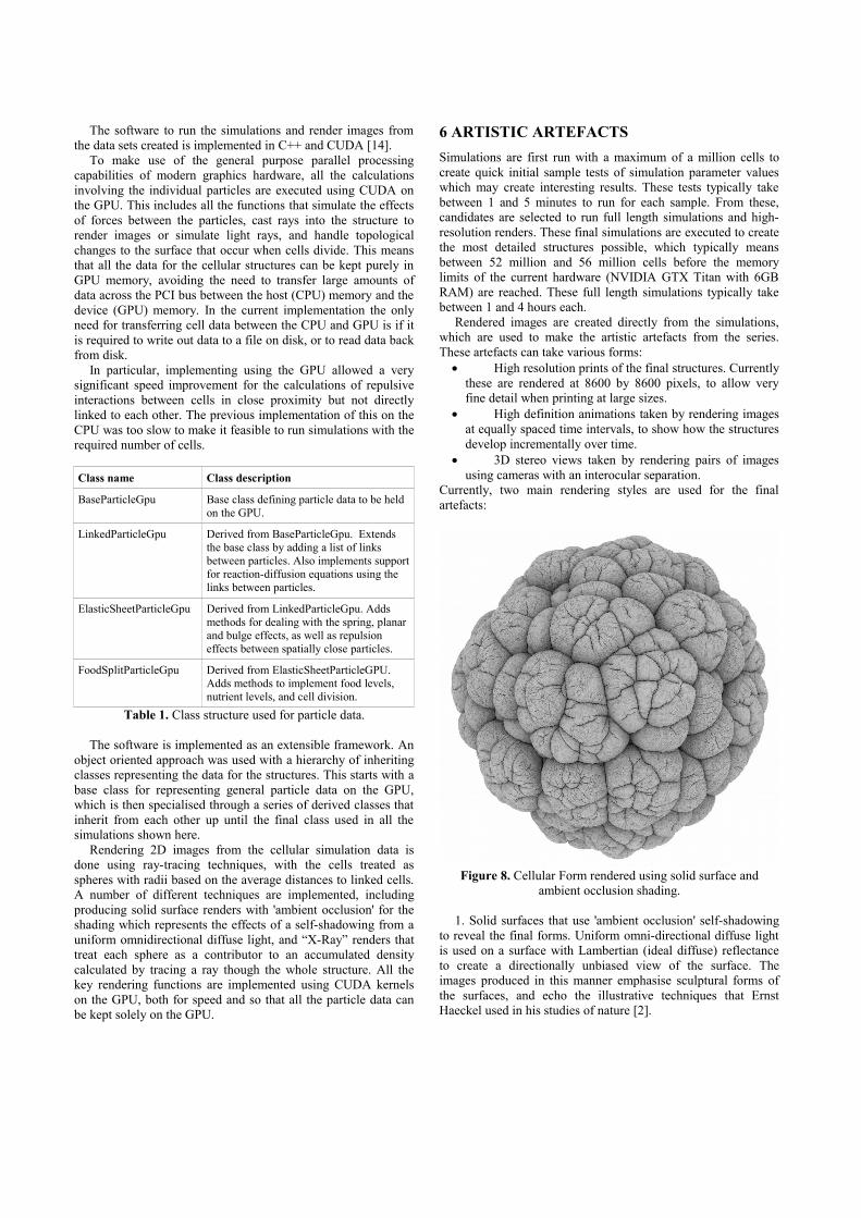
1. Solid surfaces that use 'ambient occlusion' self-shadowingto reveal the final forms. Uniform omni-directional diffuse lightis used on a surface with Lambertian (ideal diffuse) reflectanceto create a directionally unbiased view of the surface. Theimages produced in this manner emphasise sculptural forms ofthe surfaces, and echo the illustrative techniques that ErnstHaeckel used in his studies of nature [2].
Figure 8. Cellular Form rendered using solid surface andambient occlusion shading.
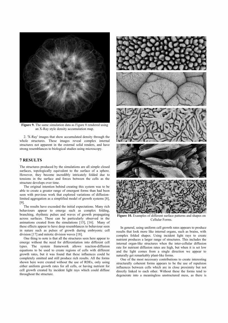
2. 'X-Ray' images that show accumulated density through thewhole structures. These images reveal complex internalstructures not apparent in the external solid renders, and havestrong resemblances to biological studies using microscopy.
7 RESULTS
The structures produced by the simulations are all simple closedsurfaces, topologically equivalent to the surface of a sphere.However, they become incredibly intricately folded due totensions in the surface and forces between the cells as thestructure develops over time.
The original intention behind creating this system was to beable to create a greater range of emergent forms than had beenseen with previous work that explored variations of diffusion-limited aggregation as a simplified model of growth systems [8],[9].
The results have exceeded the initial expectations. Many richbehaviours appear to emerge such as complex folding,branching, rhythmic pulses and waves of growth propagatingacross surfaces. These can be particularly observed in theanimations created from the simulations [15], [16]. Many ofthese effects appear to have deep resemblances to behaviour seenin nature such as pulses of growth during embryonic celldivision [17] and mitotic division waves [18].
One thing to note is that all the structures seen here appear toemerge without the need for differentiation into different celltypes. The system framework allows reaction-diffusionequations to be used to create regions of cells with differentgrowth rates, but it was found that these influences could becompletely omitted and still produce rich results. All the formsshown here were created without the use of RDEs, only usingeither uniform growth rates for all cells, or having nutrient forcell growth created by incident light rays which could diffusethroughout the structure.
In general, using uniform cell growth rates appears to produceresults that look more like internal organs, such as brains, withcomplex folded shapes. Using incident light rays to createnutrient produces a larger range of structures. This includes theinternal organ-like structures when the inter-cellular diffusionrate for nutrient diffusion rates are high, but when it is set lowand the light comes from a single direction we appear tonaturally get remarkably plant-like forms.
One of the most necessary contributions to create interestingstructurally coherent forms appears to be the use of repulsioninfluences between cells which are in close proximity but notdirectly linked to each other. Without these the forms tend todegenerate into a meaningless unstructured mess, as there is
Figure 10. Examples of different surface patterns and shapes onCellular Forms.
Figure 9. The same simulation data as Figure 8 rendered usingan X-Ray style density accumulation map.
nothing preventing the surface self-intersecting as it folds. Theseadditional influences can be seen as exerting physical constraintson the structures.
8 CONCLUSIONS & FUTURE WORK
The use of a simple system of interactions between adjacent orspatially close cells is seen to produce a wide range of complexresults.
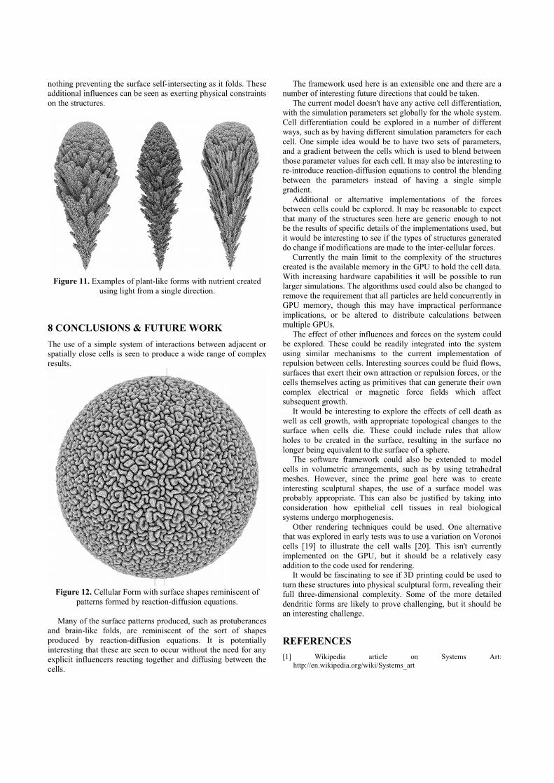
Many of the surface patterns produced, such as protuberancesand brain-like folds, are reminiscent of the sort of shapesproduced by reaction-diffusion equations. It is potentiallyinteresting that these are seen to occur without the need for anyexplicit influencers reacting together and diffusing between thecells.
The framework used here is an extensible one and there are anumber of interesting future directions that could be taken.
The current model doesn't have any active cell differentiation,with the simulation parameters set globally for the whole system.Cell differentiation could be explored in a number of differentways, such as by having different simulation parameters for eachcell. One simple idea would be to have two sets of parameters,and a gradient between the cells which is used to blend betweenthose parameter values for each cell. It may also be interesting tore-introduce reaction-diffusion equations to control the blendingbetween the parameters instead of having a single simplegradient.
Additional or alternative implementations of the forcesbetween cells could be explored. It may be reasonable to expectthat many of the structures seen here are generic enough to notbe the results of specific details of the implementations used, butit would be interesting to see if the types of structures generateddo change if modifications are made to the inter-cellular forces.
Currently the main limit to the complexity of the structurescreated is the available memory in the GPU to hold the cell data.With increasing hardware capabilities it will be possible to runlarger simulations. The algorithms used could also be changed toremove the requirement that all particles are held concurrently inGPU memory, though this may have impractical performanceimplications, or be altered to distribute calculations betweenmultiple GPUs.
The effect of other influences and forces on the system couldbe explored. These could be readily integrated into the systemusing similar mechanisms to the current implementation ofrepulsion between cells. Interesting sources could be fluid flows,surfaces that exert their own attraction or repulsion forces, or thecells themselves acting as primitives that can generate their owncomplex electrical or magnetic force fields which affectsubsequent growth.
It would be interesting to explore the effects of cell death aswell as cell growth, with appropriate topological changes to thesurface when cells die. These could include rules that allowholes to be created in the surface, resulting in the surface nolonger being equivalent to the surface of a sphere.
The software framework could also be extended to modelcells in volumetric arrangements, such as by using tetrahedralmeshes. However, since the prime goal here was to createinteresting sculptural shapes, the use of a surface model wasprobably appropriate. This can also be justified by taking intoconsideration how epithelial cell tissues in real biologicalsystems undergo morphogenesis.
Other rendering techniques could be used. One alternativethat was explored in early tests was to use a variation on Voronoicells [19] to illustrate the cell walls [20]. This isn't currentlyimplemented on the GPU, but it should be a relatively easyaddition to the code used for rendering.
It would be fascinating to see if 3D printing could be used toturn these structures into physical sculptural form, revealing theirfull three-dimensional complexity. Some of the more detaileddendritic forms are likely to prove challenging, but it should bean interesting challenge.
REFERENCES
[1] Wikipedia article on Systems Art:http://en.wikipedia.org/wiki/Systems_art
Figure 12. Cellular Form with surface shapes reminiscent ofpatterns formed by reaction-diffusion equations.
Figure 11. Examples of plant-like forms with nutrient createdusing light from a single direction.

[2] E. Haeckel, Art Forms in Nature, Dover Publications Inc.; Revisededition (2 Jan 2000).
[3] D.W. Thompson, On Growth and Form, Cambridge UniversityPress; New Ed edition (31 July 1992).
[4] A.M. Turing, “The Chemical Basis of Morphogenesis”,Philosophical Transactions of the Royal Society of London 237(641), 37–72.
[5] G. Turk, “Generating textures on arbitrary surfaces using reaction-diffusion”, Proceedings of SIGGRAPH '91, 289-298.
[6] A. Lindenmayer, “Developmental systems without cellularinteraction, their languages and grammars”. Journal of TheoreticalBiology, 30 (1971), 455-484.
[7] T.A. Witten Jr & L.M. Sander, “Diffusion-Limited Aggregation, aKinetic Critical Phenomenon”, Phys. Rev. Lett. 47, 1400 (1981).
[8] A. Lomas, “Aggregation: Complexity out of Simplicity”, sketchsession SIGGRAPH 2005,http://www.andylomas.com/sketch_0087_final.pdf
[9] A. Lomas, video: “Growth by Aggregation”,https://vimeo.com/83297099
[10] J.A. Kaandorp, “Analysis and synthesis of radiative accretivegrowth in three dimensions”, Journal of Theoretical Biology, 175(1995), 39-55.
[11] J.A. Kaandorp & J. Kübler, The Algorithmic Beauty of Seaweeds,Sponges, and Corals, Springer-Verlag (2001), 125-144.
[12] G. Hart, “Growth Forms”, Proceedings of Bridges 2009,http://georgehart.com/Growth/GrowthForms.pdf
[13] W.T. Reeves, “Particle Systems: A Technique for Modelling aClass of Fuzzy Object”, ACM Transactions on Graphics, Volume 2Issue 2, April 1983, 91-108.
[14] CUDA http://www.nvidia.com/object/cuda_home_new.html[15] A. Lomas, video: “Cellular Forms”, https://vimeo.com/82989945[16] A. Lomas, video: “Cellular Forms (X_Ray version)”,
https://vimeo.com/83294152[17] Video: “The Development of a Frog”,
http://www.youtube.com/watch?v=dXpAbezdOho[18] Video: “Early mitotic division waves in Drosophila embryo”,
http://www.youtube.com/watch?v=-Nf6CyWNodA[19] Wikipedia article on Voronoi diagrams:
http://en.wikipedia.org/wiki/Voronoi_diagram[20] A. Lomas, video: “Cellular Form Generation Test 003c5”,
http://www.youtube.com/watch?v=kcEBEIAhsuk