cells & organelles
DESCRIPTION
Cells & Organelles. A Dr. Production. Two Basic Types of Cells. Pro kary otes: prounounced: pro-carry-oats Eu karyotes Proun: you-carry-oats. Organization of Domain s Evolution of the 3 Domains. Development of the Cell Theory. Hooke (1665) named the cell - PowerPoint PPT PresentationTRANSCRIPT
Cells & Organelles
A Dr. Production
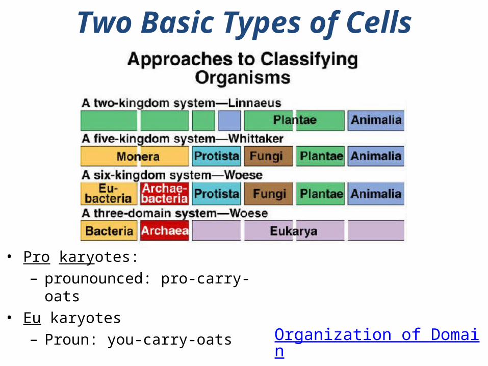
Organization of Domains
Evolution of the 3 Domains
Two Basic Types of Cells
• Pro karyotes:– prounounced: pro-carry-
oats• Eu karyotes
– Proun: you-carry-oats
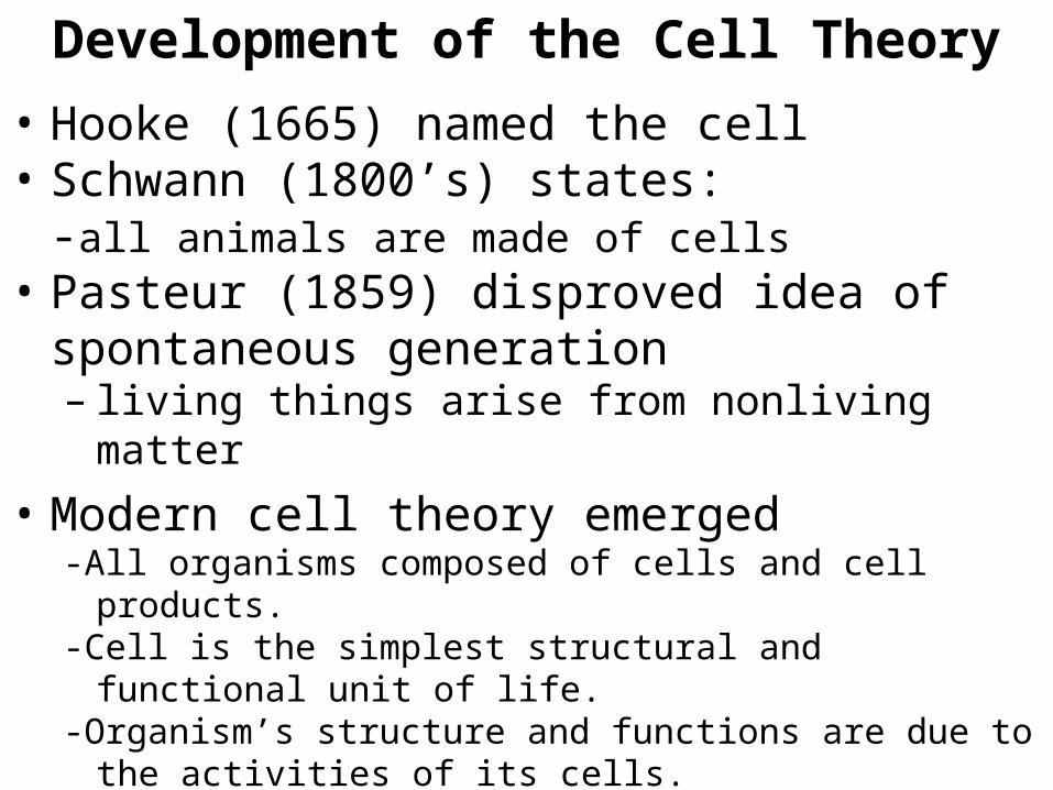
• Hooke (1665) named the cell • Schwann (1800’s) states:
-all animals are made of cells • Pasteur (1859) disproved idea of
spontaneous generation– living things arise from nonliving matter
• Modern cell theory emerged-All organisms composed of cells and cell products.-Cell is the simplest structural and functional unit of
life. -Organism’s structure and functions are due to the
activities of its cells.-Cells come only from preexisting cells.-Cells of all species have many fundamental
similarities.
Development of the Cell Theory
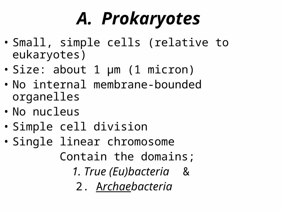
A. Prokaryotes• Small, simple cells (relative to eukaryotes)• Size: about 1 µm (1 micron)• No internal membrane-bounded organelles• No nucleus• Simple cell division• Single linear chromosome
Contain the domains; 1. True (Eu)bacteria &
2. Archaebacteria
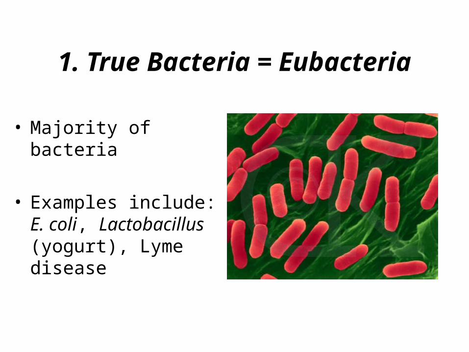
1. True Bacteria = Eubacteria
• Majority of bacteria
• Examples include: E. coli, Lactobacillus (yogurt), Lyme disease
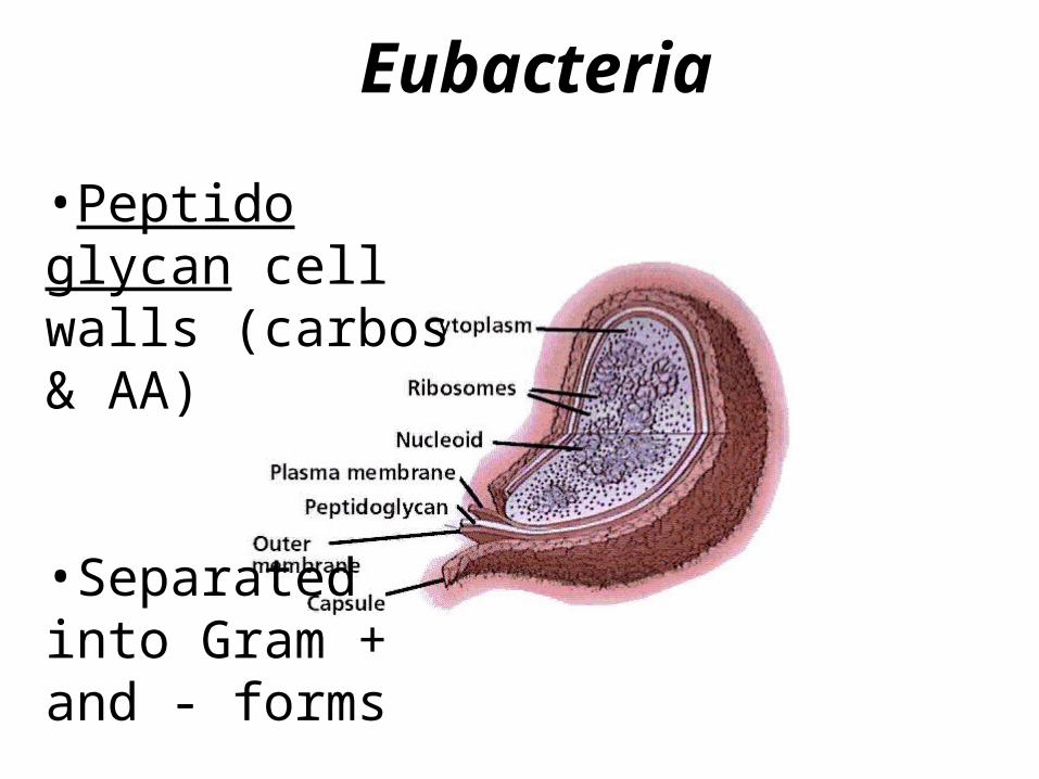
Eubacteria
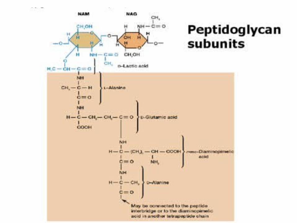
•Peptido glycan cell walls (carbos & AA)
•Separated into Gram + and - forms
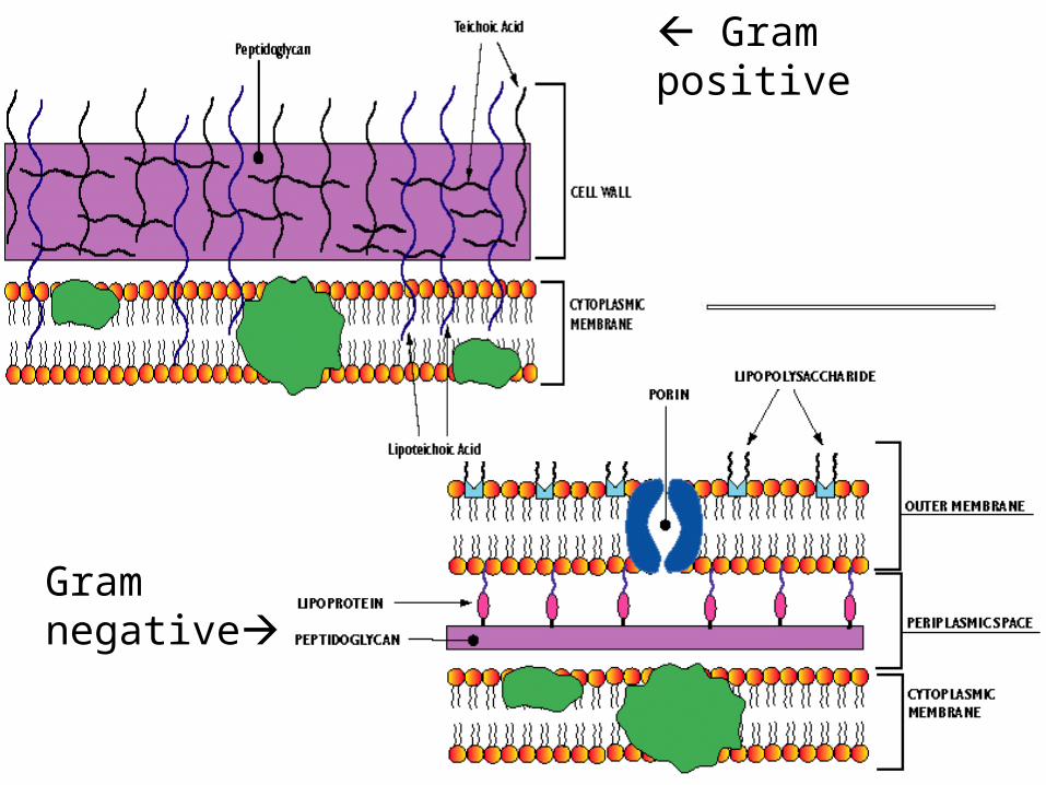
Gram negative
Gram positive
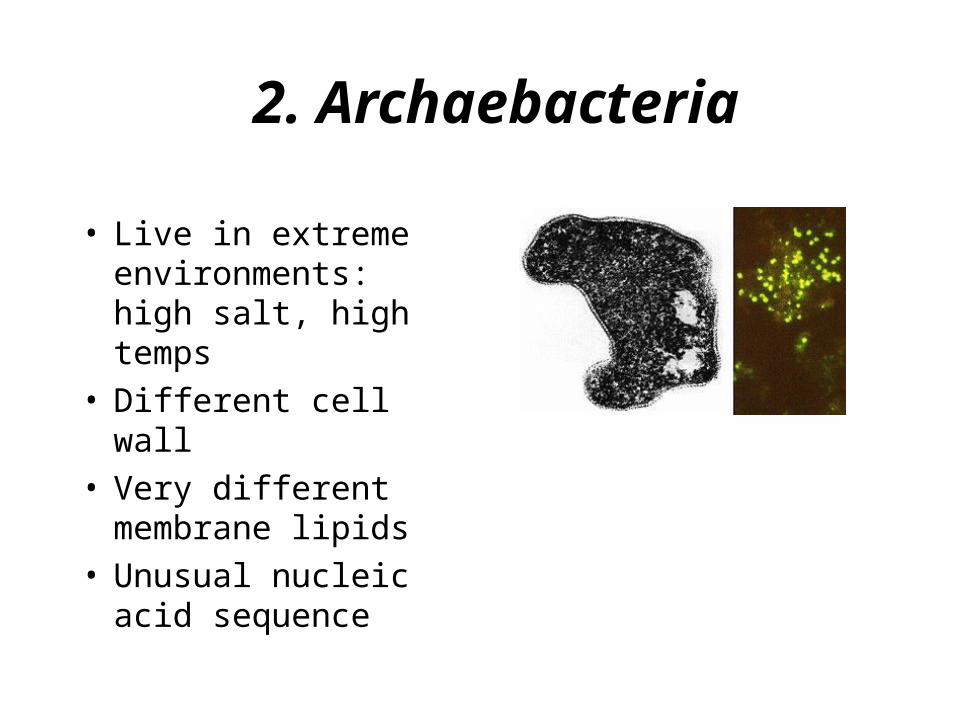
2. Archaebacteria
• Live in extreme environments: high salt, high temps
• Different cell wall• Very different
membrane lipids• Unusual nucleic
acid sequence
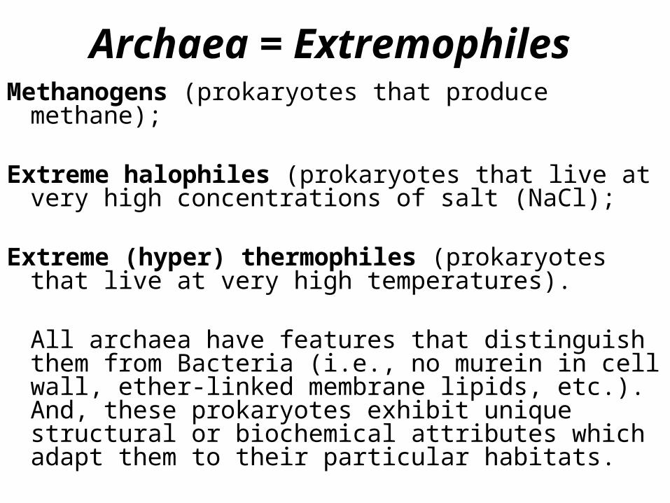
Archaea = Extremophiles
Methanogens (prokaryotes that produce methane);
Extreme halophiles (prokaryotes that live at very high concentrations of salt (NaCl);
Extreme (hyper) thermophiles (prokaryotes that live at very high temperatures).
All archaea have features that distinguish them from Bacteria (i.e., no murein in cell wall, ether-linked membrane lipids, etc.). And, these prokaryotes exhibit unique structural or biochemical attributes which adapt them to their particular habitats.
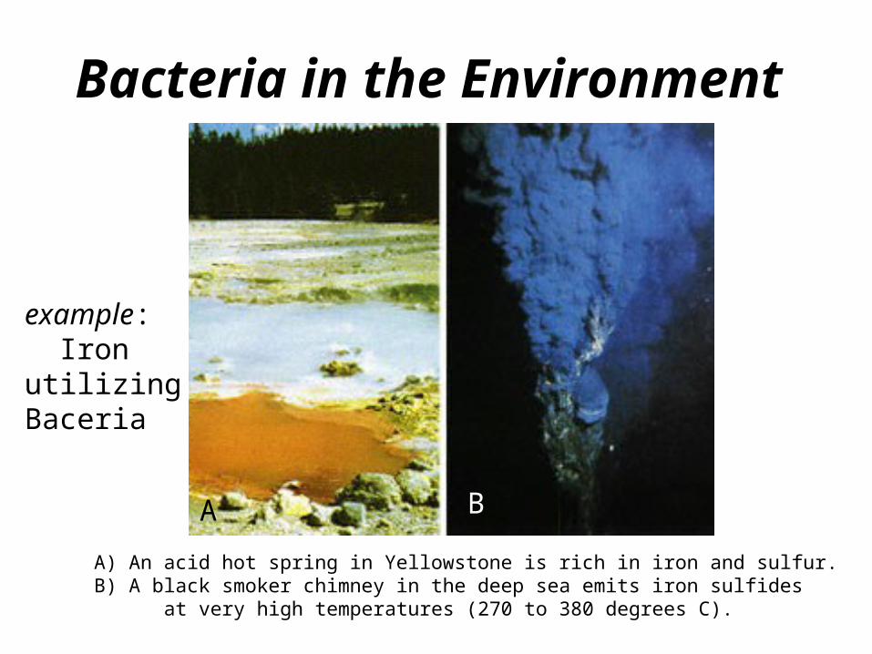
Bacteria in the Environment
A) An acid hot spring in Yellowstone is rich in iron and sulfur. B) A black smoker chimney in the deep sea emits iron sulfides at very high temperatures (270 to 380 degrees C).
example: Iron utilizing Baceria
A B
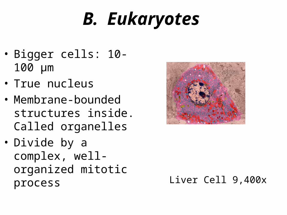
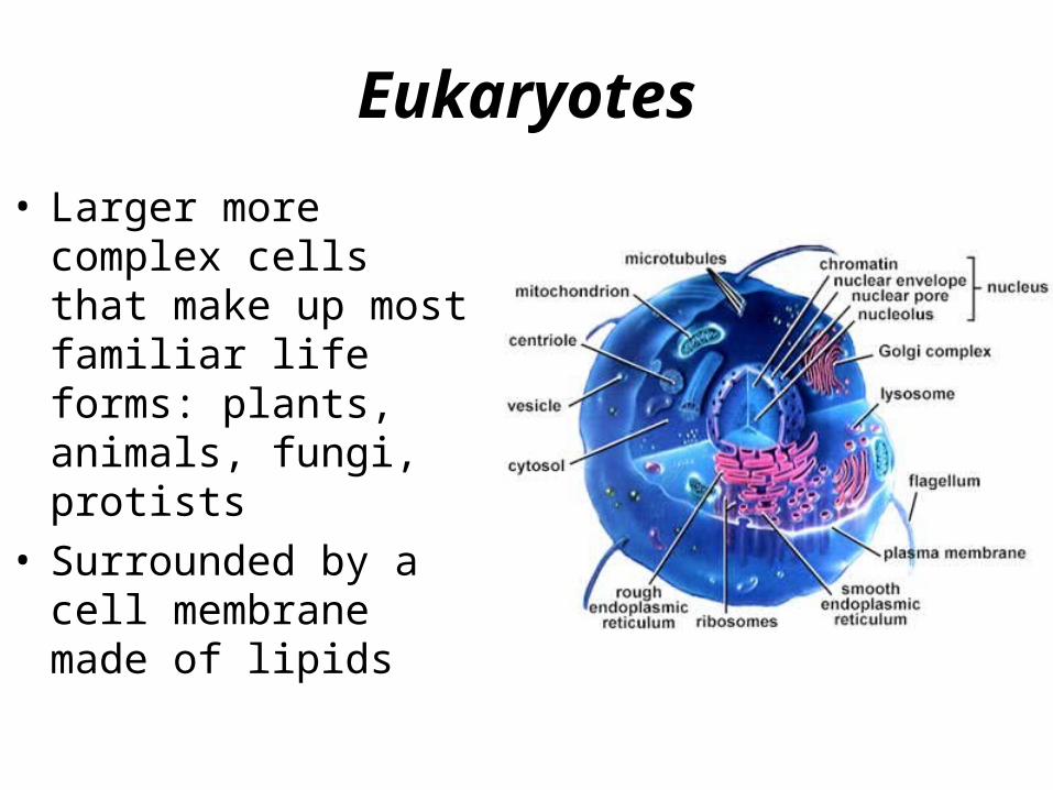
B. Eukaryotes
• Bigger cells: 10-100 µm
• True nucleus• Membrane-bounded
structures inside. Called organelles
• Divide by a complex, well-organized mitotic process
Liver Cell 9,400x
Eukaryotes
• Larger more complex cells that make up most familiar life forms: plants, animals, fungi, protists
• Surrounded by a cell membrane made of lipids
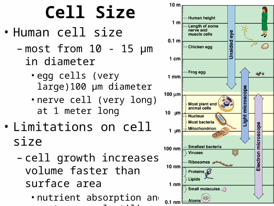
Cell Size• Human cell size
– most from 10 - 15 µm in diameter• egg cells (very large)100
µm diameter• nerve cell (very long) at 1
meter long
• Limitations on cell size– cell growth increases
volume faster than surface area• nutrient absorption and
waste removal utilize surface
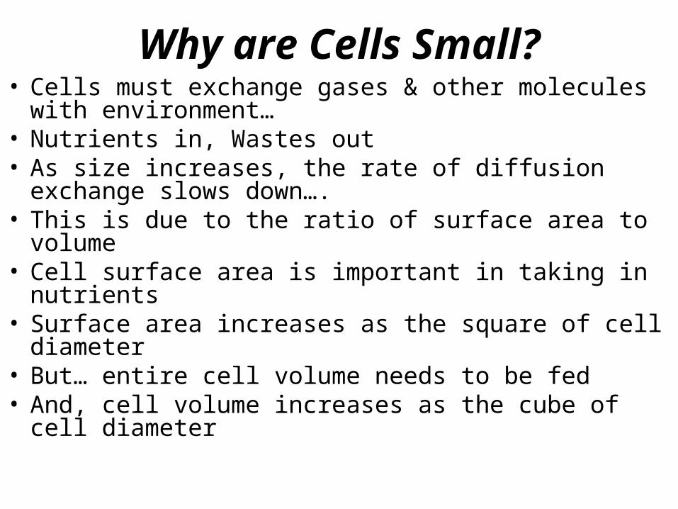
Why are Cells Small?• Cells must exchange gases & other molecules
with environment…• Nutrients in, Wastes out• As size increases, the rate of diffusion exchange
slows down….• This is due to the ratio of surface area to volume • Cell surface area is important in taking in
nutrients• Surface area increases as the square of cell
diameter• But… entire cell volume needs to be fed• And, cell volume increases as the cube of cell
diameter
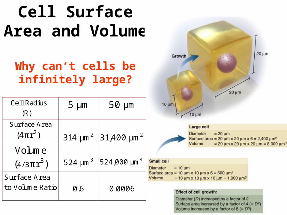
Cell Surface Area and Volume
Why can’t cells be infinitely large? Cell Radius
(R) 5 µm 50 µm
Surface Area
(4πr2) 314 µm2
31,400 µm2
Volume (4/ 3πr3)
524 µm3
524,000 µm3
Surf ace Area to Volume Ratio
0.6
0.0006
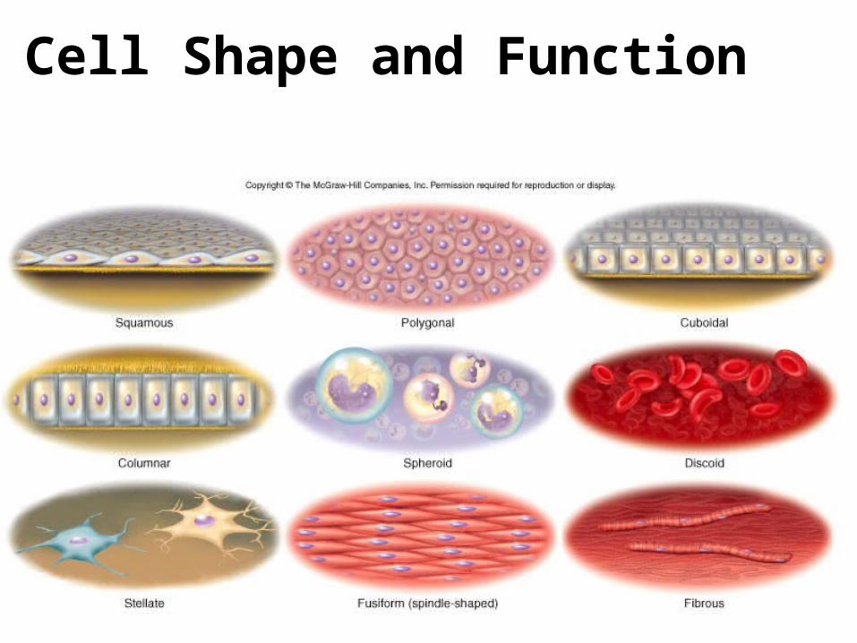
Cell Shape and Function

The Eukaryotic Cell: Components
• Cell membrane composed of lipids and proteins
• Cytosol: interior region. Composed of water & dissolved chemicals…a gel
• Numerous organelles….
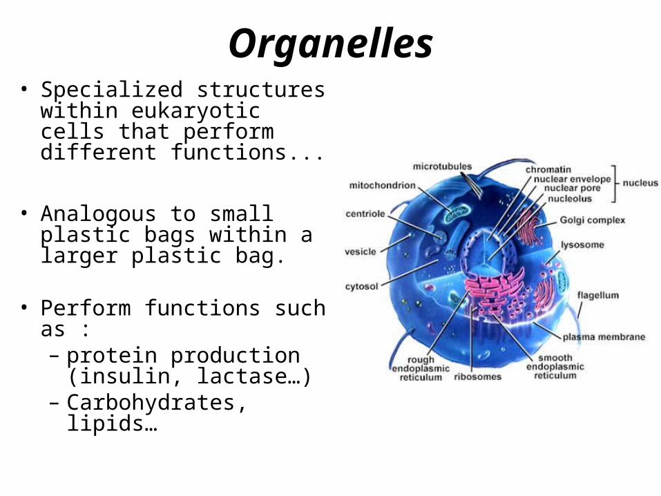
Organelles• Specialized structures
within eukaryotic cells that perform different functions...
• Analogous to small plastic bags within a larger plastic bag.
• Perform functions such as :– protein production
(insulin, lactase…)– Carbohydrates,
lipids…
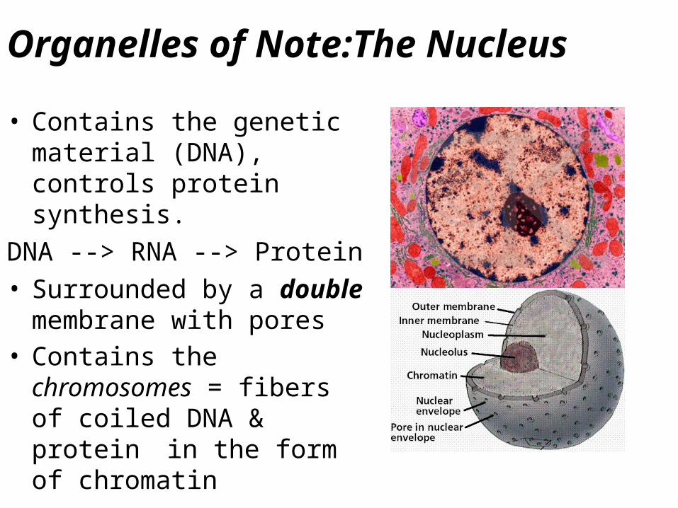
Organelles of Note:The Nucleus• Contains the genetic
material (DNA), controls protein synthesis.
DNA --> RNA --> Protein• Surrounded by a double
membrane with pores• Contains the
chromosomes = fibers of coiled DNA & protein in the form of chromatin
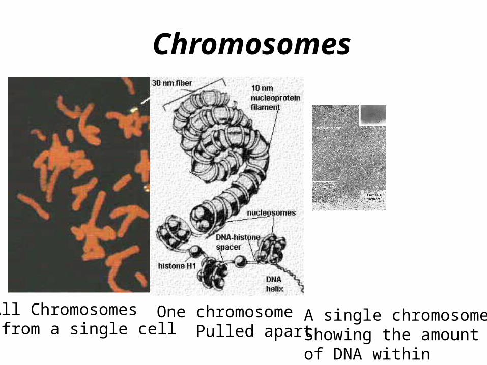
Chromosomes
All Chromosomes from a single cell
One chromosome Pulled apart
A single chromosomeShowing the amount of DNA within
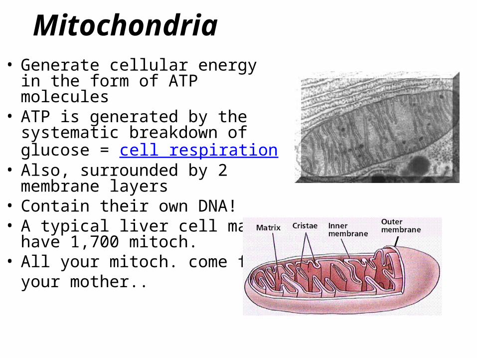
Mitochondria• Generate cellular energy in
the form of ATP molecules• ATP is generated by the
systematic breakdown of glucose = cell respiration
• Also, surrounded by 2 membrane layers
• Contain their own DNA!• A typical liver cell may
have 1,700 mitoch.• All your mitoch. come from
your mother..
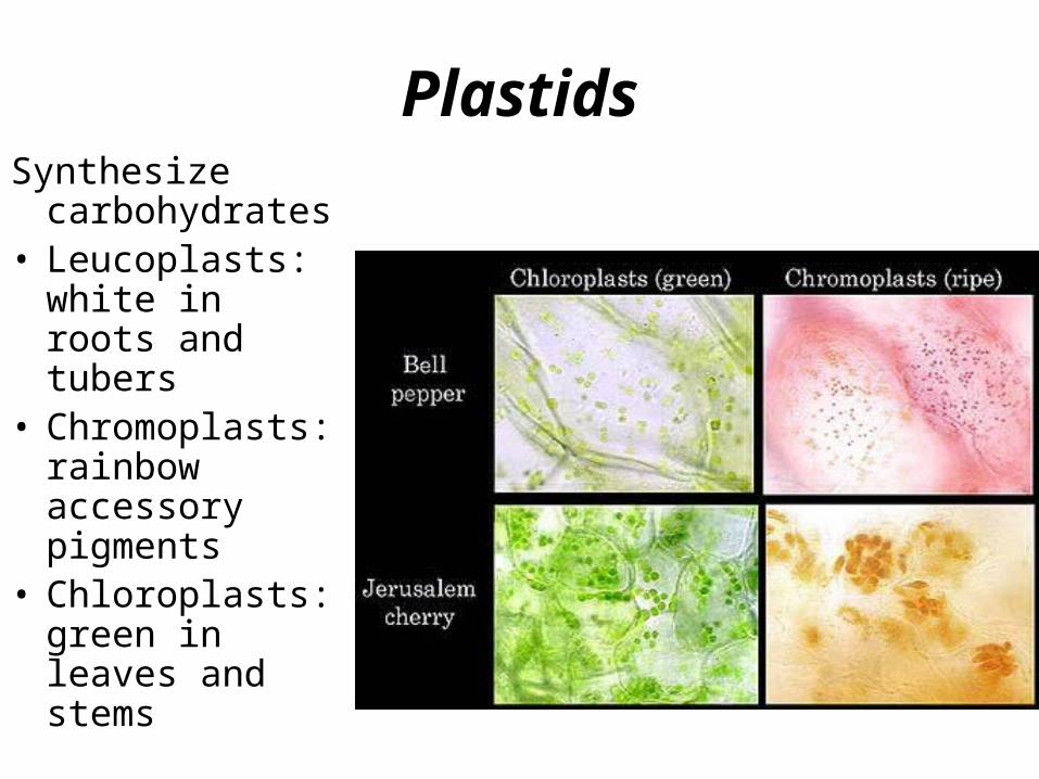
PlastidsSynthesize
carbohydrates• Leucoplasts:
white in roots and tubers
• Chromoplasts: rainbow accessory pigments
• Chloroplasts: green in leaves and stems
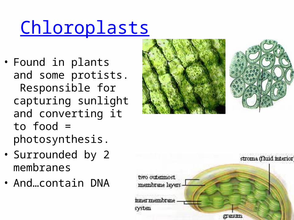
Chloroplasts
• Found in plants and some protists. Responsible for capturing sunlight and converting it to food = photosynthesis.
• Surrounded by 2 membranes
• And…contain DNA
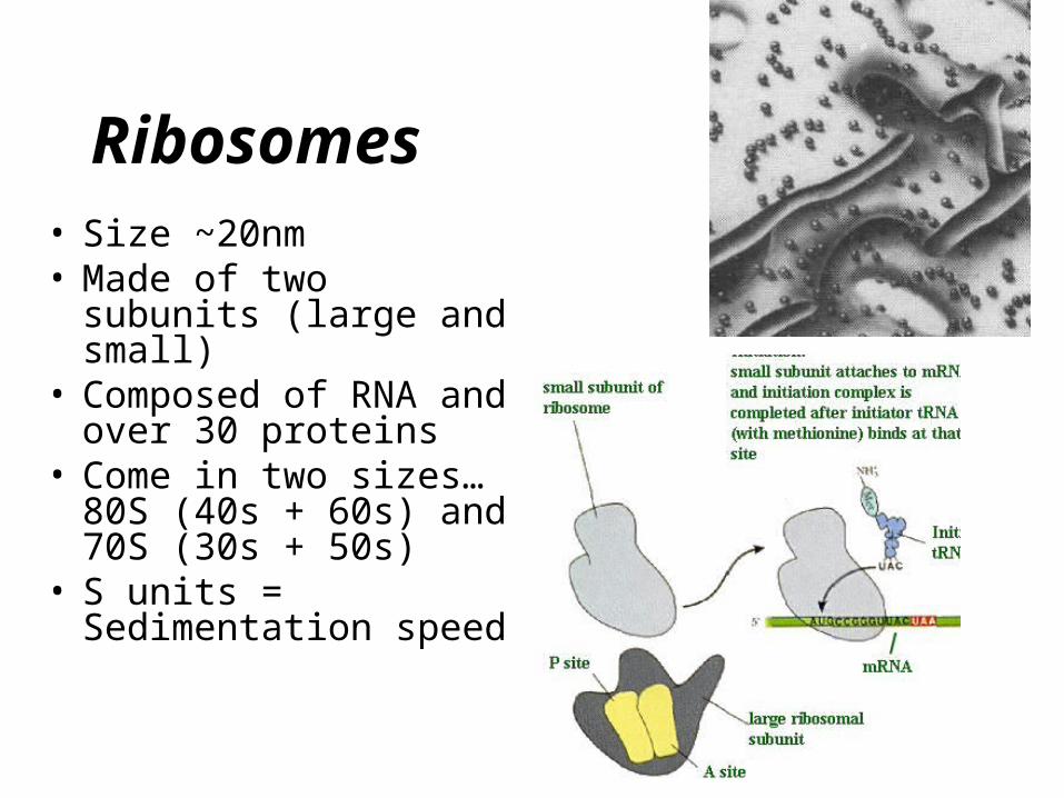
Ribosomes• Size ~20nm• Made of two subunits
(large and small)• Composed of RNA
and over 30 proteins• Come in two sizes…
80S (40s + 60s) and 70S (30s + 50s)
• S units = Sedimentation speed
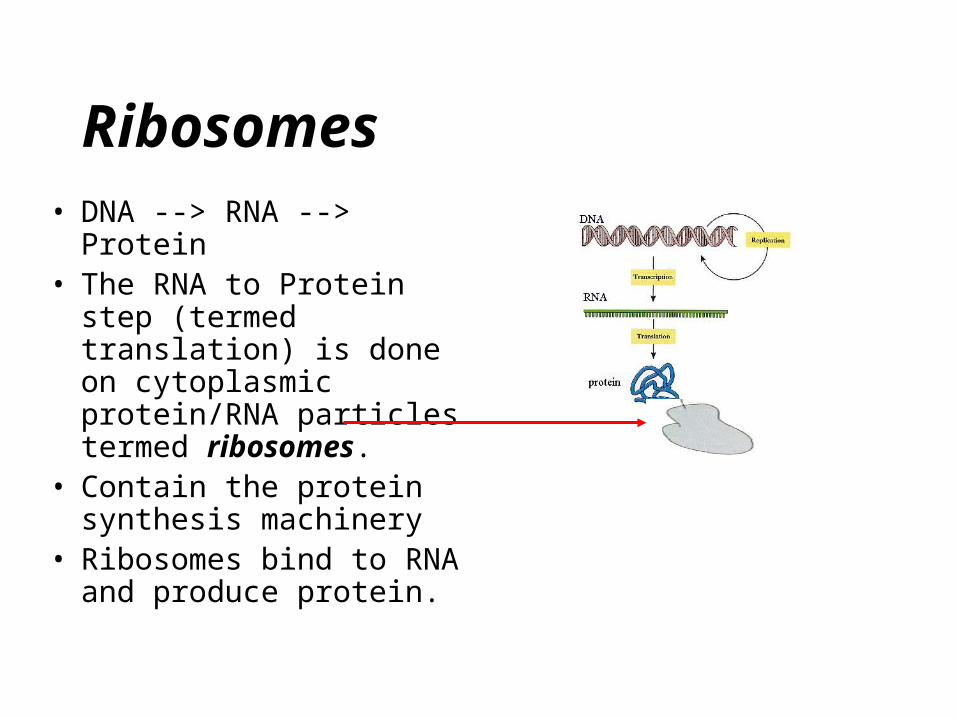
Ribosomes• DNA --> RNA --> Protein• The RNA to Protein step
(termed translation) is done on cytoplasmic protein/RNA particles termed ribosomes.
• Contain the protein synthesis machinery
• Ribosomes bind to RNA and produce protein.
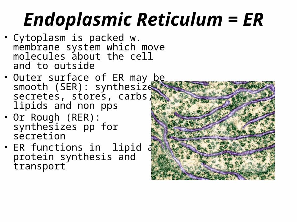
Endoplasmic Reticulum = ER• Cytoplasm is packed w.
membrane system which move molecules about the cell and to outside
• Outer surface of ER may be smooth (SER): synthesizes secretes, stores, carbs, lipids and non pps
• Or Rough (RER): synthesizes pp for secretion
• ER functions in lipid and protein synthesis and transport

Golgi Complex• Stacks of
membranes…• Involved in
modifying proteins and lipids into final form…– Adds the sugars
to make glyco-proteins and glyco-lipids
• Also, makes vesicles to release stuff from cell
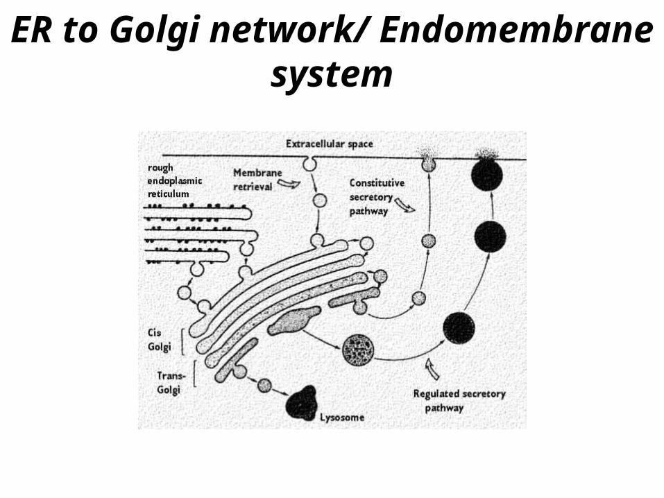
ER to Golgi network/ Endomembrane system

Membrane Flow through Golgi
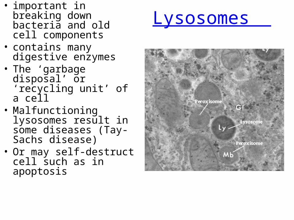
Lysosomes • important in breaking
down bacteria and old cell components
• contains many digestive enzymes
• The ‘garbage disposal’ or ‘recycling unit’ of a cell
• Malfunctioning lysosomes result in some diseases (Tay-Sachs disease)
• Or may self-destruct cell such as in apoptosis
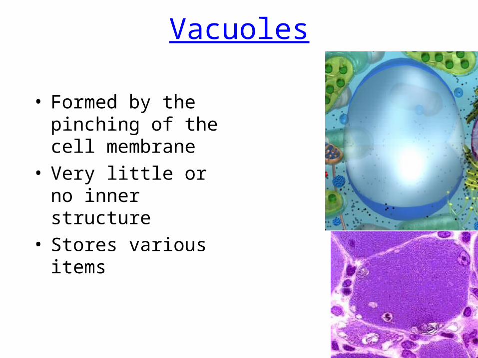
Vacuoles
• Formed by the pinching of the cell membrane
• Very little or no inner structure
• Stores various items
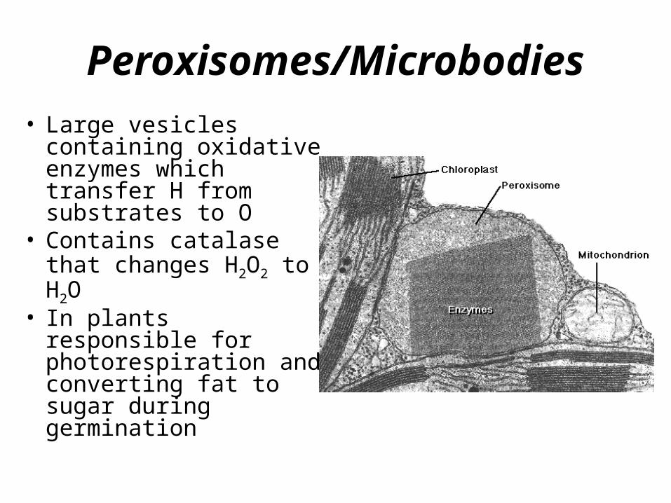
Peroxisomes/Microbodies
• Large vesicles containing oxidative enzymes which transfer H from substrates to O
• Contains catalase that changes H2O2 to H2O
• In plants responsible for photorespiration and converting fat to sugar during germination
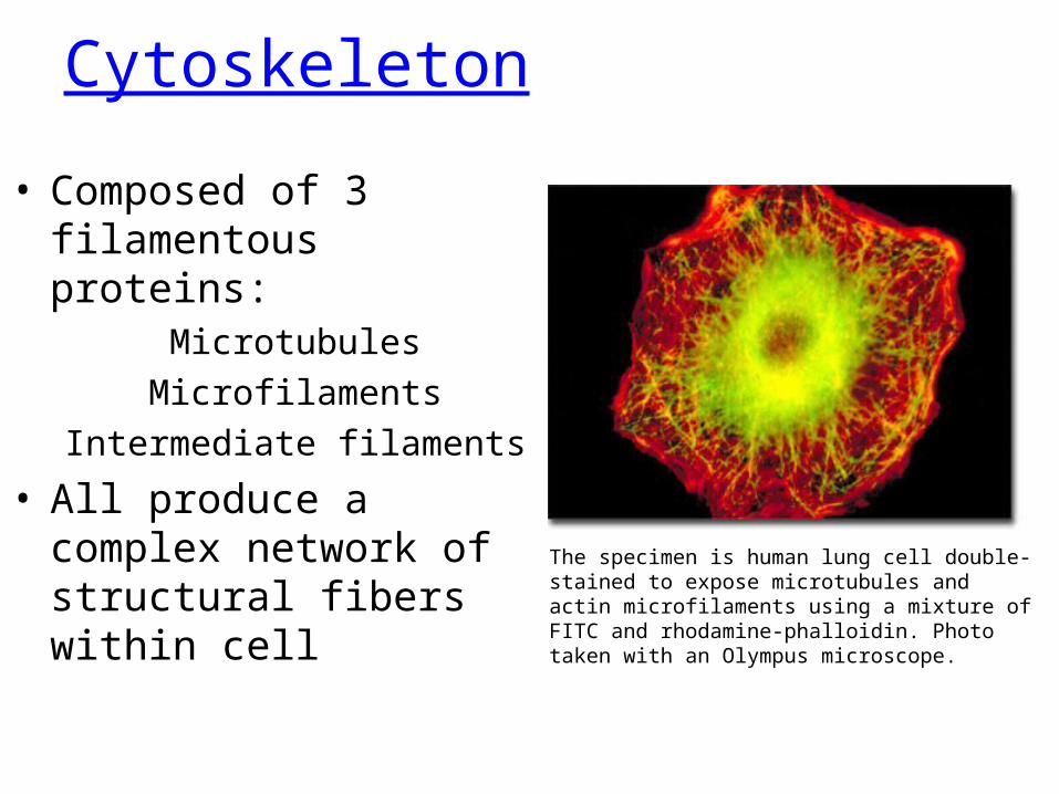
Cytoskeleton
• Composed of 3 filamentous proteins:
MicrotubulesMicrofilaments
Intermediate filaments
• All produce a complex network of structural fibers within cell The specimen is human lung cell double-
stained to expose microtubules and actin microfilaments using a mixture of FITC and rhodamine-phalloidin. Photo taken with an Olympus microscope.
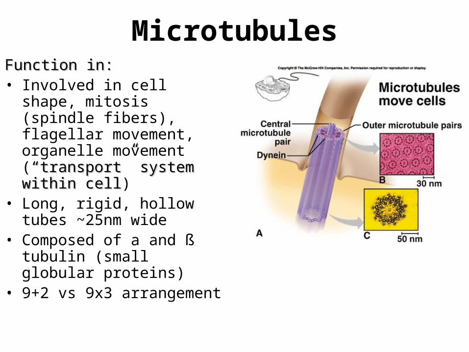
MicrotubulesFunction in:Function in:• Involved in cell shape,
mitosis (spindle fibers), flagellar movement, organelle movement (“transport” system “transport” system within cellwithin cell)
• Long, rigid, hollow tubes ~25nm wide
• Composed of a and ß tubulin (small globular proteins)
• 9+2 vs 9x3 arrangement
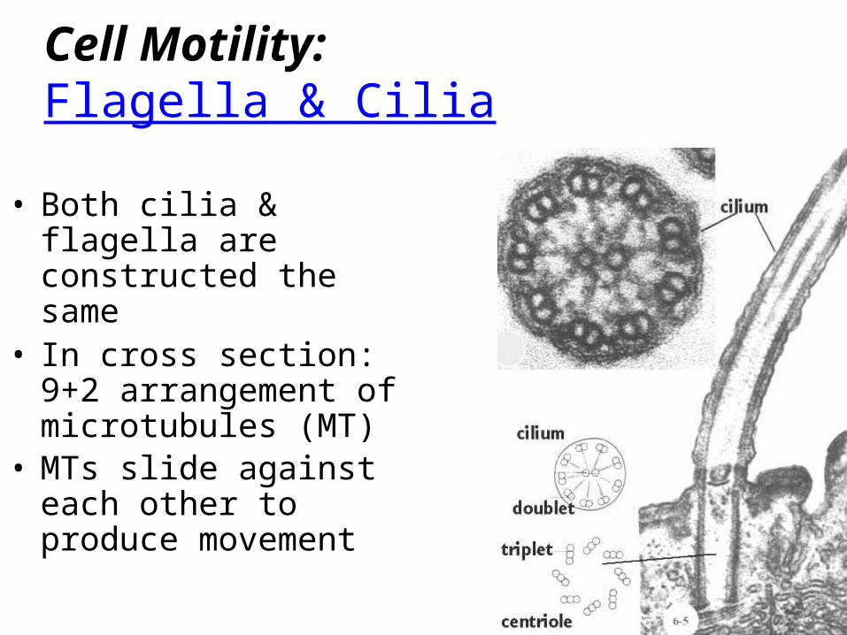
Cell Motility:Flagella & Cilia
• Both cilia & flagella are constructed the same
• In cross section: 9+2 arrangement of microtubules (MT)
• MTs slide against each other to produce movement
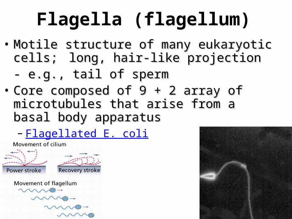
Flagella (flagellum)• Motile structure of many eukaryotic Motile structure of many eukaryotic
cells; cells; long, hair-like projectionlong, hair-like projection- e.g., tail of sperm- e.g., tail of sperm
• Core composed of 9 + 2 array of Core composed of 9 + 2 array of microtubules that arise from a basal microtubules that arise from a basal body apparatusbody apparatus– Flagellated E. coli
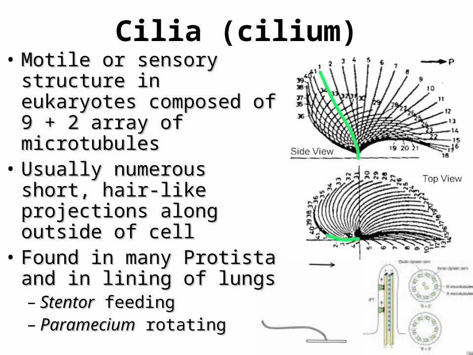
Cilia (cilium)• Motile or sensory Motile or sensory
structure in eukaryotes structure in eukaryotes composed of 9 + 2 composed of 9 + 2 array of microtubulesarray of microtubules
• Usually numerous Usually numerous short, hair-like short, hair-like projections along projections along outside of celloutside of cell
• Found in many Protista Found in many Protista and in lining of lungsand in lining of lungs– StentorStentor feeding feeding– ParameciumParamecium rotating rotating
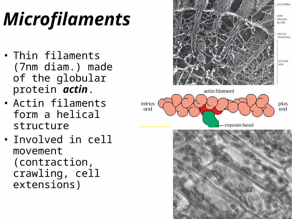
Microfilaments
• Thin filaments (7nm diam.) made of the globular protein actin.
• Actin filaments form a helical structure
• Involved in cell movement (contraction, crawling, cell extensions)
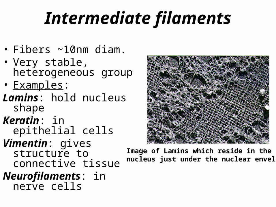
Intermediate filaments
• Fibers ~10nm diam.• Very stable,
heterogeneous group• Examples:Lamins: hold nucleus
shapeKeratin: in epithelial
cells Vimentin: gives
structure to connective tissue
Neurofilaments: in nerve cells
Image of Lamins which reside in the nucleus just under the nuclear envelope
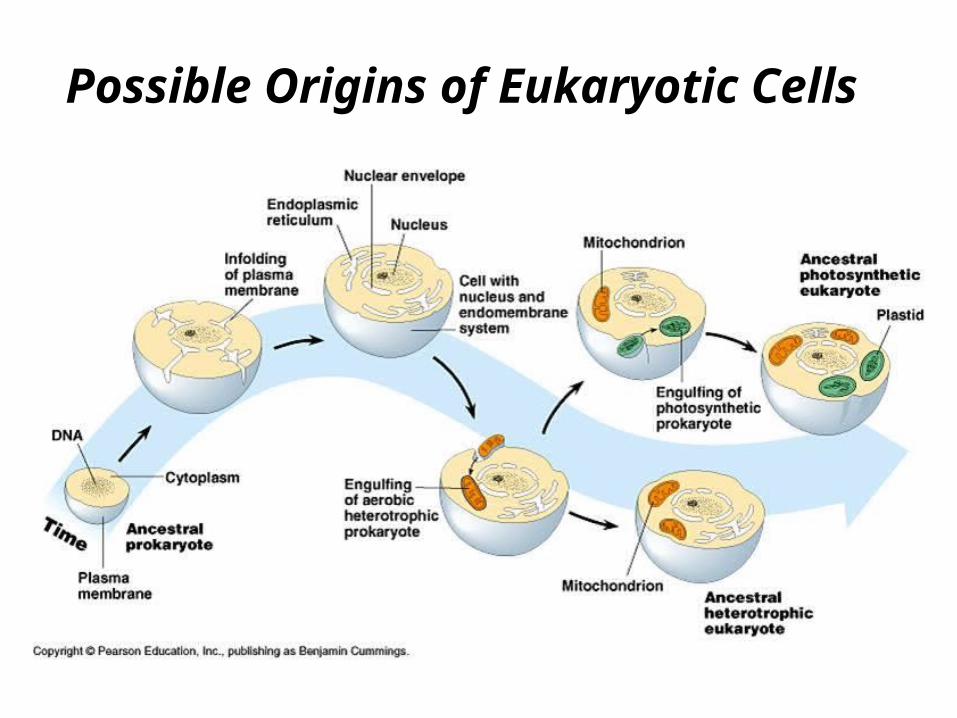
Possible Origins of Eukaryotic Cells
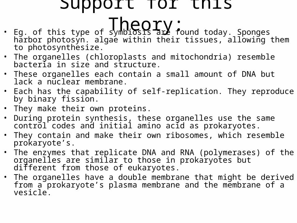
Support for this Theory:• Eg. of this type of symbiosis are found today. Sponges harbor
photosyn. algae within their tissues, allowing them to photosynthesize.
• The organelles (chloroplasts and mitochondria) resemble bacteria in size and structure.
• These organelles each contain a small amount of DNA but lack a nuclear membrane.
• Each has the capability of self-replication. They reproduce by binary fission.
• They make their own proteins.• During protein synthesis, these organelles use the same control
codes and initial amino acid as prokaryotes.• They contain and make their own ribosomes, which resemble
prokaryote’s. • The enzymes that replicate DNA and RNA (polymerases) of the
organelles are similar to those in prokaryotes but different from those of eukaryotes.
• The organelles have a double membrane that might be derived from a prokaryote’s plasma membrane and the membrane of a vesicle.
Resources • Rediscovering Biology Animation
Guide• Cell Signaling and Cell Cycle
Animations • Molecular Movies• Apoptosis Animation • Bacterial Animations