cells and tissues - fths wiki · name and describe the structure and function of cytoskeletal...
TRANSCRIPT

89

90
Cells:
Living Units

91
Cells: Living Units Unit Front Page

92
Cells: Living Units
At the end of this unit, I will:
□ Define cells and tissues and explain how they are organized. □ Explain the function of cell organelles
□ Describe how proteins are manufactured, and the function of the endomembrane system in manufacturing and modifying polypeptides or
proteins.
□ Name and describe the structure and function of cytoskeletal elements. □ Describe the chemical composition of the plasma membrane and relate it
to its membrane function. □ Compare the structure and function of tight junctions, desmosomes, and
gap junctions. □ Explain active and passive transport mechanisms of the plasma
membrane while differentiating the transport processes relative to energy source, substances transported, direction, and mechanism.
□ Describe how DNA is packaged and stored in the nucleus. Explain the various forms of DNA from nucleosome, chromatin, to chromosome.
Roots, Prefixes and Suffixes I will understand and recognize in words
are:
□ Crist-, Cyto-, Desm-, Dia-, Dys-, Flagell-, mito-, nucle-, onco-, osmo-,
permea-, phag-, philo-, phobo-, tono-,
□ -plasm, -troph, -villus

93
Composite/Generalized Cell vs. Differentiated/Diverse Cell
Definition of Composite Cell:
________________________________________________________________________________________________________________________________
________________________________________________________________
Basic Illustrated Example of Composite Cell:
Three illustrated examples of diverse/differentiated cells:
Explanation: Explanation: Explanation:

94
Reading Guide: Chapter 3
Cells, Living Units
Instructions: The specific instructions for various activities and the grading rubric for reading guides can be found on pages 11 – 19 and pages 30 – 31 of
your intNB. Refer to these pages carefully, as you will be completing reading
guides all throughout this year. 1. Read pgs. 65 – 66: 1st column on Overview of the Cellular Basis of Life On the left page 93 of your interactive notebook, define a “composite” or “generalized” cell
and draw your own interpretation of this cell based on its definition. LABEL the three main parts of this composite cell that all human cells have. Contrast your composite cell with cells that have diverse shapes and functions by drawing
three examples of cells that differ from your composite cell. Explain the different shape or function of the cells you chose to draw. Figure 3.1 may help.
2. Read pgs. 66 – Pg. 69 on the Fluid Mosaic Model Write a GIST on page 96 of your intNB
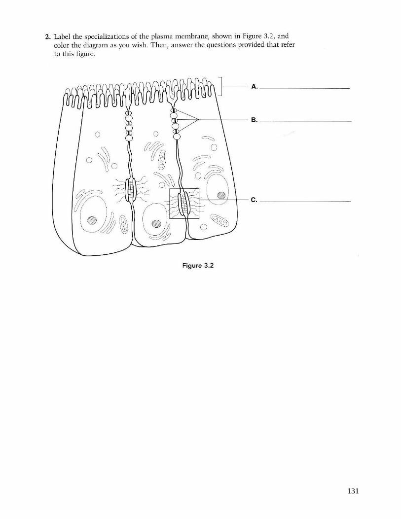
3. Read pg 69 and figure 3.5 on page 70 of your textbook on Specializations of Plasma Membrane. Identify the three types of junctions on the illustration on page 95 of your intNB. Color each
individual junction with different colors and use the same colors to color only the junctions in the larger graphic. For each junction, explain its function and identifying characteristics.
4. Read pages 70 – 81 on Membrane Transport Complete a 4-column T-chart on page 97 of your intNB that contrasts the following passive
transport mechanisms: simple diffusion, carrier-mediated facilitated diffusion, channel-mediated facilitated diffusion, and osmosis. List at least FOUR facts for each.
Complete a 3-column T-chart on page 97 of your intNB that contrasts the three main active transport mechanisms: primary active transport, secondary active transport, and vesicular transport. List at least FOUR facts for each.
5. Read pages 83 – 84 on Cell to Environment Interactions Write a GIST on page 96 of your intNB.
6. Review pages 84 – 97 on The Cytoplasm and The Nucleus. Complete the table on page 100 of your intNB.
Color and label the image of the cell on page 99 of your intNB Write a GIST explaining how DNA is organized inside the nucleus on page 98 of your intNB. (The section on Chromatin pg. 97 of your textbook and Figure 3.29 on page 98 will help you
with your GIST)

95
Membrane Junctions: Color each individual junction with different colors and use the same colors on the larger image to the left.
Type of Junction:
Function and Characteristics:
Type of Junction:
Function and Characteristics:
Type of Junction:
Function and Characteristics:
During lecture, you will be labeling specific characteristics of each junction in the above image.

96
Reading Guide Chapter 3
GIST 1 Fluid Mosaic Model

97
Passive Transport
Simple Diffusion Carrier-Mediated
facilitated diffusion
Channel-
Mediated facilitated
diffusion
Osmosis
Active Transport
Primary Active
Transport
Secondary Active
Transport
Vesicular Transport

98
GIST 3 Organization of DNA

99
Cell Labeling

100

101
Packaging of DNA into Chromosomes
In the figure below, label the following: DNA, Nucleosome, Chromatin, Looped
Domains, and Chromosome
___________________________
_____________________
__
_____________________
__
_________________
______
______________________
_

102
Date_______________
Chapter 3: Cytoplasmic Organelles and the Nucleus

103
Cell Organelle Layered Book
Create a pocket on this page to store your Cell Organelle Layered Book or glue
the back of your book onto this page.

104

105

106
Date_______________
Chapter 3: Plasma Membrane

107

108

109

110

111
Membrane Transport
Label each of the following transport methods as either 1) active or passive
then 2) specify the specific type of membrane transport:
1) ____________________________ 1) ____________________________ 2) ____________________________ 2) ____________________________
1) ____________________________ 1) ____________________________ 2) ____________________________ 2) ____________________________

112

113
1) ____________________________ 1) ____________________________
2) ____________________________ 2) ____________________________
1) ____________________________ 1) ____________________________
2) ____________________________ 2) ____________________________
1) ____________________________ 1) ____________________________
2) ____________________________ 2) ____________________________

114

115
Sodium-Potassium Pump

116

117

118

119
Diffusion and Osmosis Pre-lab
Read the Diffusion and Osmosis Lab on the following pages 120 - 124
and complete the pre-lab PRIOR to lab day. Introduction: Dialysis tubing allows molecules to diffuse through microscopic pores in the tubing. Molecules smaller than the pores can diffuse through the dialysis membrane along
their concentration gradients, while molecules larger than the pore size are prevented from crossing the dialysis membrane.
Answer the following questions. For problems, show equations and work with units and appropriate significant figures.
Part 1A: Predict whether or not each of these is expected to pass through the dialysis membrane.
Water ____________ Glucose ____________ I2KI ____________ Starch ____________ How will you know if the iodine solution (I2KI) has crossed the dialysis membrane?
Part 1B: In the following situations, assume that sucrose cannot diffuse through the dialysis membrane.
1. If a dialysis bag containing a .20 M solution of sucrose is placed in a beaker of
distilled water. Will the dialysis bag gain or lose mass? Explain why.
2. A dialysis bag has an initial mass of 30.2 g and a final mass of 26.3 g. Find the %
change in mass.
CAREFULLY read the procedure on pages 123 – 124 lab 1B. In the space below, DRAW how the beakers will be set up in the space below. Label the contents of each dialysis bag and
the contents in each beaker in your illustration. To help you, see Figure 1.1 on page 123, which provides an example of one of the beakers.

120

121

122

123

124

125

126
This is an EXAMPLE of what your graph should look like on the opposite page. Make sure to graph the CLASS DATA and to use a
LINE OF BEST FIT.
% Change in Mass of Dialysis Bags as a function of Sucrose Molarity
-5 .0%
0 .0%
5 .0%
10 .0%
15 .0%
20 .0%
25 .0%
30 .0%
35 .0%
0 0 .2 0 .4 0 .6 0 .8 1 1 .2
Sucrose Molarity (moles/L)
Pe
rce
nt
ch
an
ge
in
ma
ss o
f D
ialy
sis
Ba
gs
(%
)
C las s Data Group Data
L inear (G roup Data) L inear (C las s Data)
Analysis of Results

127

128
Chapter 3
Study Guide

129

130
Use Figure 3.1 to answer the following:
131

132
Use Figure 3.2 to answer the following:

133

134

135

136

137
Cells: Living Units Unit Back Page (See page 19 for instructions)