cells (and other stuff)
TRANSCRIPT

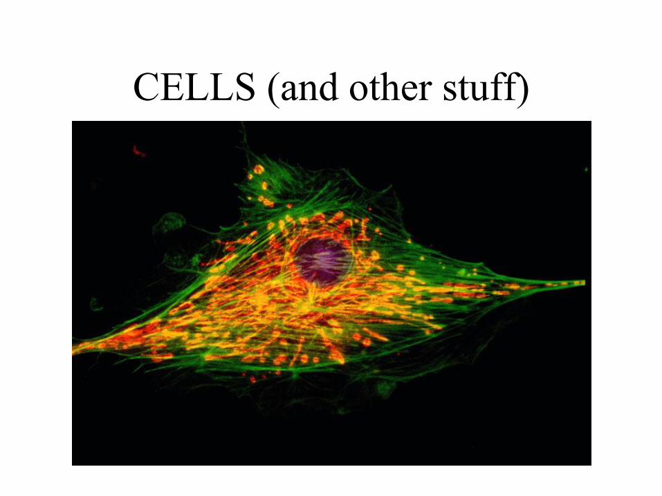
CELLS (and other stuff)

• Life’s basic characteristic is a high degree of order.
– At the lowest level are atoms that are ordered into
complex biological molecules.
– Many molecules are arranged into minute structure
called organelles, which are the components of cells.
– New properties emerge at each higher level of org.
1. Each level of biological organization has
emergent properties
Copyright © 2002 Pearson Education, Inc., publishing as Benjamin Cummings
Fig. 1.2(1) Fig. 1.2(2)

• The cell is the lowest level of structure that is capable of
performing all the activities of life (metabolism,
response, homeostasis, growth, reproduction, nutrition).
• Two big names in early cell discoveries:
• Robert Hooke
• Anton vanLeeuwenhoek
2. Cells are an organism’s basic
unit of structure and function
Copyright © 2002 Pearson Education, Inc., publishing as Benjamin Cummings

• In 1839, Matthais Schleiden and Theodor
Schwann extrapolated from their own
microscopic research and that of others to
propose the cell theory.
– 1. All living things consist of cells.
– 2. All cells come from other cells.
• This suggests a big idea, foreshadowing Darwin’s idea of
descent with modification. Pasteur’s famous experiment
contributed to this idea. Remember it?
-3.Cells are the smallest unit of life, capable of
carrying out all life functions. Everything we
do is a result of something our cells do.
Copyright © 2002 Pearson Education, Inc., publishing as Benjamin Cummings

• All cells are enclosed …
• At some point, all cells contain …
• Two major kinds of cells - prokaryotic cells and
eukaryotic cells - can be distinguished by their
structural organization.
– The cells of the microorganisms called bacteria and
archaea are prokaryotic.
– All other forms of life have the more complex
eukaryotic cells.
Copyright © 2002 Pearson Education, Inc., publishing as Benjamin Cummings

• All cells are surrounded by a plasma membrane.
– The term plasma is reserved for the outer membrane.
• The semifluid substance within the membrane is the
cytosol, containing the organelles.
• All cells at some point contain chromosomes which have
genes in the form of DNA.
• All cells also have ribosomes, tiny organelles that make
proteins using the instructions contained in genes.
1. Prokaryotic and eukaryotic cells
differ in size and complexity
Copyright © 2002 Pearson Education, Inc., publishing as Benjamin Cummings

• A major difference between prokaryotic and
eukaryotic cells is the location of chromosomes.
• In an eukaryotic cell, chromosomes are
contained in a …
• In a prokaryotic cell, the DNA is concentrated
in the nucleoid without a membrane separating
it from the rest of the cell.
• The –oid suffix means what?
Copyright © 2002 Pearson Education, Inc., publishing as Benjamin Cummings

Terms to keep straight:
• Cytoplasm:
• Nucleoplasm:

Copyright © 2002 Pearson Education, Inc., publishing as Benjamin Cummings
Fig. 7.4 The prokaryotic cell is much simpler in structure, lacking a nucleus and the other
membrane-enclosed organelles of the eukaryotic cell.

• Eukaryotic cells are generally 10 times bigger
than prokaryotic cells
• Here’s a size comparison, let’s practice some
math:
– Molecules – 1 nm (scientific notation?)
– Membrane thickness – 10 nm
– Viruses – 100 nm
– Bacteria – 1 um (how much bigger than a virus?)
– Organelles – 10 um
– Cells – up to 100 um
– See the pattern? 10 times bigger each.
Copyright © 2002 Pearson Education, Inc., publishing as Benjamin Cummings

Copyright © 2002 Pearson Education, Inc., publishing as Benjamin Cummings
Fig. 7.5
• Metabolic requirements also set an upper limit
to the size of a single cell.
• As a cell increases in size its volume increases
faster than its surface area.
– Smaller objects have a greater
ratio of surface area to volume.

• The volume of cytoplasm determines the need
for the exchange of nutrients and wastes across
the plasma membrane.
• Rates of chemical exchange may be inadequate
to maintain a cell with a very large cytoplasm.
• Why can’t a cell be as big as a left tackle for the
Seminoles?
• Larger organisms do not generally have larger
cells than smaller organisms - simply more
cells.
• How about some math practice?
Copyright © 2002 Pearson Education, Inc., publishing as Benjamin Cummings

Tips • Grid LEFT to right
• Use the formula sheet
• Don’t round until the end
• Look at HOW the answer should be given
“round to nearest…”
.123
The 1 is in the tenths place
The 2 is in the hundreds place
The 3 is in the thousandths place

Q2: Surface Area and Volume
• What is the SA/V for this cell? Round your
answer to the nearest hundredths.

Q2
SA= 4 r2
=4(3.14) 52
=314
Volume of a sphere= 4/3 r3
=4/3 (3.14)53
=523.33
SA/V=314/523.33
=.60

• A eukaryotic cell has extensive and elaborate internal
membranes, which partition the cell into compartments.
• These membranes also participate in metabolism by
providing a surface for reactions to occur on, as well as
enzymes to speed them up.
• What is the advantage of such partitioning???
2. Internal membranes compartmentalize the
functions of a eukaryotic cell
Copyright © 2002 Pearson Education, Inc., publishing as Benjamin Cummings

• The plasma membrane separates the living cell
from its nonliving surroundings.
• This thin barrier, 8 nm thick, controls traffic into
and out of the cell.
• Like other membranes, the plasma membrane is
selectively permeable, meaning what???
• Check this out:
http://www.johnkyrk.com/cellmembrane.html
Introduction
Copyright © 2002 Pearson Education, Inc., publishing as Benjamin Cummings

• The main macromolecules in membranes are
_____and _____ but include some ________.
• The most abundant lipids are _______.
• The phospholipids and proteins in membranes
create a unique physical environment, described
by the fluid ______ model.
– A membrane is a fluid structure with proteins
embedded or attached to a double layer of
phospholipids each of which has a polar “head” and
non-polar fatty acid “tails”.
Copyright © 2002 Pearson Education, Inc., publishing as Benjamin Cummings

• In 1972 (when I was your age), S.J. Singer and
G. Nicolson presented a model that proposed
that the membrane proteins are dispersed and
individually inserted into the phospholipid
bilayer.
– In this fluid mosaic
model, the hydrophilic
regions of proteins
and phospholipids are
in maximum contact
with water and the
hydrophobic regions
are in a nonaqueous
environment. Copyright © 2002 Pearson Education, Inc., publishing as Benjamin Cummings
Fig. 8.2b

• Membrane molecules are held in place by
relatively weak hydrophobic interactions.
• Most of the lipids and some proteins can drift
laterally in the plane of the membrane, but rarely
flip-flop from one layer to the other.
1. Membranes are fluid
Copyright © 2002 Pearson Education, Inc., publishing as Benjamin Cummings
Fig. 8.4a

• The steroid cholesterol is wedged between
phospholipid molecules in the plasma membrane
of animal cells, (not other kinds).
• At warm temperatures, it restrains the movement
of phospholipids and reduces fluidity.
• At cool temperatures, it maintains fluidity by
preventing tight packing.
Copyright © 2002 Pearson Education, Inc., publishing as Benjamin Cummings
Fig. 8.4c

• A membrane is a collage of different proteins
embedded in the fluid matrix of the lipid bilayer.
3. Membranes are mosaics of
structure and function
Copyright © 2002 Pearson Education, Inc., publishing as Benjamin Cummings
Fig. 8.6

• Proteins determine most of the membrane’s
specific functions.
• The plasma membrane and the membranes of
the various organelles each have unique
collections of proteins.
• There are two populations of membrane
proteins.
– Peripheral proteins are not embedded in the lipid
bilayer at all.
– Instead, they are loosely bound to the surface of the
protein, often connected to the other population of
membrane proteins.
Copyright © 2002 Pearson Education, Inc., publishing as Benjamin Cummings

– Integral proteins penetrate the hydrophobic core of
the lipid bilayer, often completely spanning the
membrane. What level of organization do you see?
Copyright © 2002 Pearson Education, Inc., publishing as Benjamin Cummings
Fig. 8.7

• The proteins in the plasma membrane may
provide a variety of major cell functions.
Copyright © 2002 Pearson Education, Inc., publishing as Benjamin Cummings
Fig. 8.9

• The membrane plays the key role in cell-cell recognition and communication
– Cell-cell recognition is the ability of a cell to distinguish one type of neighboring cell from another.
– This attribute is important in cell sorting and organization as tissues and organs in development.
– It is also the basis for rejection of foreign cells by the immune system.
– Cells recognize other cells by keying on surface molecules, often carbohydrates, on the plasma membrane.
4. Membrane carbohydrates are
important for cell-cell recognition
Copyright © 2002 Pearson Education, Inc., publishing as Benjamin Cummings

• Membrane carbohydrates are usually branched with
fewer than 15 sugar units.
• They may be covalently bonded either to lipids,
forming glycolipids, or, more commonly, to proteins,
forming glycoproteins.
• They form what is called the cell coat in animal cells
and vary from species to species, individual to
individual, and even from cell type to cell type within
the same individual.
– This variation marks each cell type as distinct.
– Blood groups example???
Copyright © 2002 Pearson Education, Inc., publishing as Benjamin Cummings

Many things, like a girl’s car, can be
identified by their surface markings.

Copyright © 2002 Pearson Education, Inc., publishing as Benjamin Cummings
Fig. 7.7

Copyright © 2002 Pearson Education, Inc., publishing as Benjamin Cummings
Fig. 7.8

• The nucleus contains most of the genes in a eukaryotic cell. – Some genes, however, are located in _____ and
_______.
• The nucleus is separated from the cytoplasm by a double membrane.
• Where the double membranes are fused, a pore formed by 8 protein molecules in a ring allows large macromolecules and particles to pass through.
1. The nucleus contains a
eukaryotic cell’s genetic library
Copyright © 2002 Pearson Education, Inc., publishing as Benjamin Cummings

Copyright © 2002 Pearson Education, Inc., publishing as Benjamin Cummings
Fig. 7.9

• Within the nucleus, the DNA and associated proteins are organized into fibrous material, chromatin. Prokaryotic cells don’t have as much protein stuck to their DNA.
• In a normal cell they appear as a diffuse mass.
• However, What happens to chromatin when a cell is getting ready to divide????
• Each eukaryotic species has a characteristic number of chromosomes.
– A typical human cell has ___ chromosomes (but so do a plum’s), fruit flies have 8, pea plants have 14.
Copyright © 2002 Pearson Education, Inc., publishing as Benjamin Cummings

• In the nucleus is a region of densely stained
fibers and granules, the nucleolus.
– What happens in this region????
• The nucleus directs protein synthesis by
synthesizing messenger RNA (mRNA).
• Can a cell have more than one? Why? None?
Examples?
Copyright © 2002 Pearson Education, Inc., publishing as Benjamin Cummings

• Cell types that synthesize large quantities of
proteins (e.g., pancreatic cells) have large
numbers of ribosomes and prominent nuclei.
• Free vs. Bound ribosomes????
Copyright © 2002 Pearson Education, Inc., publishing as Benjamin Cummings

• The endoplasmic reticulum (ER) accounts for
half the membranes in a eukaryotic cell.
• The ER includes membranous tubules and
internal, fluid-filled spaces, the cisternae.
• The ER membrane is continuous with the nuclear
envelope and the cisternal space of the ER is
continuous with the space between the two
membranes of the nuclear envelope.
1. The endoplasmic reticulum manufacturers
membranes and performs many other
biosynthetic functions
Copyright © 2002 Pearson Education, Inc., publishing as Benjamin Cummings

• There are two, albeit
connected, regions of ER
that differ in structure
and function.
– Smooth ER looks smooth
because ________.
– Rough ER looks rough
because ______ including
the outside of the nuclear
envelope.
– Watch what they do:
http://www.johnkyrk.com/
golgiAlone.html
Copyright © 2002 Pearson Education, Inc., publishing as Benjamin Cummings
Fig. 7.11

• The smooth ER is rich in enzymes and plays a
role in a variety of metabolic processes.
• Enzymes of smooth ER synthesize lipids,
including oils, phospholipids, and steroids.
– These includes the sex hormones of vertebrates and
adrenal steroids.
• The smooth ER also catalyzes a key step in the
mobilization of glucose from stored glycogen in
the liver. Smooth ER helps make everything
but protein.
Copyright © 2002 Pearson Education, Inc., publishing as Benjamin Cummings

• Other enzymes in the smooth ER of the liver
help detoxify drugs and poisons.
– These include alcohol and barbiturates.
– Frequent exposure leads to proliferation of smooth
ER, increasing tolerance to the drug.
– What do you think Michael Jackson’s liver cells
looked like?
Copyright © 2002 Pearson Education, Inc., publishing as Benjamin Cummings

• Rough ER is especially abundant in those cells that
secrete _______(like __________secreted from
pancreas cells).
– As a polypeptide is synthesized by the ribosome, it is
threaded into the cisternal space through a pore formed by a
protein in the ER membrane.
– Many of these polypeptides are glycoproteins, a polypeptide
to which an oligosaccharide is attached.
• These secretory proteins are packaged in transport
vesicles that carry them to their next stage.
Copyright © 2002 Pearson Education, Inc., publishing as Benjamin Cummings

• Rough ER is also a membrane factory.
– Membrane bound proteins are synthesized directly
into the membrane.
– Enzymes in the rough ER also synthesize
phospholipids from precursors in the cytosol.
– As the ER membrane expands, parts can be
transferred as transport vesicles to other
components of the endomembrane system.
Copyright © 2002 Pearson Education, Inc., publishing as Benjamin Cummings


• Many transport vesicles from the ER travel to the
Golgi apparatus for modification of their
contents.
• The Golgi is a center of manufacturing,
warehousing, sorting, and shipping.
• In what kind of cells would you expect to find a
lot of Golgi?
2. The Golgi apparatus finishes,
sorts, and ships cell products
Copyright © 2002 Pearson Education, Inc., publishing as Benjamin Cummings
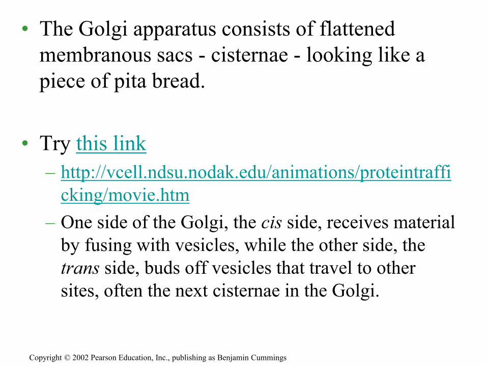
• The Golgi apparatus consists of flattened
membranous sacs - cisternae - looking like a
piece of pita bread.
• Try this link
– http://vcell.ndsu.nodak.edu/animations/proteintraffi
cking/movie.htm
– One side of the Golgi, the cis side, receives material
by fusing with vesicles, while the other side, the
trans side, buds off vesicles that travel to other
sites, often the next cisternae in the Golgi.
Copyright © 2002 Pearson Education, Inc., publishing as Benjamin Cummings

• Products from the ER are modified as they pass
through.
• The Golgi can also manufacture its own
macromolecules, including pectin and other
noncellulose polysaccharides.
• Each cisterna has its own set of enzymes.
• Finally, the Golgi tags, sorts, and packages
materials into transport vesicles.
Copyright © 2002 Pearson Education, Inc., publishing as Benjamin Cummings
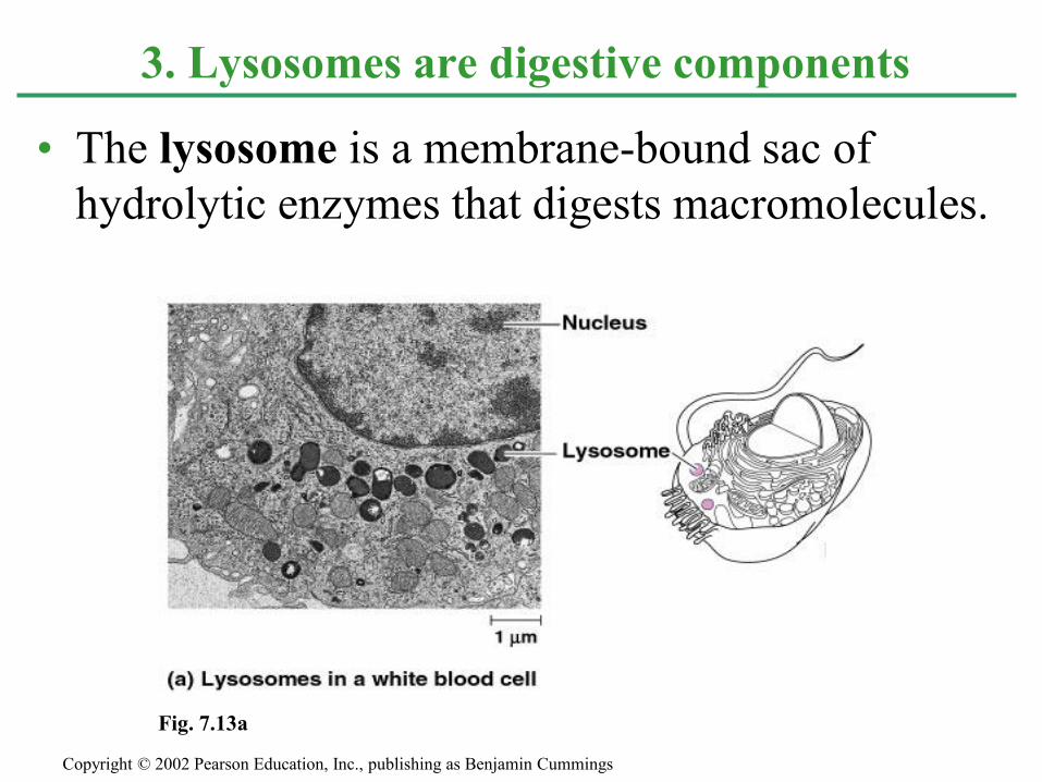
• The lysosome is a membrane-bound sac of
hydrolytic enzymes that digests macromolecules.
3. Lysosomes are digestive components
Copyright © 2002 Pearson Education, Inc., publishing as Benjamin Cummings
Fig. 7.13a

• The lysosomal enzymes and membrane are
synthesized by rough ER and then transferred to
the Golgi.
• Lysosomes
bud from
the trans
face of
the Golgi.
Copyright © 2002 Pearson Education, Inc., publishing as Benjamin Cummings
Fig. 7.14

Copyright © 2002 Pearson Education, Inc., publishing as Benjamin Cummings
• Lysosomes can fuse with food vacuoles.
• As the polymers are digested, their monomers
pass out to the cytosol to become nutrients for
the cell.
• Lysosomes can also
fuse with another
organelle or part
of the cytosol.
– This recycling, this process of autophagy, renews the cell.
Fig. 7.13b

• The lysosomes play a critical
role in apoptosis, or
programmed cell death.
–Tails of tadpoles?
–Webbing between fingers and toes?
–It’s a good thing this happens so
that we can do this….
Copyright © 2002 Pearson Education, Inc., publishing as Benjamin Cummings


• Vesicles and vacuoles – what’s the difference??
– Food vacuoles.
– Contractile vacuoles,
– Central vacuoles are found in many mature ______
cells, but none so big are ever found in ________
cells.
4. Vacuoles have diverse functions
in cell maintenance
Copyright © 2002 Pearson Education, Inc., publishing as Benjamin Cummings

Copyright © 2002 Pearson Education, Inc., publishing as Benjamin Cummings
Fig. 7.15

• Mitochondria are the sites of cellular ________, generating _____ from the catabolism of sugars, fats, and other fuels in the presence of ________.
• Chloroplasts, found in plants and eukaryotic algae, are the site of __________________.
– They convert solar energy to chemical energy and synthesize new organic compounds, mostly _______ from CO2 and _____.
1. Mitochondria and chloroplasts are the
main energy transformers of cells
Copyright © 2002 Pearson Education, Inc., publishing as Benjamin Cummings

• Mitochondria and chloroplasts are not part of
the endomembrane system.
• Their proteins come primarily from free
ribosomes in the cytosol and a few from their
own ribosomes.
• Both organelles have small quantities of
bacteria-like DNA that direct the synthesis of
the polypeptides produced by these internal
ribosomes.
• Mitochondria and chloroplasts grow and
reproduce as semiautonomous organelles.
• The Endosymbiont theory. Copyright © 2002 Pearson Education, Inc., publishing as Benjamin Cummings

• Almost all eukaryotic cells have
mitochondria. Name one that doesn’t???
– There may be one very large
mitochondrion or hundreds to thousands
of individual mitochondria.
– The number of mitochondria is correlated
with aerobic metabolic activity; muscle
cells have a lot.
– A typical mitochondrion is 1-10 microns
long, similar to a _______________ cell.
Copyright © 2002 Pearson Education, Inc., publishing as Benjamin Cummings

• Two membranes, inner one is folded into____.
– This creates a fluid-filled space between them, the
intermembrane space.
– Classic S-F connection??????
• The inner membrane encloses the
mitochondrial ________, a fluid-filled space
with DNA, ribosomes, and enzymes.
Copyright © 2002 Pearson Education, Inc., publishing as Benjamin Cummings

Copyright © 2002 Pearson Education, Inc., publishing as Benjamin Cummings
Fig. 7.17

• The chloroplast is one of several members of a
generalized class of plant structures called plastids.
– Amyloplasts store starch in roots and tubers.
– Chromoplasts store pigments for fruits and flowers.
• The chloroplast produces sugar via photosynthesis.
– Chloroplasts gain their color from high levels of the
green pigment chlorophyll.
• Chloroplasts measure about 2 microns x 5 microns and
are found in leaves and other green structures of plants
and in eukaryotic algae.
Copyright © 2002 Pearson Education, Inc., publishing as Benjamin Cummings

• The processes in the chloroplast are separated
from the cytosol by two membranes.
• Inside the inner membrane is a fluid-filled
space, the stroma, in which float membranous
sacs, the thylakoids.
– The stroma contains DNA, ribosomes, and enzymes
for part of the photosynthetic reactions..
– The thylakoids, flattened sacs, are stacked into
grana and are critical for converting light to
chemical energy.
Copyright © 2002 Pearson Education, Inc., publishing as Benjamin Cummings

Copyright © 2002 Pearson Education, Inc., publishing as Benjamin Cummings
Fig. 7.18

Theme time…
• So let’s see how many
similarities and differences
we can list between
mitochondria and
chloroplasts.

• Peroxisomes contain enzymes that transfer hydrogen from various substrates to oxygen
– An intermediate product of this process is hydrogen peroxide (H2O2), a poison, but the peroxisome has another enzyme, catalase, that converts H2O2 to water.
– Other peroxisomes in liver cells detoxify alcohol and other harmful compounds.
– Specialized peroxisomes, glyoxysomes, convert the fatty acids in seeds to sugars, an easier energy and carbon source to transport.
Peroxisomes
Copyright © 2002 Pearson Education, Inc., publishing as Benjamin Cummings

• The cytoskeleton is a network of protein fibers
extending throughout the cytoplasm.
• The cytoskeleton
organizes the
structures and
activities of
the cell.
Introduction
Copyright © 2002 Pearson Education, Inc., publishing as Benjamin Cummings
Fig. 7.20

• The cytoskeleton provides mechanical support and maintains or helps change the shape of the cell.
• The cytoskeleton provides anchorage for many organelles and cytosolic enzymes.
• The cytoskeleton is dynamic, dismantling in one part and reassembling in another to change cell shape.
Cytoskeleton functions
Copyright © 2002 Pearson Education, Inc., publishing as Benjamin Cummings

• The cytoskeleton also plays a major role in cell
motility.
– This involves both changes in cell location and
limited movements of parts of the cell.
• The cytoskeleton interacts with motor proteins.
– In cilia and flagella motor proteins pull components
of the cytoskeleton past each other.
– This is also true
in muscle cells.
Copyright © 2002 Pearson Education, Inc., publishing as Benjamin Cummings
Fig. 7.21a

• Motor molecules also carry vesicles or
organelles to various destinations along
“monorails’ provided by the cytoskeleton.
• Interactions of motor proteins and the
cytoskeleton circulates materials within a cell -
called cell streaming or cyclosis.
Copyright © 2002 Pearson Education, Inc., publishing as Benjamin Cummings
Fig. 7.21b

• There are three main types of fibers in the
cytoskeleton: microtubules, microfilaments,
and intermediate filaments.
Copyright © 2002 Pearson Education, Inc., publishing as Benjamin Cummings
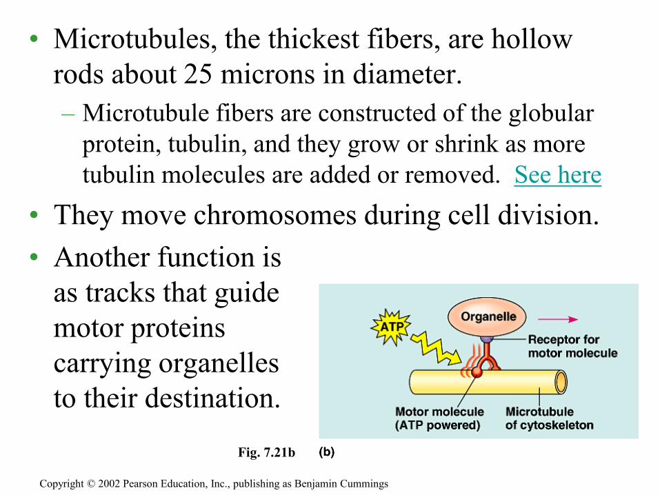
• Microtubules, the thickest fibers, are hollow
rods about 25 microns in diameter.
– Microtubule fibers are constructed of the globular
protein, tubulin, and they grow or shrink as more
tubulin molecules are added or removed. See here
• They move chromosomes during cell division.
• Another function is
as tracks that guide
motor proteins
carrying organelles
to their destination.
Copyright © 2002 Pearson Education, Inc., publishing as Benjamin Cummings
Fig. 7.21b

Copyright © 2002 Pearson Education, Inc., publishing as Benjamin Cummings
Fig. 7.22
• In animal cells, the centrosome has a pair of centrioles, each with nine triplets of microtubules arranged in a ring.
• During cell division the centrioles replicate.
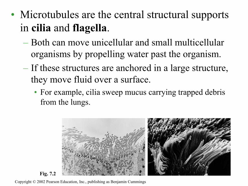
• Microtubules are the central structural supports
in cilia and flagella.
– Both can move unicellular and small multicellular
organisms by propelling water past the organism.
– If these structures are anchored in a large structure,
they move fluid over a surface.
• For example, cilia sweep mucus carrying trapped debris
from the lungs.
Copyright © 2002 Pearson Education, Inc., publishing as Benjamin Cummings
Fig. 7.2

• In spite of their differences, both cilia and
flagella have the same ultrastructure.
– Both have a core of microtubules sheathed by the
plasma membrane.
– Nine doublets of microtubules are arranged around
a pair at the center, the “9 + 2” pattern.
– Flexible “wheels” of proteins connect outer
doublets to each other and to the core.
– The outer doublets are also connected by motor
proteins.
– The cilium or flagellum is anchored in the cell by a
basal body, whose structure is identical to a
centriole.
Copyright © 2002 Pearson Education, Inc., publishing as Benjamin Cummings

Copyright © 2002 Pearson Education, Inc., publishing as Benjamin Cummings
Fig. 7.24

• The bending of cilia and flagella is driven by the
arms of a motor protein, dynein.
– Dynein arms alternately
grab, move, and release
the outer microtubules.
– Protein cross-links limit
sliding and the force is
expressed as bending.
– Watch this cilia on cells
– In a clam’s gill
Copyright © 2002 Pearson Education, Inc., publishing as Benjamin Cummings
Fig. 7.25

• Microfilaments, the thinnest class of
the cytoskeletal fibers, are solid rods of
the globular protein actin.
• Microfilaments are designed to resist
tension.
• With other proteins, they form a three-
dimensional network just inside the
plasma membrane.
Copyright © 2002 Pearson Education, Inc., publishing as Benjamin Cummings

• In plant cells (and others), actin-myosin
interactions drive cytoplasmic streaming.
– This creates a circular flow of cytoplasm in the cell.
Watch here
– This speeds the distribution of materials within the
cell.
Copyright © 2002 Pearson Education, Inc., publishing as Benjamin Cummings
Fig. 7.21c

• Intermediate filaments, intermediate in size at 8 - 12 nanometers, are specialized for bearing tension.
– Intermediate filaments are built from a diverse class of subunits from a family of proteins called keratins.
• They reinforce cell shape and fix organelle location.
• Watch how structure is related to function. Link to Intermediate Filaments
Copyright © 2002 Pearson Education, Inc., publishing as Benjamin Cummings
Fig. 7.26

• The cell wall, also found in prokaryotes, fungi,
and some protists, has multiple functions.
• In plants, the cell wall protects the cell, maintains
its shape, and prevents excessive uptake of water.
• It also supports the plant against the force of
gravity.
• The thickness and chemical composition of cell
walls differs from species to species and among
cell types.
1. Plant cells are encased by cell walls
Copyright © 2002 Pearson Education, Inc., publishing as Benjamin Cummings
• The basic design consists of microfibrils of cellulose embedded in a matrix of proteins and other polysaccharides.
– This is like steel-reinforced concrete or fiberglass.
• A mature cell wall consists of a primary cell wall and layers of secondary cell wall.
Copyright © 2002 Pearson Education, Inc., publishing as Benjamin Cummings
Fig. 7.28

Cell wall structure
• Primary cell walls contain cellulose and pectin, and is thin and flexible.
• Secondary cell walls are thicker and also contain lignin, which further stiffens it. Cells of hard plant parts such as stems have one or more layers of secondary walls.
• A middle lamella of sticky pectin separates the cell walls of adjoining cells.

• Neighboring cells in tissues, organs, or organ
systems often adhere, interact, and communicate
through direct physical contact.
• Plant cells are perforated with plasmodesmata,
channels allowing cysotol to pass betells.
3. Intracellular junctions in plant cells
Copyright © 2002 Pearson Education, Inc., publishing as Benjamin Cummings
Fig. 7.28 inset

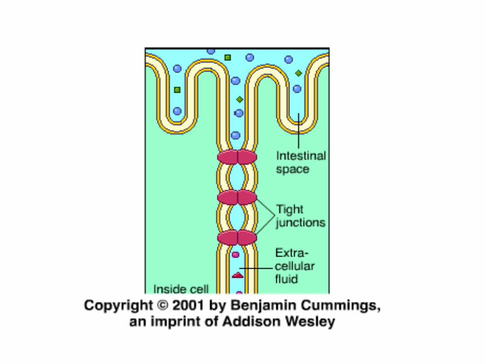
• Animal cells have 4 main types of intercellular
links: tight junctions, desmosomes, gap
junctions and the newly discovered tunnelling
nanotubes.
• In tight junctions, membranes of adjacent cells
are fused, forming continuous belts around
cells.
– This prevents leakage of extracellular fluid.
Copyright © 2002 Pearson Education, Inc., publishing as Benjamin Cummings
Fig. 7.30

• Desmosomes (or anchoring junctions) fasten cells
together into strong sheets, much like rivets.
– Intermediate filaments of keratin reinforce
desmosomes.
• Gap junctions (or communicating junctions) provide
cytoplasmic channels between adjacent cells.
– Special membrane proteins surround these pores.
– Salt ions, sugar, amino acids, and other small
molecules can pass.
– In embryos, gap junctions facilitate chemical
communication during development.
Copyright © 2002 Pearson Education, Inc., publishing as Benjamin Cummings


Practice time…
• List as many similarities and
differences as you can for:
– Prokaryotic vs. Eukaryotic cells
– Plant cells vs. animal cells
– http://multimedia.mcb.harvard.edu/a
nim_innerlife_hi.html

Tunnelling nanotubes
• Discovered in 2004, these narrow tubes can
form between all types of cells to allow
them to pass things such as protein
hormones and molecules that cause genes to
become active. Unfortunately, they also
can allow for viruses to pass from cell to
cell.
