cell structure & function there are two types of cells: 1.prokaryotic- cells that do not have a...
Post on 19-Dec-2015
217 views
TRANSCRIPT
Cell Structure & Function
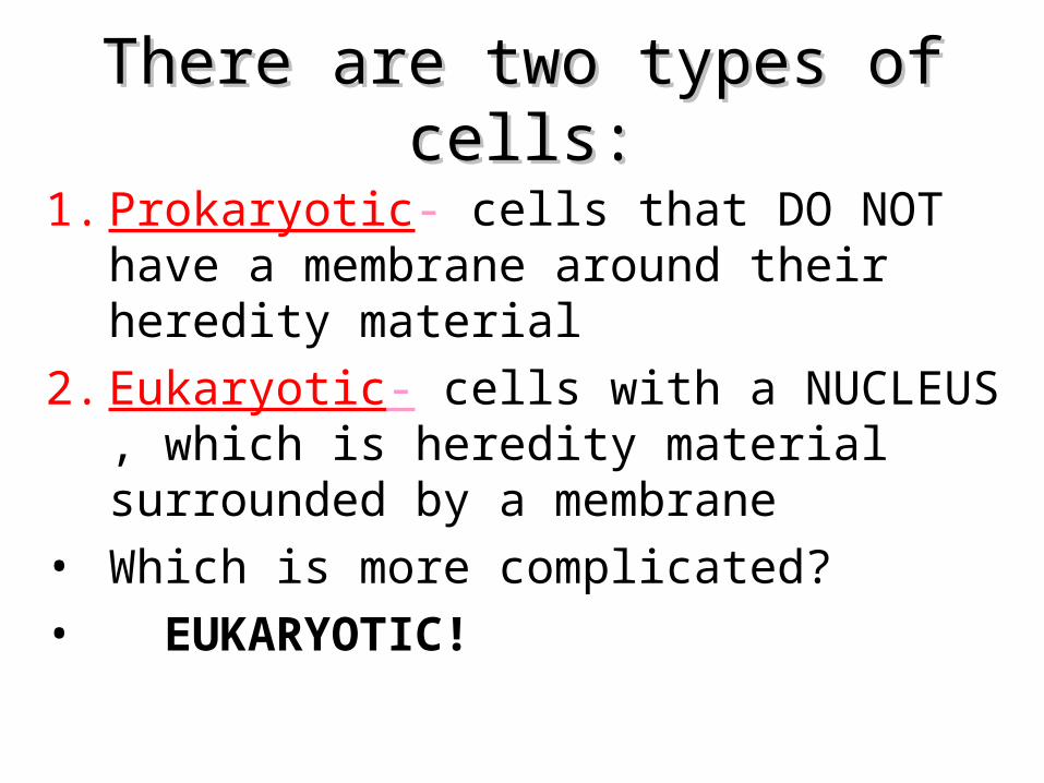
There are two types of cells:There are two types of cells:
1. Prokaryotic- cells that DO NOT have a membrane around their heredity material
2. Eukaryotic- cells with a NUCLEUS , which is heredity material surrounded by a membrane
• Which is more complicated?• EUKARYOTIC!

• Examples of Eukaryotes: • plants, animals, fungi.
• Examples of Prokaryotes: • Bacteria
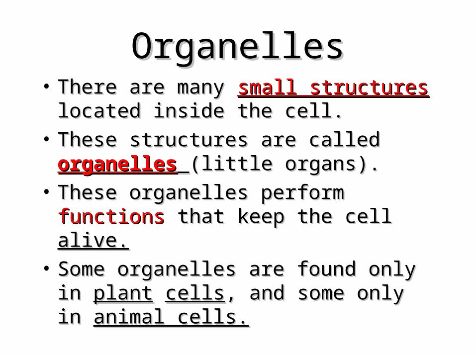
OrganellesOrganelles• There are many There are many small structuressmall structures located located
inside the cell.inside the cell.• These structures are called These structures are called organellesorganelles (little (little
organs).organs).• These organelles perform These organelles perform functionsfunctions that that
keep the cell keep the cell alive.alive.• Some organelles are found only in Some organelles are found only in plantplant
cellscells, and some only in , and some only in animal cells.animal cells.
Name some organelles inside an Name some organelles inside an animal cellanimal cell
• Nucleus• Lysosome• Golgi bodies• Mitochondrion• Ribosome• Vacuole• Cytoplasm• Endoplasmic reticulum (ER)
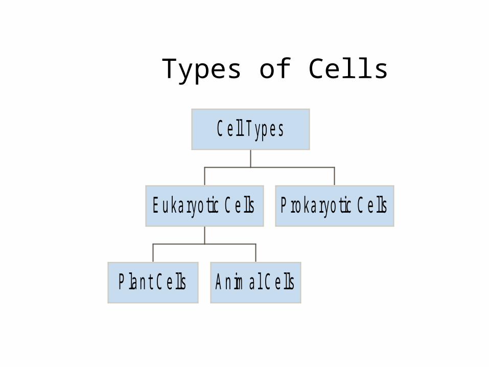
Types of Cells
P la n t C e lls A n im a l C e lls
E u ka ryo tic C e lls P ro ka ryo tic C e lls
C e ll T yp e s
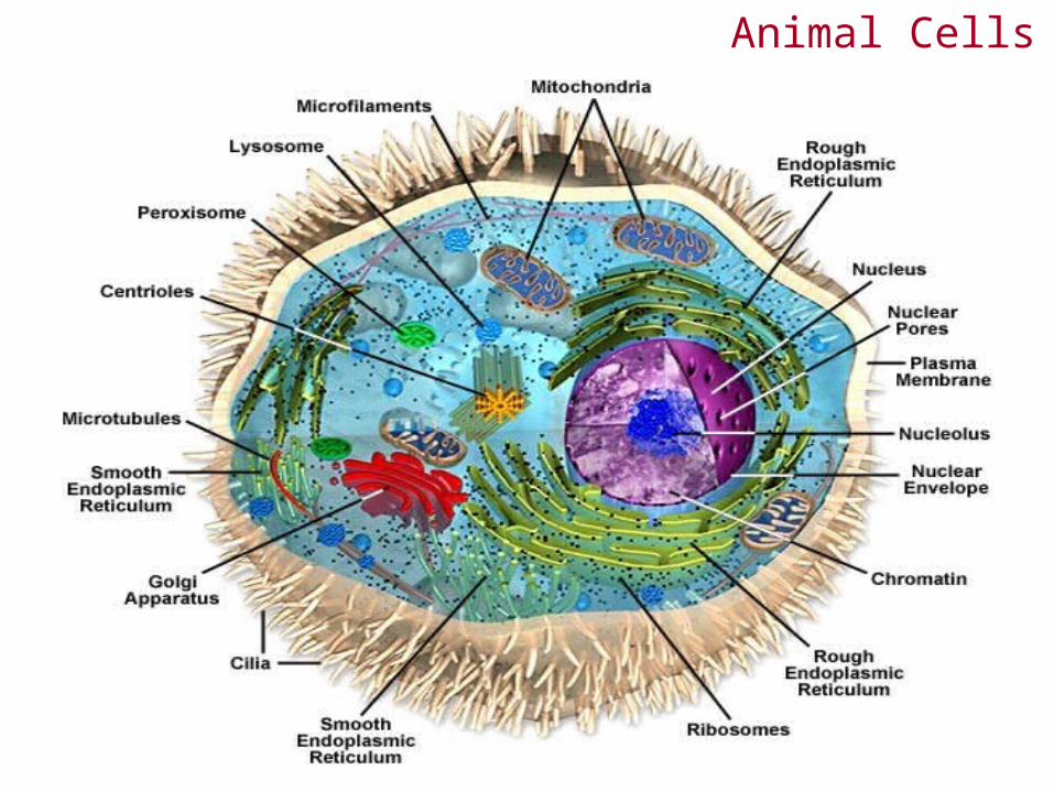
Animal Cells

Cellular Anatomy

Biochemistry and Evolution
• Prokaryotes - do not have a membrane-bounded nucleus
• Eukaryotes - possess nucleus and other complex internal structures
• Prokaryotes and eukaryotes appear to have evolved from a common ancestor over three billion years ago
The Cell is the Basic Unit of Life
• Plasma membrane - surrounds aqueous environment of the cell
• Cytoplasm - all materials enclosed by the plasma membrane (except the nucleus)
• Cytosol - aqueous portion of the cytoplasm minus subcellular structures
• Bacteriophage or phage - viruses that infect prokaryotic cells

Prokaryotic Cells: Structural Features
• Prokaryotes, or bacteria are usually single-celled organisms
• Prokaryotes lack a nucleus (their DNA is packed in a nucleoid region of the cytoplasm)
• Escherichia coli (E. coli) - one of the best studied of all living organisms
• E. coli cells are ~0.5m diameter, 1.5m long

Eukaryotic Cells: Structural Features
• Eukaryotes: plants, animals, fungi, protists
• Have a membrane-enclosed nucleus containing the chromosomes
• Are commonly 1000-fold greater in volume than prokaryotic cells
• Have an intracellular membrane network that subdivides the interior of the cell
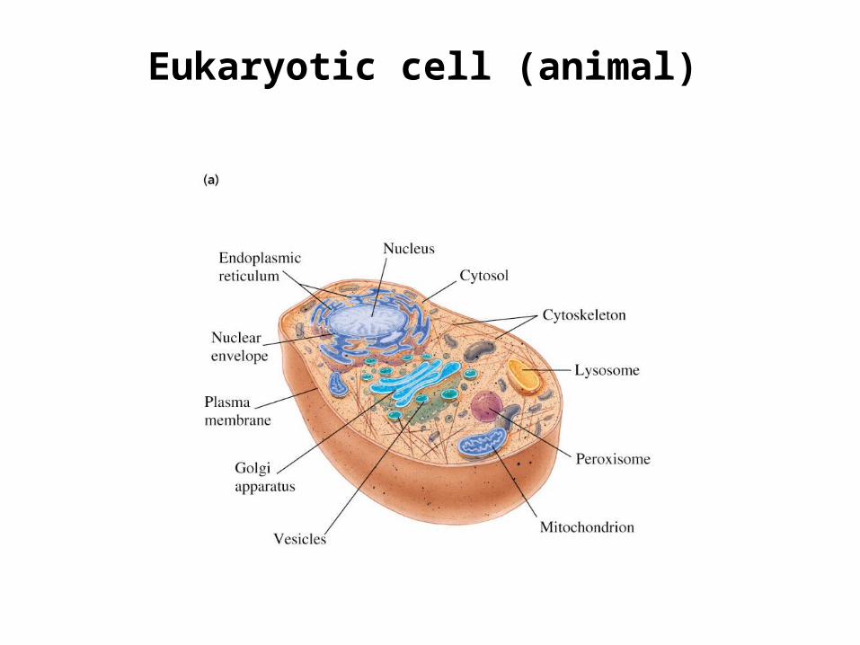
Eukaryotic cell (animal)
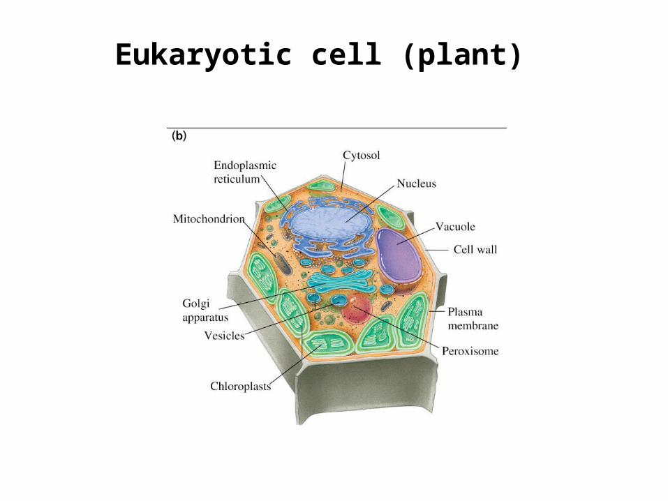
Eukaryotic cell (plant)
NUCLEUS:
• Most functions of the cell are controlled by the nucleus.
• Functions: “Brain” of the cell.
• It houses and protects the cell’s genetic information.

The Cell Nucleus
• Why have nuclear pores at all?
• What materials can pass through the nuclear envelope? What materials are retained?
• What is in the nucleolus?
• What molecules are in chromatin?

NucleusNucleus• Large round Large round structure located inside structure located inside
the cytoplasm.the cytoplasm.• Contains Contains genetic materialgenetic material (DNA). (DNA).• Has a Has a nuclear membranenuclear membrane (semi-(semi-
permeable).permeable).• Contains a Contains a NucleolusNucleolus (makes (makes
Ribosomes).Ribosomes).• Controls the Controls the activity activity of the cell.of the cell.
Name 3 parts of the nucleus
• Nuclear membrane• Chromatin• nucleolus
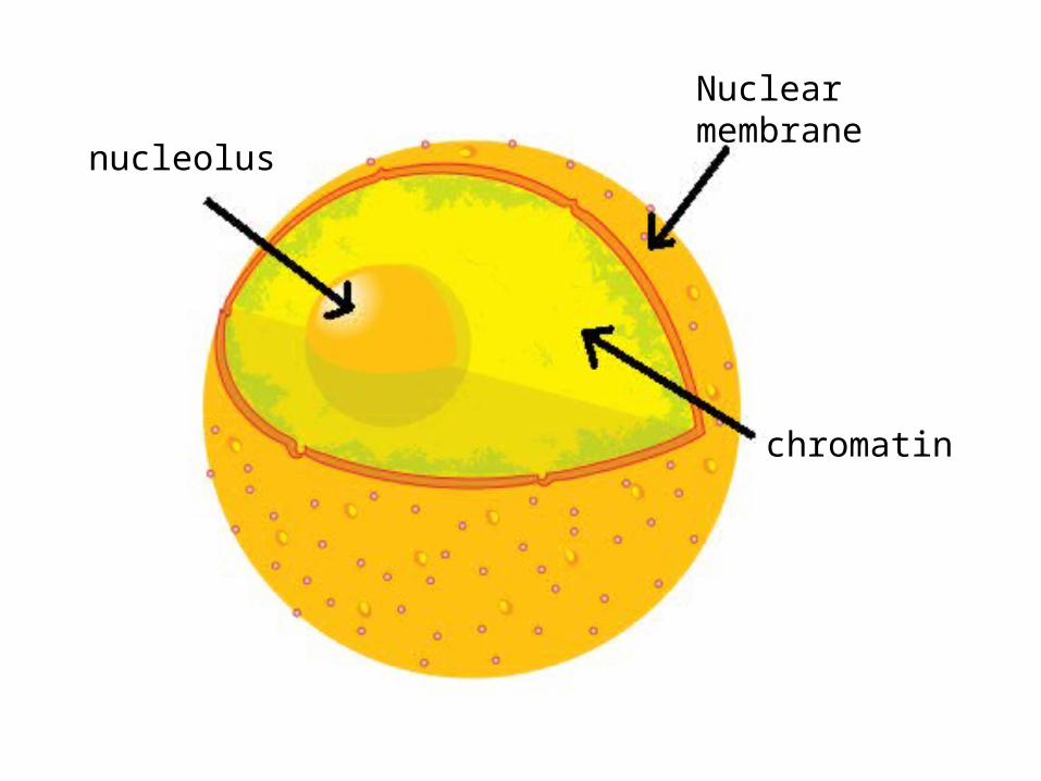
Nuclear membrane
chromatin
nucleolus
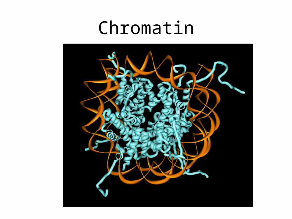
Chromatin

questions
• Where is chromatin located? • In the nucleus.• Where is DNA located? • In the chromatin.• What does DNA do? • In controls the activities in the cell.

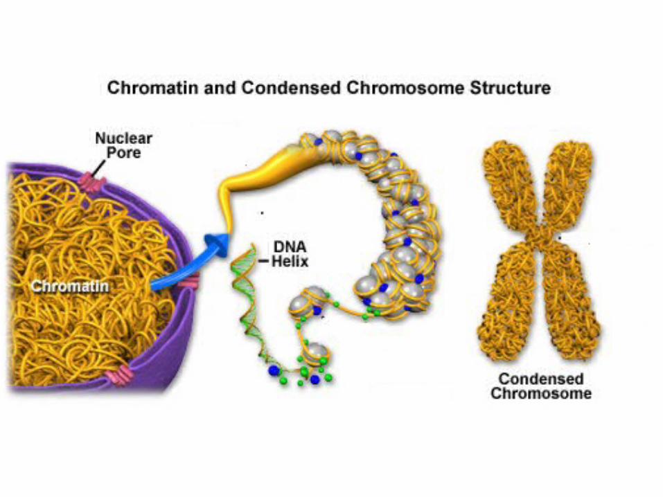
Note!
• The shape of chromatin changes when a cell begins to divide.
• When a cell begins to divide the chromatin coils and takes the form of chromosomes.

ChromosomesChromosomes• Long thread-like structures found Long thread-like structures found
in the in the nucleusnucleus of the cell.of the cell.• Contain Contain hereditaryhereditary information.information.• Genes are hereditary units made Genes are hereditary units made
up of up of DNADNA..
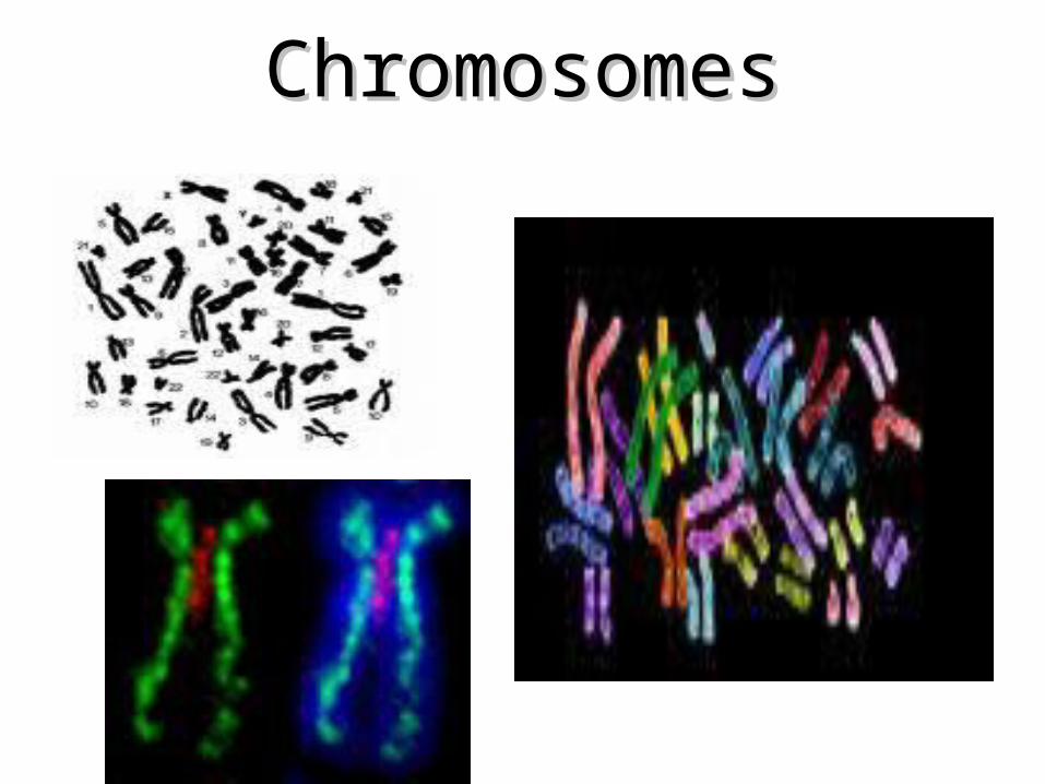
ChromosomesChromosomes

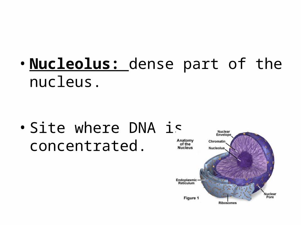
• Nucleolus: dense part of the nucleus.
• Site where DNA is concentrated.

Nuclear Envelope: double layer that covers the nucleus. Also made of 2 phospholipid
bilayers.
• Nuclear Pores: holes in the nuclear envelope that allow passageways for RNA and other things entering and leaving the nucleus.
DNA structure and replication
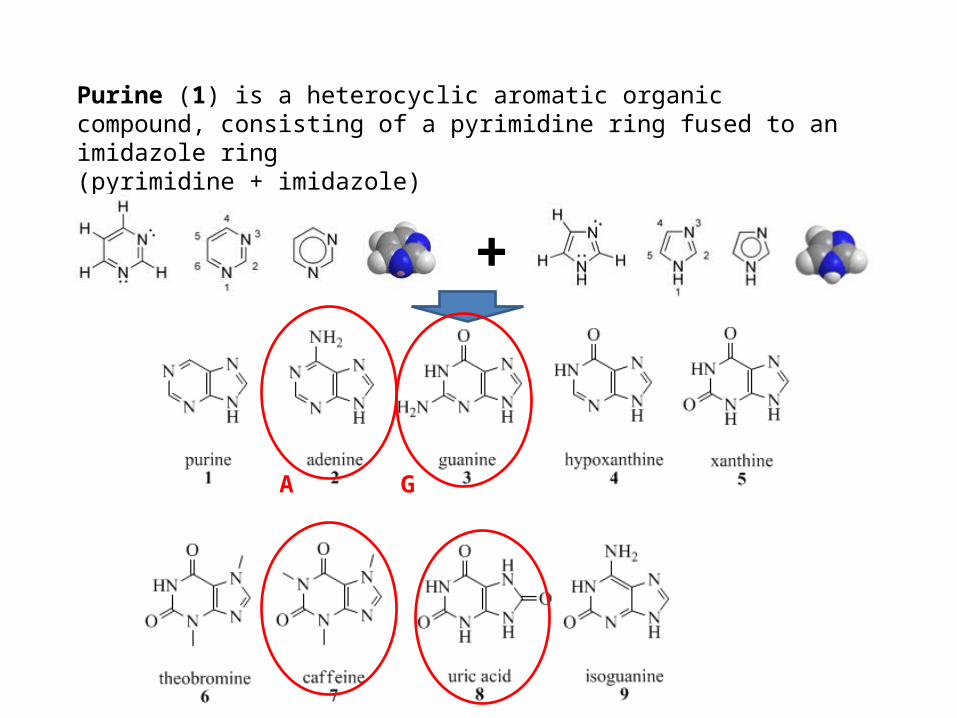
Purine (1) is a heterocyclic aromatic organic compound, consisting of a pyrimidine ring fused to an imidazole ring(pyrimidine + imidazole)
+
A G
Aside from DNA and RNA, purines are biochemically significant components in a number of other important biomolecules, such as ATP, GTP, cyclic AMP, NADH, and coenzyme A.They may also function directly as neurotransmitters, acting upon purinergic receptors. Adenosine, activates adenosine receptors.
Purines are found in high concentration in meat and meat products, especially internal organs such as liver and kidney. Plant based diet is generally low in purinesExamples of high purine sources include: sweetbreads, anchovies, liver, beef kidneys, scallopsA moderate amount of purine is also contained in beef, pork, fish and seafood, asparagus, spinach, mushrooms, green peas, beans, oatmeal, wheat bran and wheat germ.[4]

Pyrimidine is a heterocyclic aromatic organic compound similar to benzene containing two nitrogen atoms at positions 1 and 3 of the six-member ring
benzene
Pyrimidine
cytosine thymine uracil
C T U

Chromosome structure
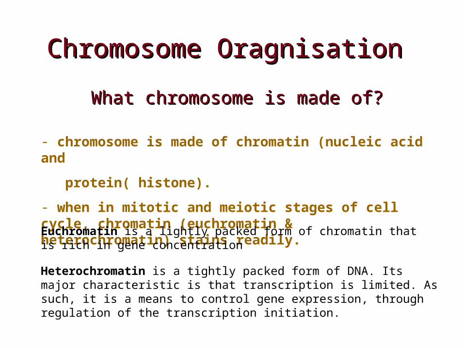
ChroChromosome Oragnisation mosome Oragnisation
What chromosome is made of?What chromosome is made of?
- chromosome is made of chromatin (nucleic acid and
protein( histone).
- when in mitotic and meiotic stages of cell cycle, chromatin (euchromatin & heterochromatin) stains readily.
Euchromatin is a lightly packed form of chromatin that is rich in gene concentration
Heterochromatin is a tightly packed form of DNA. Its major characteristic is that transcription is limited. As such, it is a means to control gene expression, through regulation of the transcription initiation.

Chromosome PackagingChromosome Packaging
Chromatin is organised on three basic levels:
- primary (nucloesome)
- secondary (solenoid)
- tertiary/quaternary (final folding into chromosome shape)
A typical eukaryotic chromosome contians 1 to 20 cm of DNA. During metaphase of meiosis/mitosis, this DNA is package into a chromosome with a length of only 1 to 10 m (a condensation of almost 104-fold in length from the naked DNA molecule).

Chromosome PackagingChromosome Packaging
beaded string – nucleosome structure of chromatin

Chromosome PackagingChromosome Packaging
- nucleosome contains histones (2 of each H2A, H2B, H3
and H4, and one H1)
- the diameter of nucleosome (bead) is 11nm.
- 200 bp of DNA associated with one bead, 23 bp protected by H1 and 8 to 114bp (depends on species and type of cells) form a linker between beads.

Chromosome PackagingChromosome Packaging
Second level of chromatin organization: solenoid
- the nucleosome is supercoiled and organised into a solenoid structure, with 6-7 nucleosomes per turn.
- H1 stabilize the structure of solenoid.
- the supercoiling produces a fibre of approximately 30nm in diameter.

Chromosome PackagingChromosome Packaging
Higher order folding of chromatin into chromosome

Transcription and Translation
Nucleic Acids
• Polynucleotides - nucleic acid biopolymers are composed of nucleotide monomers
• Nucleotide monomers are composed of:
(1) A five-carbon sugar
(2) A heterocyclic nitrogenous base
(3) Phosphate group(s)

Deoxyribose
• Deoxyribose lacks a hydroxyl group at C-2. It is the sugar found in DNA.
Nitrogenous bases
• Major Purines:
Adenine (A)
Guanine (G)
• Major Pyrimidines
Cytosine (C)
Thymine (T)
Uracil (U)
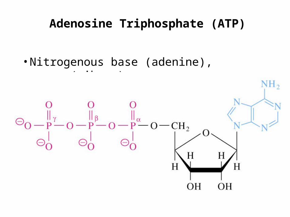
Adenosine Triphosphate (ATP)
• Nitrogenous base (adenine), sugar (ribose)

Structure of a dinucleotide
• Residues are joined by a phosphodiester linkage

Short segment of a DNA molecule
• Two polynucleotides associate to form a double helix
• Genetic information is carried by the sequence of base pairs
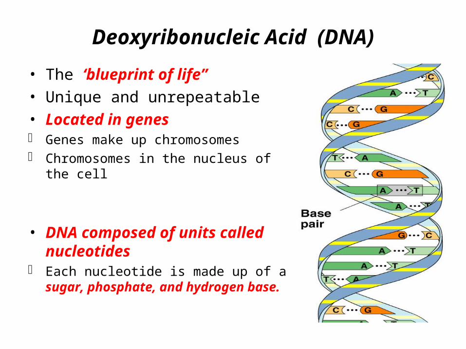
Deoxyribonucleic Acid (DNA)
• The ‘blueprint of life”• Unique and unrepeatable• Located in genes Genes make up chromosomes Chromosomes in the nucleus of the cell
• DNA composed of units called nucleotides
Each nucleotide is made up of a sugar, phosphate, and hydrogen base.
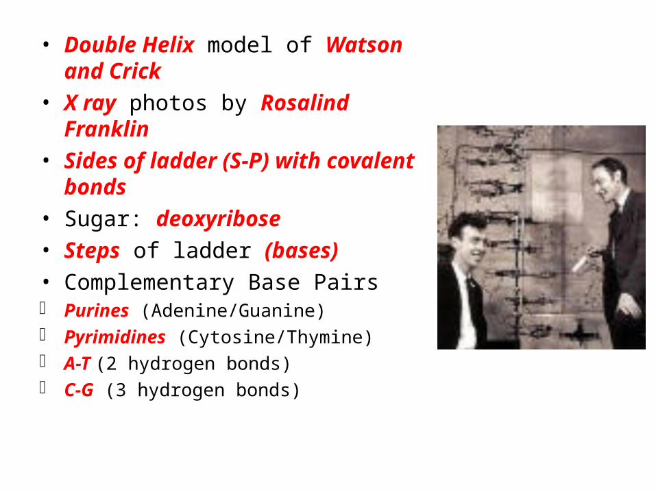
• Double Helix model of Watson and Crick• X ray photos by Rosalind Franklin• Sides of ladder (S-P) with covalent
bonds• Sugar: deoxyribose• Steps of ladder (bases)• Complementary Base Pairs Purines (Adenine/Guanine) Pyrimidines (Cytosine/Thymine) A-T (2 hydrogen bonds) C-G (3 hydrogen bonds)
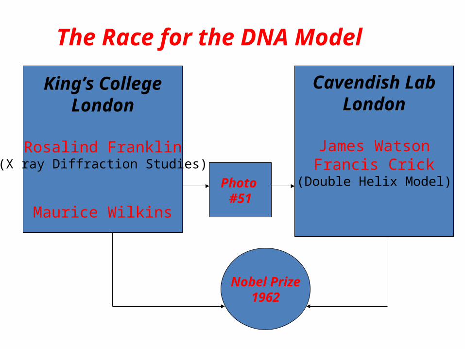
The Race for the DNA Model
King’s CollegeLondon
Rosalind Franklin(X ray Diffraction Studies)
Maurice Wilkins
Photo #51
Nobel Prize1962
Cavendish LabLondon
James WatsonFrancis Crick
(Double Helix Model)

• Mutation: a mistake in nitrogen base pairs• Pattern of bases codes for a specific amino acid• Codon: 3 letter (bases) code for 1 amino acid
• Replication of DNA (Making an identical copy)
DNA unzips and hydrogen bases separate (action of DNA polymerase)
Free-floating nucleotides match up on each side Hydrogen bonds reform Result: 2 identical DNA molecules (Each contain 1 of the
original DNA strands) Semi-Conservative Model of Replication

• Each DNA molecule with a new strand and an old strand
• DNA never leaves the nucleus

Ribonucleic Acid (RNA)
• Made up of nucleotides• Sugar called ribose• Nitrogen Bases: A, C,G,U• Single-stranded• Located in the nucleus and in the ribosomes• 3 Types: Messenger RNA (mRNA); carries message of DNA to ribosomes (3 letter
code – codon); single strand of letters Transfer RNA (tRNA): reads the mRNA code at the ribosomes (3 letter
code – anticodon); carries an amino acid; shamrock shape Ribosomal RNA (rRNA): joins mRNA to tRNA
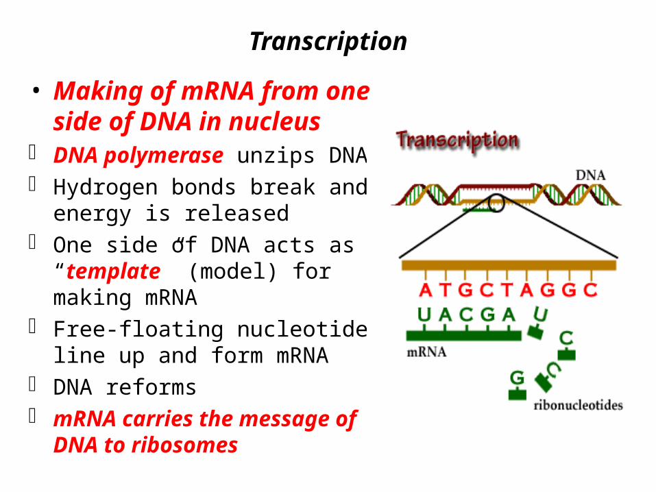
Transcription
• Making of mRNA from one side of DNA in nucleus
DNA polymerase unzips DNA Hydrogen bonds break and energy is
released One side of DNA acts as a
“template” (model) for making mRNA
Free-floating nucleotides line up and form mRNA
DNA reforms mRNA carries the message of DNA
to ribosomes
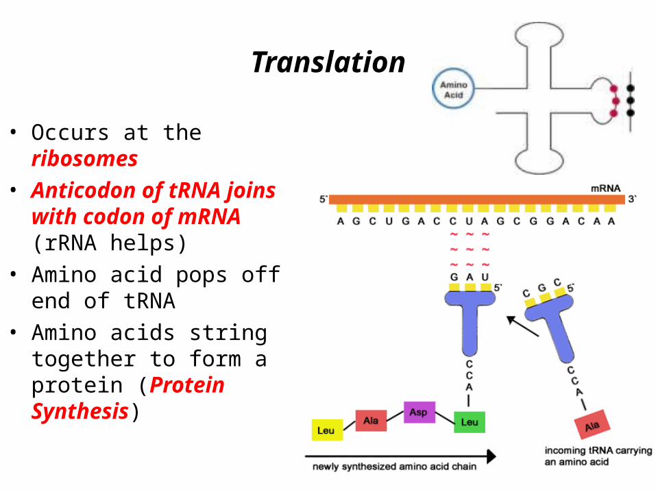
Translation
• Occurs at the ribosomes• Anticodon of tRNA joins with
codon of mRNA (rRNA helps)• Amino acid pops off end of
tRNA• Amino acids string together to
form a protein (Protein Synthesis)

Endoplasmic reticulum (ER)Endoplasmic reticulum (ER)
• cytoplasmic channels from the cell membrane cytoplasmic channels from the cell membrane to the nuclear membrane to the nuclear membrane
• associated with the associated with the storage, synthesis, and storage, synthesis, and transporttransport of materials within the cell of materials within the cell
• ““HIGHWAY” for cell transportHIGHWAY” for cell transport


Endoplasmic Reticulum
• What are the two types of ER?
• How does the role of each type differ?
• What kind of cells would have a lot of rough ER? Smooth ER?
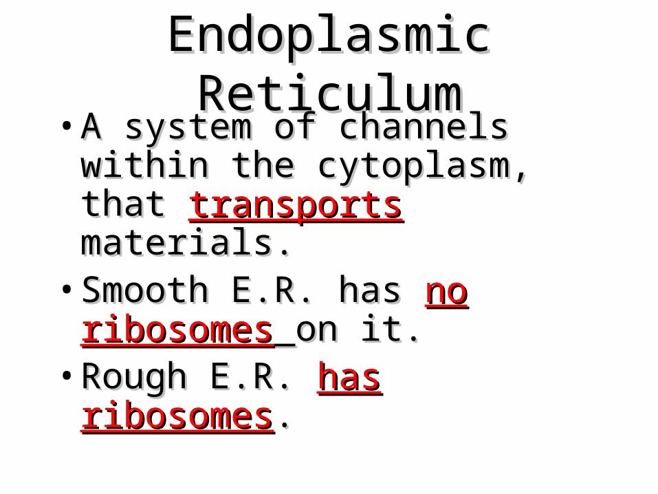
Endoplasmic ReticulumEndoplasmic Reticulum• A system of channels within the A system of channels within the
cytoplasm, that cytoplasm, that transportstransports materials.materials.
• Smooth E.R. has Smooth E.R. has nono ribosomesribosomes on on it.it.
• Rough E.R. Rough E.R. has ribosomeshas ribosomes..
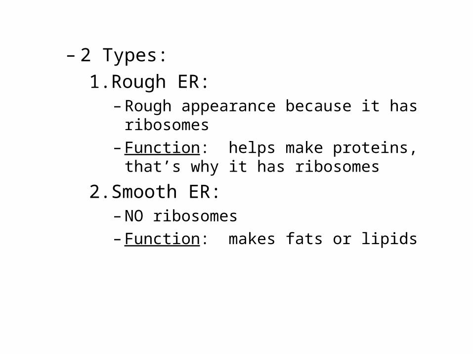
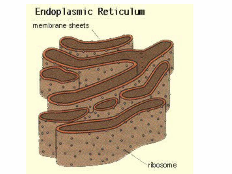
– 2 Types:1.Rough ER:
– Rough appearance because it has ribosomes
– Function: helps make proteins, that’s why it has ribosomes
2.Smooth ER:– NO ribosomes– Function: makes fats or lipids

The endomembrane system regulates protein traffic and performs metabolic functions in the cell
Endoplasmic reticulumSmooth v. Rough
Smooth ER•lacks ribosomes•Involved in synthesis of lipids, metabolism of carbohydrates, and detoxification of poisons
Rough ER•Has attached ribosomes•Makes proteins and phospholipids
Smooth ER
Rough ER
Nuclear envelope
Rough ERSmooth ER
ER lumen
200 nm
Transport vesicles-transport materials from the ER to the Golgi

Endoplasmic Reticulum (ER)
• A system of membranes that is found in a cell’s cytoplasm
• Assists in the production, processing, and transport of proteins
• Assists in the production of lipids
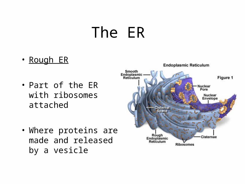
The ER
• Rough ER
• Part of the ER with ribosomes attached
• Where proteins are made and released by a vesicle
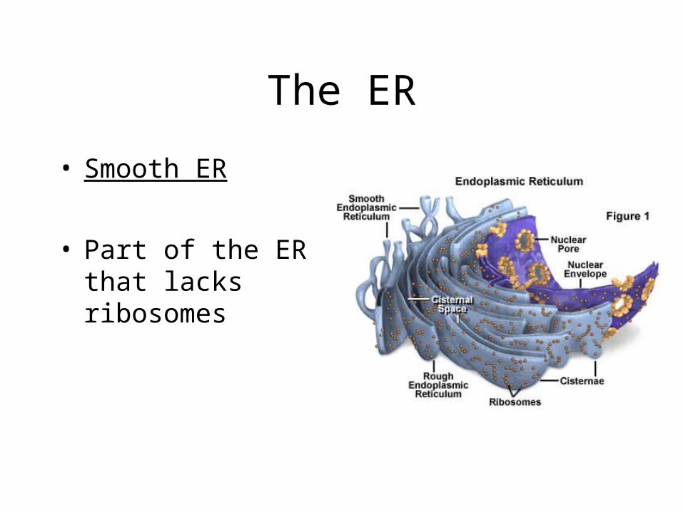
The ER
• Smooth ER
• Part of the ER that lacks ribosomes

Endoplasmic Reticulum (ER)• Network of fluid filled tubules (cisternae)• Roughly ½ of eukaryotic membrane
tissue• Continuous with the nuclear envelope• Smooth ER = lacks ribsosomes• Rough ER = ribosomes bound to
cytoplasmic side of ER membrane
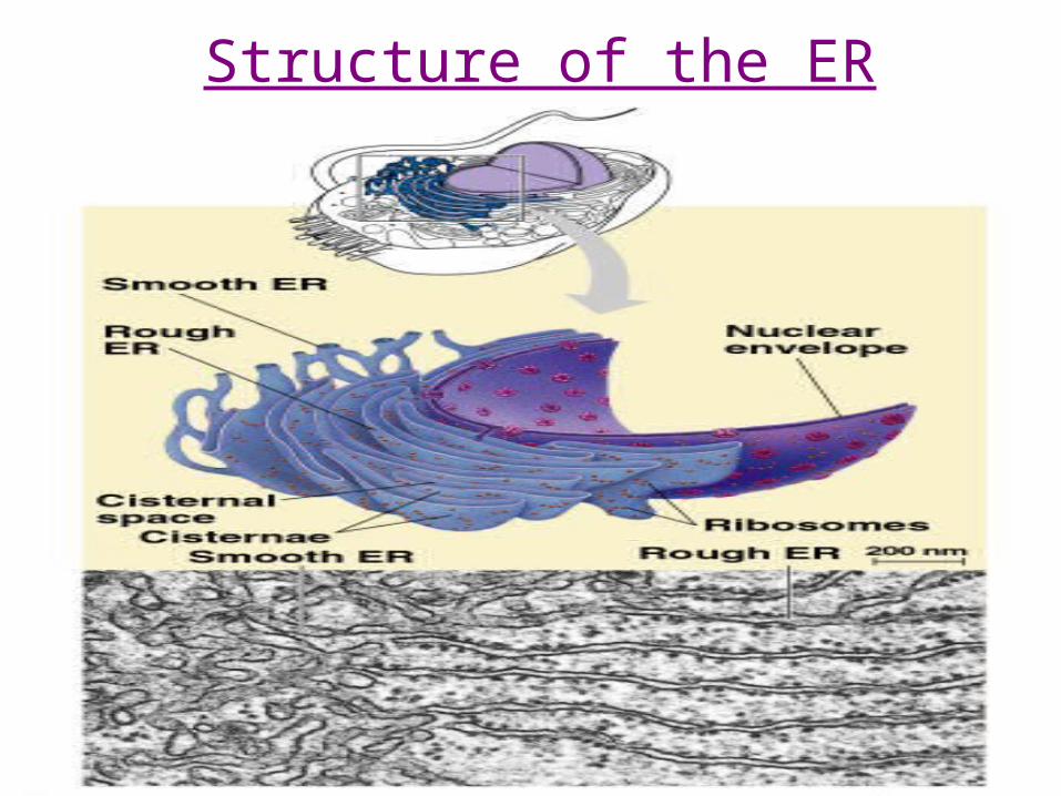
Structure of the ER

The Smooth ER• Synthesis of lipids, steroids (ie: sex hormones) • Carb metabolism (ie: liver cells hydrolysis
glycogen into glucose utilizes enzymes in smooth ER)
• Detoxification of drugs/poisons (ie: smooth ER enzymes make drugs more soluble add –OH). Tolerance = more Smooth ER
• Involved in Ca ion movement during muscle contraction.
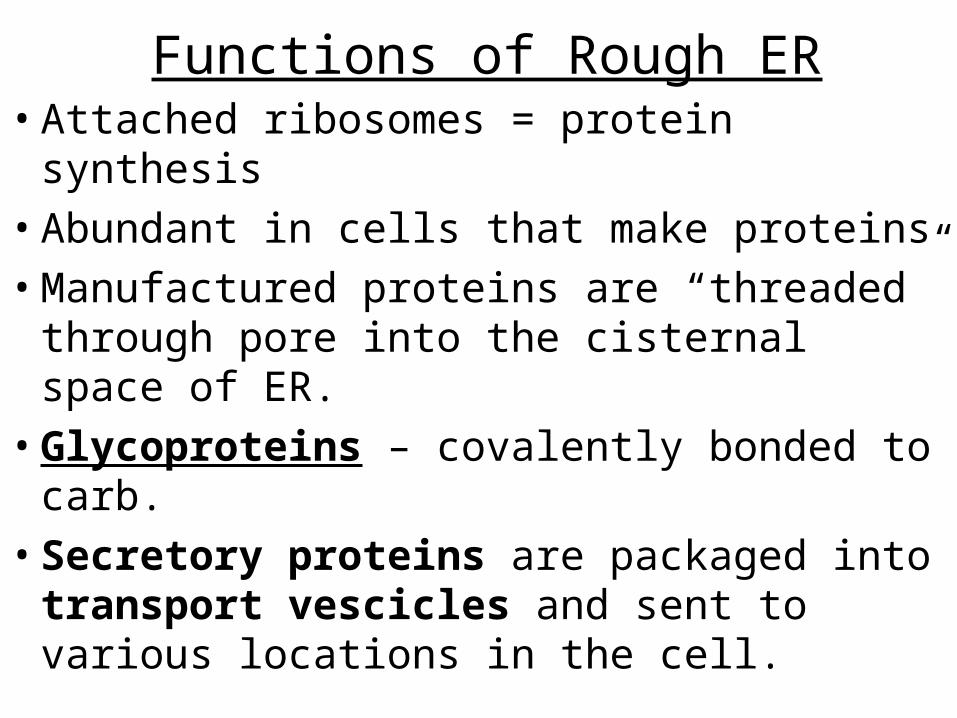
Functions of Rough ER• Attached ribosomes = protein synthesis• Abundant in cells that make proteins• Manufactured proteins are “threaded”
through pore into the cisternal space of ER.• Glycoproteins – covalently bonded to carb. • Secretory proteins are packaged into
transport vescicles and sent to various locations in the cell.

Rough ER
• Also manufactures phospholipids from precursors in cytoplasm.
• Assembles phospholipids and proteins into new membrane sections.
• ER membrane can expand or transfer new membrane via vescicles to other parts of endomembrane system.

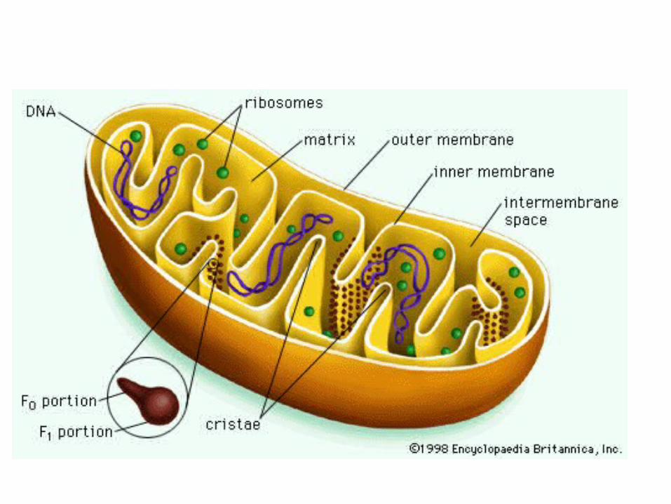
MITOCHONDRION:
tiny, double-membrane organelles that transfer ENERGY from organic molecules to ATP.
ATP powers most of the cell’s chemical reactions.
Found in large amounts in muscle cells and cells requiring ENERGY.
Function: Powerhouse of the cell.
MitochondriaMitochondria• The “The “powerhousepowerhouse” of the cell.” of the cell.• Food molecules are broken down and Food molecules are broken down and
energyenergy is released. is released. • Functions in Functions in CellularCellular RespirationRespiration..

The Citric Acid (Krebs) Cycle consists of eight steps

TCA Cycle Control
• Citrate Synthase (Synthetase)– Condensing Enzyme– Inhibited By:
• ATP• NADH• Succinyl CoA

TCA Cycle Control-Cont:
• Isocitrate Dehydrogenase– Activated By:
• ADP
– Inhibited By:• ATP• NADH
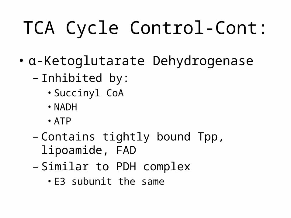
TCA Cycle Control-Cont:
• α-Ketoglutarate Dehydrogenase– Inhibited by:
• Succinyl CoA• NADH• ATP
– Contains tightly bound Tpp, lipoamide, FAD– Similar to PDH complex
• E3 subunit the same

TCA Cycle Control-Cont:
• Succinyl CoA Synthetase– Coupled reaction with GTP– Enzyme that catalyses coupled reaction is called
Nucleotidediphosphate Kinase

TCA Cycle Control-Cont:
• Succinate Dehydrogenase– Has Iron-Sulfur Centers– Covalently Bound with FAD
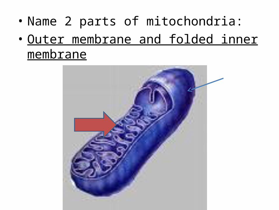
• Name 2 parts of mitochondria:• Outer membrane and folded inner membrane

• What types of cells you expect to have more mitochondria? why
• More active cells like muscle cells because they need more energy.

Mitochondrion • How many membranes? Why?
• What cells would have high numbers of mitochondria?
• What do mito. have to do with cloning?
• What is the current theory on mito. origin?

What are mitochondria?
• An intracellular organelle.• There are 100 to 1000s of mitochondria/cell.• All mitochondria come from the mother.• Mitochondria have their own DNA.• Found in all cell types, except the RBC.• Major functions of mitochondria:
– Makes energy in the form of ATP.– Programmed cell death (apoptosis).

Chemical Energy
Cars Gasoline Cells ATP
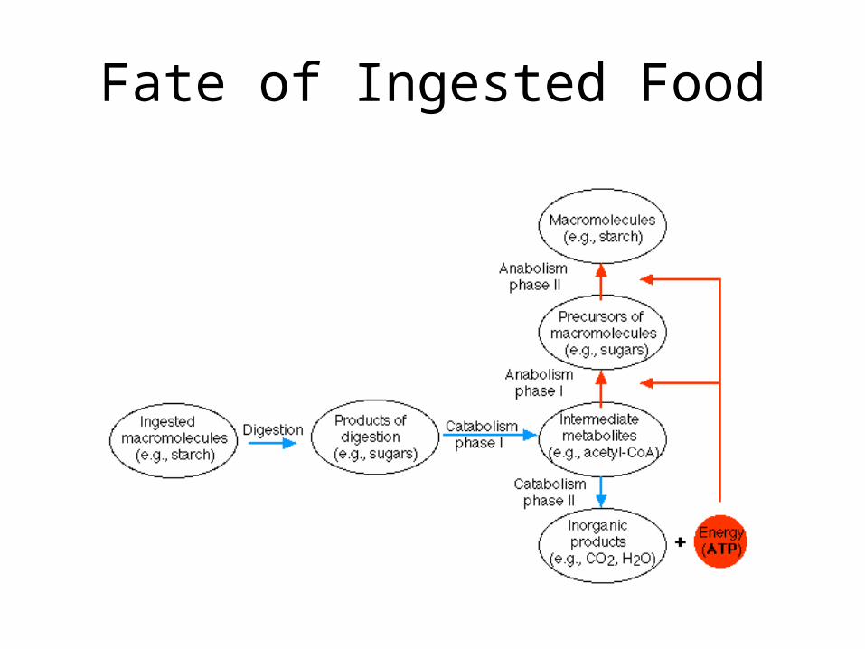
Fate of Ingested Food
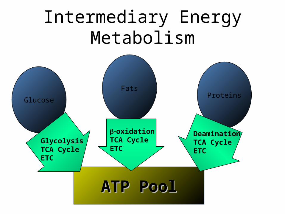
Intermediary Energy Metabolism
ATP PoolATP Pool
Glucose
FatsProteins
GlycolysisTCA CycleETC
oxidationTCA CycleETC
DeaminationTCA CycleETC
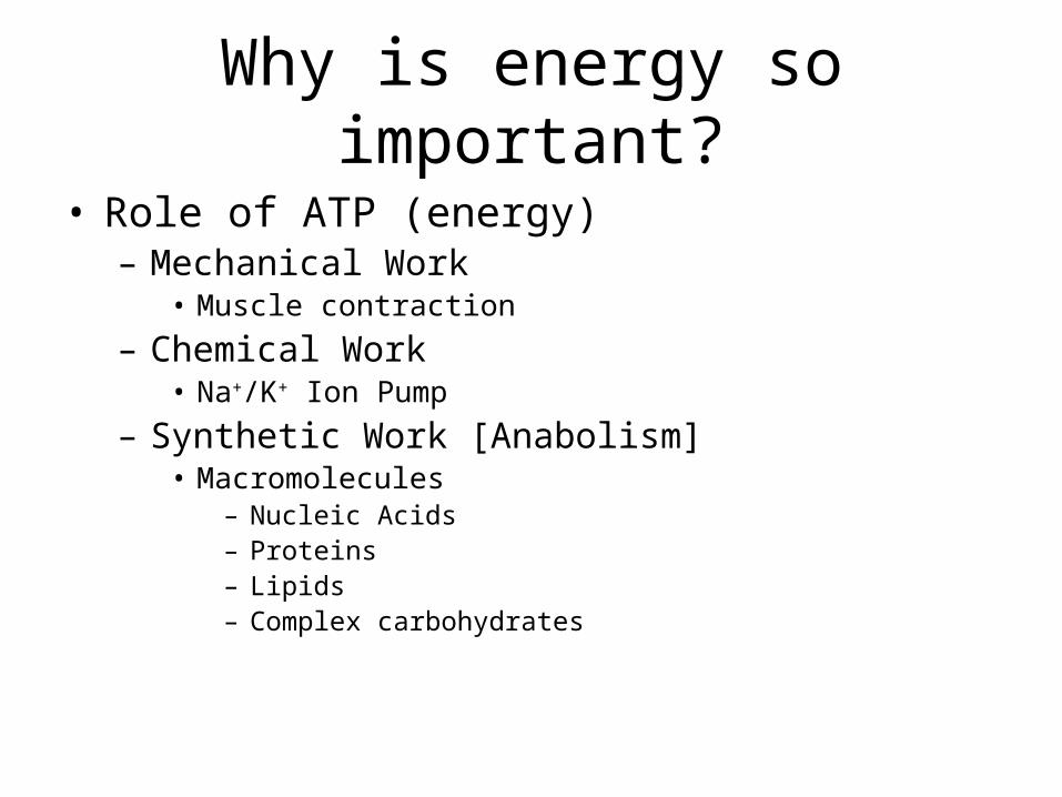
Why is energy so important?
• Role of ATP (energy)– Mechanical Work
• Muscle contraction– Chemical Work
• Na+/K+ Ion Pump– Synthetic Work [Anabolism]
• Macromolecules– Nucleic Acids– Proteins– Lipids– Complex carbohydrates

Bioenergetics: Energy
• 1 teaspoon of sugar weighs 5 gm and contains 20 calories of energy
• 1 teaspoon of sugar contains 10 X 1021 molecules of sugar or sucrose– 10,000,000,000,000,000,000,000 molecules
• 1 teaspoon of sugar forms about 3.6 X 10²³ molecules of ATP– 360,000,000,000,000,000,000,000 molecules

Bioenergetics: Energy
• At rest, the average adult male will need 3.0 x 10¹⁸ molecules of ATP per second for normal organ functioning.
• The body produces and makes approximately 70 Kg of ATP daily (average adult male).
• The brain uses approximately 70% of all ATP produced.
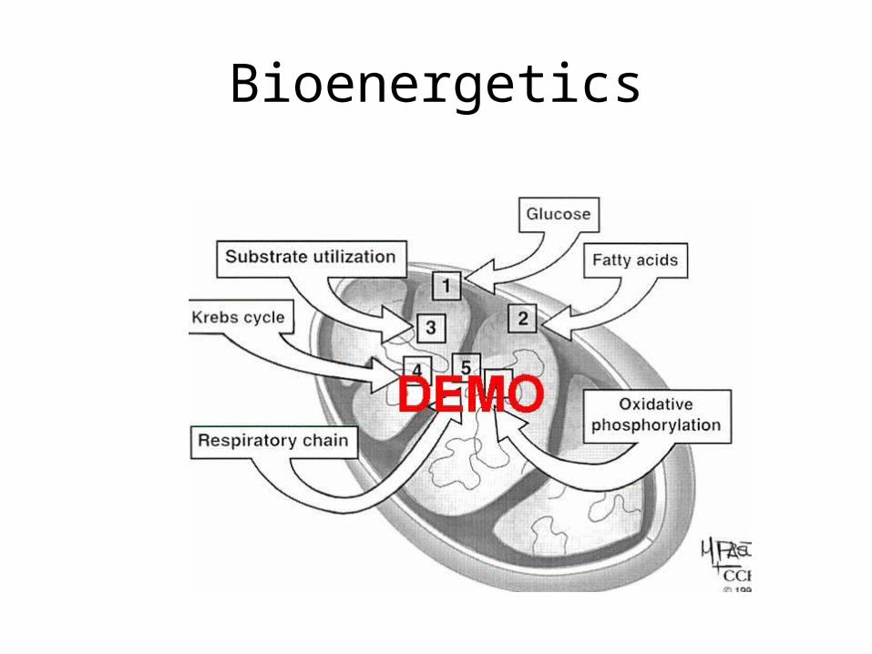
Bioenergetics
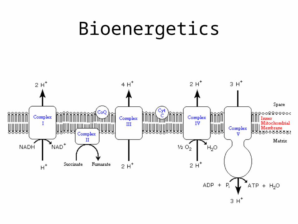
Bioenergetics
Bioenergetics: Summary• Mitochondria function is to produce ATP for energy. • The mitochondria use electrons and protons from
metabolism and molecular oxygen to reduce water and generate proton-motive force to produce ATP from ADP: oxidative phosphorylation.
• When this process is dysfunctional, then disease can occur.
• Bottomline: mitochondrial cytopathies are diseases of energy production.
Bioenergetics: Summary
• What happens when an organ does not get enough ATP or energy?– Brain dysfunction: when the brain doesn’t get it’s
70% of energy required:• Seizure• Mental Retardation• Cognitive dysfunctions• Psychological dysfunctions?
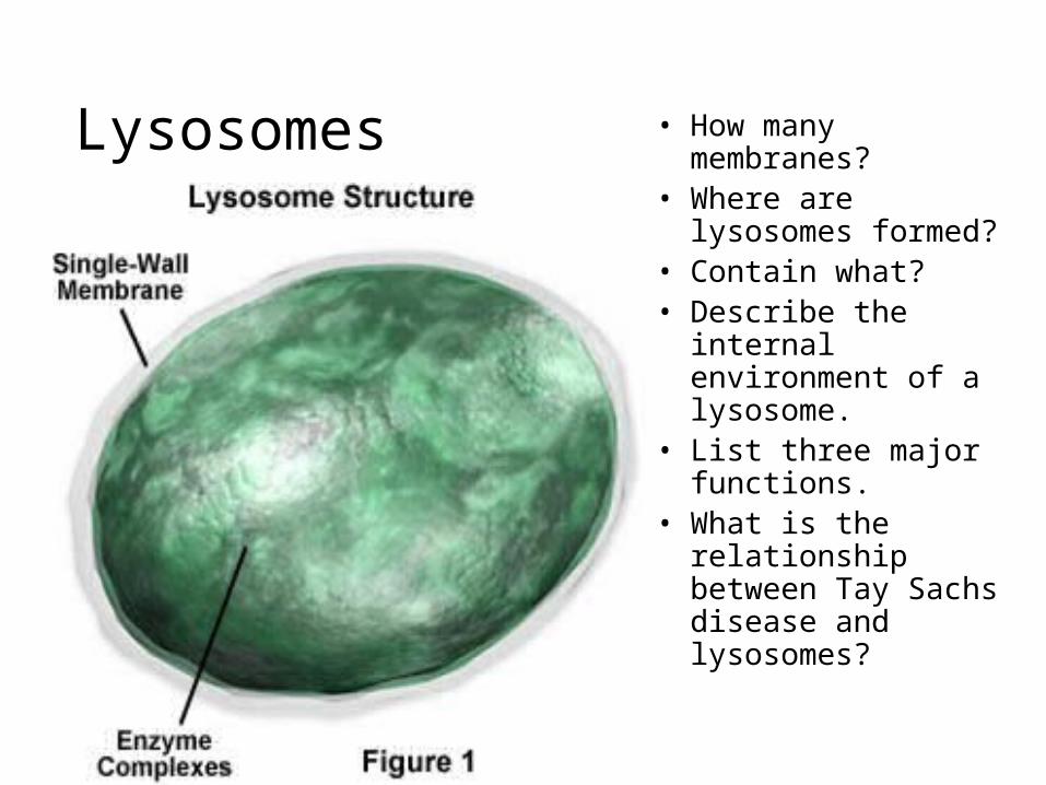
Lysosomes • How many membranes?
• Where are lysosomes formed?
• Contain what?• Describe the internal
environment of a lysosome.
• List three major functions.
• What is the relationship between Tay Sachs disease and lysosomes?

Lysosomes• Internal sacs bound by single membrane• Originate by budding from Golgi based on sorting
of mannose-6-phosphate “tags” on proteins• Responsible for degrading cell components that
have become obsolete for cell or organism—digestive sys of cells
• Internal pH about 5 (very acidic)• Compartmentalization ESSENTIAL! Failure can
lead to many known disease states that result from waste accumulation in the organelle

Lysosomes
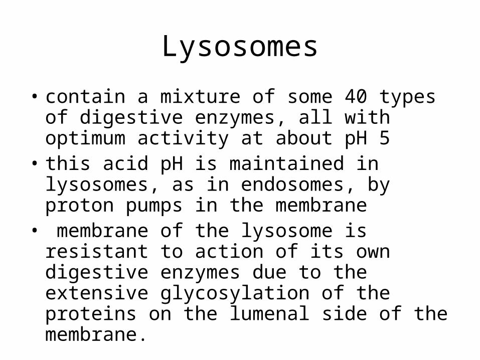
• contain a mixture of some 40 types of digestive enzymes, all with optimum activity at about pH 5
• this acid pH is maintained in lysosomes, as in endosomes, by proton pumps in the membrane
• membrane of the lysosome is resistant to action of its own digestive enzymes due to the extensive glycosylation of the proteins on the lumenal side of the membrane.
• A lysosome is a membranous sac of hydrolytic enzymes that can digest macromolecules
• Lysosomal enzymes can hydrolyze proteins, fats, polysaccharides, and nucleic acids
Endocytosis/Exocytosis
• Some types of cell can engulf another cell by phagocytosis; this forms a food vacuole– pinocytosis; exocytosis
• A lysosome fuses with the food vacuole and digests the molecules
• Lysosomes also use enzymes to recycle the cell’s own organelles and macromolecules, a process called autophagy

Fig. 6-14a
Nucleus 1 µm
Lysosome
Lysosome
Digestive enzymes
Plasma membrane
Food vacuole
Digestion
(a) Phagocytosis
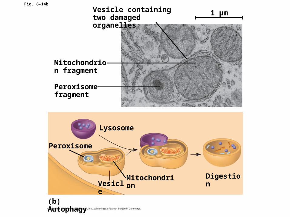
Fig. 6-14b
Vesicle containingtwo damaged organelles
Mitochondrion fragment
Peroxisome fragment
Peroxisome
Lysosome
DigestionMitochondrionVesicle
(b) Autophagy
1 µm

Peroxisome
• How do the enzymes in peroxisomes differ from the enzymes in lysosomes?
• What cells have many peroxisomes? Why?
• Plants have special peroxisomes called glyoxysomes. What is their function?

Major Metabolic Functions of the Peroxisome in Plants
1. -oxidation of fatty acids2. Glyoxylate cycle3. Photorespiration (Glycolate pathway) 4. Degradation of purines5. Decomposition of hydrogen peroxide
Two Types of Peroxisomes in Plants
• Leaves– Catalyzes oxidation of side product of CO2 fixation
in photorespiration
• Germinating seeds– Converts fatty acid in seed lipids into sugars
needed for growth in the young plant

-oxidation occurs in mitochondria and peroxisomes in mammals, but exclusively in the peroxisome in plants and yeast.

Glyoxysomes and Leaf Peroxisomes are Interconverted During
Development• Immunogold particles of
2 sizes bound to:– Enzymes of glyoxylate
cycle– Peroxisomal enzymes
• The same population of peroxisomes assumes different metabolic roles depending on developmental stage of cotelydon
Greening cotelydons

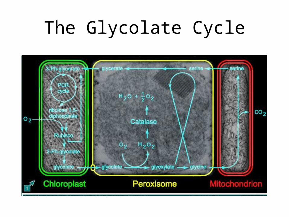
Photorespiration and Glycolate
Oxygenase activity of rubiscoConsumption of O2
Glycolate cycleProduction of CO2
Involves 3 organelles (chloroplasts, peroxisomes, & mitochondria)

Glycolic acid oxidase
The Glycolate Cycle
H2O2 production
The Glycolate Cycle

Purine Degradation
• Nucleic acid purine moieties (adenine and guanine) are degraded to uric acid
xanthine uric acid allantoin Xanthine oxidase Urate oxidase
O2 H2O2 O2 H2O2
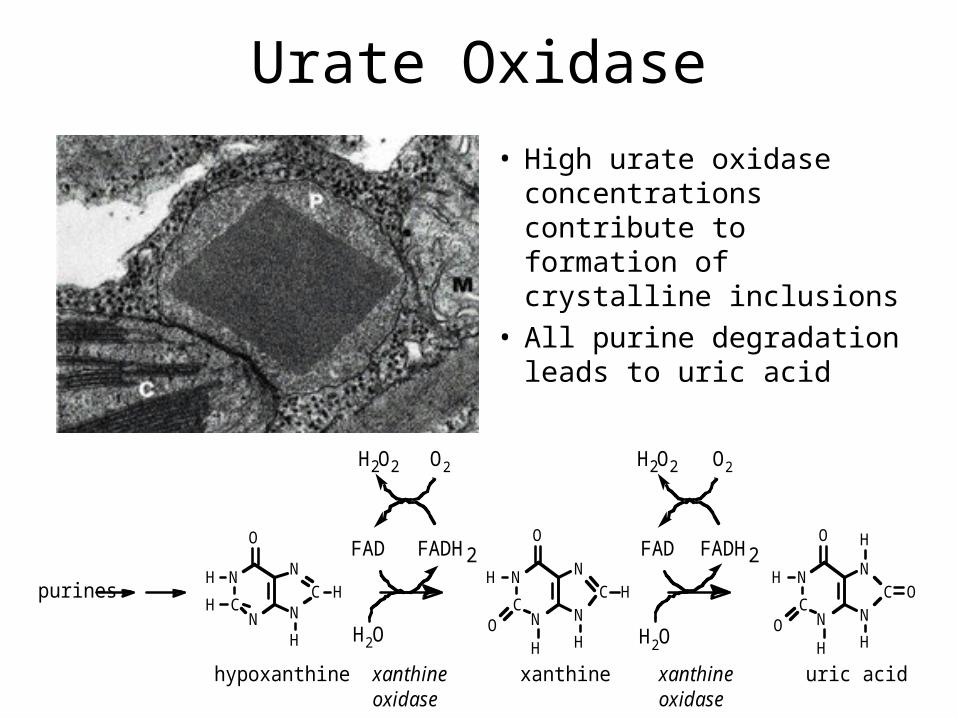
Urate Oxidase
• High urate oxidase concentrations contribute to formation of crystalline inclusions
• All purine degradation leads to uric acid
purines
hypoxanthine
N
CN
H
H
O
N
N
H
HC
H2O
xanthineoxidase
O2
FAD
H2O2
FADH2
xanthine
N
CN
H
H
O
ON
N
H
HC
xanthineoxidase
H2O
FAD FADH2
O2H2O2
uric acid
N
CN
H
H
O
ON
N
H
H
OC
Oxidases
• The oxidases use molecular oxygen to remove hydrogen atoms from specific organic substrates
• A variety of compounds, including L-amino acids, D-amino acids, polyamines, methanol, urate,
xanthine, and very-long-chain fatty acids, serve as substrates for the different oxidases
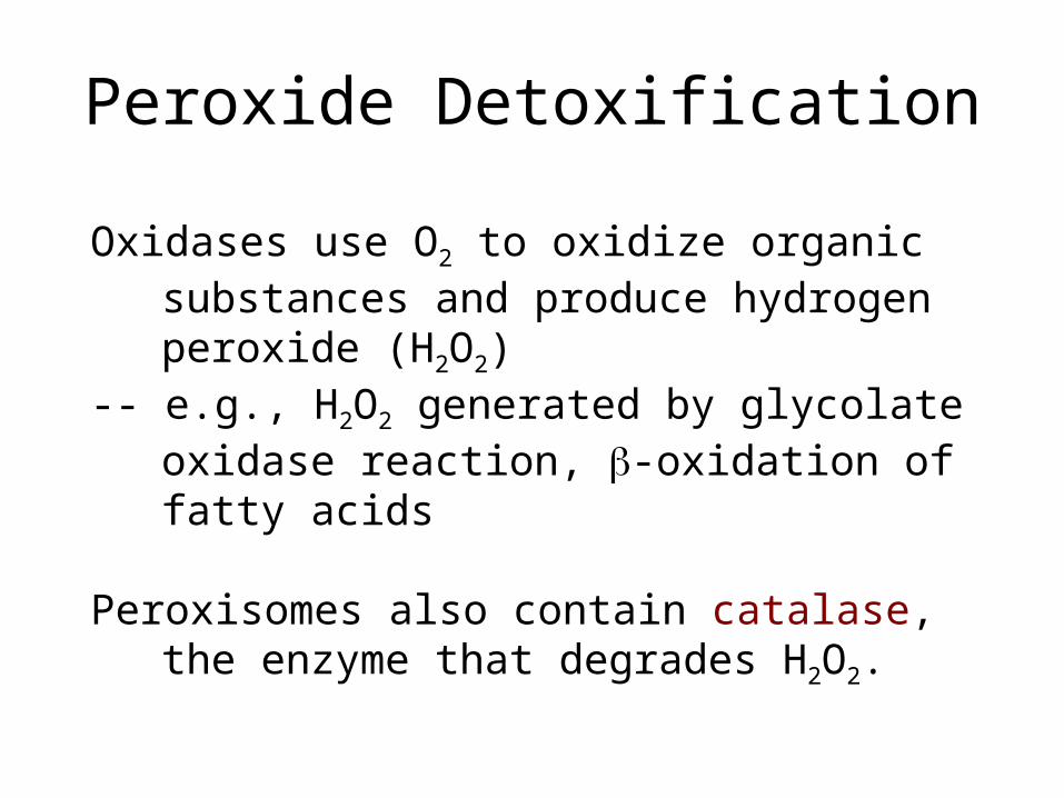
Peroxide Detoxification
Oxidases use O2 to oxidize organic substances and produce hydrogen peroxide (H2O2)
-- e.g., H2O2 generated by glycolate oxidase reaction, -oxidation of fatty acids
Peroxisomes also contain catalase, the enzyme that degrades H2O2.
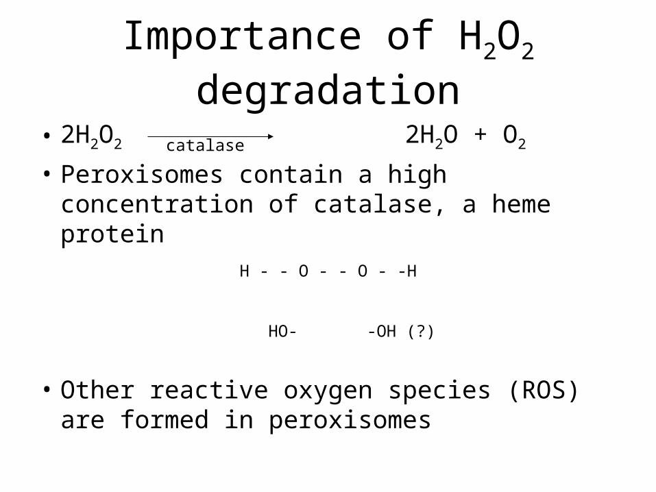
Importance of H2O2 degradation
• 2H2O2 2H2O + O2
• Peroxisomes contain a high concentration of catalase, a heme protein
• Other reactive oxygen species (ROS) are formed in peroxisomes
catalase
H - - O - - O - -H
HO- -OH (?)
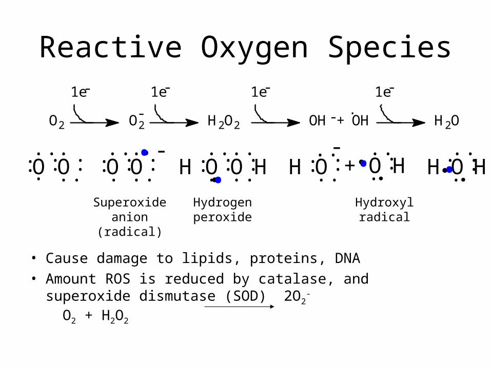
Reactive Oxygen Species
• Cause damage to lipids, proteins, DNA
• Amount ROS is reduced by catalase, and superoxide dismutase (SOD) 2O2
- O2 + H2O2
..::. .O
-....
:OHO.. :..
HH+HO.. :..
H O. .: :..-
.
..::O:..O
....
::O HO: :..
. .
+ OH·
-1e-1e-1e -1e
H2OOH--O2 O2 H2O2
•• ••
Superoxide anion (radical)
Hydrogen peroxide
Hydroxyl radical
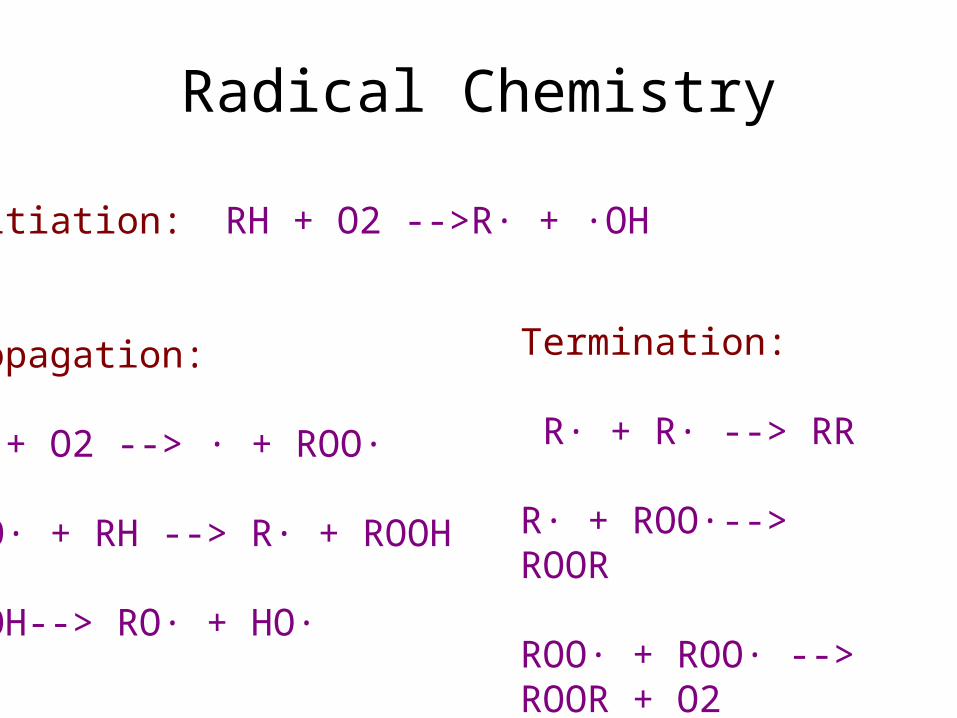
Radical Chemistry
Initiation: RH + O2 -->R· + ·OH
Propagation: R· + O2 --> · + ROO·
ROO· + RH --> R· + ROOH
ROOH--> RO· + HO·
Termination: R· + R· --> RR
R· + ROO·--> ROOR
ROO· + ROO· --> ROOR + O2

Other Peroxisomal Enzymes
Conclusions
• Compartmentalize! To protect the cell from these destructive byproducts, such reactions are segregated.

Ribosomes • Non-membrane bound!• Composed of ______ and
________.• Sites to synthesize
__________.• How are prokaryotic
ribosomes different from eukaryotic ribosomes?
• Antibiotics, including tetracycline and streptomycin, paralyze prokaryotic ribosomes. Why don’t these drugs harm eukaryotic ribosomes?