cell fate - the university of vermontbiology/classes/296b/dn_4.1.pdfr3 and r4 – from a combination...
TRANSCRIPT
Cell Fate
Chapter Four
Neuron Diversity
Neurons show enormous diversity in:• Cellular Anatomy• Physiological function• Neurochemistry
– What neurotransmitters are used• Connectivity
– Which neurons connect through synapses– Which other cells connect to
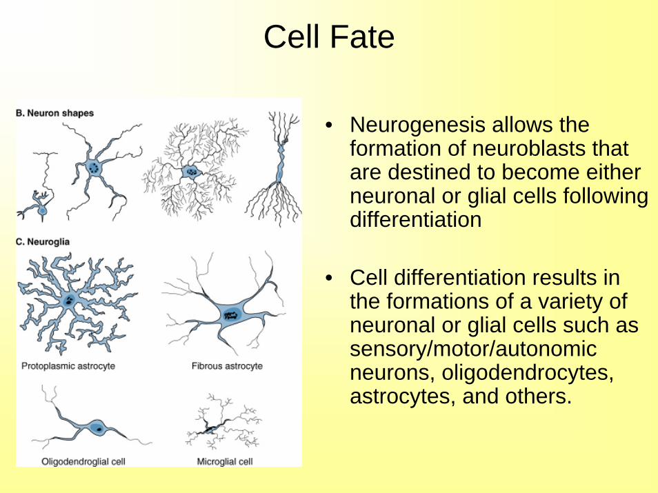
Cell Fate
• Neurogenesis allows the formation of neuroblasts that are destined to become either neuronal or glial cells following differentiation
• Cell differentiation results in the formations of a variety of neuronal or glial cells such as sensory/motor/autonomic neurons, oligodendrocytes, astrocytes, and others.

Diversity Example
• Granule Cells– Small, simple dendrites, T shaped axons– Excitatory neurotransmitter glutamate
• Purkinje Cells– Large, complex dendrites, single long axon– Inhibitory neurotransmitter GABA
• Motor Neurons– All motor neurons share morphology and physiology– Yet all are unique based on neurotransmitters and
cell types that they are connected to
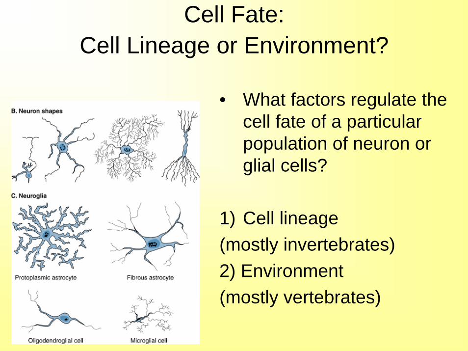
Cell Fate: Cell Lineage or Environment?
• What factors regulate the cell fate of a particular population of neuron or glial cells?
1) Cell lineage (mostly invertebrates)2) Environment (mostly vertebrates)
European vs. American• Sydney Brenner suggested neurons are
either European or American• In Europe your fate is largely determined
by your lineage– These neurons have intrinsic signals
• In America your fate is largely determined by your neighborhood– These neurons respond to extrinsic signals
• In reality – both factors are important
Intrinsic vs. Extrinsic
• Intrinsic signals:– Internal control of gene expression– Limits the possible cell fates
• Extrinsic signals:– Diffusible molecules– Cell surface proteins, extracellular matrix– Promotes a change of cell fate
• In reality these factors work together to determine cell fate of a neuron
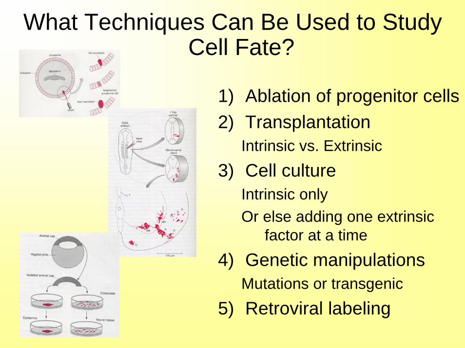
What Techniques Can Be Used to Study Cell Fate?
1) Ablation of progenitor cells2) Transplantation
Intrinsic vs. Extrinsic3) Cell culture
Intrinsic onlyOr else adding one extrinsic
factor at a time4) Genetic manipulations
Mutations or transgenic5) Retroviral labeling
C. elegans• Every neuron is determined from an
almost invariant lineage• Ablation of one progenitor:
– Removes all daughters cells normally born from there on in that branch
– Therefore neighboring cells cannot fill in missing fates
• Mutants have been found that interfere with development of particular neurons– Can be used to identify necessary genes
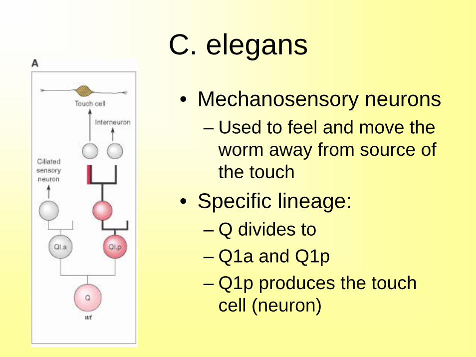
C. elegans
• Mechanosensory neurons– Used to feel and move the
worm away from source of the touch
• Specific lineage:– Q divides to – Q1a and Q1p– Q1p produces the touch
cell (neuron)
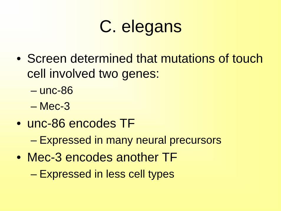
C. elegans
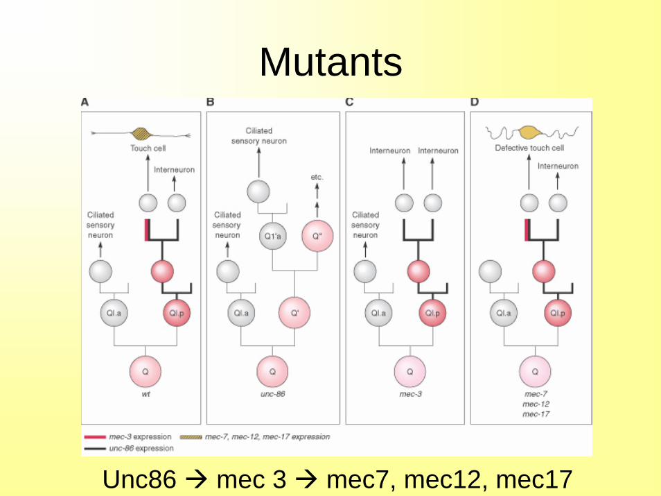
• Screen determined that mutations of touch cell involved two genes:– unc-86– Mec-3
• unc-86 encodes TF – Expressed in many neural precursors
• Mec-3 encodes another TF– Expressed in less cell types
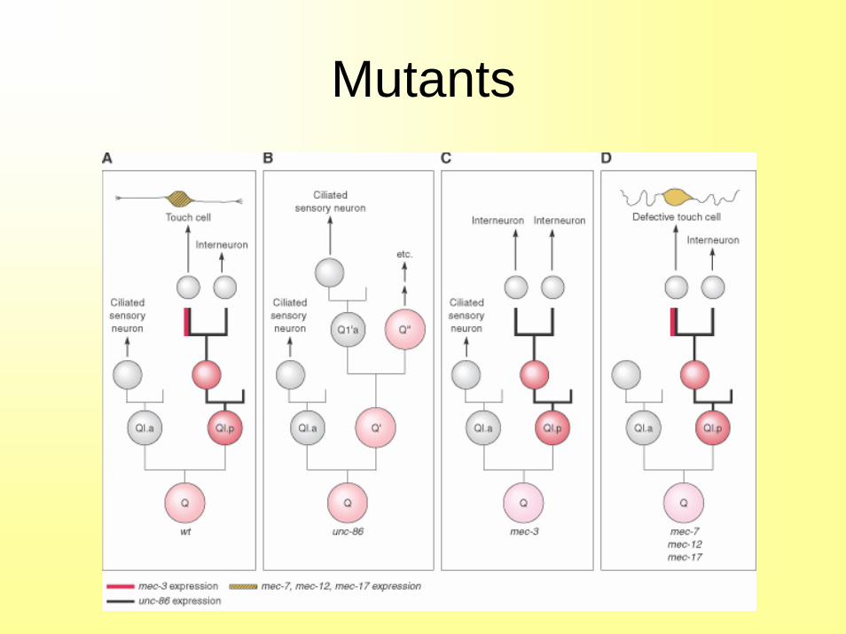
Mutants
Mutants
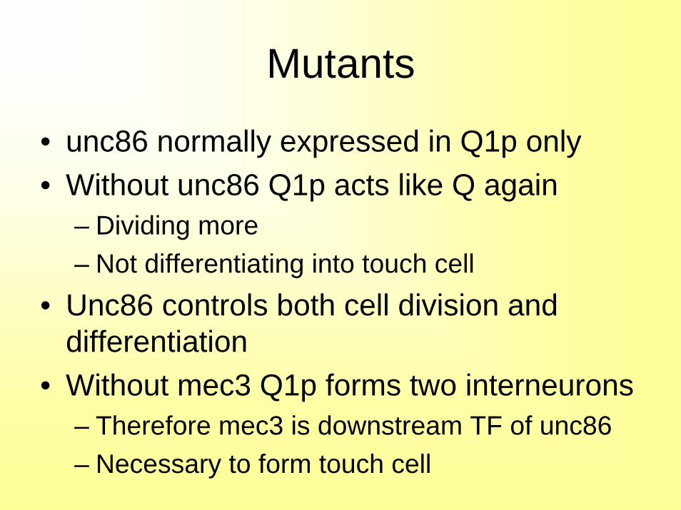
• unc86 normally expressed in Q1p only• Without unc86 Q1p acts like Q again
– Dividing more– Not differentiating into touch cell
• Unc86 controls both cell division and differentiation
• Without mec3 Q1p forms two interneurons– Therefore mec3 is downstream TF of unc86 – Necessary to form touch cell
Mutants
• Without mec7, mec12 and mec17 form only a defective touch cell
• Therefore we can see the progression of more and more specific TF’s being activated:– Unc86 determines Q1p to form precursor to
touch cell– Mec3 determines touch cell vs. interneuron– Mec7, etc determine specific cell anatomy of
touch cell
Mutants
Unc86 mec 3 mec7, mec12, mec17
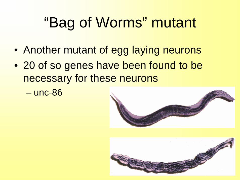
“Bag of Worms” mutant
• Another mutant of egg laying neurons• 20 of so genes have been found to be
necessary for these neurons– unc-86
Constant in C. elegans
• What has been found to be constant between different mutant phenotypes
• Neurons are always developed:– Hierarchical pathway– Rich in TF’s– Operate through lineage restrictions– Extrinsic TF’s regulate expression of intrinsic
TF’s
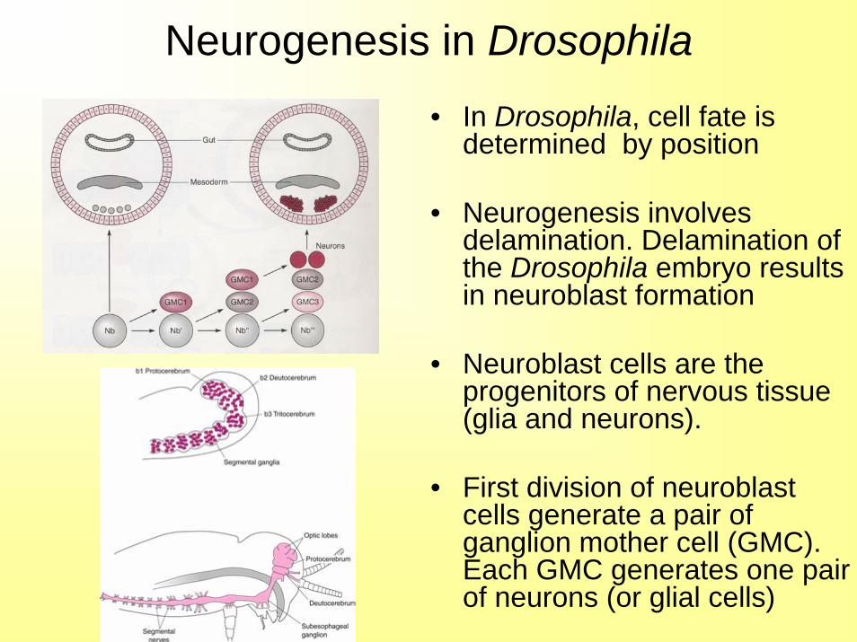
• In Drosophila, cell fate is determined by position
• Neurogenesis involves delamination. Delamination of the Drosophila embryo results in neuroblast formation
• Neuroblast cells are the progenitors of nervous tissue (glia and neurons).
• First division of neuroblast cells generate a pair of ganglion mother cell (GMC). Each GMC generates one pair of neurons (or glial cells)
Neurogenesis in Drosophila
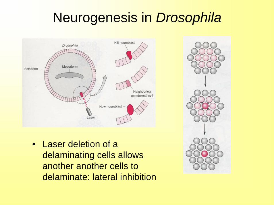
Neurogenesis in Drosophila
• Laser deletion of a delaminating cells allows another another cells to delaminate: lateral inhibition
Cell Fate in Drosophila
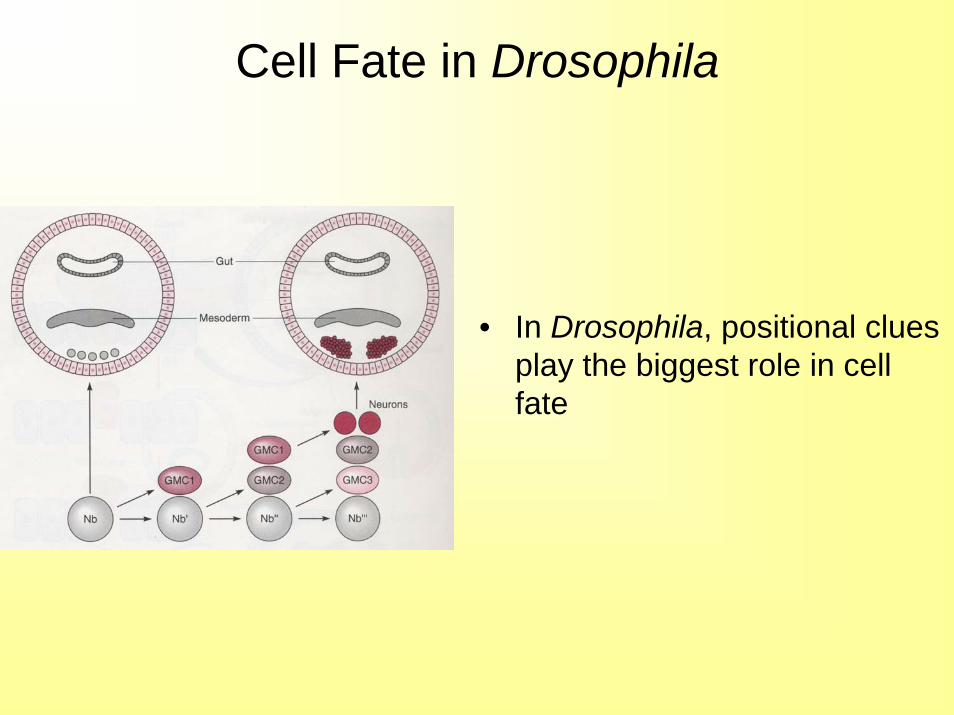
• In Drosophila, positional clues play the biggest role in cell fate
Neurogenesis in Drosophila
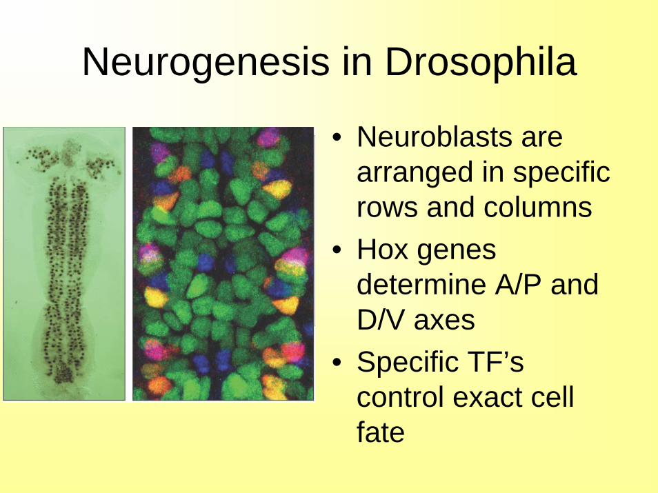
• Neuroblasts are arranged in specific rows and columns
• Hox genes determine A/P and D/V axes
• Specific TF’s control exact cell fate
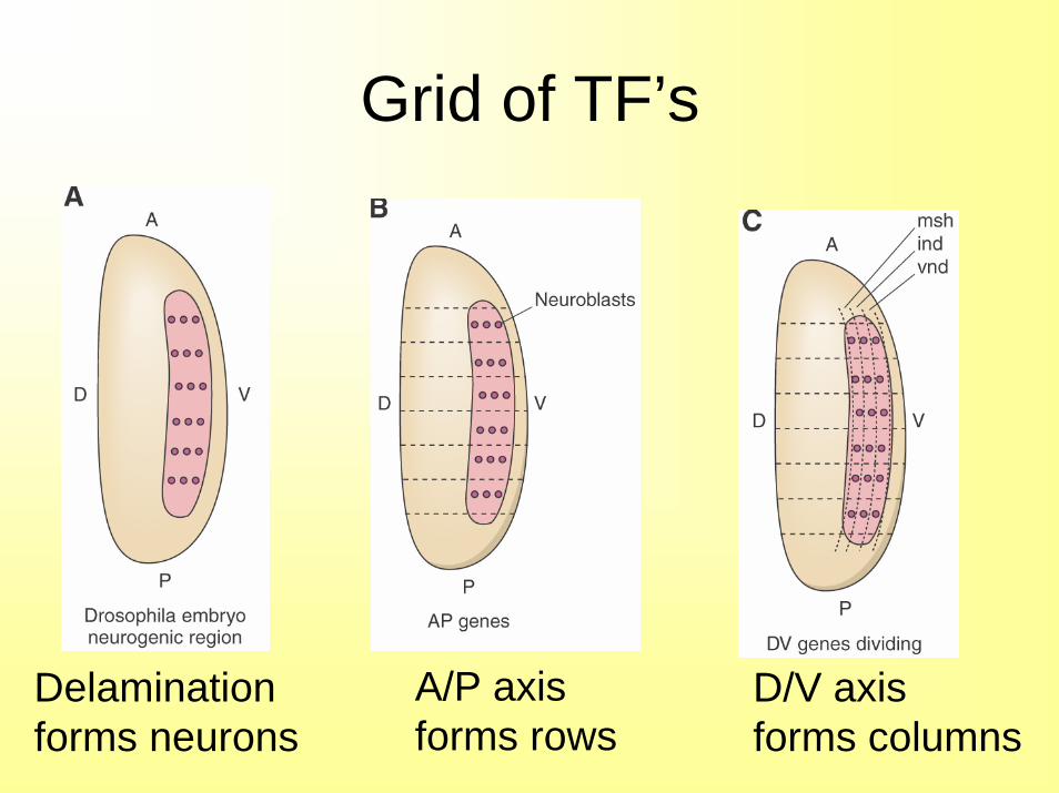
Grid of TF’s
Delaminationforms neurons
A/P axisforms rows
D/V axis forms columns
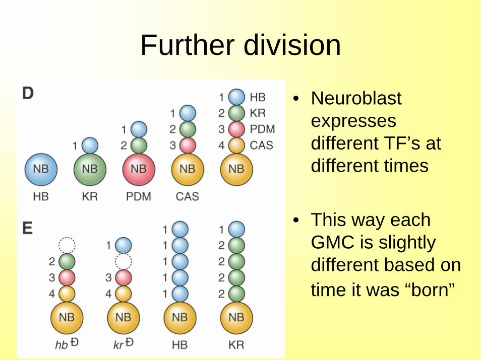
Further division
• Neuroblast expresses different TF’s at different times
• This way each GMC is slightly different based on time it was “born”
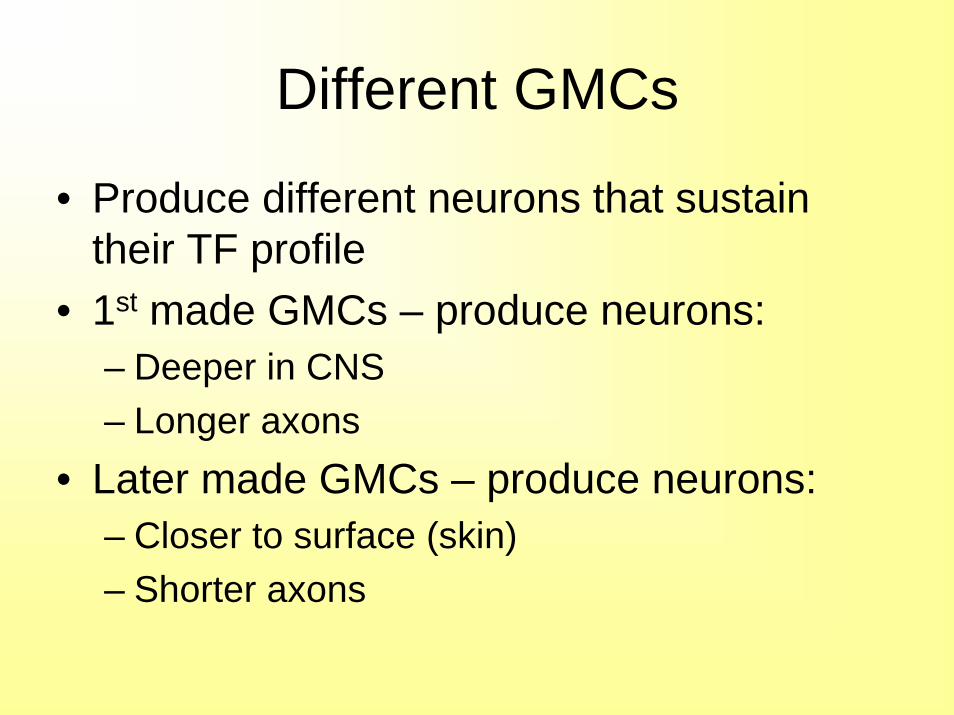
Different GMCs
• Produce different neurons that sustain their TF profile
• 1st made GMCs – produce neurons:– Deeper in CNS– Longer axons
• Later made GMCs – produce neurons:– Closer to surface (skin)– Shorter axons
TF’s in Cell Fate
Expression of an exact combination of genes is controlled by:
• Positionally controlled TFs– A/P and D/V position
• Temporally controlled TFs– Clock counting number of cell divisions
• Cell fate of any neuron is determined by the combination of TF’s that in turn influence cell’s phenotype
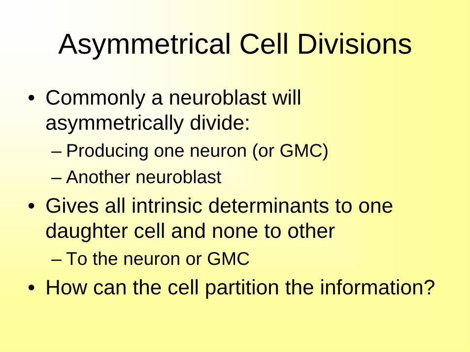
Asymmetrical Cell Divisions
• Commonly a neuroblast will asymmetrically divide:– Producing one neuron (or GMC)– Another neuroblast
• Gives all intrinsic determinants to one daughter cell and none to other– To the neuron or GMC
• How can the cell partition the information?
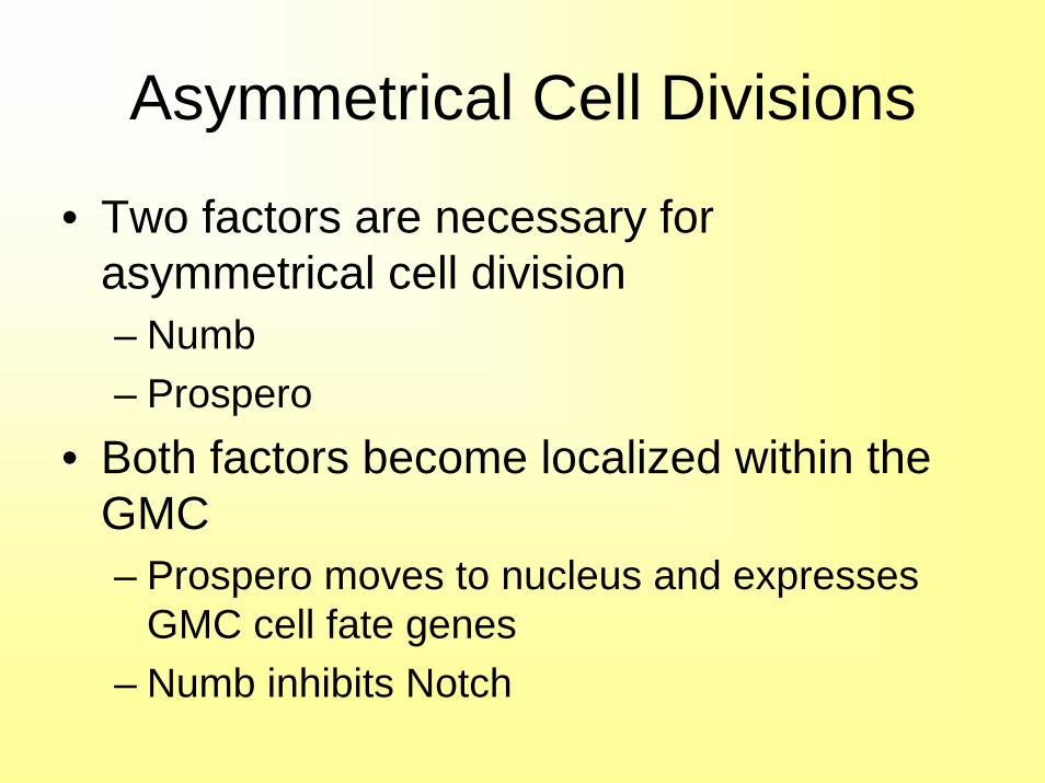
Asymmetrical Cell Divisions
• Two factors are necessary for asymmetrical cell division– Numb– Prospero
• Both factors become localized within the GMC– Prospero moves to nucleus and expresses
GMC cell fate genes– Numb inhibits Notch
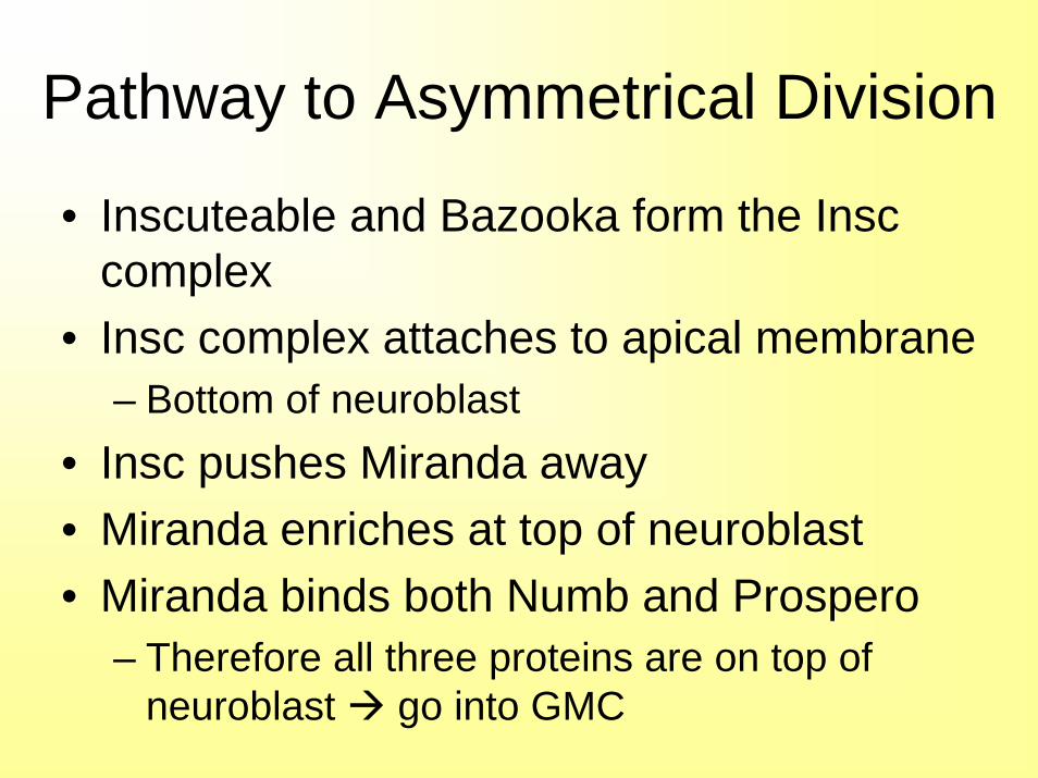
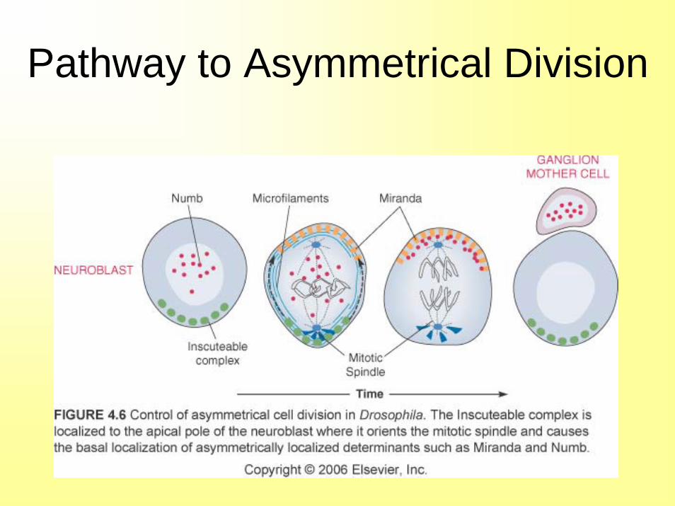
Pathway to Asymmetrical Division
• Inscuteable and Bazooka form the Insc complex
• Insc complex attaches to apical membrane– Bottom of neuroblast
• Insc pushes Miranda away• Miranda enriches at top of neuroblast• Miranda binds both Numb and Prospero
– Therefore all three proteins are on top of neuroblast go into GMC
Pathway to Asymmetrical Division
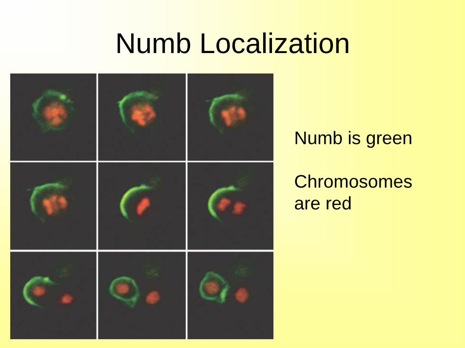
Numb Localization
Numb is green
Chromosomes are red
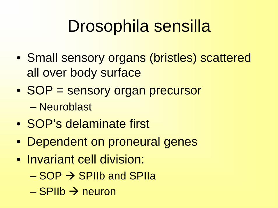
Drosophila sensilla
• Small sensory organs (bristles) scattered all over body surface
• SOP = sensory organ precursor– Neuroblast
• SOP’s delaminate first• Dependent on proneural genes• Invariant cell division:
– SOP SPIIb and SPIIa– SPIIb neuron
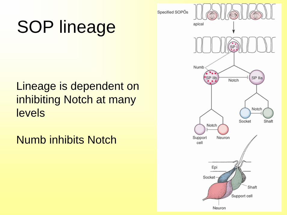
SOP lineage
Lineage is dependent oninhibiting Notch at many levels
Numb inhibits Notch
SOP mutants
• If SPIIb is ablated SPIIa becomes SPIIb and forms neuron
• If Notch is deleted no bristles or sockets– Or if Numb is overexpressed
• If Notch is overexpressed no neurons or glial cells– Or if Numb is deleted – hence the name
• Intrinsic Numb signaling cell fate
Compound Eye
• Composed of 800 identical units• Ommatidia• Each ommatidia composed of 8
photoreceptors and 12 accessory cells• Eight photoreceptors:
– R1 to R8• Extrinsic signals are involved in
determining cell fate for these cells
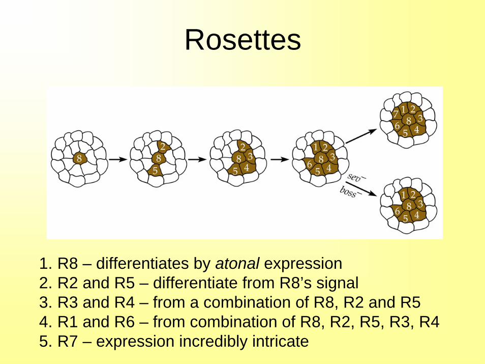
Rosettes
1. R8 – differentiates by atonal expression2. R2 and R5 – differentiate from R8’s signal3. R3 and R4 – from a combination of R8, R2 and R54. R1 and R6 – from combination of R8, R2, R5, R3, R45. R7 – expression incredibly intricate
R cell type
• Controlled by extrinsic signals coming off other cells
• In addition – photoreceptors have overlapping set of intrinsic determinants
• If you knock out certain combinations of the intrinsic determinants specific cells types will not form
• However, the cells are terminally determined by extrinsic signals
R7
• Only R7 is sensitive to UV light• Screen was done to mutagenize flies
unless found mutants that were blind to UV light
• Called “sevenless”• An incredible number of genes were
involved in the sevenless pathway– Mostly kinases and TFs
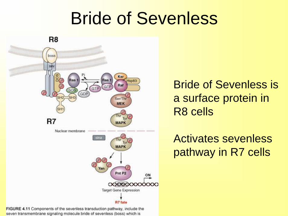
Bride of Sevenless
Bride of Sevenless isa surface protein inR8 cells
Activates sevenlesspathway in R7 cells
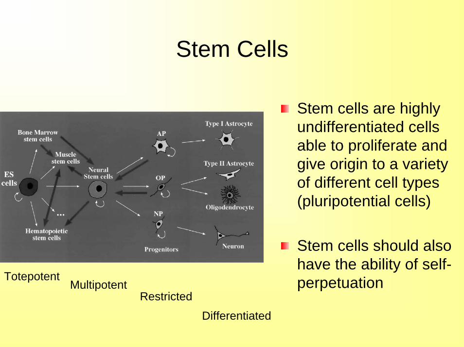
Stem Cells
Stem cells are highly undifferentiated cells able to proliferate and give origin to a variety of different cell types (pluripotential cells)
Stem cells should also have the ability of self-perpetuationTotepotent
MultipotentRestricted
Differentiated
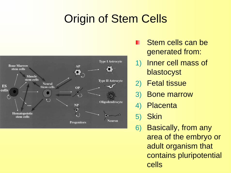
Origin of Stem Cells
Stem cells can be generated from:
1) Inner cell mass of blastocyst
2) Fetal tissue3) Bone marrow4) Placenta5) Skin6) Basically, from any
area of the embryo or adult organism that contains pluripotential cells
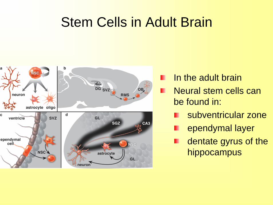
Stem Cells in Adult Brain
In the adult brain Neural stem cells can be found in:
subventricular zoneependymal layerdentate gyrus of the hippocampus
Differentiation of Stem Cells
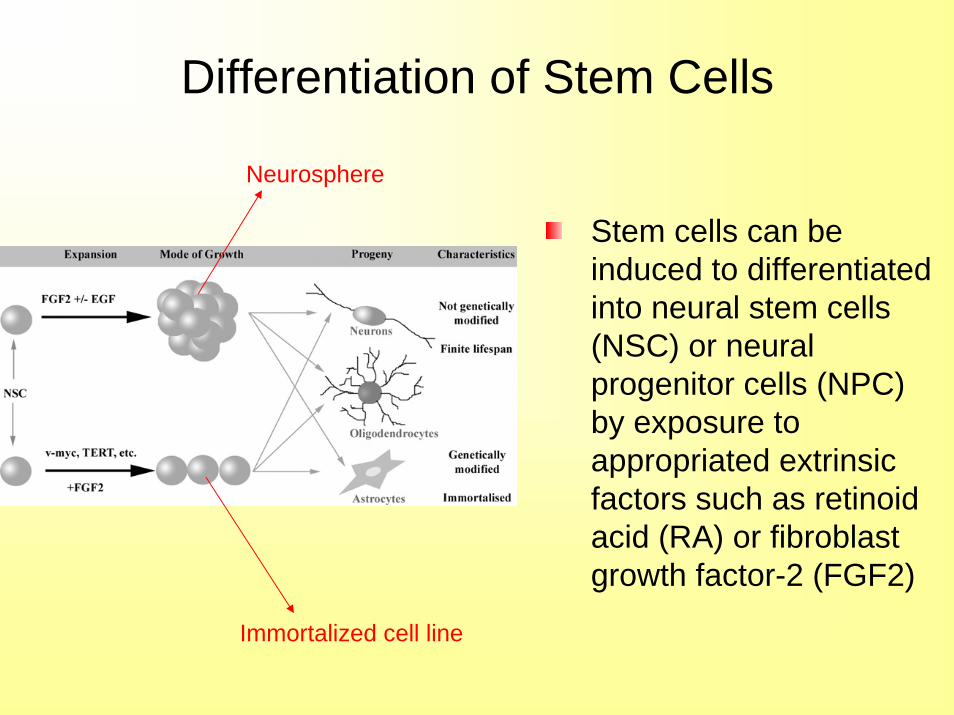
Stem cells can be induced to differentiated into neural stem cells (NSC) or neural progenitor cells (NPC) by exposure to appropriated extrinsic factors such as retinoid acid (RA) or fibroblast growth factor-2 (FGF2)
Neurosphere
Immortalized cell line
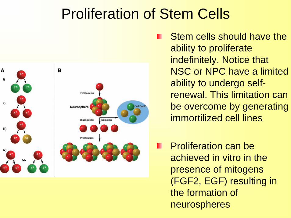
Proliferation of Stem CellsStem cells should have the ability to proliferate indefinitely. Notice that NSC or NPC have a limited ability to undergo self-renewal. This limitation can be overcome by generating immortilized cell lines
Proliferation can be achieved in vitro in the presence of mitogens (FGF2, EGF) resulting in the formation of neurospheres
Differentiation of Stem Cells
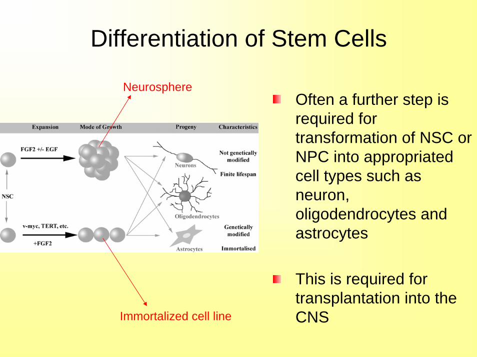
Often a further step is required for transformation of NSC or NPC into appropriated cell types such as neuron, oligodendrocytes and astrocytes
This is required for transplantation into the CNS
Neurosphere
Immortalized cell line
Differentiation of Stem Cells
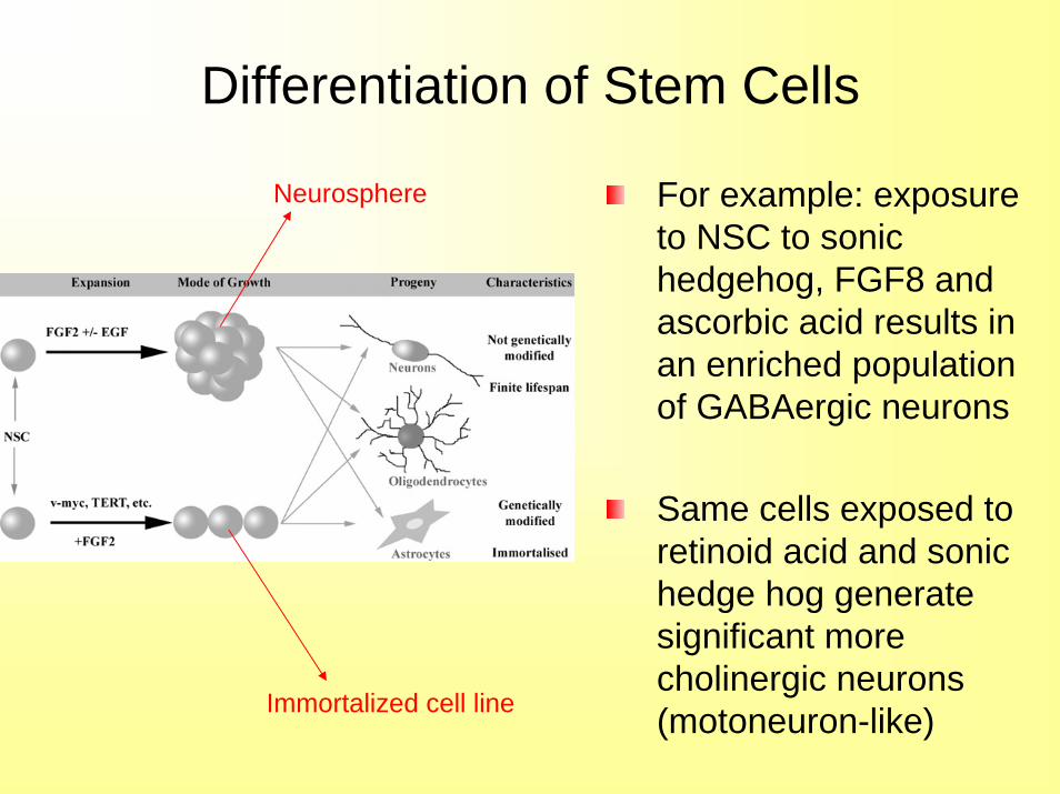
For example: exposure to NSC to sonic hedgehog, FGF8 and ascorbic acid results in an enriched population of GABAergic neurons
Same cells exposed to retinoid acid and sonic hedge hog generate significant more cholinergic neurons (motoneuron-like)
Neurosphere
Immortalized cell line
Differentiation of Stem Cells
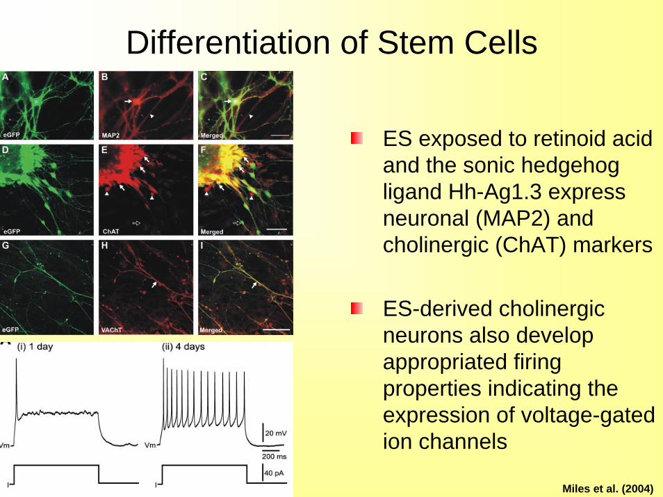
ES exposed to retinoid acid and the sonic hedgehog ligand Hh-Ag1.3 express neuronal (MAP2) and cholinergic (ChAT) markers
ES-derived cholinergic neurons also develop appropriated firing properties indicating the expression of voltage-gated ion channels
Miles et al. (2004)
Differentiation of Stem Cells
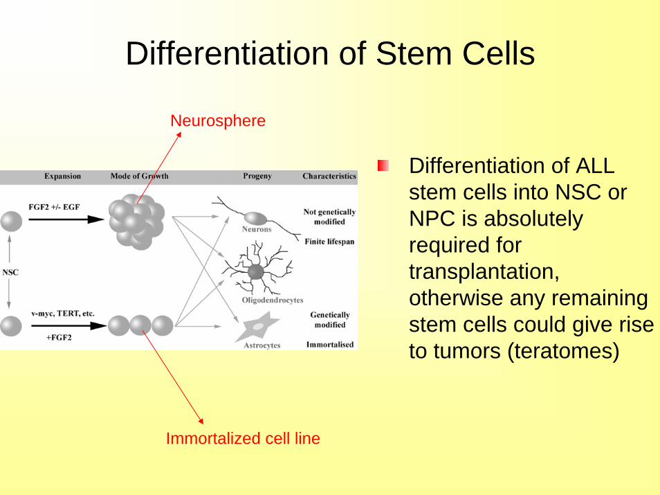
Differentiation of ALL stem cells into NSC or NPC is absolutely required for transplantation, otherwise any remaining stem cells could give rise to tumors (teratomes)
Neurosphere
Immortalized cell line
Potential Use of Stem Cells
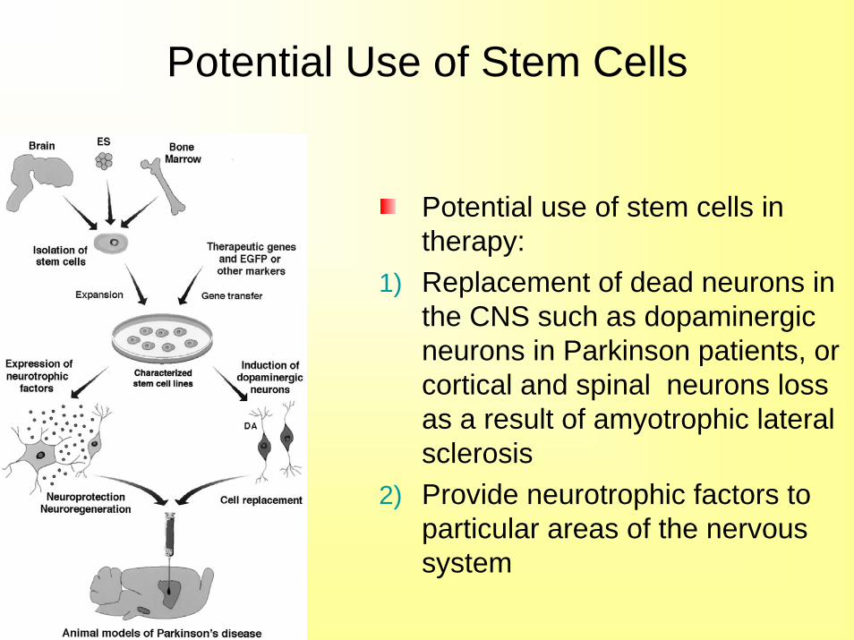
Potential use of stem cells in therapy:
1) Replacement of dead neurons in the CNS such as dopaminergic neurons in Parkinson patients, or cortical and spinal neurons loss as a result of amyotrophic lateral sclerosis
2) Provide neurotrophic factors to particular areas of the nervous system
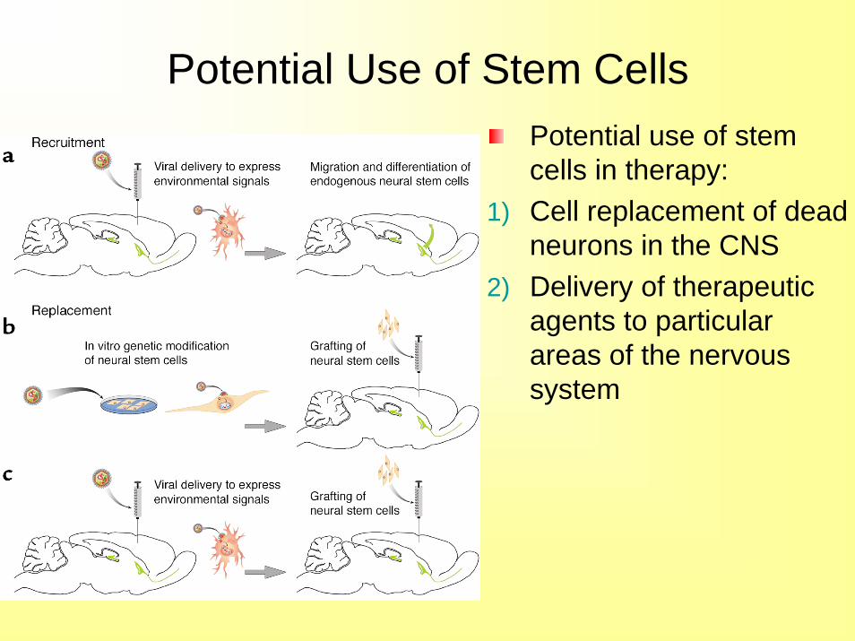
Potential Use of Stem CellsPotential use of stem cells in therapy:
1) Cell replacement of dead neurons in the CNS
2) Delivery of therapeutic agents to particular areas of the nervous system
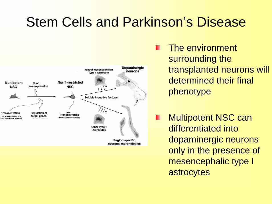
Stem Cells and Parkinson’s Disease
The environment surrounding the transplanted neurons will determined their final phenotype
Multipotent NSC can differentiated into dopaminergic neurons only in the presence of mesencephalic type I astrocytes
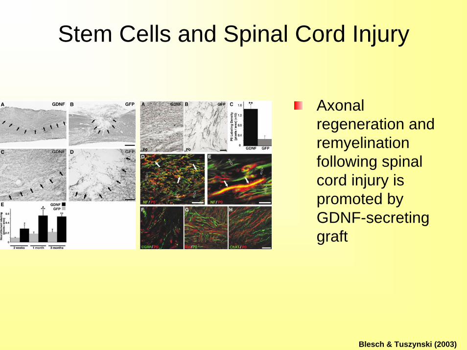
Stem Cells and Spinal Cord Injury
Axonal regeneration and remyelination following spinal cord injury is promoted by GDNF-secreting graft
Blesch & Tuszynski (2003)

Any Questions?
Read Chapter Four