cell death and autophagy in prion diseases (transmissible ... · cell death and autophagy in prion...
TRANSCRIPT
Folia Neuropathologica 2008; 46/1 1
Cell death and autophagy in prion diseases (transmissible spongiform encephalopathies)
PPaawweełł PP.. LLiibbeerrsskkii11,, DDaavviidd RR.. BBrroowwnn22,, BBeeaattaa SSiikkoorrsskkaa11,, BByyrroonn CCaauugghheeyy33,, PPaauull BBrroowwnn44
1Department of Molecular Pathology and Neuropathology, Chair of Oncology, Medical University of Lodz, Poland; 2Department
of Biology and Biochemistry, University of Bath, Bath, UK; 3Laboratory of Persistent Viral Diseases, Rocky Mountain Laboratories,
National Institute for Allergy and Infectious Diseases, National Institutes of Health, Hamilton, Montana, USA; 4retired, USA
Folia Neuropathol 2008; 46 (1): 1-25
Review article
A b s t r a c t
Neuronal autophagy, like apoptosis, is one of the mechanisms of programmed cell death. In this review, wesummarize current information about autophagy in naturally occurring and experimentally induced scrapie,Creutzfeldt-Jakob disease and Gerstmann-Sträussler-Scheinker syndrome against the broad background of neuraldegenerations in transmissible spongiform encephalopathies (TSEs). Typically a sequence of events is observed:from a part of the neuronal cytoplasm sequestrated by concentric arrays of double membranes (phagophores);through the enclosure of the cytoplasm and membrane proliferation; to a final transformation of the large area ofthe cytoplasm into a collection of autophagic vacuoles of different sizes. These autophagic vacuoles form not onlyin neuronal perikarya but also in neurites and synapses. On the basis of ultrastructural studies, we suggest thatautophagy may play a major role in transmissible spongiform encephalopathies and may even participate in theformation of spongiform change.
KKeeyy wwoorrddss:: autophagy, apoptosis, prion diseases, neurons, ultrastructure.
CCoommmmuunniiccaattiinngg aauutthhoorr::
Prof. Paweł P. Liberski, Department of Molecular Pathology and Neuropathology, Chair of Oncology, Medical University of Lodz,
Czechosłowacka Str. 8/10, PL 92-216 Lodz, Poland, tel./fax: +48 42 679 14 77, Email: [email protected]
Introduction
The transmissible spongiform encephalopathies
(TSEs), or prion diseases, are a group of neuro-
degenerative disorders which include kuru (Fig. 1)
[73,89,135], Creutzfeldt-Jakob disease (CJD) [76],
Gerstmann-Sträussler-Scheinker (GSS) disease [157],
and fatal familial insomnia (FFI) in humans [147],
natural scrapie in sheep (Fig. 2), goats [53,54,57], and
mouflons [224], transmissible mink encephalopathy
(TME) in ranch-reared mink [35], chronic wasting
disease (CWD) of mule deer and elk in North America
[137,221,222], bovine spongiform encephalopathy
(BSE) or “mad cow disease” [218], and its analogues in
several exotic species of antelopes [55,68,106,115] and
wild felids in zoological gardens [223], and feline
spongiform encephalopathy (FSE) in domestic cats
[226].
The cause of these disorders is still not completely
understood. Despite a wide acceptance for the prion

Folia Neuropathologica 2008; 46/1 2
Paweł P. Liberski, David R. Brown, Beata Sikorska, Byron Caughey, Paul Brown
FFiigg.. 11.. Kuru-affected children. Courtesy of DrCarleton Gajdusek, Amsterdam, the Netherlands
FFiigg.. 22.. Scrapie in a Romanov sheep. Courtesy ofProf. Jeanne Brugere- Picoux, Paris, France
theory, the name of the infectious agent (a “prion”)
still reflects our ignorance or at the very least, our
uncertainties of its nature [44,139,152]. Those who
prefer to view this pathogen as composed predo-
minantly or exclusively of a pathologically misfolded
protein use the term “prion” [184]; hence the term
“prion diseases” [67,217]. In this review, we will use the
term “PrPc” to designate the normal soluble and prote-
inase K-sensitive cellular protein, and the term “PrPTSE”
to designate all of its pathological forms, including
intermediate species that are sensitive to proteinase K
(PK) digestion as well as the mature amyloid species
that is insoluble and partially PK-resistant [32].
Other hypotheses, albeit less widely accepted, are
still not formally rejected. The “virino” hypothesis
suggests that the pathogen is a molecular chimera
composed of a yet-to-be-discovered nucleic acid and
a shell-protein, which is host-encoded (could be PrP)
[113]. The fact that RNA enhances conversion of PrPc
into PrPTSE in vitro is interesting in this context [56].
The “unified theory” of Weissmann [216], not unlike
the virino theory, suggests that the agent is
a molecular chimera in which PrPTSE confers infectivity
and an unidentified oligonucleotide specifies strain
characteristics. The virus hypothesis simply suggests
that the pathogen is a yet-to-be-identified uncon-
ventional virus [58,72,216]. To this end, virus-like
particles have been repeatedly shown in all TSEs
studied so far [140,144,153].
PrP, the PrP gene, the “prion hypothesis”and strains of the pathogen
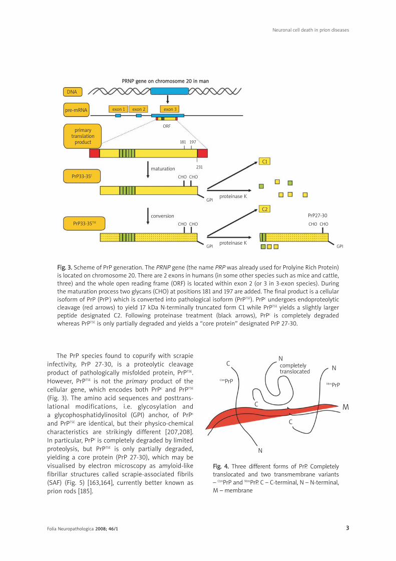
PrPc is a highly conserved sialoglycoprotein
encoded by a cellular gene mapped to chromosome
20 in man (Prion Protein; PRNP) and chromosome 2
in mouse (Fig. 3) [7,16,45,90,176,202,204]. The gene
is ubiquitous [193,194,225]; it has been cloned from
numerous mammalian species, included marsupials,
and there are analogues of this gene in birds [71,93],
reptiles [200], amphibians [209], and even fish [177]. The molecular biology of PrP has been extensively
studied. Human PrPc contains 253 amino acidsencoded by an intronless open reading frame (ORF)(Fig. 3) [7]. As a result, alternative splicing does notoccur. Three forms of PrPc exist – one completelytranslocated and two transmembrane variants, CtmPrPand NtmPrP [94,95,214] (Fig. 4) – and the sequenceencoding residues 151-165 that form the transmem-brane region is highly conserved [214]. Furthermore,PrPc undergoes endoproteolytic cleavage to yield 17 kDa N-terminally truncated form C1, while PrPTSE
yields a slightly larger peptide designated C2 (Fig. 3)[43].
The “prion” hypothesis, which is deeply rooted inthe association between PrPTSE and infectivity, was formulated by Stanley B. Prusiner in 1982[10,59,161,181,182]. The hypothesis postulated thatthe scrapie agent was a proteinaceous infectiousparticle, because infectivity was dependent onprotein but resistant to methods known toinactivate nucleic acids. A similar proposal waspresented a decade earlier by Gibbons and Hunter[75], Griffith [85], and Levine [130], who all expandedthe earlier suggestion of Alper and her co-workers[1], based on irradiation studies, that scrapie agentwas devoid of disease-specific nucleic acid.Furthermore, other investigators in the late 1970sand early 1980s had found that scrapie infectivitywas sensitive to proteolytic digestion [47,166].
Folia Neuropathologica 2008; 46/1 3
Neuronal cell death in prion diseases
DNA
PPRRNNPP ggeennee oonn cchhrroommoossoommee 2200 iinn mmaann
pre-mRNA
maturation
proteinase K
C1
C2PrP27-30
proteinase K
conversion
exon 1 exon 2 exon 3
ORF
181 197
231
CHO CHO
CHO CHO CHO CHO
GPI
GPI GPI
primarytranslation
product
PrP33-35C
PrP33-35TSE
FFiigg.. 33.. Scheme of PrP generation. The PRNP gene (the name PRP was already used for Prolyine Rich Protein)is located on chromosome 20. There are 2 exons in humans (in some other species such as mice and cattle,three) and the whole open reading frame (ORF) is located within exon 2 (or 3 in 3-exon species). Duringthe maturation process two glycans (CHO) at positions 181 and 197 are added. The final product is a cellularisoform of PrP (PrPc) which is converted into pathological isoform (PrPTSE). PrPc undergoes endoproteolyticcleavage (red arrows) to yield 17 kDa N-terminally truncated form C1 while PrPTSE yields a slightly largerpeptide designated C2. Following proteinase treatment (black arrows), PrPc is completely degradedwhereas PrPTSE is only partially degraded and yields a “core protein” designated PrP 27-30.
The PrP species found to copurify with scrapie
infectivity, PrP 27-30, is a proteolytic cleavage
product of pathologically misfolded protein, PrPTSE.
However, PrPTSE is not the primary product of the
cellular gene, which encodes both PrPc and PrPTSE
(Fig. 3). The amino acid sequences and posttrans-
lational modifications, i.e. glycosylation and
a glycophosphatidylinositol (GPI) anchor, of PrPc
and PrPTSE are identical, but their physico-chemical
characteristics are strikingly different [207,208].
In particular, PrPc is completely degraded by limited
proteolysis, but PrPTSE is only partially degraded,
yielding a core protein (PrP 27-30), which may be
visualised by electron microscopy as amyloid-like
fibrillar structures called scrapie-associated fibrils
(SAF) (Fig. 5) [163,164], currently better known as
prion rods [185].
C
C
C
N
N
N
M
completelytranslocated
CtmPrP NtmPrP
FFiigg.. 44.. Three different forms of PrP. Completelytranslocated and two transmembrane variants – CtmPrP and NtmPrP. C – C-terminal, N – N-terminal,M – membrane

Folia Neuropathologica 2008; 46/1 4
Paweł P. Liberski, David R. Brown, Beata Sikorska, Byron Caughey, Paul Brown
Cellular trafficking of PrPc and PrPTSE
PrPc, as is true of all cell surface glycoproteins, is
synthesized first in the endoplasmic reticulum,
matured in the Golgi apparatus, and transported in
membrane-bound vesicles to the plasma membrane,
where it is usually anchored by the GPI moiety (Fig. 6).
Once it is on the cell surface, PrPc can be subjected to
endocytosis and either degraded in lysosomes or
recycled to the plasma membrane via recycling
endosomes [92,149,150,180,198]. In peripheral (PNS)
and central nervous system (CNS) neurons fast
anterograde and retrograde axonal transport of PrPc
have also been detected [11,170,171], facilitating
movement to and from neural extremities.
Coincident with becoming infected, cells can bind
and internalize exogenous PrPTSE aggregates [97,98].
Depending on cell type, PrPTSE internalization can
involve heparan sulphate [97,98], laminin receptor
[169], and/or ferritin transporters [167]. However, the
presence of PrPc does not seem to be required as
a receptor for exogenous PrPTSE [97,149].
FFiigg.. 55.. Scrapie-associated fibrils (lower arrow) visualized by negative-staining electron microscopy. An upperarrow points to protofilaments. Original magn. × 30 000; bar = 100 nm
Folia Neuropathologica 2008; 46/1 5
Neuronal cell death in prion diseases
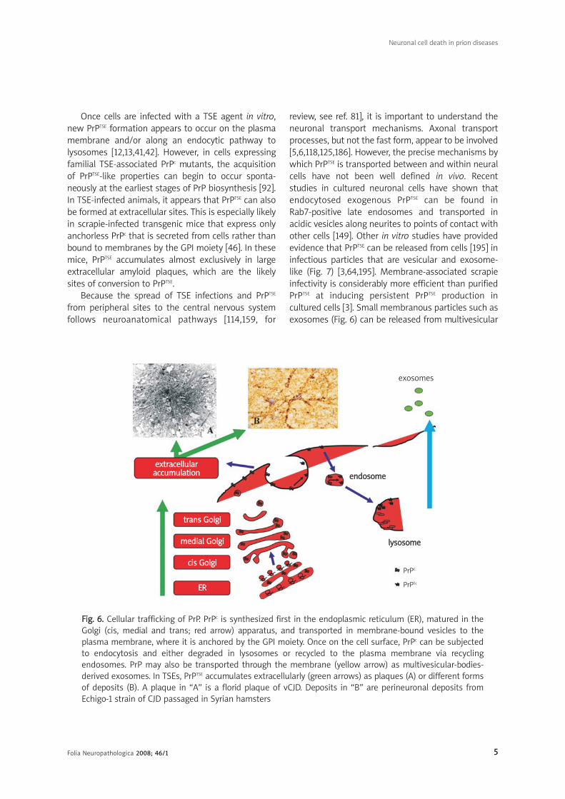
Once cells are infected with a TSE agent in vitro,
new PrPTSE formation appears to occur on the plasma
membrane and/or along an endocytic pathway to
lysosomes [12,13,41,42]. However, in cells expressing
familial TSE-associated PrPc mutants, the acquisition
of PrPTSE-like properties can begin to occur sponta-
neously at the earliest stages of PrP biosynthesis [92].
In TSE-infected animals, it appears that PrPTSE can also
be formed at extracellular sites. This is especially likely
in scrapie-infected transgenic mice that express only
anchorless PrPc that is secreted from cells rather than
bound to membranes by the GPI moiety [46]. In these
mice, PrPTSE accumulates almost exclusively in large
extracellular amyloid plaques, which are the likely
sites of conversion to PrPTSE.
Because the spread of TSE infections and PrPTSE
from peripheral sites to the central nervous system
follows neuroanatomical pathways [114,159, for
review, see ref. 81], it is important to understand the
neuronal transport mechanisms. Axonal transport
processes, but not the fast form, appear to be involved
[5,6,118,125,186]. However, the precise mechanisms by
which PrPTSE is transported between and within neural
cells have not been well defined in vivo. Recent
studies in cultured neuronal cells have shown that
endocytosed exogenous PrPTSE can be found in
Rab7-positive late endosomes and transported in
acidic vesicles along neurites to points of contact with
other cells [149]. Other in vitro studies have provided
evidence that PrPTSE can be released from cells [195] in
infectious particles that are vesicular and exosome-
like (Fig. 7) [3,64,195]. Membrane-associated scrapie
infectivity is considerably more efficient than purified
PrPTSE at inducing persistent PrPTSE production in
cultured cells [3]. Small membranous particles such as
exosomes (Fig. 6) can be released from multivesicular
FFiigg.. 66.. Cellular trafficking of PrP. PrPc is synthesized first in the endoplasmic reticulum (ER), matured in theGolgi (cis, medial and trans; red arrow) apparatus, and transported in membrane-bound vesicles to theplasma membrane, where it is anchored by the GPI moiety. Once on the cell surface, PrPc can be subjectedto endocytosis and either degraded in lysosomes or recycled to the plasma membrane via recyclingendosomes. PrP may also be transported through the membrane (yellow arrow) as multivesicular-bodies-derived exosomes. In TSEs, PrPTSE accumulates extracellularly (green arrows) as plaques (A) or different formsof deposits (B). A plaque in “A” is a florid plaque of vCJD. Deposits in “B” are perineuronal deposits fromEchigo-1 strain of CJD passaged in Syrian hamsters
exosomes
eexxttrraacceelllluullaarraaccccuummuullaattiioonn
ttrraannss GGoollggii
mmeeddiiaall GGoollggii
cciiss GGoollggii
EERR
eennddoossoommee
llyyssoossoommee
PrPC
PrPSc
Folia Neuropathologica 2008; 46/1 6
Paweł P. Liberski, David R. Brown, Beata Sikorska, Byron Caughey, Paul Brown
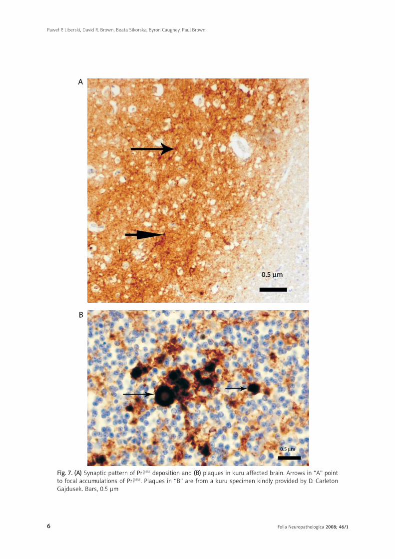
FFiigg.. 77.. ((AA)) Synaptic pattern of PrPTSE deposition and ((BB)) plaques in kuru affected brain. Arrows in “A” pointto focal accumulations of PrPTSE. Plaques in “B” are from a kuru specimen kindly provided by D. CarletonGajdusek. Bars, 0.5 μm
AA
BB

Folia Neuropathologica 2008; 46/1 7
bodies and fuse with the membranes of other cells,
providing a potential mechanism for the spread of
infectivity between cells [3,4,64].
Deposition of pathologically misfolded protein
Deposits of misfolded PrPTSE that accumulate
within the central (CNS) and peripheral nervous
system (PNS) and lymphatic tissues [138] correlate
with infectivity in most but not all situations [127,154].
Immunohistochemistry (IHC) has become the major
diagnostic tool for human prion diseases, by allowing
the detection of PrPTSE in fixed tissue sections [33].
More refined approaches, i.e. the histoblot [211] and
paraffin-embedded blot (PET) techniques, can also be
helpful [196]. A major hurdle with IHC is the
elimination PrPc labelling as no available antibodies,
including the widely used and commercially available
antibodies, can discriminate between PrPc and
misfolded PrPTSE in tissue sections.
Several patterns of PrPTSE expression are revealed
by IHC including synaptic (the most difficult to
visualize; Fig. 7a), perivacuolar, perineuronal (Fig. 7)
and plaque-like [33]. If amyloid is visualized by routine
neuropathology (H & E, Congo red, PAS- or Alcian blue
staining), it is designated as “plaques” (Fig. 6, 7b);
if detected only by IHC and not visible by routine
techniques, it is called “plaque-like deposits”.
Neuronal cell death in TSEs
The premature, primary death of nerve cells
underlies the clinical symptoms of prion diseases.
Unfortunately, despite the great efforts of researchers,
the cellular pathways leading to this neuronal loss are
not entirely clear. What is more, the question whether
there is a direct relation between the deposits of PrPTSE
and the loss of neurons still remains conjectural. As in
other neurodegenerative diseases, in TSEs apoptosis
has become the most popular concept of cell death.
However, there is no direct and convincing evidence of
apoptosis of nerve cells in most of the neurodege-
nerative diseases. In addition, the term “apoptosis” is
used in a wider sense than it was originally coined and
it has become synonymous with non-necrotic cell
death or even with programmed cell death. The data
on the role of apoptosis in prion diseases are
conflicting. Among recently recognized other types of
programmed cell death only autophagy has been
reported in TSEs but its role in prion diseases
pathology is not established.
There is currently no consensus on the classifi-
cation of different types of programmed cell death.
One of the oldest but also still considered most
accurate classifications is based on morphology.
According to this classification introduced by
Schweichel and Merker [197], three types of
programmed cell death (PCD) are discriminated
[116,189,215,230]:
1) apoptosis,
2) autophagy,
3) cytoplasmic cell death.
Although it must be stressed that this
categorization was based on the ultrastructural
features of embryonic cells during morphogenesis,
such a classification is widely accepted for deve-
loping and mature organisms.
1. The term “apoptosis” in its original meaning refers
to a morphological phenomenon [111] characterized
by chromatin condensation, cell shrinkage, pykno-
sis, plasma membrane blebbing and fragmentation
of the nucleus (karyorrhexis). There is little or no
ultrastructural modification of other subcellular
organelles. The integrity of plasma membrane is
maintained until the late stages of the process [124].
In the end-stage, the cell breaks into small
membrane-bound fragments, called apoptotic
bodies, which are phagocytosed by macrophages or,
in the case of neurons, by microglial cells without
inciting any inflammatory response [111]. Apoptosis
is regulated by a highly conservative network of
molecules consisting of Bcl-2 family and caspases
and Apaf-1 [116,189,215,230]. The apoptotic cell
death process has a very rapid time course and is
complete within a few hours.
2. Autophagic cell death is also one of the
programmed cell death mechanisms, sometimes
called “type II programmed cell death”, in contrast
to “type I programmed cell death” (apoptosis).
Contrary to apoptosis, autophagic cell death is
characterized by abundant autophagic vacuoles in
the cytoplasm, mitochondrial dilatation, and
enlargement of both the Golgi apparatus and
endoplasmatic reticulum, which precedes nuclear
destruction. Intermediate filaments and micro-
filaments are largely preserved [36,37,99].
3. A third type of programmed cell death is called
“cytoplasmic cell death” and it was subsequently
Neuronal cell death in prion diseases
Folia Neuropathologica 2008; 46/1 8
divided into two subtypes, 3A and 3B [49]. Type 3A
of neuronal death that occurs during development
is characterized by swelling of subcellular
organelles, formation of large empty spaces within
the cytoplasm, and fusion of these spaces to
extracellular space and, finally, disintegration of
the cellular structures. There are no features of
autophagic or heterophagic activity. In the 3B
subtype of cell death there are similar vacuoles
and empty spaces in the cytoplasm but, in
addition, there is a retraction of plasma membrane
and karyolysis.
According to a recent review [162], it is now
possible to discriminate eleven pathways of cell
death occurring in mammals. These types of cell
death include: 1) necrosis, 2) apoptosis, 3) anoikis ,
4) caspase-independent apoptosis, 5) autophagy,
6) Wallerian degeneration, 7) excitotoxicity, 8) ery-
thropoiesis, 9) platelet cell death, 10) cornification
and 11) lens cell death. All of them, except for
necrosis, are genetically programmed; however,
morphological features of necrosis are occasionally
observed during the active cell processes [158].
Some of those subtypes occur merely in one type of
cell but others are more common. The first seven
types are observed in nerve cells. The latter
classification is also based on morphological
features because in the majority of cases the
molecular mechanism is not known [162]. Other
investigators have discriminated more types of cell
death, e.g. paraptosis, pyroptosis, oncosis, abortosis,
aposklesis and many more [66,168,188,205,228].
Recently, the Nomenclature Committee on Cell
Death has been established and according to its
suggestions the whimsical names of cell death
should be replaced by more descriptive terms [123].
Prion proteins and the cell death
Mechanistically, neuronal cell death in prion
disease has a remarkable feature that distinguishes
it from neuronal loss in other neurodegenerative
disorders. In the absence of cellular expression of
PrPc, neuronal death does not occur [103]. This was
initially shown in cell culture systems with neurones
derived from PrP-knockout mice [23,78], and later
confirmed using an animal model in which PrPTSE was
transplanted into PrP-deficient tissue without
evidence of neuronal death [15]. Even more
complicated genetically engineered models have
demonstrated that when cellular expression of PrPc
is switched off, cell loss does not occur, and disease
progress is abated; illness is entirely prevented when
neurones cannot produce PrPc, even when non-
neuronal tissue contains dense deposits of PrPTSE.
Neuronal protection against cell death can also
occur in an animal model in which a different form of
PrPc is expressed that is more difficult to convert to
PrPTSE because of a species barrier. In transgenic mice
co-expressing hamster and mouse PrPc, infection with
hamster prion results in production of PrPTSE without
any pathology or neuronal death [187]. This suggests
that expression of a form of PrPc resistant to protein
conversion protects cells from neurotoxicity. However,
if the transgenic mice only express hamster PrPc in
astrocytes, then infection with hamster prions results
in prion diseases with associated neuronal death.
In this case, the neuronal death is possibly indirect as
a result of increased neuronal sensitivity to glutamate
toxicity [18].
The potential for PrPc expression to protect against
neuronal cell death runs counter to the finding that
expression is necessary for susceptibility to neuronal
death in prion disease. The only logical explanation is
that its normal cellular function is protective. Loss of
that function due to loss of expression of the protein
would result in alterations in the expression of
proteins with a similar function, resulting in
a compensatory effect. However, loss of function of
the protein with continued expression, as occurs in
prion disease, does not result in an increase in the
compensatory mechanisms. It has been postulated
that PrPc is an antioxidant (increased expression of
other antioxidants has been noted in PrP-knockout
mice) [26,219]. Simple loss of function is clearly not the
cause of cell death in prion disease, as PrP-knockout
mice do not show altered cell survival in vivo [34];
however, neurons in culture are more susceptible to
a range of neurotoxic insults when they are derived
from PrP-deficient mice [24,26,27,126,219].
Aggresomes
It is well known that protein aggregates are
generally difficult to unfold or to degrade. Misfolded
and aggregated proteins are usually handled in the
cell through chaperone-mediated refolding or, when
this is impossible, they are destroyed by proteasomal
degradation. Recent findings suggest that there is
Paweł P. Liberski, David R. Brown, Beata Sikorska, Byron Caughey, Paul Brown
Folia Neuropathologica 2008; 46/1 9
a third way for a cell to deal with misfolded proteins.
This pathway involves the sequestration of aggre-
gated proteins into specialized “holding stations”, as
they are sometimes called, or aggresomes. In this
mechanism proteins form small aggregates that are
transported along microtubules (MTs) towards
a microtubule organizing centre (MTOC) by a process
mediated by the minus-end motor protein dynein. At
the organizing centre the particles form a spherical
structure, usually 1-3 μm in diameter, called an
aggresome. Aggresomes are not just static garbage
deposits; they recruit various chaperones, ubiqui-
tination enzymes, and proteasome components. They
are also supposed to trigger autophagy [74].
A recent report of Kristiansen et al. [122] suggests
that neuronal propagation of prions invokes a neuro-
toxic mechanism involving intracellular formation of
PrPTSE aggresomes. The authors showed that only in
prion-infected cells did mild proteasome impairment
result in formation of large cytosolic, perinuclear
structures, containing PrPTSE, heat shock protein 70,
ubiquitin, proteasome subunits and vimentin. These
structures are consistent with the definition of
aggresomes. Those authors also claimed to show
aggresomes in vivo in brains of prion-infected mice,
but it is well known that vimentin is present in
neurons only in trace quantities while it is robust
in glial cells. This means that showing aggresomes
in vivo needs further studies. A few years earlier
Cohen and Taraboulos [50] showed that hampering
the activity of cyclophilin isomerases with the fungal
immunosuppressant CsA in different cell lines led to
accumulation of a PrP population with prion-like
properties that was not ubiquitylated and partially
resisted proteasomal degradation. These aggregated
molecules formed perinuclear aggresomes. Although
a growing body of evidence seems to confirm the
formation of aggresomes in prion diseases, it must be
mentioned that the majority of the studies were
performed in vitro and PrPTSE in human or animal
diseased brains does not intracellular aggregates
reminiscent of aggresomes. Similar aggregates are
only observed by electron microscopy, but they are
extremely rare.
PrP and Oxidative Stress
Recent work on cell death mechanisms has
focused on pathways involving PrPTSE. Numerous cell
culture lines exposed to neurotoxic PrP fragments
have not always given consistent results, although it
appears that caspases are involved [96,122,191,201],
and that levels of ERK proteins are increased when
cells are treated with toxic forms of PrP [129]. Other
proteins have also been suggested to play a role, such
as p38 [52], JNK, and Bax. However, none of these
observations have been independently confirmed.
Several of the studies have also suggested that
calcium entry is involved [24,172,175,213], but this is
a common event of many cell death pathways and the
true intracellular pathway has yet to be determined.
It seems likely that some extracellular event must
trigger the intracellular pathway that leads to cell
death, and many researchers have attempted to
identify binding partners on the cell surface that could
initiate the intracellular process. Several proteins have
been suggested and include the laminin receptor
[190], stress inducible protein-1 [231], and PrPc [20].
Binding of PrP106-126 to PrPc causes direct inhibition
of the antioxidant activity of PrPc [20]. Treatment of
neurons with PrP106-126 causes a marked reduction
in the resistance of neurons to oxidative stress [94],
and a decrease in the activity of other antioxidant
enzymes such as Cu/Zn superoxide dismutase
[30,219]. In addition, the peptide causes a decrease in
the uptake of Cu by neuronal cells [20], and a decrease
of Cu incorporation into Cu/Zn superoxide dismutase
[21]. It is unclear how these changes are brought
about. However, there is evidence that PrP106-126 can
enter cells [160] and might interact with intracellular
proteins in microtubules [28], and aggregates of
PrP106-126 can cause PrPc to become trapped in the
aggregates [20].
Interestingly, a subset of antibodies to PrP can
also cause in vitro apoptosis, suggesting that
inhibition of protein catabolism or its interaction
with other proteins might be sufficient to trigger cell
death [29]. Recently, it has been shown that similar
antibodies injected in vivo can also cause neuronal
apoptosis [203]. As antibodies to PrP increase the
toxicity of Cu ions to cells, it is possible that inter-
ference with the protein’s role in Cu metabolism
might be central to the ability of PrP106-126 to
initiate cell death. However, there is evidence to
suggest that the direct effect of PrP106-126 on
neurons is insufficient to complete its execution.
PrP106126 also has the effect of compromising the
neurons’ ability to deal with stressful conditions
[27], and in this way gives neurons a phenotype like
that observed for PrP-deficient neurones [26].
Neuronal cell death in prion diseases

Folia Neuropathologica 2008; 46/1 10
Execution of cell death then comes about as a result
of this compromised phenotype and one of a number
of different stress events such as the production of
superoxide [27].
Several studies have identified markers of
oxidative stress in the brains of rodents with prion
disease. There are increased levels of oxidised lipids
in the brains of scrapie-infected hamsters [86].
Another study [87] has shown increased levels of
nitrotyrosine and hemeoxygenase-1 in the brains of
scrapie-infected mice. These observations imply that
significant free radical damage is being generated in
the brains of scrapie-infected mice. Additionally,
there is evidence for mitochondrial damage in cells
from brains of scrapie-infected hamsters and mice
[48,128]. These changes include reduction in the
activity of mitochondrial enzymes and structural
abnormalities in the mitochondria. Other enzymes
known to be associated with resistance to oxidative
stress, such as catalase and glutathione-S-transfe-
rase, show increased expression [128].
Taken together, these results suggest that
oxidative stress is involved in the pathology of prion
diseases. It is possible that oxidative damage to the
brain in scrapie might be a result of damage to the
mitochondria, which can generate superoxide.
However, the measured level of oxygen radicals
detected with dichlorofluorescein in mitochondrial
fractions from the brains of scrapie-infected mice
was not greatly increased above that of controls
[112]. Reactive oxygen species such as superoxide
are generated by microglia and the implication of
this is that microglia cause damage in the brain of
scrapie-infected mice. In vitro studies have de-
monstrated that microglia can mediate the cell
death caused by PrPTSE or PrP106-126 [27,78,155,
156]. However, microglia activation has also been
postulated to be a response to neuronal damage
rather than the cause of it. Even if neuronal damage
was the sole cause of the microglial response (which
is unlikely given the complexity of cell-cell
interactions), then the microglial activation is still
likely to cause significant production of toxic
substances to trigger neuronal apoptosis.
In summary, the neurotoxic mechanism involved
in prion disease remains unresolved. However, it is
clear that PrPc plays a major role in the mechanism,
making cells susceptible to cell death. Cell death
could involve loss or subversion of the normal
cellular function of the protein and could make cells
susceptible to the toxicity of a variety of agents such
as superoxide released by microglia. Therefore
a combination of direct and indirect effects caused
by PrPTSE remains the most likely explanation of the
cell death mechanism.
Apoptosis and (macro)autophagy
Using TUNEL methodology, apoptotic neurons
have been repeatedly identified in both naturally
occurring and experimentally induced TSEs [60,69,
70,107-109], and some investigators believe that at
least a proportion of “dark neurons” that are
shrunken, with homogeneously dark cytoplasm, may
represent cells undergoing apoptosis (other workers
regard them as fixation artefacts). The data on
whether neurons, and perhaps even glia, die of
apoptosis in TSE are conflicting, however. While
Migheli et al. [165] found no evidence of apoptosis in
scrapie-infected mouse brains, Giese et al. [79,80],
Lucassen et al. [146], Fraser et al. [70], Williams et al.
[220], Kretzschmar et al. [119-121], and Jamieson
et al. [100] readily found apoptosis in various
scrapie-infected rodent models. The characteristic
DNA fragmentation ladder has been seen in both
BSE [212] and natural scrapie [62], and in situ
end-labelling (ISEL) revealed apoptotic cells in human
and experimental CJD [63,84,107]. Collectively, these
data strongly suggest that neurons in TSEs die
because of apoptosis.
It is evident that PrPTSE accumulation in TSE-
affected brain largely precedes development of other
changes – i.e. spongiform change and astrogliosis,
for which PrP serves as a signal for proliferation
[17,18,22,25,88,229]. At the ultrastructural level,
apparently normal looking neurons secrete PrPTSE,
which later fibrillizes and becomes neurotoxic [104],
and in vitro experiments highlight some possible
intermediate steps.
As mentioned, several synthetic peptides which
form amyloid fibrils, e.g. PrP106126 [19] and PrP118-135
[179], induce apoptosis in a dose-dependent manner.
PrP106-126 may exert its pro-apoptotic characteristics
via disruption of mitochondrial membranes with
subsequent release of cytochrome-c and caspase
activation [175]. Next, intracellular Ca2+ concentration
rises and another family of proteases, calpains, are
activated. In contrast, Bounhar et al. [14] found that
PrP may serve as an anti-apoptotic factor protecting
neurons in vitro from Bax-induced apoptosis. Removal
Paweł P. Liberski, David R. Brown, Beata Sikorska, Byron Caughey, Paul Brown
Folia Neuropathologica 2008; 46/1 11
of four of five octarepeats (codon 51-91 of PrP) or
D178N and T183A PRNP mutations completely abo-
lished this effect. It is apparent that a functional gain of
PrP (as PrPTSE) may result in changes of anti-apoptotic
into pro-apoptotic properties of PrP, which is vaguely
reminiscent of changes of anti-oncogenic into
oncogenic functions of p53 protein (also involved in
apoptosis).
Moreover, there is no consistent correlation
between PrPTSE, pathology and neuronal death.
Following transmission of BSE from cattle to mice,
Lasmezas et al. [127] found two lines of BSE-infected
mice, one with PrPTSE and typical TSE pathology
(vacuolation and astrogliosis), and a second without
PrPTSE but with prominent apoptosis of neurons.
These observations suggest that PrPTSE is not directly
responsible for apoptosis.
However, in another experiment, where neuronal
PrPc was depleted in NFH-Cre/MloxP transgenic mice,
after early spongiform change had developed
following scrapie neuroinvasion, Collinge’s group [151]
found a reversal of spongiform change, while
PrPTSE, released probably from glial cells, steadily
accumulated. These PrPc-depleted Tg mice also
became disease-free; thus, the lack of neuronal PrPTSE
and/or PrPc protected them from the final prion-
related neuronal degeneration. In contrast, in
transgenic mice in which PrP transgene was
exclusively expressed in astrocytes under the control
of GFAP promoter, spongiform change and neuronal
decay were observed following scrapie infection [103].
In an independent experiment, the highest
densities of apoptotic cells were observed in those
neuroanatomical areas in which spongiform change is
minimal or absent – viz. retina and cerebellum [120].
Thus, apoptosis and the presence of PrPTSE may not
only be uncoupled under specific experimental
conditions but may be directly (not mediated through
PrPTSE) linked to scrapie infectivity.
Neurons may only degenerate and eventually die
through a limited number of pathologic pathways.
Cellular necrosis is caused by a sudden brain insult,
Neuronal cell death in prion diseases
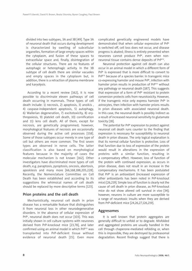
FFiigg.. 88.. An autophagic vacuole containing a part of the cytoplasm. Phagophore is indicated by arrows; mt,microtubules. A hamster brain infected with the Echigo-1 strain of CJD. Original magn. × 50 000; bar = 100 nm
Folia Neuropathologica 2008; 46/1 12
leading to destruction of the entire cell, the remnants
of which attract inflammatory cells. Apoptosis is
a programmed molecular process following a “suicidal”
stimulus, which leads to a hierarchical gene response.
Apoptotic cells, in contrast to necrotic cells, attract no
inflammatory response. As the relative lack of classical
immunological response in TSE-affected brain is
paradigmatic for the whole group of these diseases
[31], neurons, by definition, should die by apoptosis
and not necrosis.
A determinant for apoptotic cell detection is the
time over which neurons die. Even when the number
of neurons in dorsal lateral geniculate nucleus (dLGN)
following intraocular inoculation drops from over
22 000 to less than 2000 [101], the number of
apoptotic nuclei detected by TUNEL method is low
[70]. Because apoptotic cells are most readily
detected in highly structured neuronal systems such
as the retina and hippocampus, what is observable in
less structured regions may represent only a minute
proportion of all apoptotic events occurring in
TSE-affected brains.
As an evolutionarily ancient cellular response to
intra- and extracellular noxious stimuli, autophagy
may precede or co-exist with apoptosis, and the
process may be induced by apoptotic stimuli [38,227].
Furthermore, the level of autophagy may define the
sensitivity of a given neuronal population to apoptotic
Paweł P. Liberski, David R. Brown, Beata Sikorska, Byron Caughey, Paul Brown
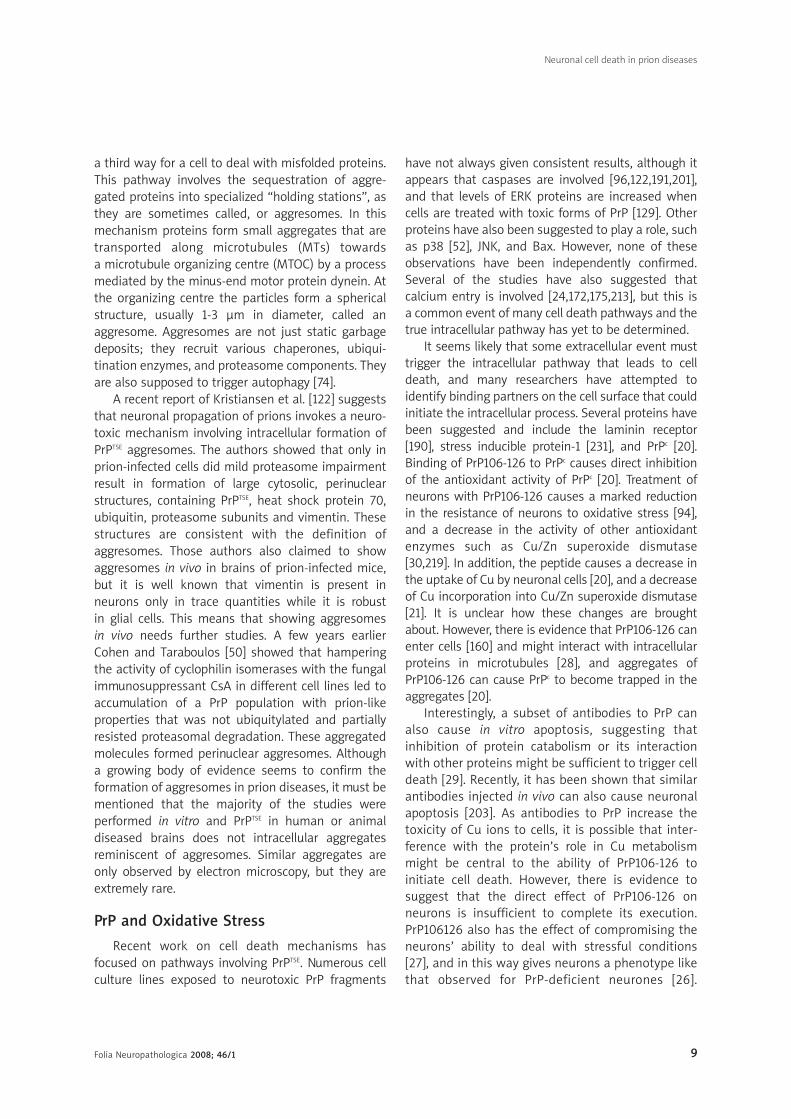
FFiigg.. 99.. Neuronal autophagic vacuoles in an early stage of development. Note that sequestrated cytoplasm is ofhigh electron density (arrow). SV – synaptic vesicles. A hamster brain infected with the 263K strain of scrapie.Original magn. × 12 000; bar = 200 nm
Folia Neuropathologica 2008; 46/1 13
stimuli, which may underlie the phenomenon of
“selective neuronal vulnerability”. Thus, autophagy
and apoptosis are often interwoven [40].
Cellular autophagy is a physiological degradation
process employed, like apoptosis, in embryonic
growth and development, cellular remodelling and
the biogenesis of some subcellular organelles – viz.
multilamellar bodies [65,91,192]. Autophagosomes
coalesce with lysosomes to form degraded auto-
phagic vacuoles, and as in apoptosis, only excessive
or misdirected autophagy causes a pathological
process. Autophagy is highly enhanced in other brain
amyloidoses or conformational disorders [83],
Alzheimer’s disease [206], Parkinson’s disease [2],
and Huntington’s disease, in which the signal for
autophagy is huntingtin [110]. Here, we extend
these observations using different models of scrapie
and CJD.
Neuronal autophagy in TSEs
Data on autophagy in TSEs are very limited,
consisting of a few electron-microscopic papers,
including reports from our own laboratories
[132,136,143-145,199]. The pioneering work was
published in Acta Neuropathologica by Boellaard et
al. [9]. Our initial experimental approach using the
hamster-adapted 263K or 22C-H strains of scrapie
Neuronal cell death in prion diseases
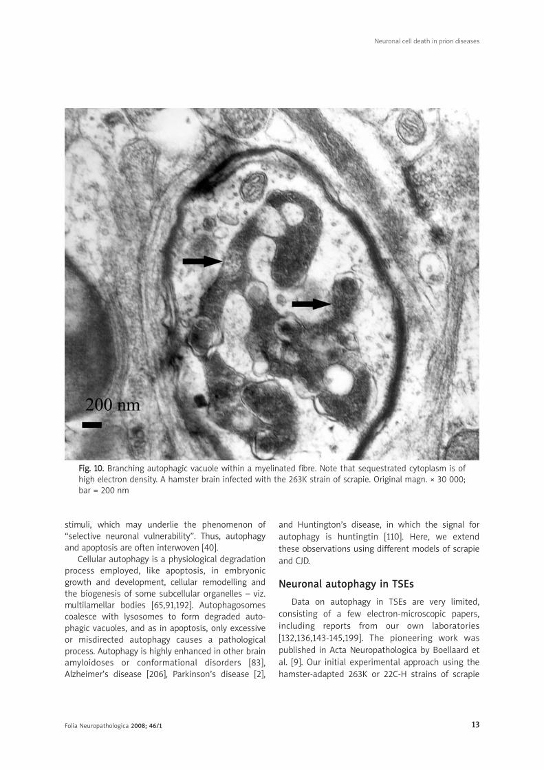
FFiigg.. 1100.. Branching autophagic vacuole within a myelinated fibre. Note that sequestrated cytoplasm is ofhigh electron density. A hamster brain infected with the 263K strain of scrapie. Original magn. × 30 000;bar = 200 nm

Folia Neuropathologica 2008; 46/1 14
[132,136,145] was subsequently extended by studies
of human brain biopsies from patients with sporadic
CJD, variant CJD, and FFI [144]. Experimentally
infected animal models are widely used because
of their relatively short incubation periods that, for
mice, range from 16 to 18 weeks, and for hamsters
from 9 to 10 weeks for the 263K strain and 24-26
weeks for the 22C-H strain.
Formation of autophagic vacuoles in TSEs-affected brains
Autophagic vacuoles are areas of the cytoplasm
sequestered within double or multiple membranes
(phagophores) of unknown origin; one possible
source is the endoplasmic reticulum (Fig. 8). They
contain ribosomes, small secondary vacuoles, and
occasional mitochondria. Some vacuoles present
a homogeneously dense appearance.
We observed neuronal autophagic vacuoles in
different stages of formation in the same specimens
and our interpretation of the ‘maturity’ of their
formation may or may not equate to actual
developmental stages. Initially, a part of the neuronal
cytoplasm was sequestered within double or multiple
membranes (phagophores) and often exhibited
increased electron-density (Figs. 9-11). The intra-
cytoplasmic membranes multiplied in a labyrinth-like
Paweł P. Liberski, David R. Brown, Beata Sikorska, Byron Caughey, Paul Brown
FFiigg.. 1111.. Fully developed autophagic vacuole. Phagophores are pointed to by arrows. Note that part of thesequestrated cytoplasm is of high electron density. A hamster brain infected with the 263K strain ofscrapie. Original magn. × 12 000; bar = 200 nm

Folia Neuropathologica 2008; 46/1 15
Neuronal cell death in prion diseases
FFiigg.. 1122.. Section through a myelinated fibre showing formation of autophagic vacuoles (hearts). A hamsterbrain infected with the 263K strain of scrapie. Original magn. × 8300; bar = 100 nm
manner (Fig. 11). The autophagic vacuoles thenexpanded and eventually a vast area of the cytoplasmwas transformed into a merging mass of autophagicvacuoles. Occasionally, a single large autophagicvacuole was visible. Autophagic vacuoles developednot only in neuronal perikarya but also in neuronalprocesses, eventually replacing the whole cross-section of affected neurites (Fig. 9, 12). In a fewspecimens we found round electron-dense structuresthat we identified as aggresomes. In general, therewas little qualitative difference between these twomodels, although hamsters inoculated with the 263Kstrain showed a more robust pathology.
Conclusions and hypotheses
One of the major problems of TSEs pathogenesisis the cause of neuronal degeneration with eventualneuronal loss [69,136]. Whether the prion is or is notthe pathogen, it is widely accepted that the basic
underlying pathological event is the conversion of
a normal isoform of prion protein (PrPc) into its
pathological misfolded isoform (PrPTSE) [15,51,178,
183], involving an α-helical to β-pleated sheath (or
β-helical) transformation. Using a highly sophisti-
cated mathematical model, Stumpf and Krakauer
[210] tried to reason whether PrPTSE causes neurons
to die because of neurotoxic effect of PrPTSE (gain of
function), or loss of function of PrPc. They assumed
that if cells die of apoptosis because of neurotoxic
gain of function of PrPTSE, the cells should die rapidly,
and the amount of PrPTSE should be low. Indeed, in
both CJD and FFI, there are more apoptotic cells and
a lower amount of PrPTSE [107] than in GSS, where
the amount of amyloid is vast and the number of
apoptotic cells is low [84].
As already mentioned, three types of programmed
cell death (PCD) are known: apoptosis, autophagy and
swelling of intracellular organelles. Apoptosis has

Folia Neuropathologica 2008; 46/1 16
Paweł P. Liberski, David R. Brown, Beata Sikorska, Byron Caughey, Paul Brown
FFiigg.. 1133.. “Spongiform” vacuoles within a dendrite (because a synaptic terminal with vesicles (SV) is attachedto this vacuole) containing a structure at the top suggestive of undergoing autophagy. Note thatsequestrated cytoplasm is of high electron density. Curled membrane fragments are indicated by arrows.A hamster brain infected with the 263K strain of scrapie. Original magn. × 8300; bar = 100 nm
been discussed already, and in this part we willconcentrate on autophagy in regard to TSEs.
Whereas apoptosis in TSE is relatively wellunderstood, autophagy is not. As already mentioned,autophagic cell death or type II PCS is a process usedby a cell to remove the bulk of organelles, e.g. duringgrowth and development, but which becomespathological if too robust or wrongly placed [39].According to Bursch et al. [39], the appearance of“autophagic vacuoles in dying cells by electronmicroscopy is taken as condition sine qua non (i.e. anabsolute prerequisite) to denote cell death asautophagic/type II PCD”. To this end, the merepresence of autophagic vacuoles is relatively welldemonstrated in TSE.
There are several uncertainties in our thinking on
neuronal autophagy in TSEs. First, autophagy is
regarded as a short-term response to nutrient
limitations [61], which is, by definition, not the case in
slow transmissible diseases like TSEs. However, it
seems that autophagy is activated to prevent
apoptosis; when autophagy is blocked, apoptosis
ensues. On the other hand, when apoptosis is blocked
as in Bax/Bak-deficient mice, autophagy is activated
as a cell-survival mechanism [148]. However, when all
subcellular organelles are self-eaten, the cell
eventually dies. An analogous situation was observed
when inhibition of autophagosomes and lysosomes
was accomplished via targeting of LAMP2 by RNA
interference [82]. A dual role for autophagy was
envisaged – i.e. protective role against apoptosis and
detrimental role in cell death – and both of these are
mediated by the same set of ATG genes [61]. Thus, the
Folia Neuropathologica 2008; 46/1 17
Neuronal cell death in prion diseases
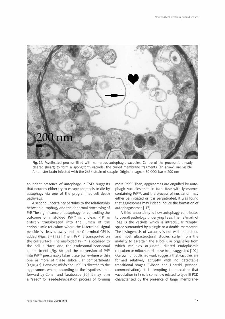
FFiigg.. 1144.. Myelinated process filled with numerous autophagic vacuoles. Centre of the process is alreadycleared (heart) to form a spongiform vacuole; the curled membrane fragments (an arrow) are visible. A hamster brain infected with the 263K strain of scrapie. Original magn. × 30 000; bar = 200 nm
abundant presence of autophagy in TSEs suggests
that neurons either try to escape apoptosis or die by
autophagy via one of the programmed-cell death
pathways.
A second uncertainty pertains to the relationship
between autophagy and the abnormal processing of
PrP. The significance of autophagy for controlling the
outcome of misfolded PrPTSE is unclear. PrPc is
entirely translocated into the lumen of the
endoplasmic reticulum where the N-terminal signal
peptide is cleaved away and the C-terminal GPI is
added (Figs. 3-4) [92]. Then, PrPc is transported on
the cell surface. The misfolded PrPTSE is localized to
the cell surface and the endosomal-lysosomal
compartment (Fig. 6); and the conversion of PrPc
into PrPTSE presumably takes place somewhere within
one or more of these subcellular compartments
[13,41,42]. However, misfolded PrPTSE is directed to the
aggresomes where, according to the hypothesis put
forward by Cohen and Taraboulos [50], it may form
a “seed” for seeded-nucleation process of forming
more PrPTSE. Then, aggresomes are engulfed by auto-
phagic vacuoles that, in turn, fuse with lysosomes
containing PrPTSE, and the process of nucleation may
either be initiated or it is perpetuated. It was found
that aggresomes may indeed induce the formation of
autophagosomes [117].
A third uncertainty is how autophagy contributes
to overall pathology underlying TSEs. The hallmark of
TSEs is the vacuole which is intracellular “empty”
space surrounded by a single or a double membrane.
The histogenesis of vacuoles is not well understood
and most ultrastructural studies suffer from the
inability to ascertain the subcellular organelles from
which vacuoles originate; dilated endoplasmic
reticulum or mitochondria have been suggested [102].
Our own unpublished work suggests that vacuoles are
formed relatively abruptly with no detectable
transitional stages [Gibson and Liberski, personal
communication]. It is tempting to speculate that
vacuolation in TSEs is somehow related to type III PCD
characterized by the presence of large, membrane-

Folia Neuropathologica 2008; 46/1 18
Paweł P. Liberski, David R. Brown, Beata Sikorska, Byron Caughey, Paul Brown
FFiigg.. 1155.. Two dystrophic neurites filled with numerous autophagic vacuoles (arrows) and lysosomal electron-dense bodies. A hamster brain infected with the 263K strain of scrapie. Original magn. × 30 000; bar = 200 nm
bound intracellular empty spaces without the
participation of lysosomes. The other option, that
tissue destruction by autophagy results in vacuolation
(Figs. 13, 14), would imply that autophagic vacuoles
should be detected prior to spongiform vacuoles.
Indeed, in the terminal stages of sheep scrapie
[M. Jeffrey – personal communication] and in human
TSEs (CJD, GSS and FFI) [144], little or no autophagy is
seen but robust vacuolation is typical if not
pathognomic. In contrast, when earlier stages of TSE
have been monitored in experimental rodent models,
both autophagy and vacuolation have been reported
[105,142,145]. Jeffrey et al. [105] have suggested that
their presence reflects the robust accumulation of
misfolded PrPTSE, overloading of the neuronal catabolic
machinery, and, eventually, bulk removal of damaged
neurons via autophagy.
If this scenario is correct, the pathology of TSEs is
akin to the reactivation of certain embryonic
processes in which bulk removal of cells are present;
i.e. remodelling of insect larvae [173]. Nearly 20 years
ago, Elizabeth Beck [8] suggested that robust
vacuolation like that characteristic of TSEs is present
when rats are inoculated with a suspension of cells
from normal immature cerebellum. Neurons of
inoculated rats not only demonstrated intracyto-
plasmic vacuoles but, at the ultrastructural level,
showed abundant coated pits and vesicles – pheno-
mena of widespread appearance in TSEs.
The other form of neuronal degeneration is
neuroaxonal dystrophy [77,131,133,134]. The ultrastruc-
tural correlate of NAD is the dystrophic neurite – an
axon or dendrite filled with electron-dense inclusions,
many of which were recently recognized as small
autophagic vacuoles (Fig. 15) [174]. Both immature
autophagic vacuoles immunogold-labelled with abs
against Cat-D and mature vacuoles containing
catepsin were observed within those neurites [174]. As
dystrophic neurites are abundant in TSEs [131,141], it is
plausible that (macro)autophagy plays a role in
neuronal degeneration in TSEs. However, taking into
account the evidence that autophagy may prevent

Folia Neuropathologica 2008; 46/1 19
apoptosis, it is also possible that abundant presence of
autophagic vacuoles within dystrophic neurites
actually reflects neurons struggling to survive in the
noxious environment of misfolded PrPTSE.
In summary, autophagy certainly does occur in
TSE, but its pathogenetic role as a cause of cell
death is uncertain. In particular, more research will
be necessary to determine the connection, if any,
between programmed cell death and the formation
of spongiform change.
AAcckknnoowwlleeddggeemmeennttss
This paper is supported in part by a Ministry of
Science and Higher Education grant and it is a part
of “NeuroPrion” Network of Excellence (project
leader: Prof. Herbert Budka). Ms. Lucyna Ciesielska,
Mr. Ryszard Kurczewski, Ms. Elzbieta Naganska,
Ms. Leokadia Romanska and Mr. Kazimierz Smoktuno-
wicz are kindly acknowledged for skilful technical
assistance.
RReeffeerreenncceess
1. Alper T, Cramp WA, Haig DA, Clarke MC. Does the agent of scrapiereplicate without nucleic acid? Nature 1967; 214: 764-766.
2. Anglade P, Vyas S, Javoy-Agid F, Herrero MT, Michel PP,Marquez J, Mouatt-Prigent A, Ruberg M, Hirsch EC, Agid Y.Apoptosis and autophagy in nigral neurons of patients withParkinson’s disease. Histol Histopathol 1997; 12: 25-31.
3. Baron GS, Magalhaes AC, Prado MA, Caughey B. Mouse-adapted scrapie infection of SN56 cells: greater efficiency withmicrosome-associated versus purified PrP-res. J Virol 2006; 80:2106-2117.
4. Baron GS, Wehrly K, Dorward DW, Chesebro B, Caughey B.Conversion of raft associated prion protein to the protease-resistant state requires insertion of PrP-res (PrP(Sc)) intocontiguous membranes. EMBO J 2002; 21: 1031-1040.
5. Bartz JC, Kincaid AE, Bessen RA. Retrograde transport oftransmissible mink encephalopathy within descending motortracts. J Virol 2002; 76: 5759-5768.
6. Bartz JC, Kincaid AE, Bessen RA. Rapid prion neuroinvasionfollowing tongue infection. J Virol 2003; 77: 583-591.
7. Basler K, Oesch B, Scott M, Westaway D, Wälchli M, Groth DF,McKinley MP, Prusiner SB, Weissmann C. Scrapie and cellularPrP isoforms are encoded by the same chromosomal gene. Cell 1986; 46: 417-428.
8. Beck E. Lesions akin to transmissible spongiform encephalo-pathy in the brains of rats inoculated with immaturecerebellum. Acta Neuropathol 1988; 76: 295-305.
9. Boellaard JW, Kao M, Schlote W, Diringer H. Neuronal autophagyin experimental scrapie. Acta Neuropathol 1991; 82: 225-228.
10. Bolton DC, McKinley MP, Prusiner SB. Identification of a proteinthat purifies with the scrapie prion. Science 1982; 218: 1309-1311.
11. Borchelt DR, Koliatsos VE, Guarnieri M, Pardo CA, Sisodia SS,Price DL. Rapid anterograde axonal transport of the cellular
prion glycoprotein in the peripheral and central nervoussystems. J Biol Chem 1994; 269: 14711-14714.
12. Borchelt DR, Scott M, Taraboulos A, Stahl N, Prusiner SB. Scrapieand cellular prion proteins differ in their kinetics of synthesisand topology in cultured cells. J Cell Biol 1990; 110: 743-752.
13. Borchelt DR, Taraboulos A, Prusiner SB. Evidence for synthesisof scrapie prion protein in the endocytic pathway. J Biol Chem1992; 267: 16188-16199.
14. Bounhar Y. Prion protein protects human neurons against Bax-mediated apoptosis. J Biol Chem 2001; 276: 39145-39149.
15. Brandner S, Isenmann S, Raeber A, Fischer M, Sailer A, Kobayashi Y,Marino S, Weissmann C, Aguzzi A. Normal host prion proteinnecessary for scrapie-induced neurotoxicity. Nature 1996; 379: 339-343.
16. Bratosiewicz-Wasik J, Wasik T, Liberski PP. Molecular approaches tomechanisms of prion diseases. Folia Neuropathol 2004; 42: 33-46.
17. Brown DR. Prion protein-overexpressing cells show alteredresponse to a neurotoxic prion protein peptide. J Neurosci Res1998; 54: 331-340.
18. Brown DR. Prion protein peptide neurotoxicity can be mediatedby astrocytes. J Neurochem 1999; 73: 1105-1113.
19. Brown DR. Prion protein peptides: optimal toxicity and peptideblockade of toxicity. Mol Cell Neurosci 2000; 15: 66-78.
20. Brown DR. PrPSc-like prion protein peptide inhibits the functionof cellular prion protein. Biochem J 2000; 352: 511-518.
21. Brown DR, Besinger A. Prion protein expression and superoxidedismutase activity. Biochem J 1998; 334: 423-429.
22. Brown DR, Besinger A, Herms JW, Kretzschmar HA. Microglialexpression of the prion protein. Neuroreport 1998; 9: 1425-1429.
23. Brown DR, Herms J, Kretzschmar HA. Mouse cortical cellslacking cellular PrP survive in culture with a neurotoxic PrPfragment. Neuroreport 1994; 5: 2057-2060.
24. Brown DR, Herms JW, Schmidt B, Kretzschmar HA. PrP and beta-amyloid fragments activate different neurotoxic mechanisms incultured mouse cells. Eur J Neurosci 1997; 9: 1162-1169.
25. Brown DR, Mohn CM. Astrocytic glutamate uptake and prionprotein expression. Glia 1999; 25: 282-292.
26. Brown DR, Nicholas RS, Canevari L. Lack of prion proteinexpression results in a neuronal phenotype sensitive to stress.J Neurosci Res 2002; 67: 211-224.
27. Brown DR, Schmidt B, Kretzschmar HA. Role of microglia andhost prion protein in neurotoxicity of a prion protein fragment.Nature 1996; 380: 345-347.
28. Brown DR, Schmidt B, Kretzschmar HA. A prion proteinfragment interacts with PrP-deficient cells. J Neurosci Res 1998;52: 260-267.
29. Brown DR, Schmidt B, Kretzschmar HA. Effects of copper onsurvival of prion protein knockout neurons and glia. J Neurochem1998; 70: 1686-1693.
30. Brown DR, Schultz-Schaeffer WJ, Schmidt B, Kretzschmar HA. Prionprotein-deficient cells show altered response to oxidative stressdue to decreased SOD-1 activity. Exp Neurol 1997; 146: 104-112.
31. Brown P. The phantasmagoric immunology of transmissiblespongiform encephalopathy. In: Waksman BH (ed.). ImmunologicMechanisms in Neurologic and Psychiatric Disease. Raven Press,Ltd, New York, 1990; pp. 305-313.
32. Brown P, Cervenáková L. A prion lexicon (out of control). Lancet2005; 365: 122.
Neuronal cell death in prion diseases

Folia Neuropathologica 2008; 46/1 20
33. Budka H, Head MW, Ironside JW, Gambetti P, Parchi P, ZeidlerM, Tagliavini F. Prion Disorders: CJD: Sporadic Creutzfeldt-Jakobdisease. In: Dickson D (ed.). Neurodegeneration: The MolecularPathology of Dementia and Movement Disorders. ISNNeuropath Press, Basel, 2003: pp. 287-297.
34. Bueler H, Fischer M, Lang Y, Bluethmann H, Lipp HP, DeArmondSJ, Prusiner SB, Aguet M, Weissmann C. Normal developmentand behaviour of mice lacking the neuronal cell-surface PrPprotein. Nature 1992; 356: 577-582.
35. Burger D, Hartsough GR. A scrapie-like disease of mink. In: Reportof a Scrapie Seminar. United States Department of Agriculture,paper 27, 1964; pp. 225-227.
36. Bursch W. The autophagosomal-lysosomal compartment inprogrammed cell death. Cell Death Differ 2001; 8: 569-581.
37. Bursch W. Multiple cell death programs: Charon’s lifts to Hades.FEMS Yeast Res 2004; 5: 101-110.
38. Bursch W, Ellinger A. Autophagy – a basic mechanism and a potential role for neurodegeneration. Folia Neuropathol 2005;43: 297-310.
39. Bursch W, Ellinger A, Gerner C, Schulte-Hermann R. Autophago-cytosis and programmed cell death. In: Klionsky D (ed.).Autophagy. Landes Bioscience, Georgetown, Texas, USA, 2004;pp. 290-306.
40. Bursch W, Hochegger K, Torok L, Marian B, Ellinger A, HermannRS. Autophagic and apoptotic types of programmed cell deathexhibit different fates of cytoskeletal filaments. J Cell Sci 2000;113: 1189-1198.
41. Caughey B, Raymond GJ. The scrapie-associated form of PrP ismade from a cell surface precursor that is both protease- andphospholipase-sensitive. J Biol Chem 1991; 266: 18217-18223.
42. Caughey B, Raymond GJ, Ernst D, Race RE. N-terminaltruncation of the scrapie-associated form of PrP by lysosomalprotease(s): implications regarding the site of conversion of PrPto the protease-resistant state. J Virol 1991; 65: 6597-6603.
43. Chen SG, Teplow DB, Parchi P, Teller JK, Gambetti P, Autilio-Gambetti L. Truncated forms of the human prion protein in normalbrain and in prion diseases. J Biol Chem 1995; 270: 19173-19180.
44. Chesebro B. Introduction to the transmissible spongiformencephalopathies or prion disease. Brit Med Bull 2003; 63: 1-20.
45. Chesebro B, Race R, Wehrly K, Nishio J, Bloom M, Lechner D,Bergstrom S, Robbins K, Mayer L, Keith JM, et al. Identificationof scrapie prion protein-specific mRNA in scrapie-infected anduninfected brain. Nature 1985; 315: 331-333.
46. Chesebro B, Trifilo M, Race R, Meade-White K, Teng C, LaCasse R,Raymond L, Favara C, Baron G, Priola S, Caughey B, Masliah E,Oldstone M. Anchorless prion protein results in infectious amyloiddisease without clinical scrapie. Science 2005; 308: 1435-1439.
47. Cho HJ. Requirement of a protein component for scrapieinfectivity. Intervirology 1980; 14: 213-216.
48. Choi SI, Ju WK, Choi EK, Kim J, Lea HZ, Carp RI, Wisniewski HM,Kim YS. Mitochondrial dysfunction induced by oxidative stressin the brains of hamsters infected with the 263 K scrapie agent.Acta Neuropathol 1998; 96: 279-286.
49. Clarke PG. Developmental cell death: morphological diversity andmultiple mechanisms. Anat Embryol (Berl) 1990; 181: 195-213.
50. Cohen E, Taraboulos A. Scrapie-like prion protein accumulatesin aggresomes of cyclosporin A-treated cells. EMBO J 2003; 22: 404-417.
51. Collinge J. Prion diseases of humans and animals: their causesand molecular basis. Annu Rev Neurosci 2001; 24: 519-550.
52. Corsaro A, Thellung S, Villa V, Principe DR, Paludi D, Arena S,Millo E, Schettini D, Damonte G, Aceto A, Schettini G, Florio T.Prion protein fragment 106-126 induces a p38 MAP kinase-dependent apoptosis in SH-SY5Y neuroblastoma cellsindependently from the amyloid fibril formation. Ann N Y AcadSci 2003; 1010: 610-622.
53. Cuille J, Chelle P-L. Pathologie animale – la maladie ditetremblante du mouton est-elle inoculable? Comptes rendus desSeances de l’Academie des Sciences (Paris) 1936; 203: 1552-1554.
54. Cuille J, Chelle P-L. La „tremblante” du mouton est bieninoculable. Comptes rendus des Seances de l’Academie desSciences (Paris) 1938; 206: 78-79.
55. Cunningham AA, Wells GAH, Scott AC, Kirkwood JK, Barnett JEF.Transmissible spongiform encephalopathy in greater kudu(Tragelaphus strepsiceros). Vet Rec 1993; 132: 68.
56. Deleault NR, Lucassen RW, Supattapone S. RNA moleculesstimulate prion protein conversion. Nature 2003; 425: 717-720.
57. Dickinson AG. Scrapie in sheep and goats. In: Kimberlin RH(ed.). Slow Virus Diseases of Animals and Man. North-HollandPubl Comp, Amsterdam, 1976; pp. 209-241.
58. Diringer H. Hidden amyloidoses. Exp Clin Immunogenet 1992;9: 212-229.
59. Diringer H, Gelderblom H, Hilmert H, Ozel M, Edelbluth C,Kimberlin RH. Scrapie infectivity, fibrils and low molecularweight protein. Nature 1983; 306: 476-478.
60. Dorandeu A, Wingertsmann L, Chretien F, Delisle M-B, Vital C,Parchi P, Montagna P, Lugaresi E, Ironside JW, Budka H, Gambetti P, Gray F. Neuronal apoptosis in fatal familial insomnia.Brain Pathol 1998; 8: 531-537.
61. Eskelinen EL. Doctor Jekyll and Mister Hyde: autophagy canpromote both cell survival and cell death. Cell Death Differ2005; 12 (Suppl 2): 1468-1472.
62. Fairbairn DW, Carnahan KG, Thwaits RN, Grigsby RV, HolyoakGR, O’Neill KL. Detection of apoptosis induced DNA cleavage inscrapie-infected sheep brain. FEMS Microbiol Lett 1994; 115: 341-346.
63. Ferrer I. Nuclear DNA fragmentation in Creutzfeldt-Jakobdisease: does a mere positive in situ nuclear end-labelingindicate apoptosis? Acta Neuropathol (Berl) 1994; 97: 5-12.
64. Fevrier B, Vilette D, Archer F, Loew D, Faigle W, Vidal M, Laude H,Raposo G. Cells release prions in association with exosomes.Proc Natl Acad Sci USA 2004; 101: 9683-9688.
65. Filonova LH, Bozhkov PV, Brukhin VB, Daniel G, Zhivotovsky B,von Arnold S. Two waves of programmed cell death occurduring formation and development of somatic embryos in thegymnosperm, Norway spruce. J Cell Sci 2000; 113: 4399-4411.
66. Fink SL, Cookson BT. Apoptosis, pyroptosis, and necrosis:mechanistic description of dead and dying eukaryotic cells.Infect Immun 2005; 73: 1907-1916.
67. Flechsig E, Weissmann C. The role of PrP in health and disease.Curr Mol Med 2004; 4: 337-353.
68. Fleetwood AJ, Furley CW. Spongiform encephalopathy in aneland. Vet Rec 1990; 126: 408-409.
69. Fraser JR. What is the basis of transmissible spongiformencephalopathy induced neurodegeneration and it berepaired? Neuropathol Appl Neurobiol 2002; 28: 1-11.
Paweł P. Liberski, David R. Brown, Beata Sikorska, Byron Caughey, Paul Brown

Folia Neuropathologica 2008; 46/1 21
70. Fraser JR, Halliday WG, Brown D, P Belichenko P, Jeffrey M.Mechanisms of scrapie-induced neuronal cell death. In: Court L, Dodet B (eds.). Transmissible subacute spongiformencephalopathies: prion disease. IIIrd International Symposiumon transmissible subacute spongiform encephalopathies: priondisease, Val-de-Grace Paris, France. Elsevier, Amsterdam-Oxford-Paris, 1996; pp. 107-112.
71. Gabriel JM, Oesch B, Kretzschmar H, Scott M, Prusiner SB.Molecular cloning of a candidate chicken prion protein. ProcNatl Acad Sci USA 1992; 89: 9097-9101.
72. Gajdusek DC. Unconventional viruses and the origin anddisappearance of kuru. Science 1977; 197: 943-960.
73. Gajdusek DC, Gibbs CJ, Alpers M. Experimental transmission of a kuru-like syndrome to chimpanzees. Nature 1966; 209: 794-796.
74. Garcia-Mata R, Gao YS, Sztul E. Hassles with taking out thegarbage: aggravating aggresomes. Traffic 2002; 3: 388-396.
75. Gibbons RA, Hunter GD. Nature of the scrapie agent. Nature1967; 215: 1041-1043.
76. Gibbs CJ Jr, Gajdusek DC, Asher DM, Alpers MP, Beck E, DanielPM, Matthews WB. Creutzfeldt-Jakob disease (spongiformencephalopathy): transmission to chimpanzee. Science 1968;161: 388-389.
77. Gibson PH, Liberski PP. An electron and light microscopic studyof the numbers of dystrophic neurites and vacuoles in thehippocampus of mice infected intracerebrally with scrapie. ActaNeuropathol 1987; 73: 379-382.
78. Giese A, Brown DR, Groschup MH, Feldmann C, Haist I,Kretzschmar HA. Role of microglia in neuronal cell death inprion disease. Brain Pathol 1998; 8: 449-457.
79. Giese A, Groschup MH, Hess B, Kretzschmar HA. Neuronal celldeath in scrapie-infected mice is due to apoptosis. Brain Pathol1995; 5: 213-221.
80. Giese A, Kretzschmar HA. Prion-induced neuronal damage – themechanisms of neuronal destruction in the subacutespongiform encephalopathies. Curr Top Microbiol Immunol2001; 253: 203-217.
81. Glatzel M, Giger O, Braun N, Aguzzi A. The peripheral nervoussystem and the pathogenesis of prion diseases. Curr Mol Med2004; 4: 355-359.
82. Gonzalez-Polo RA, Boya P, Pauleau AL, Jalil A, Larochette N,Souquere S, Eskelinen EL, Pierron G, Saftig P, Kroemer G. Theapoptosis/autophagy paradox: autophagic vacuolization beforeapoptotic death. J Cell Sci 2005; 118: 3091-3102.
83. Graeber MB, Moran LB. Mechanisms of cell death inneurodegenerative diseases: fashion, fiction, and facts. BrainPathol 2002; 12: 385-390.
84. Gray F, Chretien F, Adle-Biassette H, Dorandeu A, Ereau T,Delisle M-B, Kopp N, Ironside JW, Vital C. Neuronal apoptosis inCreutzfeldt-Jakob disease. J Neuropathol Exp Neurol 1999; 58: 321-328.
85. Griffith JS. Self-replication and scrapie. Nature 1967; 215: 1043-1044.86. Guan Z, Soderberg M, Sindelar P, Prusiner SB, Kristensson K,
Dallner G. Lipid composition in scrapie-infected mouse brain:prion infection increases the levels of dolichyl phosphate andubiquinone. J Neurochem 1996; 66: 277-285.
87. Guentchev M, Voigtländer T, Haberler C, Groschup MH, BudkaH. Evidence for oxidative stress in experimental prion disease.Neurobiol Dis 2000; 7: 270-273.
88. Hafiz FB, Brown DR. A model for the mechanism of astrogliosisin prion disease. Mol Cell Neurosci 2000; 16: 221-232.
89. Hainfellner H, Liberski PP, Guiroy DC, Cervenakova L, Brown P,Gajdusek DC, Budka H. Pathology and immunohistochemistryof a Kuru brain. Brain Pathol 1997; 7: 547-554.
90. Hardy J, Scholz S, Evans W, Goldfarb L, Singleton A. Priongenotypes in Central America suggest selection for the V129allele. Am J Med Genet B Neuropsychiatr Genet 2006; 141: 33-35.
91. Hariri M, Millane G, Guimond M-P, Dennis JW, Nabi IR.Biogenesis of multilamellar bodies via autophagy. Mol Biol Cell2000; 11: 255-268.
92. Harris DA. Trafficking, turnover and membrane topology of PrP.Br Med Bull 2003; 66: 71-85.
93. Harris DA, Falls DL, Johnson FA, Fischbach GD. A prion-like proteinfrom chicken brain copurifies with an acetylcholine receptor-inducing activity. Proc Natl Acad Sci USA 1991; 88: 7664-7668.
94. Hedge RS, Mastrianni JA, Scott MR, DeFea KA, Tremblay P, TorchiaM, DeArmond SJ, Prusiner SB, Lingappa VR. A transmembrane formof the prion protein in neurodegenerative disease. Science 1998;279: 827-834.
95. Hegde RS, Voigt S, Lingappa VR. Regulation of protein topologyby trans-acting factors at the endoplasmic reticulum. Mol Cell1998; 2: 85-91.
96. Hetz C, Russelakis-Carneiro M, Maundrell K, Castilla J, Soto C.Caspase-12 and endoplasmic reticulum stress mediateneurotoxicity of pathological prion protein. EMBO J 2003; 22: 5435-5445.
97. Hijazi N, Kariv-Inbal Z, Gasset M, Gabizon R. PrPScincorporation to cells requires endogenous glycosaminoglycanexpression. J Biol Chem 2005; 280: 17057-17061.
98. Horonchik L, Tzaban S, Ben Zaken O, Yedidia Y, Rouvinski A,Papy-Garcia D, Barritault D, Vlodavsky I, Taraboulos A.Heparan sulfate is a cellular receptor for purified infectiousprions. J Biol Chem 2005; 280: 17062-17067.
99. Inbal B, Bialik S, Sabanay I, Shani G, Kimchi A. DAP kinase andDRP-1 mediate membrane blebbing and the formation ofautophagic vesicles during programmed cell death. J Cell Biol2002; 157: 455-468.
100. Jamieson E, Jeffrey M, Ironside JW, Fraser JR. Apoptosis anddendritic dysfunction precede prion protein accumulation in87V scrapie. Neuroreport 2001; 12: 2147-2153.
101. Jeffrey M, Fraser JR, Halliday WG, Fowler N, Goodsir CM, BrownDA. Early unsuspected neuron and axon terminal loss in scrapie-infected mice revealed by morphometry and immunohisto-chemistry. Neuropathol Appl Neurobiol 1995; 21: 41-49.
102. Jeffrey M, Goodbrand IA, Goodsir A. Pathology of thetransmissible spongiform encephalopathies with specialemphasis on ultrastructure. Micron 1995; 26: 277-298.
103. Jeffrey M, Goodsir CM, Race RE, Chesebro B. Scrapie-specificneuronal lesions are independent of neuronal PrP expression.Ann Neurol 2004; 55: 781-792.
104. Jeffrey M, McGovern G, Goodsir CM, Brown KL, Bruce ME. Sitesof prion protein accumulation in scrapie-infected mousespleen revealed by immuno-electron microscopy. J Pathol2000; 191: 323-332.
105. Jeffrey M, Scott JR, Williams A, Fraser H. Ultrastructuralfeatures of spongiform encephalopathy transmitted to micefrom three species of bovidae. Acta Neuropathol 1992; 84:559-569.
Neuronal cell death in prion diseases
Folia Neuropathologica 2008; 46/1 22
106. Jeffrey M, Wells GA. Spongiform encephalopathy in a Nyala.
Vet Pathol 1988; 25: 398-399.
107. Jesionek-Kupnicka D, Buczynski J, Kordek R, Liberski PP.
Neuronal loss and apoptosis in experimental Creutzfeldt-
Jakob disease in mice. Folia Neuropathol 1999; 37: 283-286.
108. Jesionek-Kupnicka D, Buczynski J, Kordek R, Sobow T,
Kloszewska I, Papierz W, Liberski PP. Programmed cell death
(apoptosis) in Alzheimer’s disease and Creutzfeldt-Jakob
disease. Folia Neuropathol 1997; 35: 233-235.
109. Jesionek-Kupnicka D, Kordek R, Buczynski J, Liberski PP.
Apoptosis in relation to neuronal loss in experimental
Creutzfeldt-Jakob disease in mice. Acta Neurobiol Exp (Wars)
2001; 61: 13-19.
110. Kegel KB, Kim M, Sapp E, McIntyre C, Castano JG, Aronin N,
DiFiglia M. Huntingtin expression stimulates endosomal-
lysosomal activity, endosome tubulation, and autophagy.
J Neurosci 2000; 20: 7268-7278.
111. Kerr JF, Wyllie AH, Currie AR. Apoptosis: a basic biological
phenomenon with wide-ranging implications in tissue kinetics.
Br J Cancer 1972; 26: 239-257.
112. Kim JI, Ju WK, Choi JH, Choi E, Carp RI, Wisniewski HM, Kim YS.
Expression of cytokine genes and increased nuclear factor-
kappa B activity in the brains of scrapie-infected mice. Brain
Res Mol Brain Res 1999; 73: 17-27.
113. Kimberlin RH. Scrapie and possible relationships with viroids.
Sem Virol 1999; 1: 153-162.
114. Kimberlin RH, Field HJ, Walker CA. Pathogenesis of mouse
scrapie: evidence for spread of infection from central to
peripheral nervous system. J Gen Virol 1983; 64: 713-716.
115. Kirkwood JK, Wells GAH, Cunningham AA, Jackson SI, Scott AC,
Dawson M, Wilesmith JW. Scrapie-like encephalopathy in
a greater kudu (Tragelaphus strepsiceros) which had not been
fed ruminant-derived protein. Vet Res 1992; 130: 365-367.
116. Klionsky DJ, Emr SD. Autophagy as a regulated pathway of
cellular degradation. Science 2000; 290: 1717-1721.
117. Kopito RR. Aggresomes, inclusion bodies and protein
aggregation. Trends Cell Biol 2000; 10: 524-530.
118. Kovacs GG, Preusser M, Strohschneider M, Budka H.
Subcellular localization of disease-associated prion protein in
the human brain. Am J Pathol 2005; 166: 287-294.
119. Kretzschmar HA, Giese A, Brown DR, Herms J, Keller B, Schmidt
B, Groschup MH. Cell death in prion disease. J Neural Transm
Suppl 1997; 50: 191-210.
120. Kretzschmar HA, Giese A, Brown DR, Herms J, Schmidt B,
Groschup MH. Cell death in prion disease. In: Court L, Dodet B
(eds.). Transmissible Subacute Spongiform Encephalopathies:
Prion Disease. IIIrd International Symposium on transmissible
subacute spongiform encephalopathies: prion disease. Val-de-
Grace, Paris, France. Elsevier, Amsterdam-Oxford-Paris, 1996;
pp. 97-106.
121. Kretzschmar HA, Giese A, Herms J, Brown DR. Neuronal
degeneration and cell death in prion disease. In: Morrison DR
(ed.). Prions and Brain Diseases in Animals and Humans.
Plenum Press, New York, 1998; pp. 253-268.
122. Kristiansen M, Messenger MJ, Klöhn PC, Brandner S,
Wadsworth JD, Collinge J, Tabrizi SJ. Disease-related prion
protein forms aggresomes in neuronal cells leading to caspaseactivation and apoptosis. J Biol Chem 2005; 280: 38851-38861.
123. Kroemer G, El-Deiry WS, Golstein P, Peter ME, Vaux D,Vandenabeele P, Zhivotovsky B, Blagosklonny MV, Malorni W,Knight RA, Piacentini M, Nagata S, Melino G. Classification ofcell death: recommendations of the Nomenclature Committeeon Cell Death. Cell Death Differ 2005; 12 (Suppl 2): 1463-1467.
124. Kroemer G, Jaattela M. Lysosomes and autophagy in cell deathcontrol. Nat Rev Cancer 2005; 5: 886-897.
125. Künzi V, Glatzel M, Nakano MY, Greber UF, Van Leuven F,Aguzzi A. Unhampered prion neuroinvasion despite impairedfast axonal transport in transgenic mice overexpressing four-repeat tau. J Neurosci 2002; 22: 7471-7477.
126. Kuwahara C, Takeuchi AM, Nishimura T, Haraguchi K, Kubosaki A,Matsumoto Y, Saeki K, Matsumoto Y, Yokoyama T, Itohara S,Onodera T. Prions prevent neuronal cell-line death. Nature1999; 400: 225-226.
127. Lasmezas CI, Deslys JP, Robain O, Jaegly A, Beringue V, PeyrinJM, Fournier JG, Hauw JJ, Rossier J, Dormont D. Transmission ofthe BSE agent to mice in the absence of detectable abnormalprion protein. Science 1997; 275: 402-405.
128. Lee DW, Sohn HO, Lim HB, Lee YG, Kim Y-S, Carp RI, WisniewskiHM. Alteration of free radical metabolism in the brain of miceinfected with scrapie agent. Free Radic Res 1999; 30: 499-507.
129. Lee HP, Jun YC, Choi JK, Kim JI, Carp RI, Kim YS. Activation ofmitogen-activated protein kinases in hamster brains infectedwith 263K scrapie agent. J Neurochem 2005; 95: 584-593.
130. Levine P. Scrapie: an infective polypeptide? Lancet 1972; 1: 748.131. Liberski PP. Electron microscopic observations on dystrophic
neurites in hamster brains infected with the 263K strain ofscrapie. J Comp Pathol 1987; 97: 35-39.
132. Liberski PP, Asher DM, Yanagihara R, Gibbs CJ Jr, Gajdusek DC.Serial ultrastructural studies of scrapie in hamsters. J CompPathol 1989; 101: 429-442.
133. Liberski PP, Budka H. Neuroaxonal pathology in Creutzfeldt-Jakob disease. Acta Neuropathol 1999; 97: 329-334.
134. Liberski PP, Budka H, Yanagihara R, Gajdusek DC. Neuroaxonaldystrophy in experimental Creutzfeldt-Jakob disease: electronmicroscopical and immunohistochemical demonstration ofneurofilament accumulations within affected neurites. J CompPathol 1995; 112: 243-255.
135. Liberski PP, Gajdusek DC. Kuru: forty years later. Brain Pathol1997; 7: 555-560.
136. Liberski PP, Gajdusek DC, Brown P. How do neurons degeneratein prion diseases or transmissible spongiform encephalopathies(TSEs): neuronal autophagy revisited. Acta Neurobiol Exp (Wars)2002; 62: 141-147.
137. Liberski PP, Guiroy DC, Williams ES, Walis A, Budka H.Deposition patterns of disease-associated prion protein incaptive mule deer brains with chronic wasting disease. ActaNeuropathol 2001; 102: 496-500.
138. Liberski PP, Ironside JW. An outline of the neuropathology oftransmissible spongiform encephalopathies (prion diseases).Folia Neuropathol 2004; 42 (Suppl B): 39-58.
139. Liberski PP, Jaskolski M. Prion diseases: a dual view of theprion hypothesis as seen from a distance. Acta Neurobiol Exp(Wars) 2002; 62: 197-224.
Paweł P. Liberski, David R. Brown, Beata Sikorska, Byron Caughey, Paul Brown
Folia Neuropathologica 2008; 46/1 23
140. Liberski PP, Jeffrey M. Tubulovesicular structures – theultrastructural hallmark for transmissible spongiformencephalopathies or prion diseases. Folia Neuropathol 2004;42 (Suppl B): 96-108.
141. Liberski PP, Kloszewska I, Boellaard I, Papierz W, Omulecka A,Budka H. Dystrophic neurites of Alzheimer’s disease andGerstmann-Sträussler-Scheinker’s disease dissociate from theformation of paired helical filaments. Alzheimer’s Res 1995; 1: 89-93.
142. Liberski PP, Mori S. The Echigo-1: a panencephalopathic strainof Creutzfeldt-Jakob disease: a passage to hamsters andultrastructural studies. Folia Neuropathol 1997; 35: 250-254.
143. Liberski PP, Sikorska B, Bratosiewicz-Wasik J, Gajdusek DC,Brown P. Neuronal cell death in transmissible spongiformencephalopathies (prion diseases) revisited: from apoptosis toautophagy. Int J Biochem Cell Biol 2004; 36: 2473-2490.
144. Liberski PP, Streichenberger N, Giraud P, Soutrenon M,Meyronnet D, Sikorska B, Kopp N. Ultrastructural pathology ofprion diseases revisited: brain biopsy studies. NeuropatholAppl Neurobiol 2005; 31: 88-96.
145. Liberski PP, Yanagihara R, Gibbs CJ Jr, Gajdusek DC. Neuronalautophagic vacuoles in experimental scrapie and Creutzfeldt-Jakob disease. Acta Neuropathol 1992; 83: 134-139.
146. Lucassen PJ, Williams A, Chung WC, Fraser H. Detection ofapoptosis in murine scrapie. Neurosci Lett 1995; 198: 185-188.
147. Lugaresi E, Medori R, Montagna P, Baruzzi A, Cortelli P,Lugaresi A, Tinuper P, Zucconi M, Gambetti P. Fatal familialinsomnia and dysautonomia with selective degeneration ofthalamic nuclei. New Engl J Med 1986; 315: 997-1003.
148. Lum JJ, Bauer DE, Kong M, Harris MH, Li C, Lindsten T,Thompson CB. Growth factor regulation of autophagy and cellsurvival in the absence of apoptosis. Cell 2005; 120: 237-248.
149. Magalhaes AC, Baron GS, Lee KS, Steele-Mortimer O, Dorward D,Prado MA, Caughey B. Uptake and neuritic transport of scrapieprion protein coincident with infection of neuronal cells. J Neurosci 2005; 25: 5207-5216.
150. Magalhaes AC, Silva JA, Lee KS, Martins VR, Prado VF, Ferguson SS,Gomez MV, Brentani RR, Prado MA. Endocytic intermediatesinvolved with the intracellular trafficking of a fluorescent cellularprion protein. J Biol Chem 2002; 277: 33311-33318.
151. Mallucci G, Dickinson A, Linehan J, Klohn PC, Brandner S,Collinge J. Depleting neuronal PrP in prion infection preventsdisease and reverses spongiosis. Science 2003; 302: 871-874.
152. Manuelidis L. A virus behind the mask of prions? FoliaNeuropathol 2004; 42 (Suppl B): 10-23.
153. Manuelidis L. A 25 nm virion is the likely cause of transmissiblespongiform encephalopathies. J Cell Biochem 2007; 100: 897-915.
154. Manuelidis L, Sklaviadis T, Manuelidis EE. Evidence suggestingthat PrP is not the infectious agent in Creutzfeldt-Jakob disease.EMBO J 1987; 6: 341-347.
155. Marella M, Chabry J. Neurons and astrocytes respond to prioninfection by inducing microglia recruitment. J Neurosci 2004;24: 620-627.
156. Marella M, Gaggioli C, Batoz M, Deckert M, Tartare-Deckert S,Chabry J. Pathological prion protein exposure switches onneuronal mitogen-activated protein kinase pathway resultingin microglia recruitment. J Biol Chem 2005; 280: 1529-1534.
157. Masters CL, Gajdusek DC, Gibbs CJ Jr. Creutzfeldt-Jakob disease
virus isolations from the Gerstmann-Sträussler syndrome.
With an analysis of the various forms of amyloid plaque
deposition in the virus induced spongiform encephalopathies.
Brain 1981; 104: 559-588.
158. Matsumura H, Shimizu Y, Ohsawa Y, Kawahara A, Uchiyama Y,
Nagata S. Necrotic death pathway in Fas receptor signaling.
J Cell Biol 2000; 151: 1247-1256.
159. McBride PA, Schulz-Schaeffer WJ, Donaldson M, Bruce M,
Diringer H, Kretzschmar HA, Beekes M. Early spread of scrapie
from the gastrointestinal tract to the central nervous system
involves autonomic fibers of the splanchnic and vagus nerves.
J Virol 2001; 75: 9320-9327.160. McHattie SJ, Brown DR, Bird MM. Cellular uptake of the prion
protein fragment PrP106-126 in vitro. J Neurocytol 1999; 28:149-159.
161. McKinley MP, Bolton DC, Prusiner SB. A protease resistantprotein is a structural component of the scrapie prion. Cell1983; 35: 57-62.
162. Melino G, Knight RA, Nicotera P. How many ways to die? Howmany different models of cell death? Cell Death Differ 2005; 12(Suppl 2): 1457-1462.
163. Merz PA, Rohwer RG, Kascsak R, Wisniewski HM, Somerville RA,Gibbs CJ Jr, Gajdusek DC. Infection specific particle from theunconventional slow virus diseases. Science 1984; 225: 437-440.
164. Merz PA, Somerville RA, Wisniewski HM, Iqbal K. Abnormal fibrilsfrom scrapie-infected brain. Acta Neuropathol 1981; 54: 63-74.
165. Migheli A, Attanasio A, Lee WH, Bayer SA, Ghetti B. Detection ofapoptosis in weaver cerebellum by electron microscopic in situend-labeling of fragmented DNA. Neurosci Lett 1995; 199: 53-56.
166. Millson GC, Hunter GD, Kimberlin RH. The physico-chemicalnature of the scrapie agent. In: Kimberlin RH (ed.). Slow VirusDiseases of Animals and Man. North-Holland Publ Comp,Amsterdam, 1976; pp. 243-266.
167. Mishra RS, Basu S, Gu Y, Luo X, Zou WQ, Mishra R, Li R, ChenSG, Gambetti P, Fujioka H, Singh N. Protease-resistant humanprion protein and ferritin are cotransported across Caco-2epithelial cells: implications for species barrier in prion uptakefrom the intestine. J Neurosci 2004; 24: 11280-11290.
168. Moran LB, Kosel S, Spitzer C, Schwaiger FW, Riess O,Kreutzberg GW, Graeber MB. Expression of alpha-synuclein innon-apoptotic, slowly degenerating facial motoneurones. J Neurocytol 2001; 30: 515-521.
169. Morel E, Andrieu T, Casagrande F, Gauczynski S, Weiss S,Grassi J, Rousset M, Dormont D, Chambaz J. Bovine prion isendocytosed by human enterocytes via the 37 kDa/67 kDalaminin receptor. Am J Pathol 2005; 167: 1033-1042.
170. Moya KL, Hässig R, Breen KC, Volland H, Di Giamberardino L.Axonal transport of the cellular prion protein is increasedduring axon regeneration. J Neurochem 2005; 92: 1044-1053.
171. Moya KL, Hässig R, Créminon C, Laffont I, Di Giamberardino L.Enhanced detection and retrograde axonal transport of PrPc inperipheral nerve. J Neurochem 2004; 88: 155-160.
172. Muller WE, Ushijima H, Schroder HC, Forrest JM, Schatton WF,Rytik PG, Heffner-Lauc M. Cytoprotective effect of NMDAreceptor antagonists on prion protein (PrionSc)-induced toxicityin rat cortical cell cultures. Eur J Pharmacol 1993; 246: 261-267.
173. Myohara M. Real-time observation of autophagic programmedcell death of Drosophila salivary glands in vitro. Dev GenesEvol 2004; 214: 99-104.
Neuronal cell death in prion diseases

Folia Neuropathologica 2008; 46/1 24
174. Nixon RA, Wegiel J, Kumar A, Yu WH, Peterhoff C, Cataldo A,Cuervo AM. Extensive involvement of autophagy in Alzheimerdisease: an immuno-electron microscopy study. J NeuropatholExp Neurol 2005; 64: 113-122.
175. O’Donovan CN, Tobin D, Cotter TG. Prion protein fragment PrP-(106-126) induces apoptosis via mitochondrial disruption inhuman neuronal SH-SY5Y cells. J Biol Chem 2001; 276: 43516-43523.
176. Oesch B, Westaway D, Wälchli M, McKinley MP, Kent SB,Aebersold R, Barry RA, Tempst P, Teplow DB, Hood LE, et al. A cellular gene encodes scrapie PrP 27-30 protein. Cell 1985;40: 735-746.
177. Oidtmann B, Simon D, Holtkamp N, Hoffmann R, Baier M.Identification of cDNAs from Japanese pufferfish (Fugu rubripes)and Atlantic salmon (Salmo salar) coding for homologues totetrapod prion proteins. FEBS Lett 2003; 538: 96-100.
178. Peretz D, Williamson RA, Kaneko K, Vergara J, Leclerc E,Schmitt-Ulms G, Mehlhorn IR, Legname G, Wormald MR, RuddPM, Dwek RA, Burton DR, Prusiner SB. Antibodies inhibit prionpropagation and clear cell cultures of prion infectivity. Nature2001; 412: 739-743.
179. Pillot T, Drouet B, Pinçon-Raymond M, Vandekerckhove J,Rosseneu M, Chambaz J. A nonfibrillar from of the fusogenicprion protein fragment (118-135) induces apoptotic cell deathin rat cortical neurons. J Neurochem 2000; 75: 2298-2308.
180. Prado MA, Alves-Silva J, Magalhaes AC, Prado VF, Linden R,Martins VR, Brentani RR. PrPc on the road: trafficking of thecellular prion protein. J Neurochem 2004; 88: 769-781.
181. Prusiner SB. Novel proteinaceous infection particles causescrapie. Science 1982; 216: 136-144.
182. Prusiner SB. Terminology. In: Prusiner SB, McKinley MP (eds.).Prions. Novel Infectious Pathogens Causing Scrapie andCreutzfeldt-Jakob Disease. Academic Press, New York, 1987;pp. 38-53.
183. Prusiner SB. Shattuck lecture – neurodegenerative diseasesand prions. N Engl J Med 2001; 344: 1516-1526.
184. Prusiner SB (ed.). Prion Biology and Diseases. University ofCalifornia, San Francisco, 2004.
185. Prusiner SB, McKinley MP, Bowman KA, Bolton DC, BendheimPE, Groth DF, Glenner GG. Scrapie prions aggregate to formamyloid-like birefringent rods. Cell 1983; 35: 349-358.
186. Race R, Oldstone M, Chesebro B. Entry versus blockade ofbrain infection following oral or intraperitoneal scrapieadministration: role of PrP expression in peripheral nerves andspleen. J Virol 2000; 74: 828-833.
187. Raeber AJ, Race RE, Brandner S, Priola SA, Sailer A, Bessen RA,Mucke L, Manson J, Aguzzi A, Oldstone MB, Weissmann C,Chesebro B. Astrocyte-specific expression of hamster prionprotein (PrP) renders PrP knockout mice susceptible tohamster scrapie. EMBO J 1997; 16: 6057-6065.
188. Raina AK, Zhu X, Smith MA. Alzheimer’s disease and the cellcycle. Acta Neurobiol Exp (Wars) 2004; 64: 107-112.
189. Reggiori F, Klionsky DJ. Autophagy in the eukaryotic cell.Eukaryot Cell 2002; 1: 11-21.
190. Rieger R, Edenhofer F, Lasmezas CI, Weiss S. The human 37-kDa laminin receptor precursor interacts with the prionprotein in eukaryotic cells. Nat Med 1997; 3: 1383-1388.
191. Saez-Valero J, Angeretti N, Forloni G. Caspase-3 activation bybeta-amyloid and prion protein peptides is independent fromtheir neurotoxic effect. Neurosci Lett 2000; 293: 207-210.
192. Sattler T, Mayer A. Cell-free reconstitution of microautophagicvacuole invagination and vesicle formation. J Cell Biol 2000;151: 529-538.
193. Schatzl HM, Da Costa M, Taylor L, Cohen FE, Prusiner SB. Prionprotein gene variation among primates. J Mol Biol 1995; 45:362-374.
194. Schatzl HM, Da Costa M, Taylor L, Cohen FE, Prusiner SB. Prionprotein gene variation among primates. J Mol Biol 1997; 265: 257.
195. Schatzl HM, Laszlo L, Holtzman DM, Tatzelt J, DeArmond SJ,Weiner RI, Mobley WC, Prusiner SB. A hypothalamic neuronalcell line persistently infected with scrapie prions exhibitsapoptosis. J Virol 1997; 71: 8821-8831.
196. Schulz-Schaeffer WJ, Tschoke S, Kranefuss N, Drose W, Hause-Reitner D, Giese A, Groschup MH, Kretzschmar HA. Theparaffin-embedded tissue blot detects PrP(Sc) early in theincubation time in prion diseases. Am J Pathol 2000; 156: 51-56.
197. Schweichel JU, Merker HJ. The morphology of various types ofcell death in prenatal tissues. Teratology 1973; 7: 253-266.
198. Shyng S-L, Huber MT, Harris DA. A prion protein cycles betweenthe cell surface and an endocytic compartment in culturedneuroblastoma cells. J Biol Chem 1993; 268: 15922-15928.
199. Sikorska B, Liberski PP, Giraud P, Kopp N, Brown P. Autophagyis a part of ultrastructural synaptic pathology in Creutzfeldt-Jakob disease: a brain biopsy study. Int J Biochem Cell Biol2004; 36: 2563-2573.
200. Simonic T, Duga S, Strumbo B, Asselta R. cDNA cloning ofturtle prion protein. FEBS Lett 2000; 469: 33-38.
201. Siso S, Puig B, Varea R, Vidal E, Acin C, Prinz M, Montrasio F,Badiola J, Aguzzi A, Pumarola M, Ferrer I. Abnormal synapticprotein expression and cell death in murine scrapie. ActaNeuropathol 2002; 103: 615-626.
202. Soldevila M, Andres AM, Ramirez-Soriano A, Marques-Bonet T,Calafell F, Navarro A, Bertranpetit J. The prion protein gene inhumans revisited: lessons from a worldwide resequencingstudy. Genome Res 2006; 16: 231-239.
203. Solforosi L, Criado JR, McGavern DB, Wirz S, Sanchez-AlavezM, Sugama S, DeGiorgio LA, Volpe BT, Wiseman E, Abalos G,Masliah E, Gilden D, Oldstone MB, Conti B, Williamson RA.Cross-linking cellular prion protein triggers neuronalapoptosis in vivo. Science 2004; 303: 1514-1516.
204. Sparkes RS, Simon M, Cohn VH, Fournier RE, Lem J, Klisak I,Heinzmann C, Blatt C, Lucero M, Mohandas T, et al. Assignmentof the human and mouse prion protein genes to homologouschromosomes. Proc Natl Acad Sci USA 1986; 83: 7358-7362.
205. Sperandio S, de Belle I, Bredesen DE. An alternative, nonapoptoticform of programmed cell death. Proc Natl Acad Sci USA 2000; 97: 14376-14381.
206. Stadelmann C, Deckwerth TL, Srinivasan A, Bancher C, BruckW, Jellinger K, Lassmann H. Activation of caspase-3 in singleneurons and autophagic granules of granulovacuolardegeneration in Alzheimer’s disease. Evidence for apoptoticcell death. Am J Pathol 1999; 155: 1459-1466.
207. Stahl N, Baldwin MA, Hecker R, Pan K-M, Burlingame AL,Prusiner SB. Glycosylinsoitol phospholipid anchors of thescrapie and cellular prion proteins contain sialic acid.Biochemistry 1992; 31: 5043-5053.
208. Stahl N, Borchelt DR, Prusiner SB. Differential release of cellularand scrapie prion proteins from cellular membranes byphosphatidylinositol-specific phospholipase C. Biochemistry1990; 29: 5405-5412.
Paweł P. Liberski, David R. Brown, Beata Sikorska, Byron Caughey, Paul Brown

Folia Neuropathologica 2008; 46/1 25
209. Strumbo B, Ronchi S, Bolis LC, Simonic T. Molecular cloning ofthe cDNA coding for Xenopus laevis prion. FEBS Lett 2001;508: 170-174.
210. Stumpf MP, Krakauer DC. Mapping the parameters of prion-induced neuropathology. Proc Natl Acad Sci USA 2000; 97: 10573-10577.
211. Tarabulos A, Jendroska K, Serban D, Yang S-L, DeArmond SJ,Prusiner SB (1992) Regional mapping of prion protein in brain.Proc Natl Acad Sci USA 89: 7620-7624.
212. Theil D, Fatzer R, Meyer R, Schobesberger M, Zurbriggen A,Vandevelde M. Nuclear DNA fragmentation and immunereactivity in bovine spongiform encephalopathy. J Comp Pathol1999; 121: 357-367.
213. Thellung S, Florio T, Villa V, Corsaro A, Arena S, Amico C,Robello M, Salmona M, Forloni G, Bugiani O, Tagliavini F,Schettini G. Apoptotic cell death and impairment of L-typevoltage-sensitive calcium channel activity in rat cerebellargranule cells treated with the prion protein fragment 106-126.Neurobiol Dis 2000; 7: 299-309.
214. van Rheede T, Smolenaars MM, Madsen O, de Jong WW.Molecular evolution of the mammalian prion protein. Mol BiolEvol 2003; 20: 111-121.
215. Wang C-W, Klionsky DJ. The molecular mechanism of autophagy.Mol Med 2003; 9: 65-76.
216. Weissmann C. A “unified theory” of prion propagation. Nature2000; 352: 679-683.
217. Weissmann C. The state of the prion. Nat Rev Microbiol 2004;2: 861-871.
218. Wells GA, Scott AC, Johnson CT, Gunning RF, Hancock RD, Jeffrey M, Dawson M, Bradley R. A novel progressive spongiformencephalopathy in cattle. Vet Rec 1987; 121: 419-420.
219. White AR, Collins SJ, Maher F, Jobling MF, Stewart LR, Thyer JM,Beyreuther K, Masters CL, Cappai R. Prion protein-deficientneurons reveal lower glutathione reductase activity andincreased susceptibility to hydrogen peroxide toxicity. Am J Pathol 1999; 155: 1723-1730.
220. Williams A, Lucassen PJ, Ritchie D, Bruce M. PrP deposition,microglial activation, and neuronal apoptosis in murinescrapie. Exp Neurol 1997; 144: 433-438.
221. Williams ES, Young S. Chronic wasting disease of captive muledeer: a spongiform encephalopathy. J Wildl Dis 1980; 16: 89-98.
222. Williams ES, Young S. Spongiform encephalopathy of RockyMountain elk. J Wildl Dis 1982; 18: 465-471.
223. Willoughby K, Kelly DF, Lyon DG, Wells GA. Spongiformencephalopathy in a captive puma (Felis concolor). Vet Rec1992; 131: 431-434.
224. Wood JL, Lund LJ, Done SH. The natural occurrence of scrapiein moufflon. Vet Rec 1992; 130: 25-27.
225. Wopfner F, Weidenhofer G, Schneider R, von Brunn A, Gilch S,Schwarz TF, Werner T, Schatzl HM. Analysis of 27 mammalianand 9 avian PrPs reveals high conservation of flexible regionsof the prion protein. J Mol Biol 1999; 289: 1163-1178.
226. Wyatt JM, Pearson GR, Smerdon TN, Gruffydd-Jones TJ, Wells GA,Wilesmith JW. Naturally occurring scrapie-like spongiformencephalopathy in five domestic cats. Vet Rec 1991; 129: 233-236.
227. Xue L, Fletcher GC, Tolkovsky AM. Autophagy is activated byapoptotic signalling in sympathetic neurons: an alternativemechanism of death execution. Mol Cell Neurosci 1999; 14: 180-198.
228. Yakovlev AG, Faden AI. Mechanisms of neural cell death:implications for development of neuroprotective treatmentstrategies. NeuroRx 2004; 1: 5-16.
229. Ye X, Scallet AC, Kascsak RJ, Carp RI. Astrocytosis and amyloiddeposition in scrapie-infected hamsters. Brain Res 1998; 809:277-287.
230. Yuan J, Lipinski M, Degterev A. Diversity of the mechanisms ofneuronal cell death. Neuron 2003; 40: 401-413.
231. Zanata SM, Lopes MH, Mercadante AF, Hajj GN, Chiarini LB,Nomizo R, Freitas AR, Cabral AL, Lee KS, Juliano MA, de OliveiraE, Jachieri SG, Burlingame A, Huang L, Linden R, Brentani RR,Martins VR. Stress-inducible protein 1 is a cell surface ligandfor cellular prion that triggers neuroprotection. EMBO J 2002;21: 3307-3316.
Neuronal cell death in prion diseases