cell-based screen for discovering lipopolysaccharide
TRANSCRIPT

Cell-based screen for discovering lipopolysaccharidebiogenesis inhibitorsGe Zhanga, Vadim Baidina, Karanbir S. Pahila, Eileen Moisona, David Tomaseka,b, Nitya S. Ramadossc,Arnab K. Chatterjeec, Case W. McNamarac, Travis S. Youngc, Peter G. Schultzc,1, Timothy C. Meredithd,e,1,and Daniel Kahnea,f,1
aDepartment of Chemistry and Chemical Biology, Harvard University, Cambridge, MA 02138; bDepartment of Molecular and Cellular Biology, HarvardUniversity, Cambridge, MA 02138; cCalifornia Institute for Biomedical Research, La Jolla, CA 92037; dDepartment of Microbiology and Immunobiology,Harvard Medical School, Boston, MA 02115; eDepartment of Biochemistry and Molecular Biology, Pennsylvania State University, University Park, PA 16802;and fDepartment of Biological Chemistry and Molecular Pharmacology, Harvard Medical School, Boston, MA 02115
Edited by Christopher T. Walsh, Stanford University, Stanford, CA, and approved April 19, 2018 (received for review March 16, 2018)
New drugs are needed to treat gram-negative bacterial infections.These bacteria are protected by an outer membrane whichprevents many antibiotics from reaching their cellular targets. Theouter leaflet of the outer membrane contains LPS, which isresponsible for creating this permeability barrier. Interfering withLPS biogenesis affects bacterial viability. We developed a cell-based screen that identifies inhibitors of LPS biosynthesis andtransport by exploiting the nonessentiality of this pathway in Aci-netobacter. We used this screen to find an inhibitor of MsbA, anATP-dependent flippase that translocates LPS across the innermembrane. Treatment with the inhibitor caused mislocalizationof LPS to the cell interior. The discovery of an MsbA inhibitor,which is universally conserved in all gram-negative bacteria, vali-dates MsbA as an antibacterial target. Because our cell-basedscreen reports on the function of the entire LPS biogenesis path-way, it could be used to identify compounds that inhibit othertargets in the pathway, which can provide insights into vulnera-bilities of the gram-negative cell envelope.
LPS biogenesis | ABC transporter | high-throughput screening |MsbA inhibitor | Acinetobacter
With the increase in antibiotic resistance, treatment ofbacterial infections has become a major unmet clinical
need today (1, 2). It is more difficult to kill gram-negative bac-teria than gram-positive bacteria because they contain a secondmembrane, the outer membrane, which prevents antibiotics fromreaching their targets inside the cell (3). This outer membrane isan asymmetric bilayer containing phospholipids in the innerleaflet and LPS in the outer leaflet (4, 5). LPS is a complexglycolipid containing a highly acylated diglucosamine (lipid A)that is connected to repeating sugars (O-antigen) through coreoligosaccharides (Fig. 1A). Adjacent LPS molecules at the cellsurface are stabilized by electrostatic interactions with divalentcations and strong lateral interactions between neighboring hy-drophobic acyl chains. This tight packing of LPS at the cellsurface makes the outer membrane an effective permeabilitybarrier, which makes gram-negative bacteria insensitive to manyantibiotics (6).The biogenesis of LPS, which includes its biosynthesis and
transport, involves hundreds of proteins that are spread acrossthree compartments of gram-negative bacteria (Fig. 1B). Thebulk of the LPS molecule is synthesized in the cytoplasm beforebeing translocated across the inner membrane by an ATP-dependent transporter (MsbA) (7, 8). The biosynthesis of LPSis completed at the outer leaflet of the inner membrane fol-lowing translocation, and subsequently LPS is extracted from theinner membrane and delivered to the cell surface (7, 9, 10). LPStransport to the outer membrane requires a seven-protein LPStransport machine (Lpt) that has been shown to form a trans-envelope complex to accomplish periplasmic transit and trans-location through the outer membrane (10–13). Strains with
mutations that impair LPS biogenesis either are not viable orhave a leaky outer membrane (14–16). Therefore, it has beenassumed that targeting this pathway may kill gram-negative bacteriaor overcome their intrinsic insensitivity to a broad range of antibi-otics. Since the proteins involved in LPS biogenesis are highlyconserved across gram-negative bacteria, the entire pathway rep-resents an attractive target for novel antibiotic development.Despite the considerable promise of and interest in developing
inhibitors against the LPS pathway (17–19), success has largelybeen confined to a single biosynthetic target, LpxC (20). LpxC isa zinc-dependent deacetylase that catalyzes the first committedstep of lipid A biosynthesis (7). LpxC is a soluble protein with
Significance
The outer membrane of gram-negative bacteria contains LPSon the cell surface. The presence of LPS creates an effectivepermeability barrier that protects gram-negative bacteria fromsmall hydrophobic molecules. Because the entire LPS bio-genesis pathway, including biosynthesis and transport, ishighly conserved, proteins involved are attractive targets forantibiotic discovery. Historically, it has been challenging totarget LPS biogenesis since many of the components aremembrane proteins with hard-to-assay activities. Utilizing thenonessentiality of this pathway in a gram-negative pathogen,we developed a cell-based screen specific to LPS biogenesis.We identified a small-molecule inhibitor targeting an essentialcomponent of the pathway, MsbA, and validated it as an anti-bacterial target using a combination of genetics, biochemistry,and cellular assays.
Author contributions: G.Z., V.B., T.C.M., and D.K. designed experiments; G.Z. and T.C.M.constructed mutant strains in A. baumannii; V.B. constructed and characterized all re-ported A. baylyi strains; G.Z. optimized the A. baumannii assay for high throughput withhelp of V.B.; G.Z. performed the high-throughput screen and hit confirmation; N.S.R.,A.K.C., C.W.M., T.S.Y., and P.G.S. analyzed the screening hits; G.Z. characterized drug–drug interactions, selected for lpx resistance mutations, and analyzed whole-genomesequences; V.B. developed the strategy to isolate on-target resistant mutants; G.Z. andV.B. selected for msbA resistance mutations and analyzed whole genome sequences; G.Z.,V.B., and K.S.P. measured MICs; G.Z. constructed complementation strains with helpfrom V.B.; V.B. generated E. coli strains reported in this work; D.T. developed the protocolfor expressing and purifying the MsbA protein; K.S.P. synthesized analogs and performedbiochemical reconstitution; E.M. optimized conditions with G.Z. for microscopy and per-formed fluorescent imaging; G.Z. prepared and submitted samples for TEM; T.C.M. andD.K. designed and supervised the project; G.Z., V.B., K.S.P., and D.K. wrote the manuscriptwith input for specific sections from E.M., P.G.S, T.S.Y., C.W.M., and T.C.M.
The authors declare no conflict of interest.
This article is a PNAS Direct Submission.
Published under the PNAS license.
See Commentary on page 6530.1To whom correspondence may be addressed. Email: [email protected], [email protected], or [email protected].
This article contains supporting information online at www.pnas.org/lookup/suppl/doi:10.1073/pnas.1804670115/-/DCSupplemental.
Published online May 7, 2018.
6834–6839 | PNAS | June 26, 2018 | vol. 115 | no. 26 www.pnas.org/cgi/doi/10.1073/pnas.1804670115
Dow
nloa
ded
by g
uest
on
Feb
ruar
y 26
, 202
2

easy-to-assay enzymatic activity, making it a readily tractabletarget for the design of new antibiotics. In addition to LpxC,there are many other proteins in the LPS biogenesis pathwaythat may be promising drug targets. However, many of these aremembrane proteins operating in complexes that transport LPSwithout chemically modifying it. This makes it difficult to assaytheir functions, which is necessary to develop and implementassays robust enough to interrogate large chemical libraries forinhibition in a high-throughput format. Moreover, since theproteins involved in LPS transport are believed to function asa single transenvelope complex, assays using isolated proteincomponents are limited to measuring binding affinity in the ab-sence of function. To look for inhibitors in this pathway, we reporthere the development of a high-throughput, cell-based screenwhich allows us to look at all of the targets (biosynthesis andtransport) in the LPS pathway in a simple growth-based assay.
ResultsValidating Conditional Essentiality of Certain LPS Biogenesis Steps inAcinetobacter. In many gram-negative organisms, the LPS path-way genes involved in biosynthesis and transport are essential(21). Recently it has been found that several gram-negativepathogens including Acinetobacter can survive without LPS(22–24). Paradoxically, certain steps of the LPS biogenesis path-way appear to remain essential in Acinetobacter despite the factthat LPS is not essential (25–27).We studied the essentiality of the LPS-pathway genes in
Acinetobacter baylyi, an organism that allows for highly efficientgenetic manipulation (28) (Fig. 1). Genes encoding LpxC, whichperforms the first committed step in LPS biogenesis, as wellas LptD/E, which carry out the final step of the pathway, could bereadily removed without loss of viability. Unlike the lpxC knock-out, which grew at the same rate as the WT, the lptD knockoutgrew more slowly and formed clumps under the microscope(SI Appendix, Fig. S1 A and B). However, these deficiencies
disappeared when we knocked out lpxC in the ΔlptD single-deletion background, suggesting that inactivation of LPS bio-synthesis resolves problems caused by blockage of LPS assemblyat a late stage. In contrast, we could not remove the 10 in-termediate genes (lpxH, lpxB, lpxK, waaA, msbA, lptB, lptF, lptG,lptC, and lptA) despite repeated attempts. However, deletions ofthese 10 genes succeeded in the ΔlpxC strain background. At-tempts to reintroduce LpxC on a plasmid into these doubleknockouts were not successful (SI Appendix, Fig. S1C). Takentogether, these data showed intermediate genes of LPS bio-genesis to be conditionally essential in A. baylyi, likely becausewhen they are inactivated there is toxic accumulation of LPSintermediates. We leveraged our findings to develop a screen inAcinetobacter baumannii, a species closely related to A. baylyi. A.baumannii is one of the ESKAPE pathogens and has developedmultidrug resistance in the clinic (29). We reasoned that small-molecule inhibitors of intermediate steps of the LPS biogenesispathway would behave similarly to genetic knockouts of thecorresponding genes and become nontoxic if flux into the path-way is abolished. Furthermore, because removal of the LPSpathway significantly sensitizes cells to small molecules in gen-eral, only inhibitors of a component of the pathway would beexpected to inhibit the growth of LPS+ strains in preference tomore permeable LPS− strains (Fig. 1C).
Establishing a Pathway-Directed Screen Based on ConditionalEssentiality. To assess the ability of such a screen to distinguishon-pathway compounds from off-pathway compounds, weconstructed an LPS-deficient strain by knocking out lpxC. Asexpected, removal of LPS made the bacterium much more sus-ceptible to a range of antibiotics relative to WT but conferred 32-fold resistance to colistin (30), which acts by binding to LPS(Table 1). We reasoned that such distinct responses could beutilized to establish a selective screen for discovering on-pathwayinhibitors, while eliminating off-pathway compounds. Specifically,
ALptBFGCIM complex
ADP+PiATP
LptA
LPS
MsbA
ADP+PiATP
LptDE OM translocon
IM
Periplasm
OM
UDP-GlcNAc
B B
F G
C
LpxC
Cor
e ol
igos
acch
arid
eLi
pid
A
Kdo
Kdo
Kdo
O-antigen
GlcN
GlcNAcA GalN
Glc
Glc
Glc
Glc
OH
HO OO
O
OO
NH
O
O
OOP
O
-O OHOO
O
HO
NHO
OP
O
O-
O
O
O
O
HO
D
E
A B
LpxDLpxHLpxBLpxKWaaA LpxA
CStrains
Compound category
Inactive On-pathway Off-pathway
LPS+ Growth No Growth Growth
LPS- Growth Growth No Growth
Fig. 1. Certain genes in the LPS biogenesis pathwayof Acinetobacter are conditionally essential. (A) LPSis a complex glycolipid and consists of hepta-acylatedlipid A (in Acinetobacter), core oligosaccharides, andO-antigen. (B) LPS biogenesis starts with biosynthesisin the cytoplasm and transport of LPS from the innermembrane to the outer membrane, followed by as-sembly at the cell surface. Nonessential genes initi-ating lipid A biosynthesis (lpxA/C/D, red) and at thefinal step of the transport process (lptD/E, red), alongwith conditionally essential intermediate steps (lpxH/B/K, waaA, msbA, and lptA/B/C/F/G, blue) are in-dicated. Genes (bold) were experimentally verified inA. baylyi (SI Appendix, Fig. S1). The specific acylationpattern and core sugar composition of LPS are basedon published reports (45, 46). O-antigen–relatedproteins are omitted for simplicity. Abbreviations:GalN, galactosamine; Glc, glucose; GlcN, glucosamine;GlcNAcA, 2-acetamido-2-deoxy-glucopyranosylur-onic acid; Kdo, 3-deoxy-D-manno-oct-2-ulopyranosonicacid. (C) LPS biogenesis inhibitors are distinguishedfrom off-pathway or inactive compounds based ondistinct sensitivities of strains with and without LPS.
Zhang et al. PNAS | June 26, 2018 | vol. 115 | no. 26 | 6835
MICRO
BIOLO
GY
SEECO
MMEN
TARY
Dow
nloa
ded
by g
uest
on
Feb
ruar
y 26
, 202
2

novel compounds targeting a conditionally essential step of LPSbiogenesis should inhibit growth of the WT strain at lowerconcentrations compared with the LPS-null strain. At the sametime, we expect loss of LPS to make off-pathway targets moreaccessible, allowing library compounds that target other path-ways to inhibit the LPS-null strain at lower concentrations thanthe WT strain.The outer membrane and efflux pumps of gram-negative
bacteria constitute a significant challenge in whole-cell screen-ing, which is particularly important when screening the broadrange of pharmacologically unoptimized library compounds thatare typically present in compound collections. To overcome thisproblem, we engineered a screening strain background by under-mining the permeability barrier of WT A. baumannii. Specifically,we removed three major efflux pumps that are known to causemultidrug resistance in A. baumannii (31–33), as well as two LPS-modifying enzymes: a secondary acyltransferase that we found tobe important for maintaining the barrier function (SI Appendix,Table S1) and a phosphoethanolamine transferase that conferscolistin resistance when activated (34–36). This allowed us to usecolistin as a positive control and to avoid cross-resistance tosimilarly acting hits. Next, we knocked out lpxA to inactivate LPSbiosynthesis. We found that ΔlpxA A. baumannii grows signifi-cantly more poorly in LB broth than its LPS-containing parentstrain, making it difficult to score hits by direct comparison ofgrowth profiles. Therefore, we evolved LPS-null A. baumanniiby serial passage in LB broth until its growth became comparableto that of WT (hereinafter Δ5 ΔlpxA). To eliminate the con-founding effect of adaptive mutations that arose during passaging,we reintroduced the WT lpxA allele into Δ5 ΔlpxA, producing theotherwise isogenic Δ5 strain. This allowed us to confidently com-pare the growth of the two strains in the presence of compounds.To ensure that our engineered background performs as
expected, we retested our panel of antibiotics against Δ5 and Δ5ΔlpxA and compared their sensitivities to those of parental WTand ΔlpxC (Table 1). The Δ5 strain was four- to eightfold moresensitive than the unmodified WT, consistent with the lack ofefflux and compromised permeability barrier. Nonetheless, ab-sence of LPS in the isogenic Δ5 ΔlpxA strain more stronglysensitized cells to these compounds, causing 23- to 100-fold in-creased susceptibility. However, these modifications did not changethe colistin sensitivity of our engineered screening strains; Δ5 is assensitive to colistin as WT and Δ5 ΔlpxA remained highly resistantto colistin. Therefore, we conclude that removing the efflux pumpsand LPS-modifying enzymes should increase the sensitivity of ourscreen to on-pathway compounds, allowing us to screen at lowconcentrations to minimize potential off-target toxic effects. In themeantime, our engineered screening strains should not erodethe ability of the assay to eliminate off-pathway compounds giventhe significantly impaired barrier function upon loss of LPS,while maintaining selectivity toward on-pathway compounds.
Identifying and Optimizing Positive Hits to Achieve Selectivity andPotency. We used our engineered background to screen 150,000commercially available compounds and bioactives against Δ5 (SI
Appendix, Table S2). The top 1,100 compounds that inhibitedΔ5 were retested in a counterscreen against Δ5 ΔlpxA. Thosecompounds (11 in total) that were better inhibitors of Δ5 than of Δ5ΔlpxA at a single concentration were tested in dose–response againstboth strains. We thereby identified a tetrahydrobenzothiophenescaffold that demonstrated both selectivity (inhibiting Δ5 24-foldbetter than Δ5 ΔlpxA) and potency (inhibiting Δ5 at 0.5 μM).We tested minimal inhibitory concentrations (MICs) of several
analogs of the scaffold against a strain panel (Fig. 2). Our bestcompound 1 displayed 24-fold on-pathway selectivity (0.5 μM and12 μM for Δ5 and Δ5 ΔlpxA, respectively). The difference inpreferential inhibition of the less-permeable Δ5 strain was en-couraging given that the actual differential is likely much higherwhen corrected for intracellular exposure levels. Compound 1 wasalso active against the parent WT strain. The lack of strong activityagainst this strain was due to efflux by AdeIJK, since AdeIJKremoval is sufficient to achieve the sensitization observed in Δ5.The ethyl ester of 1 (compound 2) had no antibacterial activity,suggesting that the carboxylic acid is important for target en-gagement. In contrast, a bulky, hydrophobic substituent (t-Bu) atthe 6-position of the tetrahydrobenzothiophene ring in compound3 reversed selectivity. Therefore, 6-tBu erodes on-pathway po-tency, while increasing off-pathway toxicity. This compound wasalso completely inactive against WT. When we installed a smallersubstituent at the 6-position (Me; compound 4), we found itmaintained selectivity and potency against all strains. Hence, rel-atively moderate changes on the scaffold can cause significantchanges in activity, suggesting that this scaffold has a specificcellular target and can be improved.We reasoned that if compound 1 targets a conditionally es-
sential step of the LPS pathway, then its antibacterial activityagainst Δ5 could be modulated in a dose-dependent mannerusing a small-molecule inhibitor of LpxC. Unsurprisingly, treat-ment of Δ5 with increasing doses of a known LpxC inhibitor (PF-5081090) (37) protected cells against the antibacterial effect of 1(SI Appendix, Fig. S2A). A similar pattern was found betweencolistin and the LpxC inhibitor (SI Appendix, Fig. S2B). Theseresults support the hypothesis that the antibacterial effect of 1 isdue to inhibition of a conditionally essential target in the LPSbiogenesis pathway.
Identifying Mutations That Inactivate the LPS Pathway to ConferResistance. We selected for mutants on plates containing com-pound 1 at 4 μM, which was significantly above the MIC of theΔ5 strain, but below the MIC of the Δ5 ΔlpxA strain to minimize
Table 1. Acinetobacter strains with or without LPS displaydistinct susceptibilities to known antibiotics
Strains Rifampicin Bacitracin Vancomycin Colistin
WT 0.36 (0.3) 75 (110) 86 (128) 1.6 (2)ΔlpxC 0.0003 (0.0002) 0.06 (0.09) 0.17 (0.25) 50 (64)Δ5 0.07 (0.06) 9 (13) 22 (32) 1.6 (2)Δ5 ΔlpxA 0.0007 (0.0006) 0.4 (0.6) 0.3 (0.5) 50 (64)
MICs are measured in micromolar or micrograms per milliliter (shown inparentheses).
Compound StructureMICs (μM; μg/ml in parentheses)
Δ5 Δ5 ΔlpxA WT ΔadeIJK
1 0.5(0.2)
12(4)
60(20)
0.5(0.2)
2 >300(>100)
>300(>100)
>300(>100)
>300(>100)
3 9.4(3.7)
0.6(0.2)
>300(>100)
19(7.4)
4 0.5(0.2)
5.5(1.9)
60(20)
1(0.3)
SNH
OCl
OHO
12
3456
7
SNH
OCl
OO
SNH
OCl
OHO
t-Bu
SNH
OCl
OHO
Fig. 2. Structure–activity relationship of compounds across four Acineto-bacter strains. MIC values in parentheses are in micrograms per milliliter.
6836 | www.pnas.org/cgi/doi/10.1073/pnas.1804670115 Zhang et al.
Dow
nloa
ded
by g
uest
on
Feb
ruar
y 26
, 202
2
off-target effects and favor mutations in the LPS biogenesispathway. Six resistant colonies (frequency of resistance ∼1 × 10⁻⁶)were isolated and analyzed by whole-genome sequencing.All contained mutations in the genes encoding LpxA, LpxC,or LpxD, the first three enzymes in lipid A biosynthesis (SIAppendix, Table S3). We found that MICs of the resistant mu-tants for colistin and rifampicin were in agreement with those ofLPS-deficient strains (Table 2). Expressing WT LpxA, LpxC, orLpxD in trans restored their sensitivity profile to that observed inΔ5 (Table 2). Therefore, we conclude that these mutationsconfer resistance to compound 1 by resulting in loss of functionin the pathway.Although these preliminary experiments validated the princi-
ple of the screen, they did not identify the specific target of ourcompound. Characterizations of additional resistant coloniesonly yielded more Lpx nulls. This is not unexpected since thereare many more possible mutations that remove the pathway thanthose that preserve the function of the pathway while generatingresistance in the target.
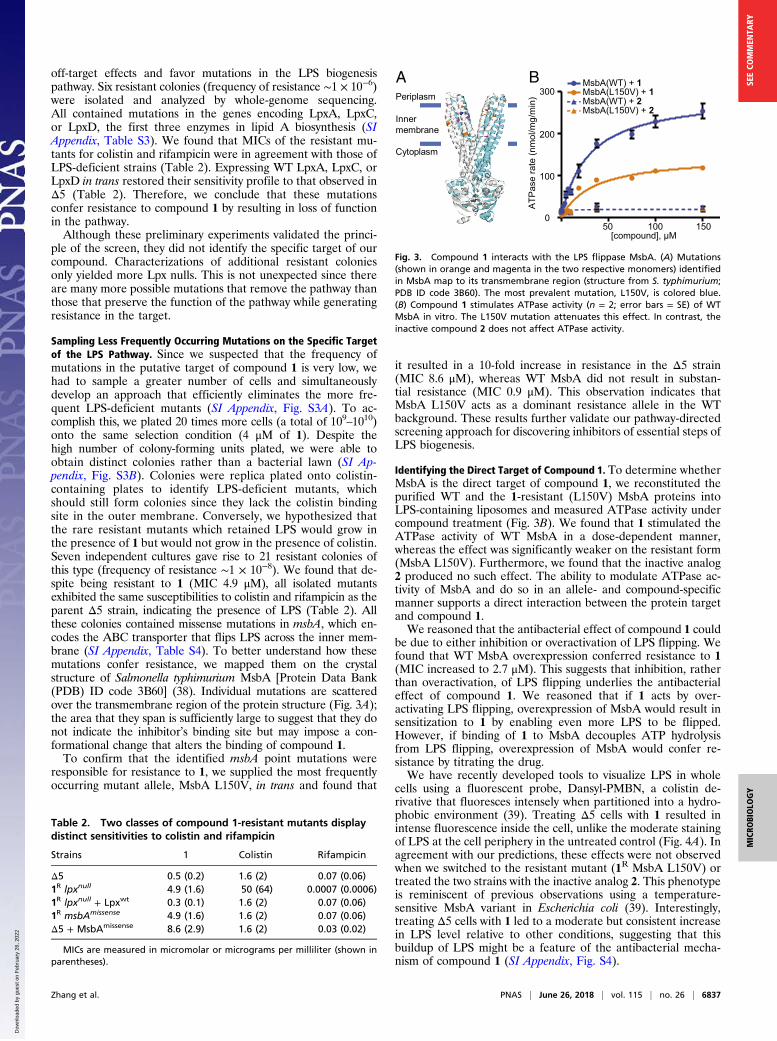
Sampling Less Frequently Occurring Mutations on the Specific Targetof the LPS Pathway. Since we suspected that the frequency ofmutations in the putative target of compound 1 is very low, wehad to sample a greater number of cells and simultaneouslydevelop an approach that efficiently eliminates the more fre-quent LPS-deficient mutants (SI Appendix, Fig. S3A). To ac-complish this, we plated 20 times more cells (a total of 109–1010)onto the same selection condition (4 μM of 1). Despite thehigh number of colony-forming units plated, we were able toobtain distinct colonies rather than a bacterial lawn (SI Ap-pendix, Fig. S3B). Colonies were replica plated onto colistin-containing plates to identify LPS-deficient mutants, whichshould still form colonies since they lack the colistin bindingsite in the outer membrane. Conversely, we hypothesized thatthe rare resistant mutants which retained LPS would grow inthe presence of 1 but would not grow in the presence of colistin.Seven independent cultures gave rise to 21 resistant colonies ofthis type (frequency of resistance ∼1 × 10⁻⁸). We found that de-spite being resistant to 1 (MIC 4.9 μM), all isolated mutantsexhibited the same susceptibilities to colistin and rifampicin as theparent Δ5 strain, indicating the presence of LPS (Table 2). Allthese colonies contained missense mutations in msbA, which en-codes the ABC transporter that flips LPS across the inner mem-brane (SI Appendix, Table S4). To better understand how thesemutations confer resistance, we mapped them on the crystalstructure of Salmonella typhimurium MsbA [Protein Data Bank(PDB) ID code 3B60] (38). Individual mutations are scatteredover the transmembrane region of the protein structure (Fig. 3A);the area that they span is sufficiently large to suggest that they donot indicate the inhibitor’s binding site but may impose a con-formational change that alters the binding of compound 1.To confirm that the identified msbA point mutations were
responsible for resistance to 1, we supplied the most frequentlyoccurring mutant allele, MsbA L150V, in trans and found that
it resulted in a 10-fold increase in resistance in the Δ5 strain(MIC 8.6 μM), whereas WT MsbA did not result in substan-tial resistance (MIC 0.9 μM). This observation indicates thatMsbA L150V acts as a dominant resistance allele in the WTbackground. These results further validate our pathway-directedscreening approach for discovering inhibitors of essential steps ofLPS biogenesis.
Identifying the Direct Target of Compound 1. To determine whetherMsbA is the direct target of compound 1, we reconstituted thepurified WT and the 1-resistant (L150V) MsbA proteins intoLPS-containing liposomes and measured ATPase activity undercompound treatment (Fig. 3B). We found that 1 stimulated theATPase activity of WT MsbA in a dose-dependent manner,whereas the effect was significantly weaker on the resistant form(MsbA L150V). Furthermore, we found that the inactive analog2 produced no such effect. The ability to modulate ATPase ac-tivity of MsbA and do so in an allele- and compound-specificmanner supports a direct interaction between the protein targetand compound 1.We reasoned that the antibacterial effect of compound 1 could
be due to either inhibition or overactivation of LPS flipping. Wefound that WT MsbA overexpression conferred resistance to 1(MIC increased to 2.7 μM). This suggests that inhibition, ratherthan overactivation, of LPS flipping underlies the antibacterialeffect of compound 1. We reasoned that if 1 acts by over-activating LPS flipping, overexpression of MsbA would result insensitization to 1 by enabling even more LPS to be flipped.However, if binding of 1 to MsbA decouples ATP hydrolysisfrom LPS flipping, overexpression of MsbA would confer re-sistance by titrating the drug.We have recently developed tools to visualize LPS in whole
cells using a fluorescent probe, Dansyl-PMBN, a colistin de-rivative that fluoresces intensely when partitioned into a hydro-phobic environment (39). Treating Δ5 cells with 1 resulted inintense fluorescence inside the cell, unlike the moderate stainingof LPS at the cell periphery in the untreated control (Fig. 4A). Inagreement with our predictions, these effects were not observedwhen we switched to the resistant mutant (1R MsbA L150V) ortreated the two strains with the inactive analog 2. This phenotypeis reminiscent of previous observations using a temperature-sensitive MsbA variant in Escherichia coli (39). Interestingly,treating Δ5 cells with 1 led to a moderate but consistent increasein LPS level relative to other conditions, suggesting that thisbuildup of LPS might be a feature of the antibacterial mecha-nism of compound 1 (SI Appendix, Fig. S4).
Table 2. Two classes of compound 1-resistant mutants displaydistinct sensitivities to colistin and rifampicin
Strains 1 Colistin Rifampicin
Δ5 0.5 (0.2) 1.6 (2) 0.07 (0.06)1R lpxnull 4.9 (1.6) 50 (64) 0.0007 (0.0006)1R lpxnull + Lpxwt 0.3 (0.1) 1.6 (2) 0.07 (0.06)1R msbAmissense 4.9 (1.6) 1.6 (2) 0.07 (0.06)Δ5 + MsbAmissense 8.6 (2.9) 1.6 (2) 0.03 (0.02)
MICs are measured in micromolar or micrograms per milliliter (shown inparentheses).
ATP
ase
rate
(nm
ol/m
g/m
in)
A MsbA(WT) + 1MsbA(L150V) + 1MsbA(WT) + 2MsbA(L150V) + 2
[compound], μM50 100 150
0
100
200
300B
Innermembrane
Cytoplasm
Periplasm
Fig. 3. Compound 1 interacts with the LPS flippase MsbA. (A) Mutations(shown in orange and magenta in the two respective monomers) identifiedin MsbA map to its transmembrane region (structure from S. typhimurium;PDB ID code 3B60). The most prevalent mutation, L150V, is colored blue.(B) Compound 1 stimulates ATPase activity (n = 2; error bars = SE) of WTMsbA in vitro. The L150V mutation attenuates this effect. In contrast, theinactive compound 2 does not affect ATPase activity.
Zhang et al. PNAS | June 26, 2018 | vol. 115 | no. 26 | 6837
MICRO
BIOLO
GY
SEECO
MMEN
TARY
Dow
nloa
ded
by g
uest
on
Feb
ruar
y 26
, 202
2

To further probe the mechanism of compound 1, we examinedcells by transmission electron microscopy to detect changescaused by compound treatment (Fig. 4B). Consistent with in-hibition of LPS flipping, we found that treating Δ5 cells with 1caused extra membrane buildup in the cytoplasm, a defectreminiscent of disruption of MsbA (40). By contrast, treatingresistant mutant (1R MsbA L150V) by 1 or both strains by 2 didnot lead to such phenotype. Taken together, these results illus-trate that the antibacterial activity of 1 arises from inhibition ofMsbA’s flippase activity.
DiscussionIn this study, we have established that a subset of the genes re-sponsible for biosynthesis of LPS and its transport to the cell surfaceare only essential when there is active LPS flux into the pathway.We observed that either genetic inactivation or small-molecule in-hibition of essential steps of the pathway can be tolerated when fluxinto the pathway is abolished. Finally, and importantly, we haveexploited this conditional essentiality to develop a cell-based screenthat enabled us to discover inhibitors of MsbA, the protein re-sponsible for translocating LPS across the inner membrane duringbiogenesis. This establishes MsbA as an antibacterial target. Wehave established that the potency of our compounds can be signif-icantly improved by knocking out a specific efflux pump.Our compound stimulates ATPase activity of MsbA, while
decoupling it from LPS translocation. Similar mechanisms havebeen reported for inhibitors of other ABC transporters. For ex-ample, tariquidar is known to inhibit drug efflux by P-glycoprotein,an efflux pump in mammalian cells, by trapping P-glycoprotein ina closed conformation that results in ATPase activation (41). Thiseffect has also been observed for an inhibitor of a bacterial ABCtransporter, LolCDE (42). It is possible that compound 1 locksMsbA in a similar state that stimulates ATP hydrolysis whilepreventing LPS translocation.In addition to discovering a small molecule that targets
MsbA, our study provides insights into an important question:Why are certain steps of LPS biogenesis essential in Acineto-bacter when it is also known that the entire pathway can beremoved? (21, 25, 27). Our results show that the organism doesnot down-regulate LPS biosynthesis upon MsbA inhibition al-though the most frequenct resistance mechanism we observedis to block LPS biosynthesis. We reason that when essentialsteps in the pathway are inactivated, the cells are unable to haltLPS biosynthesis. However, aside from lpxACD, inactivation ofnonessential steps down-regulates LPS biosynthesis. In partic-ular, it has been reported that inactivation of lptD results insignificant decrease of LPS levels (43), suggesting a feedbackresponse that down-regulates LPS biosynthesis. In contrast, ourresults suggest that MsbA inhibition is unable to induce thesame response. We speculate that the elevated levels of LPS atthe inner membrane produced by MsbA inhibition alter theproperties of the membrane, thereby interfering with one ormore of essential processes that rely on its normal state. Thismay be exacerbated by dissipation of ATP or depletion ofcommon precursors from other essential pathways, such as fattyacid biosynthesis.The pathway-directed screen developed in this study combines
the advantages of traditional cell-based and target-based methods(44). The assay uses bacterial growth as readout; therefore, it iseasy and inexpensive to implement. Discovery of LPS biogenesisinhibitors has been difficult because the pathway involves a seven-protein transenvelope complex and complex substrates with lim-ited solubility. By using whole cells, the screen we described hereallows for interrogation of all of the possible targets of the LPSbiogenesis pathway in a relevant cellular context.Establishing a cell-based, pathway-directed screen requires a
distinct phenotype that is specific to the pathway of interest. Acommon approach is to screen in cells that have been compromised
12
Δ5 1R MsbA(L150V)
No
trea
tmen
tD
ICD
a nsy
l
Δ5
0 μM 2 μM 2 μM
1 2
1R
MsbA
(L150V)
A
B
0 μM 2 μM 2 μM
1 2
DIC
Dan
syl
Fig. 4. Compound treatment causes mislocalization of LPS to the cell interior.(A) Whole-cell staining with Dansyl-PMBN reflected LPS mislocalization incompound 1-treated Δ5 cells. Cells were treated by compound 1 or 2 at 2 μM asindicated for 14 h before being stained by Dansyl-PMBN at 12 μM. DIC, dif-ferential interference contrast. (Scale bar: 2 μm.) (B) Transmission electron mi-croscopy showed intracellular accumulation of densely stained extra membranein compound 1-treated Δ5 cells, a phenotype characteristic of MsbA inhibition.Arrows indicate the buildup of extra membrane materials. Compound 1-treated Δ5 cells also showed a loss of capsular polysaccharides, which could berelated to inhibition of LPS targeting to the cell surface (47). The same effectswere absent in other conditions. (Scale bar: 100 nm.)
6838 | www.pnas.org/cgi/doi/10.1073/pnas.1804670115 Zhang et al.
Dow
nloa
ded
by g
uest
on
Feb
ruar
y 26
, 202
2

in the pathway of interest on the assumption that inhibitors of thatpathway will further push the weakened strain toward death.However, a potential problem with this approach is that cellsweakened in the pathway of interest are also weakened in otherways that increase susceptibility to off-pathway compounds; this canresult in a large number of false positives which must be painstak-ingly triaged using secondary assays. In contrast, in the screen wepresent here, we demand that compounds be able to kill the rele-vant WT strain in preference to a more permeable, LPS-deficientstrain. The requirement that the removal of the LPS pathway, whichsignificantly impairs general fitness, confers resistance rather thansensitivity is a much more specific criterion to define hits. There-fore, this approach excels at filtering out compounds with off-pathway activities. With such a stringent initial filter, the assay fa-cilitates the discovery of on-pathway small molecules at an earlystage. This improvement is particularly important to justify drugdiscovery efforts at later stages, as taking a large number of primaryhits into follow-up studies is both time-consuming and expensive.These unique benefits of the pathway-directed screen thus position
it as an ideal approach for drug discovery of LPS biogenesis ingram-negative bacteria.
Materials and MethodsMethods of strain construction and characterization, plasmid construction,high-throughput screening and hit follow-up, isolation of resistant mutants,protein overexpression and purification, ATPase assay, microscopy, andchemical synthesis of analogs are decribed in detail in SI Appendix, SI Ma-terials and Methods.
ACKNOWLEDGMENTS. We thank ICCB-Longwood Screening Facility forproviding access to their equipment, the Bauer Core Facility at HarvardUniversity for the use of equipment and sequencing services, and ClaudioZambaldo (The Scripps Research Institute) for providing reagents. We alsothank Maria Ericsson, Louise Trakimas, and Elizabeth Benecchi at theHarvard Medical School Electron Microscopy Facility for providing trans-mission electron microscopy services and for helpful discussions. This workwas supported by NIH Grants U19 AI109764 and R01 AI081059 (to D.K.) andthe Blavatnik Biomedical Accelerator at Harvard University. Fluorescencemicroscopy was performed at the Nikon Imaging Center at Harvard MedicalSchool. Electron microscopy was performed at the Harvard Medical SchoolElectron Microscopy Facility.
1. Boucher HW, et al. (2009) Bad bugs, no drugs: No ESKAPE! An update from the In-fectious Diseases Society of America. Clin Infect Dis 48:1–12.
2. Hersh AL, Newland JG, Beekmann SE, Polgreen PM, Gilbert DN (2012) Unmet medicalneed in infectious diseases. Clin Infect Dis 54:1677–1678.
3. Bladen HA, Mergenhagen SE (1964) Ultrastructure of Veillonella and morphologicalcorrelation of an outer membrane with particles associated with endotoxic activity.J Bacteriol 88:1482–1492.
4. Kamio Y, Nikaido H (1976) Outer membrane of Salmonella typhimurium: Accessibilityof phospholipid head groups to phospholipase C and cyanogen bromide activateddextran in the external medium. Biochemistry 15:2561–2570.
5. Mühlradt PF, Golecki JR (1975) Asymmetrical distribution and artifactual reorientation oflipopolysaccharide in the outer membrane bilayer of Salmonella typhimurium. EurJ Biochem 51:343–352.
6. Nikaido H (2003) Molecular basis of bacterial outer membrane permeability revisited.Microbiol Mol Biol Rev 67:593–656.
7. Raetz CR, Whitfield C (2002) Lipopolysaccharide endotoxins. Annu Rev Biochem 71:635–700.
8. Doerrler WT (2006) Lipid trafficking to the outer membrane of Gram-negative bac-teria. Mol Microbiol 60:542–552.
9. Ruiz N, Kahne D, Silhavy TJ (2009) Transport of lipopolysaccharide across the cellenvelope: The long road of discovery. Nat Rev Microbiol 7:677–683.
10. Simpson BW, May JM, Sherman DJ, Kahne D, Ruiz N (2015) Lipopolysaccharidetransport to the cell surface: Biosynthesis and extraction from the inner membrane.Philos Trans R Soc Lond B Biol Sci 370:20150029.
11. Okuda S, Sherman DJ, Silhavy TJ, Ruiz N, Kahne D (2016) Lipopolysaccharide transportand assembly at the outer membrane: The PEZ model. Nat Rev Microbiol 14:337–345.
12. May JM, Sherman DJ, Simpson BW, Ruiz N, Kahne D (2015) Lipopolysaccharidetransport to the cell surface: Periplasmic transport and assembly into the outermembrane. Philos Trans R Soc Lond B Biol Sci 370:20150027.
13. Sherman DJ, et al. (2018) Lipopolysaccharide is transported to the cell surface by amembrane-to-membrane protein bridge. Science 359:798–801.
14. Sampson BA, Misra R, Benson SA (1989) Identification and characterization of a newgene of Escherichia coli K-12 involved in outer membrane permeability. Genetics 122:491–501.
15. Vaara M (1993) Antibiotic-supersusceptible mutants of Escherichia coli and Salmo-nella typhimurium. Antimicrob Agents Chemother 37:2255–2260.
16. Wu T, et al. (2006) Identification of a protein complex that assembles lipopolysac-charide in the outer membrane of Escherichia coli. Proc Natl Acad Sci USA 103:11754–11759.
17. Onishi HR, et al. (1996) Antibacterial agents that inhibit lipid A biosynthesis. Science274:980–982.
18. Srinivas N, et al. (2010) Peptidomimetic antibiotics target outer-membrane biogenesisin Pseudomonas aeruginosa. Science 327:1010–1013.
19. Gronenberg LS, Kahne D (2010) Development of an activity assay for discovery ofinhibitors of lipopolysaccharide transport. J Am Chem Soc 132:2518–2519.
20. Erwin AL (2016) Antibacterial drug discovery targeting the lipopolysaccharide bio-synthetic enzyme LpxC. Cold Spring Harb Perspect Med 6:a025304.
21. Zhang G, Meredith TC, Kahne D (2013) On the essentiality of lipopolysaccharide toGram-negative bacteria. Curr Opin Microbiol 16:779–785.
22. Steeghs L, et al. (1998) Meningitis bacterium is viable without endotoxin. Nature 392:449–450.
23. Peng D, Hong W, Choudhury BP, Carlson RW, Gu XX (2005) Moraxella catarrhalisbacterium without endotoxin, a potential vaccine candidate. Infect Immun 73:7569–7577.
24. Moffatt JH, et al. (2010) Colistin resistance in Acinetobacter baumannii is mediated bycomplete loss of lipopolysaccharide production. Antimicrob Agents Chemother 54:4971–4977.
25. Richie DL, et al. (2016) Toxic accumulation of LPS pathway intermediates underlies therequirement of LpxH for growth of Acinetobacter baumannii ATCC 19606. PLoS One11:e0160918.
26. de Berardinis V, et al. (2008) A complete collection of single-gene deletion mutants ofAcinetobacter baylyi ADP1. Mol Syst Biol 4:174.
27. Wei JR, et al. (2017) LpxK is essential for growth of Acinetobacter baumannii ATCC19606: Relationship to toxic accumulation of lipid A pathway intermediates.MSphere2:e00199-17.
28. Metzgar D, et al. (2004) Acinetobacter sp. ADP1: An ideal model organism for geneticanalysis and genome engineering. Nucleic Acids Res 32:5780–5790.
29. Willyard C (2017) Drug-resistant bacteria ranked. Nature 543:15.30. Velkov T, Thompson PE, Nation RL, Li J (2010) Structure–Activity relationships of
polymyxin antibiotics. J Med Chem 53:1898–1916.31. Damier-Piolle L, Magnet S, Brémont S, Lambert T, Courvalin P (2008) AdeIJK, a re-
sistance-nodulation-cell division pump effluxing multiple antibiotics in Acinetobacterbaumannii. Antimicrob Agents Chemother 52:557–562.
32. Coyne S, Rosenfeld N, Lambert T, Courvalin P, Périchon B (2010) Overexpression ofresistance-nodulation-cell division pump AdeFGH confers multidrug resistance inAcinetobacter baumannii. Antimicrob Agents Chemother 54:4389–4393.
33. Magnet S, Courvalin P, Lambert T (2001) Resistance-nodulation-cell division-type ef-flux pump involved in aminoglycoside resistance in Acinetobacter baumannii strainBM4454. Antimicrob Agents Chemother 45:3375–3380.
34. Adams MD, et al. (2009) Resistance to colistin in Acinetobacter baumannii associatedwith mutations in the PmrAB two-component system. Antimicrob Agents Chemother53:3628–3634.
35. Beceiro A, et al. (2011) Phosphoethanolamine modification of lipid A in colistin-resistant variants of Acinetobacter baumannii mediated by the pmrAB two-compo-nent regulatory system. Antimicrob Agents Chemother 55:3370–3379.
36. Arroyo LA, et al. (2011) The pmrCAB operon mediates polymyxin resistance in Aci-netobacter baumannii ATCC 17978 and clinical isolates through phosphoethanol-amine modification of lipid A. Antimicrob Agents Chemother 55:3743–3751.
37. Montgomery JI, et al. (2012) Pyridone methylsulfone hydroxamate LpxC inhibitors forthe treatment of serious Gram-negative infections. J Med Chem 55:1662–1670.
38. Ward A, Reyes CL, Yu J, Roth CB, Chang G (2007) Flexibility in the ABC transporterMsbA: Alternating access with a twist. Proc Natl Acad Sci USA 104:19005–19010.
39. Moison E, et al. (2017) A fluorescent probe distinguishes between inhibition of earlyand late steps of lipopolysaccharide biogenesis in whole cells. ACS Chem Biol 12:928–932.
40. Doerrler WT, Reedy MC, Raetz CR (2001) An Escherichia coli mutant defective in lipidexport. J Biol Chem 276:11461–11464.
41. Loo TW, Clarke DM (2014) Tariquidar inhibits P-glycoprotein drug efflux but activatesATPase activity by blocking transition to an open conformation. Biochem Pharmacol92:558–566.
42. Nickerson NN, et al. (2018) A novel inhibitor of the LolCDE ABC transporter essentialfor lipoprotein trafficking in Gram-negative bacteria. Antimicrob Agents Chemother62:e02151-17.
43. Bojkovic J, et al. (2015) Characterization of an Acinetobacter baumannii lptD deletionstrain: Permeability defects and response to inhibition of lipopolysaccharide and fattyacid biosynthesis. J Bacteriol 198:731–741.
44. Castoreno AB, Eggert US (2011) Small molecule probes of cellular pathways andnetworks. ACS Chem Biol 6:86–94.
45. Boll JM, et al. (2015) Reinforcing lipid A acylation on the cell surface of Acinetobacterbaumannii promotes cationic antimicrobial peptide resistance and desiccation sur-vival. MBio 6:e00478-15.
46. Vinogradov EV, Duus JO, Brade H, Holst O (2002) The structure of the carbohydratebackbone of the lipopolysaccharide from Acinetobacter baumannii strain ATCC19606. Eur J Biochem 269:422–430.
47. Whitfield C (2006) Biosynthesis and assembly of capsular polysaccharides in Escherichiacoli. Annu Rev Biochem 75:39–68.
Zhang et al. PNAS | June 26, 2018 | vol. 115 | no. 26 | 6839
MICRO
BIOLO
GY
SEECO
MMEN
TARY
Dow
nloa
ded
by g
uest
on
Feb
ruar
y 26
, 202
2