cell and tissues rebat uni
TRANSCRIPT

© Dr. Yahia Fadl Tahir. Zoology and Human Biology - 1st Year. National Rebat University, Faculty of Pharmacy -2013
1
ZOOLOGY AND HUMAN BIOLOGY
1-Introduction
Origin of life
Scientists of Astronomy hypothesize that the cosmos had been formed before 10-20
billion years ago. While the solar system originated before 5-4.5 billion years ago,
during which the earth created from the sun when it was condensing as a result of
centrifuge of hot materials of clouds, dusts and gases. The earth detached as other planets
did from the sun. The atmosphere of the earth contained high percentage of hydrogen and
the temperature was very high, which would have promoted the combination of some
available elements. In this way there could have arisen ammonia, methane and water
vapour. These materials believed to have been the first constituents of the earth
atmosphere.
Before 4 billion years life was originated and this is recorded in rocks as fossils. The first
living organism was the bacteria. The hypothesis of life origin is that: in presence of
nitrogen, ammonia, methane and water vapour in a such reducing condition and sparks of
electric shocks which nowadays comes from Jupiter and in presence of ultraviolet as a
source of energy (at that time the ozone was not formed). This hypothesis was
experimented by Miller (1952-1954) when he passed electric discharge through the
mixture of nitrogen, ammonia, methane and water vapour, as a result the fundamental
substances required by the organism (e.g. formic acid, suiccinic acid and amino acids) or
polymers of amino acids peptide-like structures.
Before 1.5 billion years (Precambrian era) more complex organism were originated
these were fungi as well as plants. At that time the earth environment was became more
suitable for complex sort of life.
Before 600 million years the invertebrates was originated. Then vertebrates originated
mainly fishes before 500 million years and the man before 0.4 million years ago.
2-Cytology
Cell Theory
The theory is summarized in the following points:
1- The cell is the unit of life structurally and functionally.
2- Most organisms contain or consist of a large number of cells.
3- Cells have essentially the same nature and units of structure.
4- Cells arise from pre-existing cells.
5- Cells sometimes transformed into bodies no longer possessing all the character of
cells.
Protoplasm
Physical Properties:
Protoplasm is a transparence colloid emulsion substance. It has the property reversible
solution and gelatin depending on the degree of the temperature and pressure e.g. cells
division and amoeboid movement. It is surrounded by plasma membrane and includes
cellular organelles.
Chemical Composition:
Chemically, the protoplasm is composed of:
Organic substances : 9-10% of the substances are:
a- carbohydrates
b- lipids and fats
c- protein
d- nucleic acids, (RNA - deoxyribonucleic acid and DNA- ribonucleic acid)
Inorganic substances: many ions are present in the protoplasm as well as salts (sodium and
potassium chloride) and mineral e.g. calcium.
© Dr. Yahia Fadl Tahir. Zoology and Human Biology - 1st Year. National Rebat University, Faculty of Pharmacy -2013
2
Water: 10-90% weight of the protoplasm is water. Water is the solvent of many
substances inside and outside the cell.
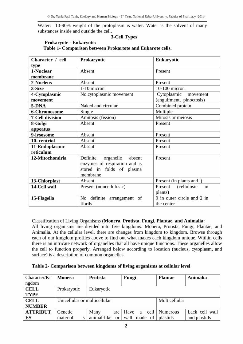
3-Cell Types
Prokaryote - Eukaryote:
Table 1- Comparison between Prokartote and Eukarote cells.
Character / cell
type
Prokaryotic Eukaryotic
1-Nuclear
membrane
Absent Present
2-Nucleus Absent Present
3-Size 1-10 micron 10-100 micron
4-Cytoplasmic
movement
No cytoplasmic movement Cytoplasmic movement
(engulfment, pinoctosis)
5-DNA Naked and circular Combined protein
6-Chromosome Single Multiple
7-Cell division Amitosis (fission) Mitosis or meiosis
8-Golgi
appeatus
Absent Present
9-lysosome Absent Present
10- centriol Absent Present
11-Endoplasmic
reticulum
Absent Present
12-Mitochondria Definite organelle absent
enzymes of respiration and is
stored in folds of plasma
membrane
Present
13-Chlorplast Absent Present (in plants and )
14-Cell wall Present (noncellulosic) Present (cellulosic in
plants)
15-Flagella No definite arrangement of
fibrils
9 in outer circle and 2 in
the center
Classification of Living Organisms (Monera, Protista, Fungi, Plantae, and Animalia:
All living organisms are divided into five kingdoms: Monera, Protista, Fungi, Plantae, and
Animalia. At the cellular level, there are changes from kingdom to kingdom. Browse through
each of our kingdom profiles above to find out what makes each kingdom unique. Within cells
there is an intricate network of organelles that all have unique functions. These organelles allow
the cell to function properly. Arranged below according to location (nucleus, cytoplasm, and
surface) is a description of common organelles.
Table 2- Comparison between kingdoms of living organisms at cellular level
Character/Ki
ngdom Monera Protista Fungi Plantae Animalia
CELL
TYPE
Prokaryotic Eukaryotic
CELL
NUMBER
Unicellular or multicellular Multicellular
ATTRIBUT
ES
Genetic
material is
Many are
animal-like or
Have a cell
wall made of
Numerous
plastids
Lack cell wall
and plastids
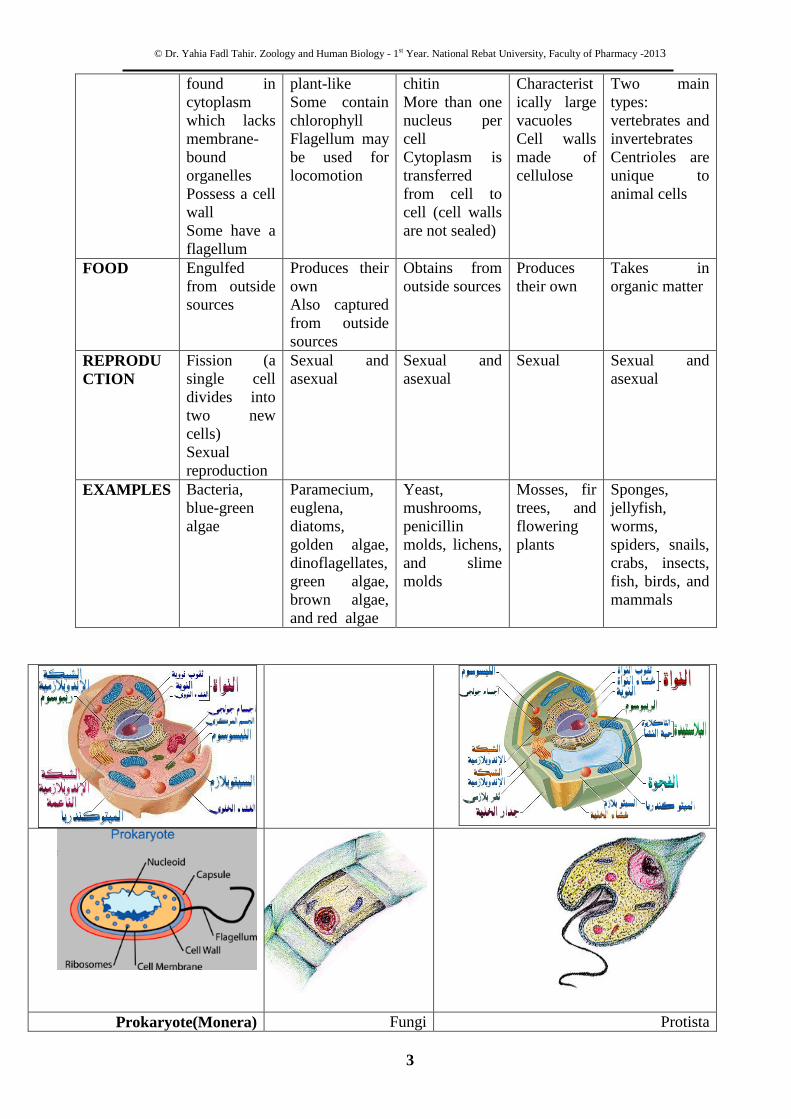
© Dr. Yahia Fadl Tahir. Zoology and Human Biology - 1st Year. National Rebat University, Faculty of Pharmacy -2013
3
found in
cytoplasm
which lacks
membrane-
bound
organelles
Possess a cell
wall
Some have a
flagellum
plant-like
Some contain
chlorophyll
Flagellum may
be used for
locomotion
chitin
More than one
nucleus per
cell
Cytoplasm is
transferred
from cell to
cell (cell walls
are not sealed)
Characterist
ically large
vacuoles
Cell walls
made of
cellulose
Two main
types:
vertebrates and
invertebrates
Centrioles are
unique to
animal cells
FOOD Engulfed
from outside
sources
Produces their
own
Also captured
from outside
sources
Obtains from
outside sources
Produces
their own
Takes in
organic matter
REPRODU
CTION
Fission (a
single cell
divides into
two new
cells)
Sexual
reproduction
Sexual and
asexual
Sexual and
asexual
Sexual Sexual and
asexual
EXAMPLES Bacteria,
blue-green
algae
Paramecium,
euglena,
diatoms,
golden algae,
dinoflagellates,
green algae,
brown algae,
and red algae
Yeast,
mushrooms,
penicillin
molds, lichens,
and slime
molds
Mosses, fir
trees, and
flowering
plants
Sponges,
jellyfish,
worms,
spiders, snails,
crabs, insects,
fish, birds, and
mammals
Protista Fungi Prokaryote(Monera)

© Dr. Yahia Fadl Tahir. Zoology and Human Biology - 1st Year. National Rebat University, Faculty of Pharmacy -2013
4
Cell Structure and Function
The protoplasm is differentiated into two parts nucleoplasm and cytoplasm; the nucleus is
surrounded by Nuclear membrane. Plasma membrane surrounds the cytoplasm.
a- Nucleoplasm:
Chromosomes:
They are the main genetic material and present in the nucleus. Usually they are in the form
of chromatin and appear only when cell dividing. and set number per species (i.e. 23 pairs
for human)
Nuclear membrane:
Double layers membrane surrounds nucleus and controls material exchange between the
cytoplasm and the nucleus. It has selective permeability.
Nuclear sap or karyolymph: Nucleoplasm or nuclear sap is colorless solution and contains organelles. It mainly
composed of water with free-floating molecule, DNA and RNA.
Nucleolus:
Spherical in shape
Visible when cell is not dividing
Contains RNA for protein manufacture
Bar body:
It is small chromatin body near the nuclear membrane in female of animal’s nervous
tissues.
b-Cytoplasm
Centrioles:
Paired cylindrical organelles Lie at right angles to each other near nucleus.
Composed of nine tubes, each with three tubules. Involve in cellular division
and cilia movement
Chloroplasts:
A plastid usually found in plant cells
Contain green chlorophyll where photosynthesis takes place

© Dr. Yahia Fadl Tahir. Zoology and Human Biology - 1st Year. National Rebat University, Faculty of Pharmacy -2013
5
Cytoskeleton:
Composed of microtubules
Supports cell and provides shape
Aids movement of materials in and out of cells
Endoplasmic reticulum:
It is of two types granular or rough and agranular or smooth endoplasmic reticulum.
Rough type the ribosomes embedded in surface and present in tissues producing proteins
like liver and pancreas, while Smooth type lacks ribosomes and present in eye retina and
its function may be sensation. Stores, separates, and serves as cell's transport system
Ribosomes:
Composes 25% of cell's mass.
Stationary type: embedded in rough endoplasmic reticulum
Mobile type: injects proteins directly into cytoplasm

© Dr. Yahia Fadl Tahir. Zoology and Human Biology - 1st Year. National Rebat University, Faculty of Pharmacy -2013
6
Golgi apparatus:
it is a membrane structure found near nucleus. It has a fundamental role in production of
zymogene (enzymes precursor), bile, mucous, vitamin G and others.
It is very sensitive to physiological condition of the animal as vitamin D deficiency
and exposure to chemical such pesticides and phosphorous.
Lysosome:
Present in organs cell digestion occurs like liver, kidney, and small intestine. Transports
undigested material to cell membrane for removal.It is very sensitive to physiological
condition as disease (cancer) and exposure pesticides where it becomes very little in
number.
Mitochondria:
It present in most tissues and the number is constant in cells of the same type.
Double-layered outer membrane with inner folds called cristae
Energy-producing chemical reactions take place on cristae
Recycles and decomposes proteins, fats, and carbohydrates, and forms urea
It is very sensitive to physiological condition as disease and exposure pesticides cyanide,
phosphorous and x-rays where it becomes fat drops.

© Dr. Yahia Fadl Tahir. Zoology and Human Biology - 1st Year. National Rebat University, Faculty of Pharmacy -2013
7
Vacuoles:
Membrane-bound sacs for storage, digestion, and waste removal. they are
Osmotic pressure regulators
Cell wall:
Most commonly found in plant cells (cellulose) and in fungi (chitin).
Controls turgity
Extracellular structure surrounding plasma membrane
Plasma membrane:
Outer membrane of cells that controls cellular traffic (passage of materials). It has
property of permeability. There pores in it.
It is very sensitive to antigen exposure heavy metals, x-rays and fat solvents that lyse
(digest) the membrane.

© Dr. Yahia Fadl Tahir. Zoology and Human Biology - 1st Year. National Rebat University, Faculty of Pharmacy -2013
8
Transport in Cells:
All cells acquire the molecules and ions they need from their surrounding. There is an
unceasing traffic of molecules and ions. This action is carried out by tow ways which are:
1-Passive Transport
1-Diffusion: Molecules and ions move spontaneously down their concentration gradient
(i.e., from a region of higher to a region of lower concentration). The plasma membrane
is differentially permeable because some particles can pass through, others cannot.
a-Osmosis
Osmosis is the diffusion of water across a semipermeable membrane. Tonicity refers to
the relative concentration of solute on either side of a membrane.
Isotonic
In an isotonic solution, the concentration of solute is the same on both sides of the
membrane (inside the cell and outside.
Hypotonic
A hypotonic solution is one that has less solute (more water). Cells in hypotonic
solution tend to gain water.
Hypertonic solution
A hypertonic solution is one that has a high solute concentration. Cells in a
hypertonic solution will lose water.
b-Facilitated Diffusion
Facilitated diffusion involves the use of a protein to facilitate the movement of molecules
across the membrane. In some cases, molecules pass through channels within the protein.
In other cases, the protein changes shape, allowing molecules to pass through.
2-Active Transport: Molecules and ions can be moved against their concentration gradient in this type, the
energy of ATP is used to enforce ions or small molecules or even particles through the
membrane against their concentration gradient.
a-Direct Active Transport
The cytoplasm of animal cells contains a concentration of potassium ions (K+) as much as
20 times higher than that in the extracellular fluid. Conversely, the extracellular fluid
contains a concentration of sodium ions (Na+) as much as 10 times greater than that
within the cell. These concentration gradients are established by the active transport of
both ions. The same transporter, called the Na+/K
+ ATPase, does both jobs.
b- Indirect Active Transport
Indirect active transport uses the downhill flow of an ion (electrochemical differences)to
pump some other molecule or ion against its gradient. The driving ion is (H+) which is
pumped out the membrane to join glucose molecule. Later the ion is pumped back to
inside the cell by the ATPase.
c-Vacoules
The processes by which a cell engulfs or get red of material, these are:
-Endocytosis: a vacuole is formed that contains the material that has been engulfed.
-Exocytosis: moves material to the outside. A vesicle fuses with the plasma
membrane and discharges its contents outside. This allows cells to secrete
molecules.
-Pinocytosis: moves solutions to the inside by means of very small vesicle.

© Dr. Yahia Fadl Tahir. Zoology and Human Biology - 1st Year. National Rebat University, Faculty of Pharmacy -2013
9
Cell Division
The unique and fundamental property of protoplasm is its power to grow and multiply by
cell division. There are three types of cell division occur in living organisms, these are:
1.Amitosis of direct cell division.
2.Mitosis or indirect cell division.
3.Meiosis or reduction division.
1. Amitosis or direct cell division:
This is found in mainly in prokaryotes like cyanobateria, bacteria, yeast etc. some
protozoa's also divide by amitosis. The process is a means of reproduction. During it the
nucleus elongates and a constriction appears in the center resulting in a dumb bell shaped
structure. The constriction deepens and the nucleus gets divided into two bits. There is no
spindle formation of chromosomes. Cytoplasmic division follows the nucleus division
and the cell gets divided into two daughter cells. Thus the amitosis is only a quantitative
division.
2. Mitosis or indirect cell division:
Mitosis occurs in the somatic cell of multicellular organisms and its chief function is to
increasing in size and in individuals. It is defined as a complex process leading to
formation of two equal daughter cells from a single parental cell. The mitosis is divided
into two main stages or sometimes called cell cycle: cytokiensis and karyokinesis.
Cell cycle
Interphase:
Cell cycle
Interphase is the time between divisions (in all types of divisions) or at which cell is not
dividing. The nucleus is surrounded by membrane as well the chromatin and nucleolus
are present. The phase is divided into three periods known as cell cycle:
-G1 during which the protein and RNA synthesis are carried out.
- S during which the DNA are replicated towards division time.
- G 2 during which the volume of the nucleus increases.
Prophase:
It is the first stage of the nuclear division and it takes longest time in mitotic division.
During which the chromatin shortens, thickens and coils into chromosomes and become
visible. Nuclear membrane disintegrates. Centriole pairs move to opposite ends of the
cell. Spindle fibers begin to form.
Metaphase:
The phase lasts only for a short duration. It is guided by the spindle fibers, the
chromosome pairs line up along the center of the spindle structure at equatorial plate.

© Dr. Yahia Fadl Tahir. Zoology and Human Biology - 1st Year. National Rebat University, Faculty of Pharmacy -2013
10
Anaphase:
The thickening of the chromosomes is increased. The chromatids begin to depart from
the centrioles. Every chromatid (now becomes chromosome) removes to opposite poles
of the cell by the spindle fibers.
Telophase:
Chromosomes return to chromatin. Spindle disintegrates. Nuclear membrane takes shape
again. Centrioles replicate. Membrane continues to pinch inward by the process called
cytokinasis (in plant cells a new cell wall is laid). When the process is complete, each cell
will have the same genetic material that the original cell had before replication. Each of
the daughter cells is also identical to each other. Note that once telophase is complete, the
cell returns to interphase.
Significance of Mitosis:
1. Mitosis is extremely regular process maintaining the qualitative and quantitative
distribution of heredity materials to daughter cells. The process takes 3-10 hours in
plant cell and18-20 hours in animal cell,
2. It maintains the diploid number of chromosome of species.
3. The process results in growth and development of the organism. Adult human body
(75 kg in weight) consists of 60 trillion (60 000 000 000 000) cells derived from one
cell. The growth is very rapid in embryo so and it has three types in adult animals:
cell not dividing (arrested) as nervous cells, dividing in some cases as liver cell and
dividing continuously as skin cell.

© Dr. Yahia Fadl Tahir. Zoology and Human Biology - 1st Year. National Rebat University, Faculty of Pharmacy -2013
11
4. The process results in replacement of dead cells e.g. epidermis, RBC and gut (50 000
000 000 cells/ day).
5. The process plays an important in the asexual reproduction.
3. Meiosis or Reduction Division
Meiosis is a special type of division. All gametes have half the number of chromosomes
that regular cells have. Gametes are created through the process of meiosis. Meiosis
involves two divisions, which create four haploid cells.
The first meiosis division:
Interphase: it consists of same G1, S, and G2 phases
Prophase I: This phase takes longest time and it is complex .All chromatin material makes a copy of
itself, then pairing of homologous chromosomes takes place. Chromosomes grouped into
tetrads i.e. chromosome having two chromatids. Some crossing over may occur at
chiasma between opposite chromatids. The tetrads line up and pull apart by a process (
terminalization ). Once the chromosome pairs are at opposite poles of the cytoplasm.
Nucleolus disappears.
Metaphase I: The nuclear membrane disappears. The centrioles are jointed by spindle
fibers, which develop from tubules. The bivalents chromosomes arrange themselves on
the equatorial plate. The centromeres lie towards poles.
Anaphase I: each homologous chromosome of bivalent moves towards the opposite
poles. The centromere does not divide (it divides in mitosis).
Telophase I: the chromosomes reach the respect poles and become indistinct. Nucleolus
appears. Cell membrane is formed in animal cells by furrowing (cytokinasis). Finally
two cells formed. The two cells formed do not have the same genetic material; they have
the half number of chromosomes.
The second meiosis division:
The second meiotic division is a mitosis in which both the chromosomes and cell are
divide. The main difference is DNA does not duplicate as interphase of mitosis.
Prophase II: the centrioles divide into two and migrate to opposite poles. Nucleolus and
nuclear membrane disintegrate.
Metaphase II: It is of very short duration. Chromosomes line up at the center (equatorial
plate). The centromeres divide separating the two chromatids. The centromeres are
attached to chromosomal spindle fibers.
Anaphase II: the daughter chromosomes migrate to opposite poles. The movement is
due to the contraction of the chromosomal spindle fibers and continuos stretching of
spindle fibers.
Telophase II: the daughter chromosomes reach the opposite poles.. Nucleolus and
nuclear membrane reappear so daughter nucleus is formed. The cytoplasm then divides
(cytokinasis). The nucleus is haploid (half number of chromosomes). As the result four
cells are formed.
In male organisms the four new cells are all the same size. In females one of the four cells
receives the bulk of the cytoplasm material. This becomes the functioning egg while the
other three smaller cells disintegrate.

© Dr. Yahia Fadl Tahir. Zoology and Human Biology - 1st Year. National Rebat University, Faculty of Pharmacy -2013
12

© Dr. Yahia Fadl Tahir. Zoology and Human Biology - 1st Year. National Rebat University, Faculty of Pharmacy -2013
13
HISTOLOGY
The structure of tissues
Histology is the study of animal and plant tissues. Tissues are defined as cells with their
ground substance acting together in the performance of a particular function. Tissues are
routinely cut into thin translucent sections that are then differentially stained and
examined under a microscope.
The primary tissue categories in animal histology are structurally there are four types of
tissue (1) epithelial tissue (2) connective tissue (3) muscular tissue (4) nervous tissue.
(1) Epithelial tissue
1-Epithelial tissues are composed of closely aggregated cells with very little
extracellular matrix.
2-The functions of epithelia are as a protective covering, absorption, secretion, sensation
and contractility. 3- Embryonically, the epithelia derive from all three embryonic tissues
4-Epithelial tissue is often associated with a basement membrane made of thin fibers and
ground substance.
5-Epithelial cells often have adaptations at their luminal surface to increase surface area
for absorption or to help move substances over the epithelial surface e.g. cilia
Structural types of epithelia:
(A) Simple, one layer and (B) Compound (stratified)
(A) Simple epithelia:
it Characterized by that cells be found as a single layer (simple). it is classified as follws:
(I) Squamous Epithelia:
These consist of greatly flattened cells cemented together to form a single layer which
under the microscope. They occur in the linings of abdominal cavities, the heart, blood
vessels, the walls of alveoli and in the outer layer of the elementary canal.
(II) Columnar Epithelia:
The cells of these are brick-shaped. The entire lining of the elementary canal (except
esophagus, buccal cavity and anus) and it’s associated with glands consisting of this type
of cells. Those of the villi of the small intestine have an outer striated border..
(III) Ciliated Columnar Epithelia:
In these the cells are columnar but possess cilia (cytoplasmic thread) on their outer
border. They are found in the linings of the trachea and larynx, the larger bronchiole
tubes in the lungs, nasal passage and fallopian tubules.
© Dr. Yahia Fadl Tahir. Zoology and Human Biology - 1st Year. National Rebat University, Faculty of Pharmacy -2013
14
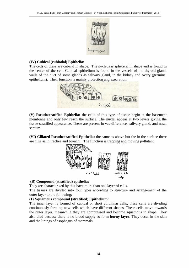
(IV) Cubical (cubiodal) Epithelia:
The cells of these are cubical in shape. The nucleus is spherical in shape and is found in
the center of the cell. Cubical epithelium is found in the vessels of the thyroid gland,
walls of the duct of some glands as salivary gland, in the kidney and ovary (germinal
epithelium). Their function is mainly protection and execration.
(V) Pseudostratified Epithelia: the cells of this type of tissue begin at the basement
membrane and only few reach the surface. The nuclei appear at two levels giving the
tissue-stratified appearance. These are present in vas-difference, salivary gland, and nasal
septum.
(VI) Ciliated Pseudostratified Epithelia: the same as above but the in the surface there
are cilia as in trachea and bronchi. The function is trapping and moving pollutant.
(B) Compound (stratified) epithelia:
They are characterized by that have more than one layer of cells.
The tissues are divided into four types according to structure and arrangement of the
outer layer to the following:
(1) Squamous compound (stratified) Epithelium:
The inner layer is formed of cubical or short columnar cells; these cells are dividing
continuously forming new cells which have different shapes. These cells move towards
the outer layer, meanwhile they are compressed and become squamous in shape. They
also died because there is no blood supply so form horny layer. They occur in the skin
and the linings of esophagus of mammals.
© Dr. Yahia Fadl Tahir. Zoology and Human Biology - 1st Year. National Rebat University, Faculty of Pharmacy -2013
15
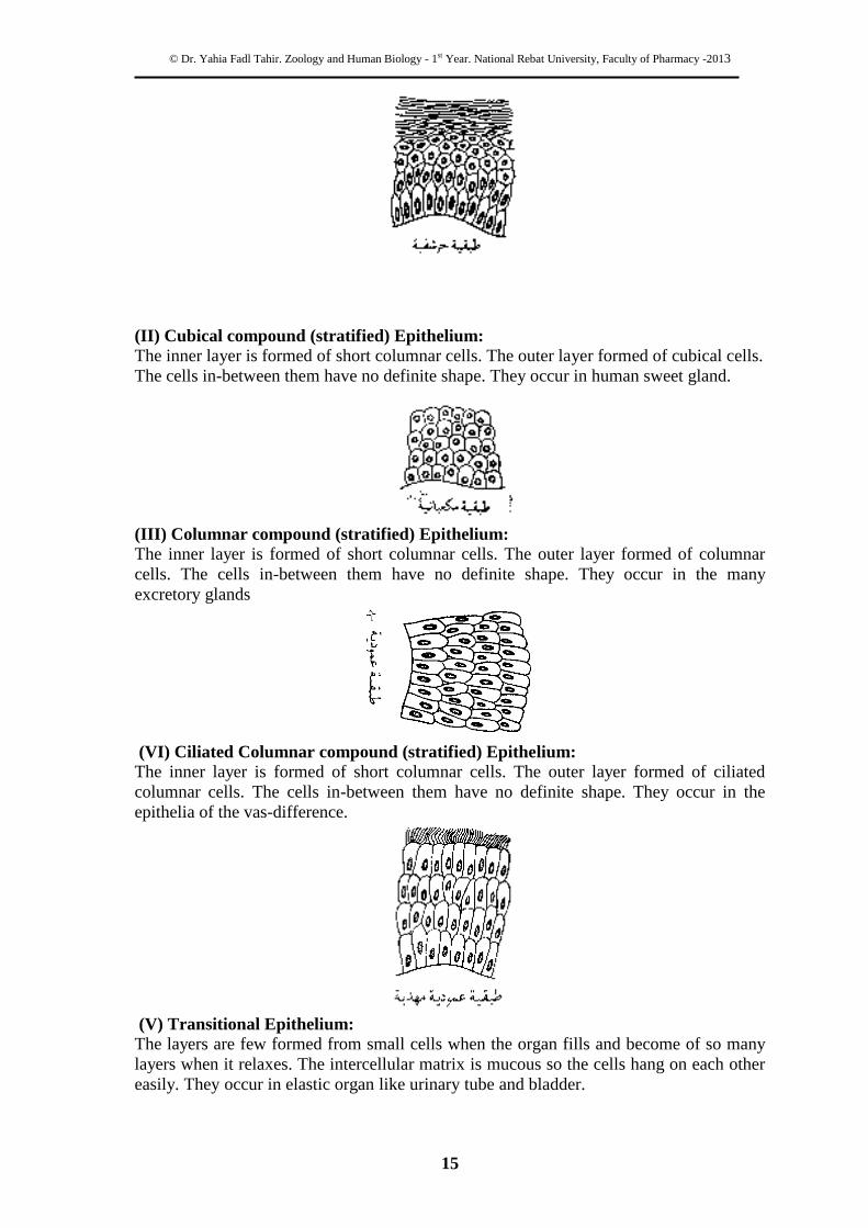
(II) Cubical compound (stratified) Epithelium:
The inner layer is formed of short columnar cells. The outer layer formed of cubical cells.
The cells in-between them have no definite shape. They occur in human sweet gland.
(III) Columnar compound (stratified) Epithelium:
The inner layer is formed of short columnar cells. The outer layer formed of columnar
cells. The cells in-between them have no definite shape. They occur in the many
excretory glands
(VI) Ciliated Columnar compound (stratified) Epithelium: The inner layer is formed of short columnar cells. The outer layer formed of ciliated
columnar cells. The cells in-between them have no definite shape. They occur in the
epithelia of the vas-difference.
(V) Transitional Epithelium: The layers are few formed from small cells when the organ fills and become of so many
layers when it relaxes. The intercellular matrix is mucous so the cells hang on each other
easily. They occur in elastic organ like urinary tube and bladder.

© Dr. Yahia Fadl Tahir. Zoology and Human Biology - 1st Year. National Rebat University, Faculty of Pharmacy -2013
16
2-Connective tissue
1-These tissues are responsible for cushioning, supporting and maintaining form within
the body.
2-The major constituent of connective tissue is the ground substance, which is composed
of protein fiber (gelatinous), fluid as in blood and solid matrix as in bones.
3-Most connective tissues are derived from the mesodermal layer.
4-There is no basement membrane.
There are three types of connective tissue as follows: -
1. Connective tissue proper:
2. Skeletal connective tissue
3. Vascular connective tissue
Types of connective tissue proper:
1-Areolar connective (loose connective tissue):
This type of the tissue has a reticular shape. Connective tissue is found in sheaths around
the blood vessels, and surrounding f muscle cells. It is well vascularized and contains
many types of cells and fibers.
Fibroblasts are the most common cells in connective tissue and are responsible for
making the fibers and the ground substance. They have a large ovoid nucleus, and well-
developed endoplasmic reticulum and Golgi apparatus.
Macrophages are resident phagocytic cells and have an oval shaped nucleus, well-
developed Golgi complex, many lysosomes and a prominent rough ER. They migrate into
the connective tissue.
Mast cells They secrete the ground substance. Secretary granules within mast cells
contain histamine, sertonin, and anticoagulant heparin. Mast cells are involved in allergic
reactions.
Plasma cells These large ovoid cells are responsible for making antibodies, they become
much more prevalent in areas subject to invasion such as the intestine or where there is a
chronic infection.
Others: there are many cells like eosinophill and lymphocytes.
Collagen (white) fiber it is stronger and less flexible and consisted of a protein called
collagen occurs in wavy bundles.
© Dr. Yahia Fadl Tahir. Zoology and Human Biology - 1st Year. National Rebat University, Faculty of Pharmacy -2013
17
Elastic (yellow) fiber it is more flexible consisted of a protein called elastin occurs in
singly though they branch and coalesce with one another forming a network.
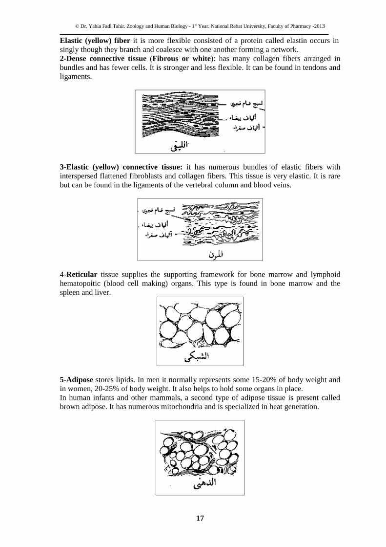
2-Dense connective tissue (Fibrous or white): has many collagen fibers arranged in
bundles and has fewer cells. It is stronger and less flexible. It can be found in tendons and
ligaments.
3-Elastic (yellow) connective tissue: it has numerous bundles of elastic fibers with
interspersed flattened fibroblasts and collagen fibers. This tissue is very elastic. It is rare
but can be found in the ligaments of the vertebral column and blood veins.
4-Reticular tissue supplies the supporting framework for bone marrow and lymphoid
hematopoitic (blood cell making) organs. This type is found in bone marrow and the
spleen and liver.
5-Adipose stores lipids. In men it normally represents some 15-20% of body weight and
in women, 20-25% of body weight. It also helps to hold some organs in place.
In human infants and other mammals, a second type of adipose tissue is present called
brown adipose. It has numerous mitochondria and is specialized in heat generation.

© Dr. Yahia Fadl Tahir. Zoology and Human Biology - 1st Year. National Rebat University, Faculty of Pharmacy -2013
18
6-Mucous is a special type of connective tissue contains a few fibers and the matrix is
gelatinous. It is found in embryonic organs and the placenta.
B- Skeletal connective tissue:
1-Cartilage
Cartilage can be found in areas where shock absorbing or sliding are needed. The cells
(chondrocytes ) are embedded in the matrix, inside capsules (lacuna). There are three
types of cartilage based on variations in matrix composition: -
1-Hyaline cartilage, the most common form is hyaline cartilage. The matrix is clear and
there are no fibers inside (glass-like). It can be found on the articulating surfaces of
bones, walls of large respiratory passages, ventral portion of the ribs. Hyaline cartilage
forms the fetal skeleton that is later ossified and becomes bone.
Chondrocytes or cartilage cells are large and rounded each lying in a space - lacuna -
enclosed by matrix. Cells often are grouped in nests of 2, 4, or 6.
2-Elastic cartilage, the matrix contains many elastic fibers. It can be found in the auricle
of the ear, ear canal, eustachian tube, and epiglottis. It is flexible than the hyaline kind.
3. Fibrocartilage Fibrocartilage contains a dense network of collagen fibers amongst which are dispersed
only a few chondrocytes in lacunae. It is located between tendon and bone, bone and
bone, hyaline cartilage and in the intervertebral (IV) disc.

© Dr. Yahia Fadl Tahir. Zoology and Human Biology - 1st Year. National Rebat University, Faculty of Pharmacy -2013
19
2-Bone
Bone, on gross observation, has two forms.
Spongy bone (Cancellous) is found in the center of flat bones and the
ends of long bones.
Compact bone is a dense tissue seen in the shafts on long bones and
surrounding the spongy bone. Red (hematogenous) bone marrow is
found in the ends of long bones and center of the flat bones of the skull
and vertebra. The bone is composed of units called Havarsian system.
The are in-between the units is called non- Havarsian.
{{{{{
C- Vascular connective tissue:
Blood might be classed as a specialized connective tissue because it is not connecting
tissues but has mesoderm origin. The matrix is plasma (90% water, 10% proteins,
inorganic salts, aminoacids, vitamins, hormones and others). it consists the following:
Red blood corpuscles/Erythrocytes (RBCs)
White blood cells/Leucocytes (WBCs)
Platelets
(A)Erythrocytes (Red Blood Cell = RBC's)
Biconcave discs; close to 7.5 µm in diameter. Comprise a flexible membrane enclosing
haemoglobin. Mature RBCs have no nucleus in mammals but it is nucleated in fishes,
reptiles, and birds. Erythrocytes also lack Golgi body, ER, ribosomes or mitochondria.
RBCs count in man is 5 millions /micoliter and 4.5/ micoliter million in women
Life span is estimated to be around 120 days, then the RBC is sequestered in the spleen,
liver or bone marrow to be phagocytosed by macrophages. The spleen takes the iron and
the other materials used by the liver in production of bile. RBC manufacture
(Haemopoiesis) takes 24-48 hours in maturation and is being in the liver and the spleen at
embryonic development then in bone marrow at maturation.
© Dr. Yahia Fadl Tahir. Zoology and Human Biology - 1st Year. National Rebat University, Faculty of Pharmacy -2013
20
(B)Leucocytes (White Blood Cells = WBC's)
These are nucleated cells, their count is 7000/ micoliter. They divided according to the
granularity of their cytoplasm into two groups - granular and agranular.
1. Granular leucocytes
All kinds appear round in a smear. They have granules in the cytoplasm. There are three
types of granular cells:
1. Neutrophil
Nucleus has three or more lobes or segments connected. They comprise 70 %of
leucocytes. They are 12-15 microns in diameter. The cytoplasm contains small granules
that are primary lysosomes containing numerous enzymes. These phagocytic cells
surround and engulf bacteria and constitute a defense against invasion.
2.Eosinophil (acidophill)
Eosinophils have a diameter of 12-15 microns and.
Nucleus is bilobed.
Cytoplasm has many large, eosinophil granules
They comprise 3 % of leucocytes.
An increase in the absolute number of eosinophils in circulation is associated with
allergic reaction and parasitic infection.
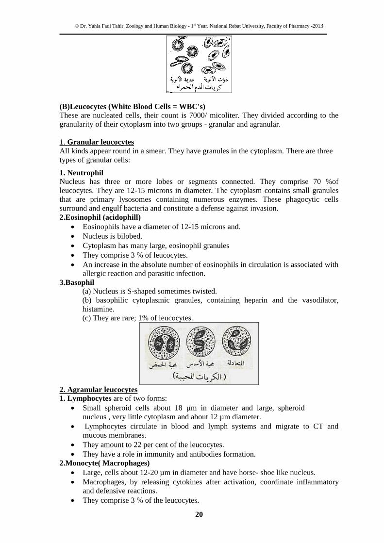
3.Basophil
(a) Nucleus is S-shaped sometimes twisted.
(b) basophilic cytoplasmic granules, containing heparin and the vasodilator,
histamine.
(c) They are rare; 1% of leucocytes.
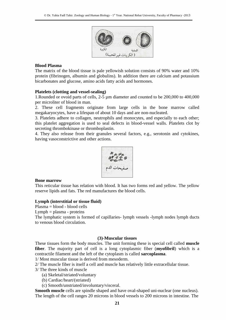
2. Agranular leucocytes
1. Lymphocytes are of two forms:
Small spheroid cells about 18 µm in diameter and large, spheroid
nucleus , very little cytoplasm and about 12 µm diameter.
Lymphocytes circulate in blood and lymph systems and migrate to CT and
mucous membranes.
They amount to 22 per cent of the leucocytes.
They have a role in immunity and antibodies formation.
2.Monocyte( Macrophages)
Large, cells about 12-20 µm in diameter and have horse- shoe like nucleus.
Macrophages, by releasing cytokines after activation, coordinate inflammatory
and defensive reactions.
They comprise 3 % of the leucocytes.
© Dr. Yahia Fadl Tahir. Zoology and Human Biology - 1st Year. National Rebat University, Faculty of Pharmacy -2013
21
Blood Plasma
The matrix of the blood tissue is pale yellowish solution consists of 90% water and 10%
protein (fibrinogen, albumin and globulins). In addition there are calcium and potassium
bicarbonates and glucose, amino acids fatty acids and hormones.
Platelets (clotting and vessel-sealing)
1.Rounded or ovoid parts of cells, 2-5 µm diameter and counted to be 200,000 to 400,000
per microliter of blood in man.
2. These cell fragments originate from large cells in the bone marrow called
megakaryocytes, have a lifespan of about 10 days and are non-nucleated.
3. Platelets adhere to collagen, neutrophils and monocytes, and especially to each other;
this platelet aggregation is used to seal defects in blood-vessel walls. Platelets clot by
secreting thrombokinase or thromboplastin.
4. They also release from their granules several factors, e.g., serotonin and cytokines,
having vasoconstrictive and other actions.
Bone marrow
This reticular tissue has relation with blood. It has two forms red and yellow. The yellow
reserve lipids and fats. The red manufactures the blood cells.
Lymph (interstitial or tissue fluid)
Plasma = blood - blood cells
Lymph = plasma - proteins
The lymphatic system is formed of capillaries- lymph vessels -lymph nodes lymph ducts
to venous blood circulation.
(3)-Muscular tissues
These tissues form the body muscles. The unit forming these is special cell called muscle
fiber. The majority part of cell is a long cytoplasmic fiber (myofibril) which is a
contractile filament and the left of the cytoplasm is called sarcoplasma.
1/ Most muscular tissue is derived from mesoderm.
2/ The muscle fiber is itself a cell and muscle has relatively little extracellular tissue.
3/ The three kinds of muscle
(a) Skeletal/striated/voluntary
(b) Cardiac/heart/(striated)
(c) Smooth/unstriated/involuntary/visceral.
Smooth muscle cells are spindle shaped and have oval-shaped uni-nuclear (one nucleus).
The length of the cell ranges 20 microns in blood vessels to 200 microns in intestine. The
© Dr. Yahia Fadl Tahir. Zoology and Human Biology - 1st Year. National Rebat University, Faculty of Pharmacy -2013
22
fiber (muscle cell) may be found as solitary as the skin or reticular as in the respiratory
tract or dense as in the alimentary canal (longitudinal and circular muscles). They Locate
at walls of hollow organs, i.e. stomach, intestine, uterus and urethra Functions of these
tissues is the involuntary movement - i.e. churning of food, movement of urine from the
kidney to the bladder.
Skeletal muscle (striated)
Skeletal muscle cells run the full length of a muscle and cylindrical in shape with the
diameter of 0.01-0.1 or up to 0.4 millimeter. Note the striation characteristics of this
muscle type. These cells, are multinuclear locate at the periphery in mammals
(syncytium). Every fiber is consisted from two types of white and dark strips, which are
chemically, are proteins called myosin and actin respectively. The fiber is surrounded by
connective tissue called perimysium.
These muscles are associated with the skeleton and are functioning as voluntary
movement. Muscles are connected to bones by tendons. Bones are connected to other
bones at their joints by ligaments.
Cardiac muscle Cardiac muscle cells are cylindrical, branched and striated but less than
above. The nucleus is oval-shaped. There are intercalated discs. The tissue Locates only
at the heart Function: involuntary, rhythmic contraction
(4) Nervous tissues
Nervous tissue derives from the embryonic ectoderm under the influence of the
notochord. The ectoderm differentiates to form the neural tube from which all of the
central nervous system derives. The two types of the cells are neuroblasts (nerve cells)
and the spongyblast called glial cells assist neurons by support, protection, defense and
nutrition of the neurons. There are about 10 times more glial cells than neurons in the
© Dr. Yahia Fadl Tahir. Zoology and Human Biology - 1st Year. National Rebat University, Faculty of Pharmacy -2013
23
brain. Glial cells create the microenvironment needed for neuronal function and
sometimes they assist in neural processing and activity. Neurons are independent
functional units responsible for the reception, transmission and processing of stimuli.
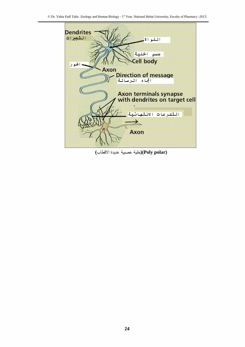
Structure of the nerve cell:
In general, neurons consist of three parts; the cell body, where the nucleus and organelles
are located; dendrites, which are processes extending from the cell body that receive
stimuli from the environment or other neurons; and the axon, which is a long single
process extending from the cell body for the transmission of nerve impulses to other
cells. The axon usually branches at its distal end and each branch terminating on another
cell has a bulbous end. The interaction of the end bulb with the adjacent cell forms a
structure called a synapse. Synapses are specialized to receive a signal and convert it into
an electrical potential.
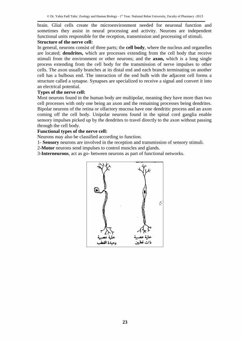
Types of the nerve cell:
Most neurons found in the human body are multipolar, meaning they have more than two
cell processes with only one being an axon and the remaining processes being dendrites.
Bipolar neurons of the retina or olfactory mucosa have one dendritic process and an axon
coming off the cell body. Unipolar neurons found in the spinal cord ganglia enable
sensory impulses picked up by the dendrites to travel directly to the axon without passing
through the cell body.
Functional types of the nerve cell: Neurons may also be classified according to function.
1- Sensory neurons are involved in the reception and transmission of sensory stimuli.
2-Motor neurons send impulses to control muscles and glands.
3-Interneurons, act as go- between neurons as part of functional networks.
© Dr. Yahia Fadl Tahir. Zoology and Human Biology - 1st Year. National Rebat University, Faculty of Pharmacy -2013
24
( عديدة االقطابخلية عصبية )(Poly polar)