cdi 13 s3 title 1. - acpgbi · colorectal disease acpgbi position statement on elective resection...
TRANSCRIPT

ColorectalDisease
ACPGBI Position Statement on Elective Resectionfor Diverticulitis
Guest editorIan Lindsey

Position Statements1
ACPGBI Position Statement on Elective Resectionfor DiverticulitisJ. B. J. Fozard, N. C. Armitage, J. B. Schofield,
O. M. Jones
CDI VOLUME 13 Suppl. 3 April 2011 � 2011 The Association of Coloproctology of Great Britain and Ireland. Colorectal Disease, 13 (Suppl. 3), 1–11
ColorectalDisease13 April 2011
View this journal online at wileyonlinelibrary.com/
journal/codi to search the articles and register for
table of contents e-mail alerts.


Position Statement doi:10.1111/j.1463-1318.2010.02531.x
ACPGBI Position Statement on Elective Resection forDiverticulitis
J. B. J. Fozard*, N. C. Armitage†, J. B. Schofield‡ and O. M. Jones§
*Royal Bournemouth Hospital, Bournemouth, UK, †Department of Colorectal Surgery, Queens Medical Centre, Nottingham, UK, ‡Department of Cellular
Pathology, Preston Hall Hospital, Kent, UK and §Surgery and Diagnostics Centre, Churchill Hospital, Oxford, UK
Introduction
Diverticular disease is common, with a frequency that
increases with age. Most patients with diverticulosis do
not develop symptoms, though a number will develop
inflammation or diverticulitis. This inflammation may
develop into an abscess or free perforation. Whilst the
prevalence of perforation has increased from 2.4 per
100 000 in 1986 to 3.8 per 100 000 in 2000 according
to one study [1], the majority of patients with divertic-
ulitis follow an indolent clinical course. There has been
controversy, therefore, as to whether elective resection
after acute diverticulitis is justified [2].
This position statement is presented in sections
dealing with the pathology, symptomatology and
investigation of diverticular disease and its conse-
quences. It then examines the evidence and indications
for surgical intervention and its timing. The issue of
laparoscopic versus open resection is then considered.
The evidence is briefly summarized under the heading
‘Findings’ and this is followed where relevant by
‘Recommendations’.
Methodology
Organized searches of the Cochrane Database, MED-
LINE and PUBMED were performed using the key-
words relevant to each section of this Position Statement.
Searches were limited predominantly to English language
articles. Additional publications were retrieved from the
references cited in articles identified from the primary
search of the literature. All evidence was classified
according to an accepted hierarchy of evidence and
recommendations graded A–C on the basis of level of
associated evidence and ⁄ or noted as Good Practice
and ⁄ or part of NICE ⁄ SIGN recommendation for Rapid
Technology Appraisal (Table 1).
The pathophysiology of diverticular disease
Disease aetiology and prevalence
In Western countries, the prevalence of diverticular
disease has dramatically increased over the last century.
There is a close link between diverticulosis and a low-fibre
diet. The refining of flour with removal of dietary fibre
that became increasingly frequent in the late 19th century
has been implicated in the aetiology of this disease [3].
Diverticular disease is much less common in the devel-
oping world where a high-roughage diet is common-
place. In western populations, vegetarians have a lower
frequency of diverticulosis, supporting the hypothesis
that a high-roughage diet protects against diverticular
disease [4]. In addition, animal studies have also shown
that a life-long low-fibre diet is associated with divertic-
ulosis [5] and studies of immigrants demonstrate devel-
opment of the western pattern of disease when a western
diet is adopted. Multiple other factors have been impli-
cated in the aetiology of diverticular disease, including
obesity [6], lack of exercise [7], smoking [8] and
immunosuppression.
The high prevalence of diverticulosis is in part due to
an increase in detection of the disorder. However,
diverticular disease of the colon increases with age and
these increases may also reflect our ageing population [9].
In western countries, over half of the population over
80 years of age has diverticula involving the colon. The
incidence increases from about 5% in the fifth decade to
60% in the ninth decade. There appears to be a small
female predominance [10] although some older papers
suggested male predominance and this might represent a
changing distribution of disease.
Pathology of diverticulosis
Colonic diverticula are formed by a combination of
increased intraluminal pressure in the colon and weakness
of the muscular wall. In the weak areas of the colonic wall
where blood vessels enter, mucosal herniation occurs. A
low-fibre diet results in small-volume stools that require
high intraluminal pressures for propulsion (‡ 150 mgHg).
Correspondence to: J. B. J. Fozard, Royal Bournemouth Hospital, Castle Lane East,
Bournemouth BH7 7DW, UK.
E-mail: [email protected]
� 2011 The Authors
Colorectal Disease � 2011 The Association of Coloproctology of Great Britain and Ireland. 13 (Suppl. 3), 1–11 1

Colonic manometry studies undertaken in the 1960s
confirmed higher luminal pressures in patients with
diverticulosis than in controls [11]. Subsequently, Painter
et al. suggested a theory of segmentation in which muscle
contraction results in a series of discrete segments of bowel
with high intraluminal pressures [12]. This disordered
motility is likely to have a major role in the pathogenesis of
both left-sided and right-sided diverticula. Patients with
ulcerative colitis have reduced bowel wall muscle tone and
contractility. This may explain the lower prevalence of
diverticulosis in patients with ulcerative colitis [13].
Increased cholinergic neural activity and decreased
noncholinergic activity has been demonstrated in diver-
ticulosis [14]. This alteration could lead to increased
tonicity but it is unclear whether this finding is causally
related [4]. Decreased numbers of glial cells and interstitial
cells of Cajal have also been recently described in patients
with diverticular disease. This finding might explain some
of the large bowel motility disturbances [15].
Solitary caecal diverticulum is an uncommon congen-
ital condition; caecal diverticula are seen more commonly
as part of generalized diverticular disease and were seen in
16% of patients in the postmortem study reported by
Hughes [16]. Right-sided diverticulosis in the absence of
left-sided disease is considered to be a different condition
with a genetic predisposition. It is seen rarely in Cauca-
sians but more commonly found in Asians and is the
predominant site in Japanese patients [17].
Pathology of diverticulitis and diverticular colitis
Diverticulitis is acute inflammation of one or more
diverticula and usually the inflammatory changes involve
the apex of the diverticulum rather than the neck. It is
likely that acute inflammation is caused by impaction of
faecal material within the diverticulum, leading to mucosal
ulceration with associated acute inflammation [7]. It has
also been suggested that a fibre-deficient diet may be
associated with a change in the bacterial flora and
alteration of local immunity, resulting in low grade
chronic inflammation. This could predispose to acute
diverticulitis [18]. Acute inflammation may rapidly involve
the serosal surface, leading to peritonitis because of the
thinness of the diverticular wall, and there may be localized
abscess formation or perforation due to damage of the
diverticular wall by acute inflammation. An inflammatory
mass may develop with or without involvement of other
pelvic organs and fistulation most often to the bladder or
vagina occurs particularly in patients who have had
previous hysterectomy. Massive rectal bleeding may occur
related to erosion of a blood vessel by the inflammatory
process, usually at the neck of the diverticulum [19].
Symptoms and signs of diverticulitis
In clinical practice, patients present with abdominal
pain, most usually in the left iliac fossa. On occasion,
pain may be in other areas of the abdomen, either
because of a long redundant sigmoid loop lying on the
right side of the abdomen or because of diverticular
disease located away from the sigmoid colon. The pain
may be associated with systemic symptoms such as
nausea and vomiting or anorexia. Physical signs will
usually include localized tenderness and guarding, fever,
and there may be a palpable mass either in the abdomen
or per rectum.
Where an inflammatory mass is present in the absence
of an abscess, this is generally termed a phlegmon. The
Table 1 Grading scheme for assessing submitted evidence. All evidence was classified according to an accepted hierarchy of evidence
that was originally adapted from the US Agency for Healthcare Policy and Research Classification.
Level of evidence Grade of recommendation
I. Evidence obtained from a single randomized controlled trial or
from a systematic review or meta-analysis of randomized
controlled trials
A. Evidence of type I or consistent findings from
multiple studies of type IIa, IIb or III
IIa. Evidence obtained from at least one well-designed controlled study
without randomization
B. Evidence of type IIa, IIb or III and generally
consistent findings
IIb. Evidence obtained from at least one other well-designed
quasi-experimental study
C. Evidence of type IIa, IIb or III but inconsistent
findings
III. Evidence obtained from well-designed nonexperimental
descriptive studies, such as comparative studies, correlation
studies and case studies
D. Little or no systemic evidence
IV. Evidence obtained from expert committee reports or opinions
and ⁄ or clinical experiences of respected authorities, case reports
GP. Recommended good practice based on the
clinical experience of the expert group and
other professionals
Poor evidence mandates a case by base decision J. B. J. Fozard et al.
� 2011 The Authors
2 Colorectal Disease � 2011 The Association of Coloproctology of Great Britain and Ireland. 13 (Suppl. 3), 1–11
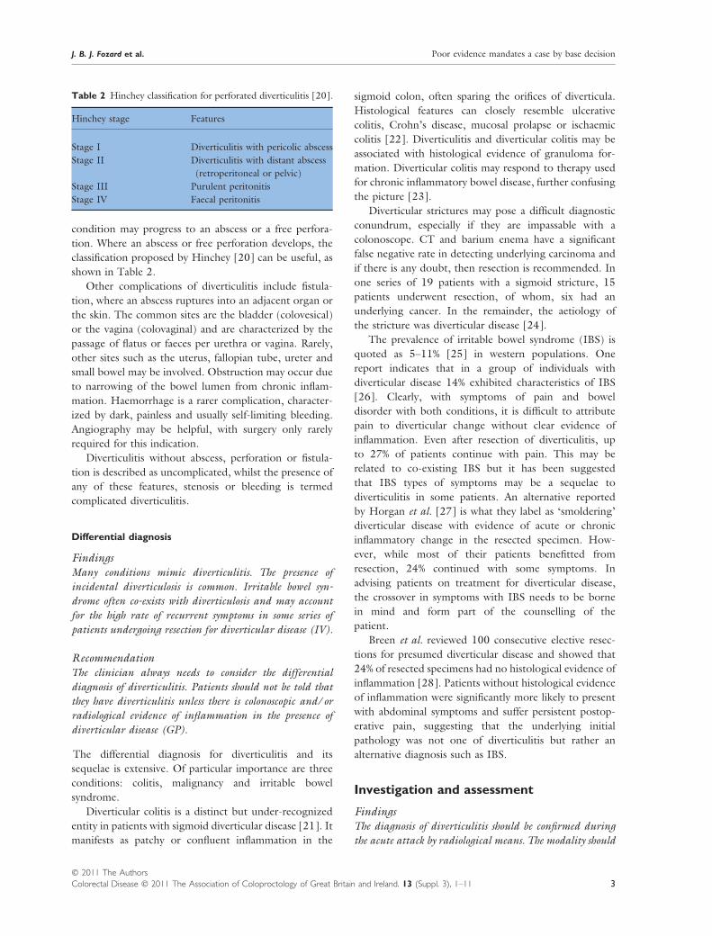
condition may progress to an abscess or a free perfora-
tion. Where an abscess or free perforation develops, the
classification proposed by Hinchey [20] can be useful, as
shown in Table 2.
Other complications of diverticulitis include fistula-
tion, where an abscess ruptures into an adjacent organ or
the skin. The common sites are the bladder (colovesical)
or the vagina (colovaginal) and are characterized by the
passage of flatus or faeces per urethra or vagina. Rarely,
other sites such as the uterus, fallopian tube, ureter and
small bowel may be involved. Obstruction may occur due
to narrowing of the bowel lumen from chronic inflam-
mation. Haemorrhage is a rarer complication, character-
ized by dark, painless and usually self-limiting bleeding.
Angiography may be helpful, with surgery only rarely
required for this indication.
Diverticulitis without abscess, perforation or fistula-
tion is described as uncomplicated, whilst the presence of
any of these features, stenosis or bleeding is termed
complicated diverticulitis.
Differential diagnosis
FindingsMany conditions mimic diverticulitis. The presence of
incidental diverticulosis is common. Irritable bowel syn-
drome often co-exists with diverticulosis and may account
for the high rate of recurrent symptoms in some series of
patients undergoing resection for diverticular disease (IV).
RecommendationThe clinician always needs to consider the differential
diagnosis of diverticulitis. Patients should not be told that
they have diverticulitis unless there is colonoscopic and ⁄ or
radiological evidence of inflammation in the presence of
diverticular disease (GP).
The differential diagnosis for diverticulitis and its
sequelae is extensive. Of particular importance are three
conditions: colitis, malignancy and irritable bowel
syndrome.
Diverticular colitis is a distinct but under-recognized
entity in patients with sigmoid diverticular disease [21]. It
manifests as patchy or confluent inflammation in the
sigmoid colon, often sparing the orifices of diverticula.
Histological features can closely resemble ulcerative
colitis, Crohn’s disease, mucosal prolapse or ischaemic
colitis [22]. Diverticulitis and diverticular colitis may be
associated with histological evidence of granuloma for-
mation. Diverticular colitis may respond to therapy used
for chronic inflammatory bowel disease, further confusing
the picture [23].
Diverticular strictures may pose a difficult diagnostic
conundrum, especially if they are impassable with a
colonoscope. CT and barium enema have a significant
false negative rate in detecting underlying carcinoma and
if there is any doubt, then resection is recommended. In
one series of 19 patients with a sigmoid stricture, 15
patients underwent resection, of whom, six had an
underlying cancer. In the remainder, the aetiology of
the stricture was diverticular disease [24].
The prevalence of irritable bowel syndrome (IBS) is
quoted as 5–11% [25] in western populations. One
report indicates that in a group of individuals with
diverticular disease 14% exhibited characteristics of IBS
[26]. Clearly, with symptoms of pain and bowel
disorder with both conditions, it is difficult to attribute
pain to diverticular change without clear evidence of
inflammation. Even after resection of diverticulitis, up
to 27% of patients continue with pain. This may be
related to co-existing IBS but it has been suggested
that IBS types of symptoms may be a sequelae to
diverticulitis in some patients. An alternative reported
by Horgan et al. [27] is what they label as ‘smoldering’
diverticular disease with evidence of acute or chronic
inflammatory change in the resected specimen. How-
ever, while most of their patients benefitted from
resection, 24% continued with some symptoms. In
advising patients on treatment for diverticular disease,
the crossover in symptoms with IBS needs to be borne
in mind and form part of the counselling of the
patient.
Breen et al. reviewed 100 consecutive elective resec-
tions for presumed diverticular disease and showed that
24% of resected specimens had no histological evidence of
inflammation [28]. Patients without histological evidence
of inflammation were significantly more likely to present
with abdominal symptoms and suffer persistent postop-
erative pain, suggesting that the underlying initial
pathology was not one of diverticulitis but rather an
alternative diagnosis such as IBS.
Investigation and assessment
FindingsThe diagnosis of diverticulitis should be confirmed during
the acute attack by radiological means. The modality should
Table 2 Hinchey classification for perforated diverticulitis [20].
Hinchey stage Features
Stage I Diverticulitis with pericolic abscess
Stage II Diverticulitis with distant abscess
(retroperitoneal or pelvic)
Stage III Purulent peritonitis
Stage IV Faecal peritonitis
J. B. J. Fozard et al. Poor evidence mandates a case by base decision
� 2011 The Authors
Colorectal Disease � 2011 The Association of Coloproctology of Great Britain and Ireland. 13 (Suppl. 3), 1–11 3

be CT or ultrasound depending on local expertise (Level
IIb). Barium enema or colonoscopy after resolution of the
acute episode is essential to rule out alternative diagnoses or
second pathologies.
RecommendationCT or ultrasound should be undertaken during the acute
presentation of diverticulitis. This helps to confirm the
diagnosis, guide management of the acute attack and
occasionally will demonstrate other pathologies. Investiga-
tion of the colonic lumen by endoscopic means or barium
enema after the acute attack is mandatory (Grade C).
Radiographic evaluation of diverticulitis during the acute
attack heightens the precision of the decision making
process. Evaluation in the first 72 h of an acute attack will
help confirm the diagnosis and may give some prognostic
information. Confirming the diagnosis is particularly
important in the younger patient group, in whom the
alternative diagnoses (particularly appendicitis) need to
be considered. In patients in whom elective resection is
being contemplated, evidence of definite inflammation
during the acute attack is essential. Colonoscopic or
barium enema evaluation some weeks after the acute
attack may show diverticulosis but will not prove the
diagnosis of diverticulitis.
The optimum radiological modality for evaluation of
acute diverticulitis has been the subject of a recent
systematic review [29]. This appraised 20 articles evalu-
ating ultrasound, CT, barium enema and MRI. It
concluded that the studies were generally of poor quality
and suggested that the best study evaluating ultrasound
and CT reached only level IIb. Whilst the best evidence in
the literature supports ultrasound, the appropriateness of
this modality will depend on local expertise. Ultrasound
and CT are superior to contrast enema or colonoscopy in
that they evaluate the extraluminal extent of the disease
and may be more useful in picking up other pathologies.
Extraluminal air ⁄ contrast and abscess may predict
poor outcome (see Table 3). The outcome, however, is
mixed and individual predictions are difficult on purely
radiological grounds, as 24% with successful conservative
management had severe CT diverticulitis and 19% with
severe CT diverticulitis, had no further problems, within
the context of this study [30].
The diagnostic and therapeutic approach, however, has
clearly changed with CT [31–33]. This particularly relates
to the confirmation of diagnosis, but also the distinction
between uncomplicated, phlegmonous diverticulitis and
complicated diverticulitis, involving fistula, peritonitis,
obstruction and abscess [34,35]. It is in the treatment of
abscesses that a difference of opinion emerges. Ambrosetti
and colleagues state that mesocolic abscesses can be
managed conservatively without percutaneous drainage
[36]. This can be either as a bridge to one-stage resection,
or more intriguingly as totally conservative treatment.
Longer term follow up of mesocolic and pelvic diverticular
abscess, with initial nonsurgical management, shows that
51% of patients with mesocolic and 71% with pelvic
abscess eventually require surgery [37]. In this respect,
CT acts as a guide to ultimate failure of conservative
treatment. Pelvic abscesses, on the other hand, will usually
require surgery [35].
Natural history of diverticular disease
FindingsThe majority of patients presenting with acute diverticulitis
can be managed with a conservative, medical approach in
the longer term. Previous blanket recommendations for
elective resection following acute diverticulitis can be
challenged (Level III).
RecommendationThe decision on elective resection should be made on an
individual basis after the assessment of the particular
circumstances of the patient (Grade C).
The evaluation of recurrent or persistent symptoms is
difficult due to the overlap of symptomatology with
conditions such as irritable bowel syndrome, as previously
described. Furthermore, it would be reasonable to argue
that the medical treatment of acute diverticulitis was
transformed in UK practice in the mid 1970s with the
introduction of metronidazole [38]. The situation was
somewhat different in the US, where the perceived
genotoxicity of metronidazole limited its early uptake.
The alternatives at that time for treatment of anaerobic
infection, lincomycin and clindamycin, were both associ-
ated with an associated risk of Clostridium difficile
Table 3 The role of CT and gastrograffin enema (GE) in the
assessment of mild and severe diverticulitis.
Mild diverticulitis Severe diverticulitis
CT
Localized wall thickening
(> 5 mm)
Inflammation of pericolic fat
CT
Same as mild + at least
one of the following
Abscess
Extraluminal air
Extraluminal enema
GE
Segmental lumen narrowing
Tethered mucosa ± mass effect
GE
Same as mild + at least
one of the following
Extraluminal air
Extraluminal enema
Poor evidence mandates a case by base decision J. B. J. Fozard et al.
� 2011 The Authors
4 Colorectal Disease � 2011 The Association of Coloproctology of Great Britain and Ireland. 13 (Suppl. 3), 1–11
infection. Care should therefore be exercised, when
interpreting historical outcomes. The pivotal paper,
informing previous recommendations, was that of Parks
in 1970 [39]. This was a hospital-based study from 1951
to 1965 with significant exclusions. One can take issue
with the diagnostic criteria employed, because barium
enema in particular has largely been supplanted by the use
of CT. Interestingly, despite the paper’s recommendation
of elective resection, the evidence at that time could have
supported the opposite view, given that only 6% of
patients following a first attack of acute diverticulitis
required surgery for subsequent recurrent attacks.
Farmakis et al. reported on GP questionnaires giving
follow-up data from a UK national audit in the 1980s and
argued the case for interval sigmoid colectomy based on
recurrent symptoms, complications and death [40].
Unfortunately, there were methodological problems
acknowledged in the discussion, particularly incomplete
follow up, deaths at home and significantly high rates of
death from intercurrent disease.
In collating all the evidence, we make a clear distinction
between elective surgery for persistence of symptoms and
the risk of recurrent acute attacks, particularly severe and
complicated, and focus especially on objective, hard
evidence of recurrent inflammation, based on CT or
surgical findings. We then factor in operative mortality
[41], morbidity including stoma formation and death
from other causes ⁄ anticipated life expectancy. Such an
approach gives a clearer picture of the natural history of
diverticulitis and best informs surgical decision making on
elective resection. The main issues are therefore prevention
of death, or an adverse outcome (e.g. permanent stoma),
due to a second or subsequent attack of diverticulitis.
The need for elective surgery is influenced in part by the
primary resection rate at index attack and this can vary from
15% to 38% [42]. With reference to UK practice, Sarin
showed that conservative medical therapy is effective in
85%, with a calculated recurrence rate of 2% per patient-
year [43]. The rate for primary surgery will differ interna-
tionally, influenced by referral practice from primary care
to secondary care, admission criteria, diagnostic criteria
(clinical, ultrasound [44], contrast enema [45] and CT),
indications (persistent diverticulitis, perforation, fistula,
stricture, bleeding and residual abscess) [46], surgical
thresholds and utilization of CT guided drainage [47,48].
A high emergency surgical mortality has been used in
the past to justify elective surgical intervention but it is
incorrect to extrapolate outcomes of primary surgery to
secondary acute surgery. Indeed the data presented by
Elliot et al. [48] can be manipulated in a contrary fashion
to support a conservative approach, as with a blanket
recommendation for interval elective colectomy, 78% of
the study group could potentially be subjected to
‘unnecessary surgery’. Furthermore, 90% of patients
who die with perforation have no prior history of
diverticulitis [49] and 60% of patients undergoing
surgery for acute diverticulitis have no antecedent history
[50]. Of patients presenting with acute diverticulitis, only
3% have had a previous episode of acute diverticulitis
[51]. The inference is that complicated diverticulitis
tends to occur de novo and that the majority of patients
have no previous history of disease, though this has not
been widely accepted.
Further confusion arises from a general failure to
distinguish truly prophylactic interval colectomy (asymp-
tomatic patients), secondary elective colectomy (patients
with symptoms or complications, e.g. fistula or obstruc-
tion) and secondary acute colectomy (recurrent divertic-
ulitis, with or without complications). Prophylactic
colectomies are unlikely to prevent late major complica-
tions. Use of radiological criteria can aid this process
(Table 3). For instance, in a paper by Ambrosetti [52] on
the evaluation of 160 patients after first hospitalization,
23 had a complication requiring operation (11 persistent
diverticulitis, 4 recurrent diverticulitis, 6 symptomatic
stenosis, 1 residual abscess and 1 colovesical fistula).
Eighteen with no radiological evidence of a residual
diverticulitis underwent surgery for persistent pain or
‘fear of recurrence’. This latter group of resections is
unlikely to prevent late major complications [53]. Com-
paring medically and surgically treated groups, 73% and
79%, respectively, did not have further problems after
treatment of the index attack [54].
The most reliable data on the role of elective resection
come from a limited number of studies with adequate
follow up of expectant management. Chautems and
Ambrosetti showed that 68% of patients whose first attack
was treated nonoperatively did not suffer a complication,
at a median follow up of 9.5 years. Death from unrelated
causes (18%) was common [55].
In a later prospective study of the prognostic role of
CT scanning, randomization to elective resection or
expectant management had to be abandoned because
only 8% of patients developed recurrent diverticulitis,
albeit after a limited follow up of 19 months [31].
Mueller and colleagues conducted telephone interview
follow up at 7 and 13 years on a retrospective cohort of
252 patients, initially treated conservatively. Only 25
patients subsequently came to surgery and lethal compli-
cations were rare [56].
Timing of elective colectomy has been subjected to
Markov modelling [57], which incorporates probabilities
of clinical end-points over time (e.g. death, recurrent
diverticulitis, death from unrelated causes, surgical mor-
tality ⁄ morbidity and stoma formation). The risk of recur-
rent diverticulitis is not eliminated by surgery (2.6–10.4%)
J. B. J. Fozard et al. Poor evidence mandates a case by base decision
� 2011 The Authors
Colorectal Disease � 2011 The Association of Coloproctology of Great Britain and Ireland. 13 (Suppl. 3), 1–11 5

[43,58,59] and elective colectomy has a mortality of 2.3%
and colostomy rate of 11.4% [60]. Overall, Markov
modelling suggests that expectant management should
be associated with lower rates of death and colostomy and
incurs lower costs. We can thus conclude that there is good
clinical and experimental evidence against the previous
recommendations for elective resection.
Clinical course in younger patients
FindingsThere is no clear evidence that younger patients presenting
with diverticulitis exhibit a more aggressive form of the
disease. The higher rates of surgery in younger patients in
some studies may relate to misdiagnosis or a perception that
they are lower risk for surgical intervention (Level III).
RecommendationWith the natural history of diverticulitis in younger
patients not being well understood, there is little evidence
to support a different management strategy in young
patients compared to older ones (Grade C).
It has previously been suggested that diverticulitis in the
younger patient pursues a more aggressive course. This
in itself has been used to justify a recommendation that
such patients should undergo elective colectomy follow-
ing medical management of a first attack of diverticulitis
[61–65]. This literature, dating up to 1997, suffers from
a number of methodological flaws that compromise the
validity of the conclusions. Over time, the age of the
reported younger group has been extended from those
aged under 40 years to those aged under 50 years.
Given the influence of comorbid factors, it may be more
appropriate to take age 50 as the cut off. The quoted
studies involve relatively small numbers and are generally
retrospective case note reviews, with variable, often
inadequate duration of follow up. There is a problem
with lack of definition of severity of disease. Later work
by Spivak et al. in 1997 [66] uses the Hinchey
classification. Of note, misdiagnosis is more apparent
in the younger age group (particularly appendicitis) and
this is likely to influence the surgical approach. It has led
to high ‘urgent’ surgery rates in the past.
The real problem, however, is a failure to address
adequately the issue of ‘decision to treat’ (i.e. the original
indications for surgical or medical management and also
indications for subsequent elective or urgent surgery).
For instance, Shauer [62] uses the proportion undergo-
ing surgery as justification for elective colectomy – a
somewhat self-fulfilling prophecy. Recurrent symptoms
and frequency of emergency presentation are other
criteria that can be inappropriately used [65].
An aggressive course in the young could be defined by
a high rate of recurrent diverticulitis, high mortality, high
rate of emergency surgery or high rates of complications.
Biondo [67] showed that the recurrence rate at 7.4–
8.4 months was similar; 25.5% (< 50 years) compared
with 22.3% (> 50 years). Furthermore, the mortality rate
for both elective and emergency colectomy is higher in
the > 50 years age group. With a low emergency colec-
tomy mortality rate for those aged < 50 years, there is
justification for adopting an expectant approach, as this is
unlikely to lead to compromise [67]. Early use of CT in
the diagnosis and management of acute diverticulitis in
the younger patient is particularly helpful [42]. It
provides a more confident diagnosis and allows for
grading between mild and severe diverticulitis (Table 3).
As a consequence, surgery was performed less frequently
in younger patients than older patients (15% vs 33%) and
this in spite of a trend towards increased severity in the
young. Ambrosetti defined poor secondary outcome as
persistent diverticulitis, recurrent diverticulitis, symptom-
atic stenosis, colovesical fistula or residual abscess. The
young were more likely to have a poor secondary
outcome (29% vs 5%) [67]. At longer term follow up
(9.5 years), there was a 32% rate of ‘remote complica-
tion’. The complication frequency (54% at 5 years) was
highest in young patients with CT severe diverticulitis
and this was used to justify a recommendation for elective
colectomy in this group [55]. However, reliable predic-
tion for the individual is elusive and timing of surgery
only truly matters if it prevents avoidable mortality or
emergency surgery. It is reassuring that of patients treated
medically for a first or subsequent attack of diverticulitis,
none suffered an untoward outcome [55].
Further evidence against elective resection is provided
by Guzzo and colleagues [69]. Of patients aged < 50
years with acute diverticulitis, 60% never had surgery and
of a total of 196 patients with medium follow up of
5 years, only one re-presented with perforation.
In conclusion, younger patients with acute divertic-
ulitis do not pursue a more aggressive course. Deci-
sions on the place of elective colectomy in the younger
patient should remain based on individual circum-
stances [70].
Resection margins in elective diverticulitissurgery
FindingsResection of diverticular disease should involve resection
back to soft compliant bowel proximally with anastomosis
onto the rectum. It may not be feasible to remove all
diverticular disease. The splenic flexure should be routinely
mobilized (Level IIb).
Poor evidence mandates a case by base decision J. B. J. Fozard et al.
� 2011 The Authors
6 Colorectal Disease � 2011 The Association of Coloproctology of Great Britain and Ireland. 13 (Suppl. 3), 1–11

RecommendationThe splenic flexure should be mobilized routinely for
diverticular disease resections. This facilitates the anasto-
mosis being made from soft, compliant bowel being brought
down to the rectum (Grade C)
The margins of resection of diverticular disease may affect
recurrence. The distal anastomosis should be made onto
the rectum, as the presence of sigmoid colon distal to the
anastomosis is an independent predictor of recurrence
[71,72]. The proximal margin of resection is more
controversial, though there is no evidence to support
radical resection of all diverticulosis. The proximal part of
the anastomosis should be made in soft, compliant colon.
This advice is in accordance with the recommendations of
the Standards Committee of the American Society of
Colon and Rectal Surgeons Practice Parameters for
Sigmoid Diverticulitis [70].
This approach is facilitated by routine mobilization of
the splenic flexure in all patients. In a recent paper
reporting higher rates of disease recurrence after both
open and laparoscopic resection, the rate of splenic
flexure mobilization was low [73]. There is some
evidence that patients with more limited diverticular
resections have a greater tendency to have recurrent
symptoms [72].
The role of laparoscopy in themanagement of diverticulitis
Elective resection of uncomplicated diverticulitis
FindingsLaparoscopic resection of uncomplicated diverticulitis con-
fers benefits to patients when performed in centres with the
appropriate expertise compared with open resection (Level
I).
RecommendationIn centres with the appropriate expertise, laparoscopic
resection should be offered for uncomplicated diverticulitis
as it is safe and provides a faster recovery time from surgery
(Grade A).
Laparoscopic resection for uncomplicated diverticulitis is a
safe alternative to open resection and may be associated
with a lower rate of morbidity and shorter hospital stay.
There have been two randomized trials comparing lapa-
roscopic and open resection of diverticular disease, one
reported in abstract form only at this time. The Sigma Trial
randomized 104 patients between open and laparoscopic
approaches [74]. Though surgery took significantly longer
and the conversion rate was 19%, there was less blood loss,
morbidity, pain and analgesic requirement and a signifi-
cant reduction in hospital stay from 10 to 8 days in the
laparoscopic group.
A further randomized trial from Geneva has been
reported [75]. After exclusions, 113 patients were ran-
domized. Median operation time was 55 min longer in
the laparoscopic group. Time to first bowel movement
was quicker in the laparoscopic group, pain was less and
hospital stay 2 days shorter (5 vs 7 days; P < 0.0001). The
conversion rate was 9%.
Similar findings have been reported for a prospective
observational study that showed a reduction in hospital
stay from 18 to 10 days in favour of laparoscopic
resection [76], though the length of stay in the open
group does appear to be excessive. A recent meta-analysis
of a number of nonrandomized studies also suggested a
reduction in hospital stay and complications [77]. This
meta-analysis included a large number of patients from a
database study by Guller et al. [78], in which an
unknown number of the patients recorded as having
had an open operation were in fact initially laparoscopic
and converted to open, potentially skewing the data in
favour of the minimally invasive approach. Both this
observational study and the meta-analysis may be subject
to bias from the easier cases (likely to attract less
morbidity) being treated laparoscopically. There has
recently been a report from a single centre of 500
consecutive patients presenting with complicated and
uncomplicated diverticulitis undergoing laparoscopic
resection [79]. Mortality was 0.2% and major morbidity
occurred in 11%. Median operating time was 120 min,
the conversion rate was 2.8% and median hospital stay
was 4 days. This study showed what can be achieved in
centres with laparoscopic expertise but the applicability of
these results is certainly not universal.
Elective resection of complicated diverticulitis
FindingsThe laparoscopic approach is appropriate for complicated
diverticulitis (Level III).
RecommendationIn centres with appropriate expertise, laparoscopic resection
could be offered for complicated diverticulitis. It may confer
benefits to patient recovery (Grade D).
Patients with complicated (fistulating or stenosing) diver-
ticulitis undergoing laparoscopic resection are at higher
risk of conversion compared with those with uncompli-
cated diverticulitis [80]. In a recent study comparing
conversion rates between 112 patients with uncompli-
cated and 91 with complicated diverticular disease, there
J. B. J. Fozard et al. Poor evidence mandates a case by base decision
� 2011 The Authors
Colorectal Disease � 2011 The Association of Coloproctology of Great Britain and Ireland. 13 (Suppl. 3), 1–11 7

was a trend towards more postoperative morbidity in
patients with complicated diverticulitis, with a greater
likelihood of conversion, though over 90% of patients in
this group were still managed laparoscopically [81].
The decision about whether to tackle complicated
diverticulitis laparoscopically requires the surgeon to take
into account his own surgical expertise. These cases can
be very challenging and though excellent results are
reported in the literature, these outcomes may not be
universally applicable.
Acute versus elective laparoscopic resection for
diverticulitis
FindingsPatients undergoing laparoscopic resection of diverticulitis
should be treated after recovery from the acute episode of
inflammation (Level III).
RecommendationWhen possible, the patient with acute diverticulitis should be
managed medically with surgery being deferred until after
recovery from the acute illness. Conversion rates are lower in
the delayed surgery patients and there may be a trend
towards a lower rate of complications (Grade D).
There are few data in the literature to support laparoscopic
resection in the acute phase of diverticulitis in preference
to delayed resection, in patients whose acute illness can be
managed conservatively. A recent retrospective, single
centre review examined 178 patients, of whom 77 had
early attempted laparoscopic resection with the remainder
having delayed resection [82]. Although morbidity and
mortality did not differ between the two groups, the
conversion rate in the early surgery group was three times
higher at nearly 38%. A similar study of 210 patients
compared 116 patients having early resection with 94
having delayed resection. It found less morbidity in the
delayed surgery group and a lower rate of conversion [81].
The roles of laparoscopic lavage in the acute setting,
and subsequent elective resection
FindingsThe role of laparoscopic intervention in the patient
presenting with diverticular peritonitis unsuitable for, or
not responding to, conservative management remains
incompletely evaluated. It is uncertain whether these
patients should undergo interval resection (Level IIb).
RecommendationLaparoscopic lavage with and without laparoscopically
placed drains may play a role in some patients with acute
diverticulitis. Whilst this is an alternative to resection in
the acute setting for some patients, it is not certain
whether it is an acute alternative to delayed resection
(Grade C).
Laparoscopy can be helpful for diagnosis of acute
diverticulitis but in practical terms is rarely required, in
the era of cross-sectional imaging. There have been a
number of reports of laparoscopic lavage for patients
with acute diverticulitis. The largest series in the
literature, by Myers et al., reports 100 patients with
perforated diverticulitis and generalized peritonitis [83].
Eight patients with Hinchey IV disease required con-
version to an open procedure. Mortality was 4% overall
(one patient in the converted group). Similar results,
albeit in smaller case series, have been reported in the
literature [84–86]. Few studies report their denominator
and so it is unclear what percentage of patients would
have settled without any surgical intervention. The
selection of patients for laparoscopic lavage and its
timing in relation to presentation vary between studies.
It has to be concluded therefore that the place of
laparoscopic washout and drain placement in the algo-
rithm of treatment for acute diverticulitis is not yet
established.
Despite the paucity of data on laparoscopic lavage for
diverticular peritonitis, this approach is becoming more
commonly employed in this setting. It avoids an acute
resection and probable stoma in most patients. It remains
unclear, however, whether an elective resection after
recovery from the acute illness should be recommended.
In the Myers series of 100 patients, this was not generally
undertaken and indeed at median follow up of 3 years,
only two patients required resection [83].
General comments
The majority of the evidence for elective resection in
diverticular disease is of poor quality. The decision
regarding whether to offer resection should be made
on an individual basis and the surgeon should involve
the radiologists and pathologists in this decision, in
addition to the patients themselves. It is difficult to
make firm comments about the decision to undertake
elective resection because of the lack of data and
consensus on the natural history of unresected diver-
ticulitis. When surgery is undertaken, laparoscopically
or open, this can be challenging due to the inflamma-
tion and fibrosis.
Conflict of interest
The authors declare no conflict of interest.
Poor evidence mandates a case by base decision J. B. J. Fozard et al.
� 2011 The Authors
8 Colorectal Disease � 2011 The Association of Coloproctology of Great Britain and Ireland. 13 (Suppl. 3), 1–11
References
1 Makela J, Kiviniemi H, Laitinen S. Prevalence of perforated
sigmoid diverticulitis is increasing. Dis Colon Rectum 2002;
45: 955–61.
2 Janes S, Meagher A, Frizelle FA. Elective surgery after acute
diverticulitis. Br J Surg 2005; 92: 133–42.
3 Painter NS, Burkitt DP. Diverticular disease of the colon, a
20th century problem. Clin Gastroenterol 1975; 4: 3–21.
4 Stollman N, Raskin JB. Diverticular disease of the colon.
Lancet 2004; 363: 631–9.
5 Hodgson WJ. An interim report on the production of
colonic diverticulosis in the rabbit. Gut 1972; 13: 802–4.
6 Aldoori WH, Giovannucci EL, Rimm EB et al. Prospective
study of physical activity and the risk of symptomatic
diverticular disease in men. Gut 1995; 36: 276–82.
7 Kohler L, Sauerland S, Neuberger E. Diagnosis and
treatment of diverticular disease: results of a consensus
development conference. The Scientific Committee of the
European Association for Endoscopic Surgery. Surg Endosoc
1999; 13: 430–6.
8 Aldoori WH, Giovannucci EL, Rimm EB, Wing AL,
Trichopoulos DV, Willett WC. A prospective study of
alcohol, smoking, caffeine, and the risk of symptomatic
diverticular disease in men. Ann Epidemiol 1995; 5: 221–8.
9 Cheskin LJ, Bohlman M, Schuster MM. Diverticular disease
in the elderly. Gastroenterol Clin N Am 1990; 19: 391–403.
10 Parks TG. Natural history of diverticular disease of the
colon. A review of 521 cases. Br Med J 1969; 4: 639.
11 Arfwidsson S, Knock NG, Lehmann L, Winberg T. Path-
ogenesis of multiple diverticula of the sigmoid colon in
diverticular disease. Acta Chir Scand 1964; 63(Suppl. 342):
1–68.
12 Painter NS, Truelove SC, Ardran GM, Tuckey M. Segmen-
tation and the localization of intraluminal pressures in the
human colon with special reference to the pathogenisis of
colonic diverticula. Gastroenterology 1965; 49: 169–77.
13 Rispo A, Pasquale L, Cozzolino A et al. Lower prevalence of
diverticulosis in patients with ulcerative colitis. Dis Colon
Rectum 2007; 50: 1164–8.
14 Golder M, Burleigh DE, Belai A et al. Smooth muscle
cholinergic denervation hypersensitivity in diverticular dis-
ease. Lancet 2003; 361: 1945–51.
15 Basotti G, Battaglia E, Bellone G et al. Interstitial cells of
Cajal, enteric nerves, and glial cells in colonic diverticular
disease. J Clin Pathol 2005; 58: 973–7.
16 Hughes LE. Post-mortem survey of diverticular diseases of
the colon. I, Diverticulosis and diverticulitis. Gut 1969; 10:
336–44.
17 Nakaji S, Danjo K, Munakata A et al. Comparison of
etiology of right-sided diverticula in Japan with that of left-
sided diverticula in the West. Int J Colorectal Dis 2002; 17:
365–73.
18 Floch MH. A hypothesis: is diverticulitis a type of inflamma-
tory bowel disease? J Clin Gastroenterol 2006; 40: S121–5.
19 Hulten L, Haboubi NY, Schofield PF. Diverticular disease.
Colorectal Dis 1999; 1: 128–36.
20 Hinchey EJ, Schaal PG, Richards GK. Treatment of
perforated diverticular disease of the colon. Adv Surg
1978; 12: 85–109.
21 West AB, Losada M. The pathology of diverticulosis coli. J
Clin Gastroenterol 2004; 38: S11–6.
22 Ludeman L, Warren BF, Shepherd NA. The pathology of
diverticular disease. Best Pract Res Clin Gastroenterol 2002;
16: 543–62.
23 Ludeman L, Shepherd NA. What is diverticular colitis?
Pathology 2002; 34: 568–72.
24 King DW, Lubowski DZ, Armstrong AS. Sigmoid stricture
at colonoscopy-an indication for surgery. Int J Colorectal
Dis 1990; 5: 161–3.
25 Spiller R, Aziz Q, Creed F et al. Guidelines on the irritable
bowel syndrome: mechanisms and practical management.
Gut 2007; 56: 1170–98.
26 Simpson J, Scholefield JH, Spiller RC. Origin of symptoms
in diverticular disease. Br J Surg 2003; 90: 899–908.
27 Horgan AF, McConnell EJ, Wolff BG, The S, Paterson C.
Atypical diverticular disease: surgical results. Dis Colon
Rectum 2001; 44: 1315–8.
28 Breen RE, Corman ML, Robertson WG, Prager ED. Are we
really operating on diverticulitis? Dis Colon Rectum 1986;
29: 174–6.
29 Liljegren G, Chabok A, Wickbom M, Smedh K, Nilsson K.
Acute colonic diverticulitis: a systematic review of diagnostic
accurancy. Colorectal Dis 2007; 9: 480–8.
30 Ambrosetti P, Grossholz M, Becker C, Terrier F, Morel P.
Computed tomography in acute left colonic diverticulitis.
Br J Surg 1997; 84: 532–4.
31 Ambrosetti P, Robert J, Witzig JA et al. Prognostic factors
from computed tomography in acute left colonic divertic-
ulitis. Br J Surg 1992; 79: 117–9.
32 Ambrosetti P, Robert JH, Witzig JA et al. Acute left colonic
diverticulitis: a prospective analysis of 226 consecutive cases.
Surgery 1994; 115: 546–50.
33 Doringer E. Computerized tomography of colonic
diverticulitis. Crit Rev Diagn Imaging 1992; 33: 421–
35.
34 Hachigian MP, Honickman S, Eisenstat TE, Rubin RJ,
Salvati EP. Computed tomography in the initial manage-
ment of acute left-sided diverticulitis. Dis Colon Rectum
1992; 35: 1123–9.
35 Detry R, Jamez J, Kartheuser A et al. Acute localized
diverticulitis: optimum management requires accurate stag-
ing. Int J Colorectal Dis 1992; 7: 38–42.
36 Ambrosetti P, Robert J, Witzig JA et al. Incidence,
outcome and proposed management of isolated abscesses
complicating acute left-sided colonic diverticulitis. A pro-
spective study of 140 patients. Dis Colon Rectum 1992;
35: 1072–6.
37 Ambrosetti P, Chautems R, Soravia C, Peiris-Waser N,
Terrier F. Long-term outcome of mesocolic and pelvic
diverticular abscesses of the left colon: a prospective study of
73 cases. Dis Colon Rectum 2005; 48: 787–91.
38 Eykyn SJ, Phillips I. Metronidazole and anaerobic sepsis. Br
Med J 1976; 2: 1418–21.
J. B. J. Fozard et al. Poor evidence mandates a case by base decision
� 2011 The Authors
Colorectal Disease � 2011 The Association of Coloproctology of Great Britain and Ireland. 13 (Suppl. 3), 1–11 9
39 Parks TG, Connell AM. The outcome in 455 patients
admitted for treatment of diverticular disease of the colon.
Br J Surg 1970; 57: 775–8.
40 Farmakis N, Tudor RG, Keighley MR. The 5-year natural
history of complicated diverticular disease. Br J Surg 1994;
81: 733–5.
41 Constantinides VA, Tekkis PP, Senapati A. Comparison of
POSSUM scoring systems and the surgical risk scale in
patients undergoing surgery for complicated diverticular
disease. Dis Colon Rectum 2006; 49: 1322–31.
42 Kang JY, Hoare J, Tinto A et al. Diverticular disease of the
colon-on the rise: a study of hospital admissions in England
between 1989 ⁄ 1990 and 1990 ⁄ 2000. Aliment Pharmacol
Ther 2003; 17: 1189–95.
43 Sarin S, Boulos PB. Long-term outcome of patients
presenting with acute complications of diverticular disease.
Ann R Coll Surg Engl 1994; 76: 117–20.
44 Schwerk WB, Schwarz S, Rothmund M. Sonography in
acute colonic diverticulitis: a prospective study. Dis Colon
Rectum 1992; 35: 1077–84.
45 Hiltunen KM, Kolehmainen H, Vuorinen T, Matikainen
M. Early water-soluble contrast enema in the diagnosis of
acute colonic diverticulitis. Int J Colorectal Dis 1991; 6:
190–2.
46 Bahadursingh AM, Virgo KS, Kaminski DL, Longo WE.
Spectrum of disease and outcome of complicated divertic-
ular disease. Am J Surg 2003; 186: 696–701.
47 Makela J, Vuolio S, Kiviniemi H, Laitinen S. Natural history
of diverticular disease: when to operate? Dis Colon Rectum
1998; 41: 1523–8.
48 Elliott TB, Yego S, Irvin TT. Five-year audit of the acute
complications of diverticular disease. Br J Surg 1997; 84:
535–9.
49 Chapman J, Davies M, Wolff B et al. Complicated divertic-
ulitis: is it time to rethink the rules? Ann Surg 2005; 242:
576–83.
50 Alexander J, Karl RC, Skinner DB. Results of changing
trends in the surgical management of complications of
diverticular disease. Surgery 1983; 94: 683–90.
51 Somasekar K, Foster ME, Haray PN. The natural history of
diverticular disease: is there a role for elective colectomy? J R
Coll Surg Edinb 2002; 47: 481–4.
52 Ambrosetti P, Robert J, Witzig JA, Mathey P, Mirescu D,
Rohner A. Value of computerized tomography in acute
diverticulitis of the left colon. Schweiz Med Wochenschr
1993; 123: 1118–20.
53 Lorimer JW. Is prophylactic resection valid as an indication
for elective surgery in diverticular disease? Can J Surg 1997;
40: 445–8.
54 Larson DM, Masters SS, Spiro HM. Medical and surgical
therapy in diverticular disease: a comparative study. Gastro-
enterology 1976; 71: 734–7.
55 Chautems RC, Ambrosetti P, Ludwig A, Mermillod B,
Morel P, Soravia C. Long-term follow-up after first acute
episode of sigmoid diverticulitis: is surgery mandatory? A
prospective study of 118 patients. Dis Colon Rectum 2002;
45: 962–6.
56 Mueller MH, Glatzle J, Kasparek MS et al. Long-term
outcome of conservative treatment in patients with diver-
ticulitis of the sigmoid colon. Eur J Gastroenterol Hepatol
2005; 17: 649–54.
57 Salem I, Veenstra DL, Sullivan SD, Flum DR. The timing of
elective colectomy in diverticulitis: a decision analysis. J Am
Coll Surg 2004; 199: 904–12.
58 Wolff BG, Ready RL, MacCarty RL, Dozois RR, Beart RW
Jr. Influence of sigmoid resection on progression of
diverticular disease of the colon. Dis Colon Rectum 1984;
27: 645–7.
59 Thorn M, Graf W, Stefansson T, Pahlman L. Clinical and
functional results after elective colonic resection in 75
consecutive patients with diverticular disease. Am J Surg
2002; 183: 7–11.
60 Netri G, Verbo A, Coco C et al. The role of surgical
treatment in colon diverticulitis: indications and results.
Ann Ital Chir 2000; 71: 209–14.
61 Ouriel K, Schwartz SI. Diverticular disease in the young
patient. Surg Gynecol Obstet 1983; 156: 1–5.
62 Schauer PR, Ramos R, Ghiatas AA, Sirinek KR. Virulent
diverticular disease in young obese men. Am J Surg 1992;
164: 443–8.
63 Acosta JA, Grebenc ML, Doberneck RC, McCarthy JD, Fry
DE. Colonic diverticular disease in patients 40 years old or
younger. Am Surg 1992; 58: 605–7.
64 Konvolinka CW. Acute diverticulitis under age forty. Am J
Surg 1994; 167: 562–5.
65 Cunningham MA, Davis JW, Kaups KL. Medical versus
surgical management of diverticulitis in patients under age
40. Am J Surg 1997; 174: 733–5.
66 Spivak H, Weinrauch S, Harvey JC, Surick B, Ferstenberg
H, Friedman I. Acute colonic diverticulitis in the young. Dis
Colon Rectum 1997; 40: 570–4.
67 Biondo S, Pares D, Marti Rague J et al. Acute diverticulitis in
patients under 50 years of age. Br J Surg 2002; 89: 1137–41.
68 Ambrosetti P, Robert JH, Witzig JA et al. Acute left colonic
diverticulitis in young patients. J Am Coll Surg 1994; 179:
156–60.
69 Guzzo J, Hyman N. Diverticulitis in young patients: is
resection after a single attack always warranted? Dis Colon
Rectum 2004; 47: 1187–90.
70 Rafferty J, Shellito P, Hyman NH, Buie WD; Standards
Committee of the American Society of Colon and Rectal
Surgeons. Practice parameters for sigmoid diverticulities.
Dis Colon Rectum 2006; 49: 939–44.
71 Benn PL, Wolff BG, Ilstrup DM. Level of anastomosis and
recurrent colonic diverticulitis. Am J Surg 1986; 151: 269–
71.
72 Thaler K, Baig MK, Berho M et al. Determinants of
recurrence after sigmoid resection for uncomplicated diver-
ticulitis. Dis Colon Rectum 2003; 46: 385–8.
73 Thaler K, Weiss EG, Nogueras JJ, Arnaud JP, Wexner SD,
Bergamaschi R. Recurrence rates at minimum five-year
follow-up: laparoscopic versus open sigmoid resection for
uncomplicated diverticulitis. Acta Chir Iugosl 2004; 51:
45–7.
Poor evidence mandates a case by base decision J. B. J. Fozard et al.
� 2011 The Authors
10 Colorectal Disease � 2011 The Association of Coloproctology of Great Britain and Ireland. 13 (Suppl. 3), 1–11

74 Klarenbeek BR, Veenhof AA, Bergamaschi R et al. Laparo-
scopic sigmoid resection for diverticulitis decreases major
morbidity rates: a randomized control trial: short term
results of the Sigma Trial. Ann Surg 2009; 249: 39–44.
75 Gervaz P, Inan I, Skala K, Morel P. A prospective,
randomised, single-blind comparison of laparoscopic versus
open sigmoidectomy for diverticulitis. Colorectal Dis 2009;
11(Suppl. 2): 1.
76 Alves A, Panis Y, Slim K et al. French multicentre prospec-
tive observational study of laparoscopic versus open colec-
tomy for sigmoid diverticular disease. Br J Surg 2005; 92:
1520–5.
77 Purkayastha S, Constantinides VA, Tekkis PP et al. Laparo-
scopic vs open surgery for diverticular disease: a meta-
analysis of nonrandomized studies. Dis Colon Rectum 2006;
49: 446–63.
78 Guller U, Jain N, Hervey S, Purves H, Pietrobon R.
Laparoscopic vs. open colectomy: outcomes comparison
based on large nationwide databases. Arch Surg 2003; 138:
1179–86.
79 Jones OM, Stevenson ARL, Clark DA, Stitz RW, Lumley
JW. Laparoscopic resection for diverticular disase: follow-up
of 500 consecutive patients. Ann Surg 2008; 248: 1092–7.
80 Le Moine MC, Fabre JM, Vacher C, Navarro F, Picot MC,
Domergue J. Factors and consequences of conversion in
laparoscopic sigmoidectomy for diverticular disease. Br J
Surg 2003; 90: 232–6.
81 Reissfelder C, Buhr HJ, Ritz JP. Can laparoscopically
assisted sigmoid resection provide uncomplicated manage-
ment even in cases of complicated diverticulitis? Surg Endosc
2006; 20: 1055–9.
82 Zingg U, Pasternak I, Guertler L, Dietrich M, Wohlwend
K-A, Metzger U. Early vs, delayed elective laparoscopic-
assisted colectomy in sigmoid diverticulitis: timing of
surgery in relation to the acute attack. Dis Colon Rectum
2007; 50: 1911–7.
83 Myers E, Hurley M, O’Sullivan GC, Kavanagh D, Wilson I,
Winter DC. Laparoscopic peritoneal lavage for generalized
peritonitis due to perforated diverticulitis. Br J Surg 2008;
95: 97–101.
84 Taylor CJ, Layani L, Ghusn MA, White SI. Perforated
diverticulitis managed by laparoscopic lavage. ANZ J Surg
2006; 76: 962–5.
85 Mutter D, Bouras G, Forgione A, Vix M, Leroy J, Marescaux
J. Two-stage totally minimally invasive approach for acute
complicated diverticulitis. Colorectal Dis 2006; 8: 501–5.
86 Franklin ME Jr, Portillo G, Trevino JM, Gonzalez JJ, Glass
JL. Long-term experience with the laparoscopic approach to
perforated diverticulitis plus generalized peritonitis. World J
Surg 2008; 32: 1507–11.
J. B. J. Fozard et al. Poor evidence mandates a case by base decision
� 2011 The Authors
Colorectal Disease � 2011 The Association of Coloproctology of Great Britain and Ireland. 13 (Suppl. 3), 1–11 11
