ccpa-dependent carbon catabolite repression in bacteria · gram-positive bacteria. during the b....
TRANSCRIPT

MICROBIOLOGY AND MOLECULAR BIOLOGY REVIEWS, Dec. 2003, p. 475–490 Vol. 67, No. 41092-2172/03/$08.00�0 DOI: 10.1128/MMBR.67.4.475–490.2003Copyright © 2003, American Society for Microbiology. All Rights Reserved.
CcpA-Dependent Carbon Catabolite Repression in BacteriaJessica B. Warner† and Juke S. Lolkema*
Molecular Microbiology, Groningen Biomolecular Sciences and Biotechnology Institute,University of Groningen, Haren, The Netherlands
INTRODUCTION .......................................................................................................................................................475STRATEGY..................................................................................................................................................................477PROTEIN FAMILIES ................................................................................................................................................477
HPr Kinase ..............................................................................................................................................................477HPr-Like Proteins...................................................................................................................................................477
DISTRIBUTION AND GENOME ANALYSIS .......................................................................................................477Firmicutes ................................................................................................................................................................477Proteobacteria .........................................................................................................................................................482Other Phyla .............................................................................................................................................................483
SEQUENCE ANALYSES ...........................................................................................................................................484Full-Length and Short-Version HPr Kinases .....................................................................................................484Phylogenetic Relationships of HPr-Like Proteins..............................................................................................484Sequence Motifs in HPr-Like Proteins................................................................................................................485
CONCLUSIONS .........................................................................................................................................................487PROSPECTS ...............................................................................................................................................................488ACKNOWLEDGMENTS ...........................................................................................................................................489REFERENCES ............................................................................................................................................................489
INTRODUCTION
In bacteria, the phosphoenolpyruvate-dependent phospho-transferase system (PTS) is the main carbohydrate uptake sys-tem and, in addition, plays an important role in the regulationof expression of catabolic genes and operons (25, 36). Thesystem is typical of bacteria and not found in the other king-doms of life, Archaea and Eucarya. PTS-mediated uptake in-volves the transfer of the phosphoryl group of the high-energymetabolite phosphoenolpyruvate to the carbohydrate througha cascade of phosphotransfer proteins including EI, HPr, andEIIABC (Fig. 1A and B, left). In PTS-mediated gene regula-tion, the phosphorylation state of the intermediate proteinscontrols the expression of genes coding for sugar-specific com-ponents of the PTS (PRD-mediated induction [35]) and fortransporters and enzymes needed for the catabolism of lessfavored carbon sources (carbon catabolite repression [CCR]).CCR is achieved by allosteric inhibition of transporters orcytoplasmic enzymes, which prevents the uptake or synthesis ofinducers, respectively (inducer exclusion), or by interactionwith transcriptional regulators (Fig. 1A and B, right). Thelatter mechanism is the topic of this review. CCR via transcrip-tional regulators follows distinct mechanisms in gram-negativeand gram-positive bacteria, using different, unrelated tran-scriptional regulators, CRP and CcpA, respectively. In gram-negative bacteria, the phosphorylation state of the glucose-specific EIIAGlc regulates the activity of adenylate cyclase and,consequently, the level of cAMP in the cell (Fig. 1A). At
sufficiently high levels, cAMP binds to the transcriptional reg-ulator CRP, which induces binding to specific DNA sequencesin the promoter region of the target genes, where it activatesthe initiation of transcription through interaction with thepolymerase (25). In gram-positive bacteria, the signaling inter-mediate is HPr and not EIIAGlc (Fig. 1B). HPr in gram-posi-tive bacteria is phosphorylated at two sites, a histidine residueand a serine residue. The histidine residue is phosphorylatedby EI at the expense of phosphoenolpyruvate, while the serineis phosphorylated by HPr kinase (HPrK) at the expense ofATP or PPi (4, 24). HPr(His�P) is involved in sugar transportand PRD-mediated regulation, and HPr(Ser-P) is involved inCCR. The primary sensor in the regulatory pathway is HPrK,which is activated by glycolytic intermediates. HPr(Ser-P)binds to the transcriptional regulator CcpA, thereby inducingthe binding of the complex to so-called cre sites (for “cataboliteresponsive element”) in the promoter region of the targetgenes, which prevents transcription of the genes (13).
The above gives a generalized scheme for the CcpA-depen-dent transcriptional control in gram-positive bacteria and doesnot take into account other functions of CcpA (e.g., activationof glycolytic enzymes), mechanistic details (e.g., the location ofthe cre site), or the diversification reported in different organ-isms (for reviews, see references 12, 37, and 41). The schemeconforms to the CCR system in Bacillus subtilis, which has beenstudied extensively and is the prototype of CCR-regulatedgene expression in gram-positives bacteria. The “prototype”contains one important variation not observed in most othergram-positive bacteria. During the B. subtilis genome-sequenc-ing project, a second gene coding for an HPr-like protein wasdiscovered (19). The protein, Crh (for “catabolite repressionHPr”) has 45% sequence identity to HPr and contains theregulatory-site serine but not the active-site histidine (9). Ac-cordingly, it was demonstrated that Crh was inactive in the PTS
* Corresponding author. Mailing address: Biological Center, Kerk-laan 30, 9751NN Haren, The Netherlands. Phone: 31-50-3632155. Fax:31-50-3632154. E-mail: [email protected].
† Present address: Medical Biology, Department of Pathology andLaboratory Medicine, University of Groningen, The Netherlands.
475
on March 30, 2019 by guest
http://mm
br.asm.org/
Dow
nloaded from

transport function but functional in CCR; Crh could be phos-phorylated at the serine residue by HPrK and then act as acorepressor of CcpA. In B. subtilis the Crh- and the HPr-mediated regulatory pathways seem to operate in parallel.Studies using mutant strains containing HPr in which the reg-ulatory-site serine was mutated to alanine showed that in vivo,Crh could (partly) take over the regulatory function of HPr inglucose-induced repression depending on the target gene(s).The regulation was not affected in a Crh knockout strain (2, 9).Apparently, Crh is redundant in glucose-potentiated cataboliterepression in B. subtilis, leaving its function obscure. Recently,we demonstrated that repression of expression of the Mg2�
citrate transporter of B. subtilis grown in a medium containingsuccinate and glutamate was specifically mediated by Crh andnot by HPr. It was suggested that Crh might be specificallyinvolved in repression of metabolic genes by nonsugars (45).
A difference between CRP-dependent CCR found in gram-negative bacteria and CcpA-dependent CCR found in gram-positive bacteria is the strictness of the coupling between thePTS transport and regulatory functions. In the CRP-depen-dent mechanism (Fig. 1A), regulation of expression is directlycoupled to turnover of IIAGlc. The level of phosphorylation ofIIAGlc is determined by the uptake rate of glucose from themedium and by the uptake of other PTS sugars which competefor P-HPr and, thereby, limit the rate of phosphorylation ofIIAGlc. In the CcpA-dependent mechanism (Fig. 1B), the pri-mary sensor is HPrK that is activated by glycolytic intermedi-ates (15, 17, 28) but, in principle, may be activated by othermetabolites as well, making the mechanism more versatile.Our studies of the regulation of expression of the Mg2� citratetransporter of B. subtilis demonstrate that, in addition to glu-cose, the pathway is potentiated by the non-PTS sugar inositoland by the nonsugars succinate and glutamate (44). Moreover,the signal transduction pathway in the HPr molecule is physi-cally separated from the phosphoryl group transfer chain of thePTS transport function, i.e., via the regulatory-site Ser residueand active-site His residue, respectively. The regulatory state ismodulated rather than being determined by the uptake systemby virtue of the phosphorylation state of the active-site histi-dine (3, 27). Crh-mediated regulation may be a manifestationof this loose coupling between regulatory and transport func-tion of the PTS; a single mutation in HPr (His to Gln) resultsin a regulatory pathway that is independent of the uptakesystem. The mechanism found in gram-positive bacteria, in-volving HPr and HPrK, may be a more general gene regulationsystem. Gram-negative bacteria may have compensated fortheir specialized but inflexible mechanism by developing theNtr (nitrogen regulation) system, a PTS-based regulatorymechanism composed of a complete phosphotransfer chainthat operates independently of the uptake system and is not
FIG. 1. Schematic representation of CRP-dependent (A) andCcpA-dependent (B) carbon catabolite repression pathways and theNtr regulatory pathway (C). (A and B) Shown on the left-hand side isPTS-mediated glucose uptake in the model organisms E. coli and B.subtilis, respectively. The phospho-carrier protein HPr is phosphory-lated at the catalytic histidine residue by enzyme EI at the expense ofphosphoenolpyruvate (PEP). The phosphoryl group is then transferredto EIIA, which is a cytoplasmic protein in E. coli (A) or part of themultidomain complex EIIABC in B. subtilis (B). From EIIA, the phos-phoryl group is transferred to EIIB, a soluble domain attached to theintegral membrane transporter domain EIIC. The glucose molecule istransported into the cell and at the same time phosphorylated by EIIB,yielding glucose-6-phosphate in the cell. Shown on the right-hand sideis transcriptional regulator-mediated CCR. The transcriptional regu-lators CRP (A) and CcpA (B) are indicated by a dark background, andthe PTS components involved, EIIA and HPr in panels A and B,respectively, are indicated by a white background. In E. coli (A), thedegree of phosphorylation of EIIA determines the activity of adenylatecyclase (AC) and, consequently, the concentration of cAMP in the cell.Binding of cAMP to CRP results in a complex that stimulates tran-scription of target genes (positive regulation). In B. subtilis (B), fruc-tose-1,6-phosphate produced from glucose-6-phosphate in glycolysisactivates HPrK that phosphorylates HPr at the regulatory-site serine atthe expense of ATP or PPi. Binding of HPr-Ser-P to CcpA results in a
complex that inhibits the transcription of target genes (negative regu-lation). The HPr molecule in HPr-mediated signal transduction is thesame as the HPr involved in glucose uptake. In Crh-mediated signaltransduction, the HPr molecule is not part of the uptake system and istermed Crh. (C) Phosphoryl group transfer chain in the nitrogen reg-ulation pathway Ntr. The HPr-like protein NPr is phosphorylated byenzyme INtr at the expense of phosphoenolpyruvate, after which thephosphoryl group is transferred to EIIANtr. The pathway is thought tooperate independently of the PTS sugar uptake pathway.
476 WARNER AND LOLKEMA MICROBIOL. MOL. BIOL. REV.
on March 30, 2019 by guest
http://mm
br.asm.org/
Dow
nloaded from

found in gram-positive bacteria (Fig. 1C) (39). The Ntr systemis involved in the regulation of nitrogen metabolism, but itsinfluence may be much broader (23), including a role in viru-lence (33, 38).
In the present study, we investigated the distribution ofCcpA-dependent CCR in the bacterial kingdom by searchingthe available sequence databases for HPrK homologues andHPr-like proteins and we investigated the evolutionary originof the pathway by analyzing the relationship between the pro-teins and searching the genome databases for evolutionarylinks between regulatory systems. It follows that homologues ofHPrK are found in many gram-negative bacteria; more impor-tantly, the results suggest an evolutionary link between CcpA-dependent CCR and the Ntr type of regulation found in gram-negative bacteria.
STRATEGY
CcpA-dependent CCR is defined as the HPrK-catalyzed,ATP-dependent phosphorylation of an HPr-like molecule thatin the seryl-phosphorylated state interacts with the transcrip-tional regulator CcpA to induce the binding of the latter to itscognate recognition site on the DNA. The signal transductionpathway may or may not be coupled to the PTS uptake system,i.e., HPr-mediated CCR or Crh-mediated CCR, respectively.The two modes differ in that in the latter, the HPr-like mole-cule Crh is not functional in carbohydrate uptake (21). B.subtilis Crh is the prototype of an HPr-like molecule that func-tions in Crh-mediated signal transduction. It is easily distin-guished from B. subtilis HPr since it lacks the active-site histi-dine that, in the latter, is essential for PTS-mediated uptake.The HPrK proteins (HPrK/P) are bifunctional enzymes thatare both kinases and phosphatases (8, 10, 17). They arethought to represent a new family of bacterial ATP-dependentprotein kinases that are functional only in the phosphorylationof HPr-like molecules at the regulatory serine residue. There-fore, they are a pivotal component of CcpA-dependent CCR.CcpA is a member of the LacI-GalR family of DNA bindingproteins that serve diverse functions in transcriptional regula-tion (13); therefore, the presence of a CcpA homologue in anorganism may not be meaningful in a search for CcpA-depen-dent CCR in that organism. Summarizing, indicators for theidentification of CcpA-dependent CCR in an organism are (i)the presence of HPrK/P, (ii) the presence of an HPr-like mol-ecule containing the regulatory-site serine residue, and (iii) thepresence of an additional HPr-like molecule missing the ac-tive-site histidine residue (Crh). Furthermore, the clustering ofthe genes coding for HPr-like proteins and HPrK/P on thegenomes will play an important role in the analysis of theevolutionary context of the pathway.
PROTEIN FAMILIES
HPr Kinase
A BLAST search of the protein database at the NationalCenter for Biotechnology Information (NCBI; http://www.ncbi.nlm.nih.gov/entrez/) (1) revealed the presence of 44unique HPrK/P sequences (Table 1) that vary in length be-tween 304 and 342 amino acid residues. The Fusobacterium
nucleatum sequence FN1012 is twice the consensus length (615residues) but represents a gene duplication with 23% sequenceidentity between the N- and C-terminal halves. Both halveshave higher sequence identity to orthologues from other bac-teria. The N-terminal half is 32% identical to HPRK of Staph-ylococcus aureus, and the C-terminal half is 34% identical toHPRK of Eubacterium acidaminophilum. All sequences are ofbacterial origin. The majority originate from the phylum Fir-micutes, containing the typical low-G�C gram-positive organ-isms, but homologues were also found in typical gram-negativebacteria in the �, �, and � subdivisions of the Proteobacteriaand in the phyla Fusobacteria, Spirochaetes, and Chlorobi (seealso references 8, 10, and 14) (Table 1). The different groups ofbacteria are indicated in the phylogenetic tree of a subset of 24typical sequences (Fig. 2). A small number of hits from theBLAST search represent proteins that are considerably smallerthan the consensus length of HPrK/P (14). They contain be-tween 141 and 159 residues, and, remarkably, all are found inbacteria belonging to the � subdivision of the Proteobacteria.Multiple sequence alignments of the full-length and short ver-sions show that the latter corresponds to an internal fragmentof the former, which corresponds to part of the catalytic do-main. This is discussed in more detail later in this review.
One hit in the BLAST search represented a protein outsidethe bacterial kingdom: MK1512 of the archaeon Methanopyruskandleri. It resembles the short-version HPrKs in that it missesthe N-terminal part of the full-length versions. The proteincorresponds to the catalytic domain of full-length HPrKs (seebelow). This sequence is not considered further here.
HPr-Like Proteins
The NCBI protein database contained 105 unique HPr-likeproteins when HPr entities that are part of multidomain pro-teins (the MTP, FruB, and FPr families) were left out (Table2). The multidomain proteins do not seem to add much to thepresent discussion and have been reviewed before (see, forexample, references 31 and 39). The HPr sequences vary inlength between 82 and 112 residues. Eight sequences miss theactive-site histidine, while no more than three (other) se-quences do not contain the regulatory site serine. The phos-phoenolpyruvate-dependent PTS is a typical bacterial uptakeand signal transduction system; accordingly, all HPr-like pro-teins were of bacterial origin. The 105 HPr proteins weredistributed over 87 different organisms, indicating that somebacteria contain more than one HPr-like molecule. The set of87 organisms contains more or less equal numbers from thephyla Firmicutes (n � 34) and Proteobacteria (n � 41) andfewer from the other phyla (n � 12). Figure 3 shows a phylo-genetic tree of a subset of the HPr-like molecules.
DISTRIBUTION AND GENOME ANALYSIS
Firmicutes
At the time of compilation of Table 2, the complete genomesequence was available for 17 of the 34 bacteria in the phylumFirmicutes. All these genomes contained a sequence coding forHPrK. Moreover, an HPrK homologue was found in 11 of theremaining 17 organisms, and it is to be expected that in the
VOL. 67, 2003 CcpA-DEPENDENT CARBON CATABOLITE REPRESSION IN BACTERIA 477
on March 30, 2019 by guest
http://mm
br.asm.org/
Dow
nloaded from
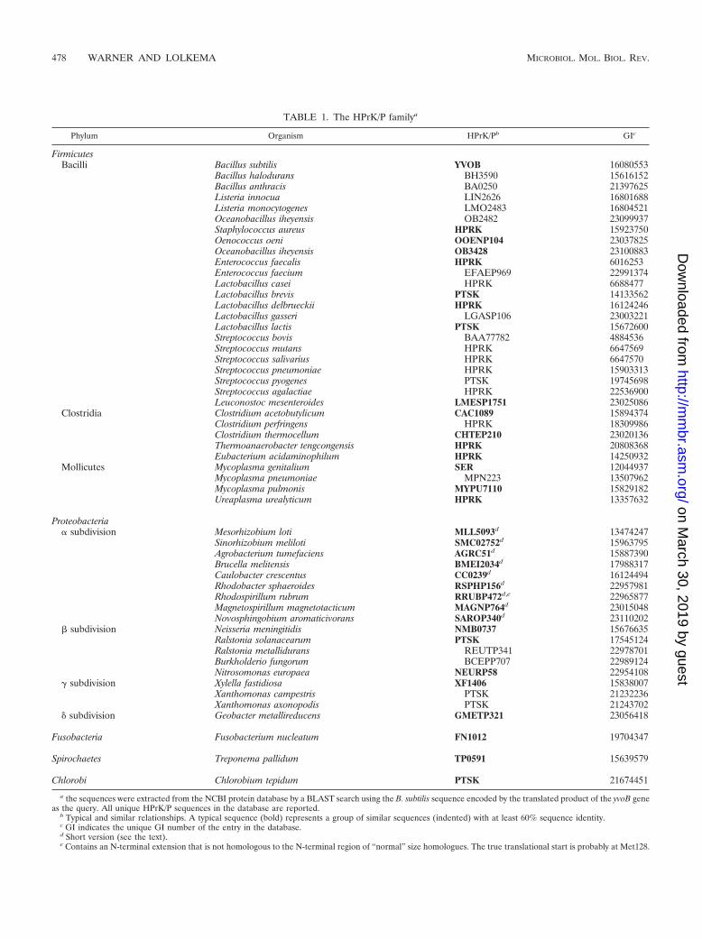
TABLE 1. The HPrK/P familya
Phylum Organism HPrK/Pb GIc
FirmicutesBacilli Bacillus subtilis YVOB 16080553
Bacillus halodurans BH3590 15616152Bacillus anthracis BA0250 21397625Listeria innocua LIN2626 16801688Listeria monocytogenes LMO2483 16804521Oceanobacillus iheyensis OB2482 23099937Staphylococcus aureus HPRK 15923750Oenococcus oeni OOENP104 23037825Oceanobacillus iheyensis OB3428 23100883Enterococcus faecalis HPRK 6016253Enterococcus faecium EFAEP969 22991374Lactobacillus casei HPRK 6688477Lactobacillus brevis PTSK 14133562Lactobacillus delbrueckii HPRK 16124246Lactobacillus gasseri LGASP106 23003221Lactobacillus lactis PTSK 15672600Streptococcus bovis BAA77782 4884536Streptococcus mutans HPRK 6647569Streptococcus salivarius HPRK 6647570Streptococcus pneumoniae HPRK 15903313Streptococcus pyogenes PTSK 19745698Streptococcus agalactiae HPRK 22536900Leuconostoc mesenteroides LMESP1751 23025086
Clostridia Clostridium acetobutylicum CAC1089 15894374Clostridium perfringens HPRK 18309986Clostridium thermocellum CHTEP210 23020136Thermoanaerobacter tengcongensis HPRK 20808368Eubacterium acidaminophilum HPRK 14250932
Mollicutes Mycoplasma genitalium SER 12044937Mycoplasma pneumoniae MPN223 13507962Mycoplasma pulmonis MYPU7110 15829182Ureaplasma urealyticum HPRK 13357632
Proteobacteria� subdivision Mesorhizobium loti MLL5093d 13474247
Sinorhizobium meliloti SMC02752d 15963795Agrobacterium tumefaciens AGRC51d 15887390Brucella melitensis BMEI2034d 17988317Caulobacter crescentus CC0239d 16124494Rhodobacter sphaeroides RSPHP156d 22957981Rhodospirillum rubrum RRUBP472d,e 22965877Magnetospirillum magnetotacticum MAGNP764d 23015048Novosphingobium aromaticivorans SAROP340d 23110202
� subdivision Neisseria meningitidis NMB0737 15676635Ralstonia solanacearum PTSK 17545124Ralstonia metallidurans REUTP341 22978701Burkholderio fungorum BCEPP707 22989124Nitrosomonas europaea NEURP58 22954108
� subdivision Xylella fastidiosa XF1406 15838007Xanthomonas campestris PTSK 21232236Xanthomonas axonopodis PTSK 21243702
� subdivision Geobacter metallireducens GMETP321 23056418
Fusobacteria Fusobacterium nucleatum FN1012 19704347
Spirochaetes Treponema pallidum TP0591 15639579
Chlorobi Chlorobium tepidum PTSK 21674451
a the sequences were extracted from the NCBI protein database by a BLAST search using the B. subtilis sequence encoded by the translated product of the yvoB geneas the query. All unique HPrK/P sequences in the database are reported.
b Typical and similar relationships. A typical sequence (bold) represents a group of similar sequences (indented) with at least 60% sequence identity.c GI indicates the unique GI number of the entry in the database.d Short version (see the text).e Contains an N-terminal extension that is not homologous to the N-terminal region of “normal” size homologues. The true translational start is probably at Met128.
478 WARNER AND LOLKEMA MICROBIOL. MOL. BIOL. REV.
on March 30, 2019 by guest
http://mm
br.asm.org/
Dow
nloaded from
final 6 organisms an HPrK homologue will be found eventu-ally. All HPr-like molecules from the phylum Firmicutes con-tained the regulatory-site serine, while all 34 bacteria, exceptfor Ureaplasma urealyticum (see below), contained at least oneHPr-like molecule with the active site histidine. Clearly, CcpA-dependent carbon catabolite repression is the mechanism inthe phylum Firmicutes.
The eight HPr-like proteins in the complete set that do notcontain the active-site histidine and, therefore, potential Crhmolecules are all found in the Firmicutes (Table 2). Five ofthese proteins are found in bacteria (B. subtilis, Bacillus halo-durans, Bacillus anthracis, Oceanobacillus iheyensis, and Ther-moanaerobacter tengcongensis) that, in addition, contain anHPr-like protein with the active-site histidine. Clostridium ther-mocellum contains three HPr-like molecules, one with and twowithout the active-site histidine. Finally, Ureaplasma urealyti-cum is the only bacterium that contains a single HPr-like mol-ecule without the active-site histidine. In B. subtilis Crh, theactive-site histidine is replaced by a glutamine while the adja-cent residues are still very similar as in HPr sequences ofsimilar organisms (see also Table 5, region A). A multiplesequence alignment of the HPr-like molecules revealed thesame sequence motif in the three HPr-like proteins of theother bacilli that therefore contain Crh in addition to HPr. Aninvestigation of the unfinished genome sequence data ofGeobacillus stearothermophilus also revealed a second HPr-likemolecule in addition to HPr, with the same characteristics of B.subtilis Crh (www.genome.ou.edu/bstearo.html) (not in Table2). T. tengcongensis, which belongs to the clostridia, containstwo HPr-like molecules, one with the active-site histidine
(FRUB) and one in which the histidine is deleted (TTE0115).Apart from the different active-site substitution, the moleculediffers from the bacillus Crhs in that the conserved sequencemotif around the site is not retained (see Table 5). Neverthe-less, the missing histidine suggests a function other than inPTS-mediated transport, possibly in Crh-mediated signaltransduction. C. thermocellum is the only bacterium in thephylum Firmicutes with three HPr-like molecules, one contain-ing the active-site histidine (CHTEP203) and two without(CHTEP180 and CHTEP182). CHTEP180 is closely related toTTE0115 of T. tengcongensis and also shows a deletion at theposition of the active-site histidine. These two sequences arethe most distant from the other members of the family (Fig. 3).In the second sequence of C. thermocellum without the active-site histidine (CHTEP182), His is replaced by Glu and, also,the surrounding region is not conserved as in the bacillus Crhs.Remarkably, the two HPr-like proteins of T. tengcongensis andC. thermocellum, FRUB and CHTEP203, respectively, thatcontain the active-site histidine residues are the closest rela-tives of the Crh proteins of the Bacillus species (Fig. 3).
The pathogen U. urealyticum contains a single HPr-like pro-tein (PTSH) in which no active-site histidine is present. Inagreement, the genome sequence does not contain a genehomologous to EI, the PTS enzyme responsible for the phos-phorylation of HPr(His) from phosphoenolpyruvate, but doescontain an HPrK/P homologue, that catalyzes the phosphory-lation of HPr/Crh(Ser) from ATP. The analysis suggests thatU. urealyticum has lost the PTS uptake system but has retainedCrh-mediated signal transduction (11). The organism belongsto the class Mollicutes, which is considered to represent mini-
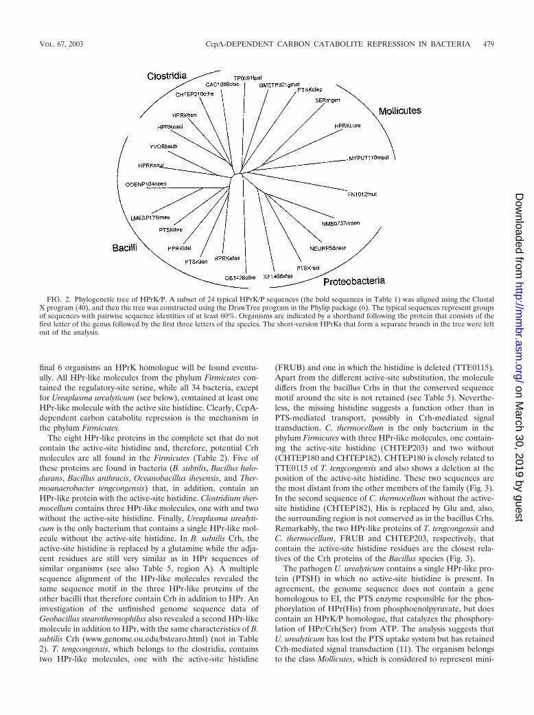
FIG. 2. Phylogenetic tree of HPrK/P. A subset of 24 typical HPrK/P sequences (the bold sequences in Table 1) was aligned using the ClustalX program (40), and then the tree was constructed using the DrawTree program in the Phylip package (6). The typical sequences represent groupsof sequences with pairwise sequence identities of at least 60%. Organisms are indicated by a shorthand following the protein that consists of thefirst letter of the genus followed by the first three letters of the species. The short-version HPrKs that form a separate branch in the tree were leftout of the analysis.
VOL. 67, 2003 CcpA-DEPENDENT CARBON CATABOLITE REPRESSION IN BACTERIA 479
on March 30, 2019 by guest
http://mm
br.asm.org/
Dow
nloaded from

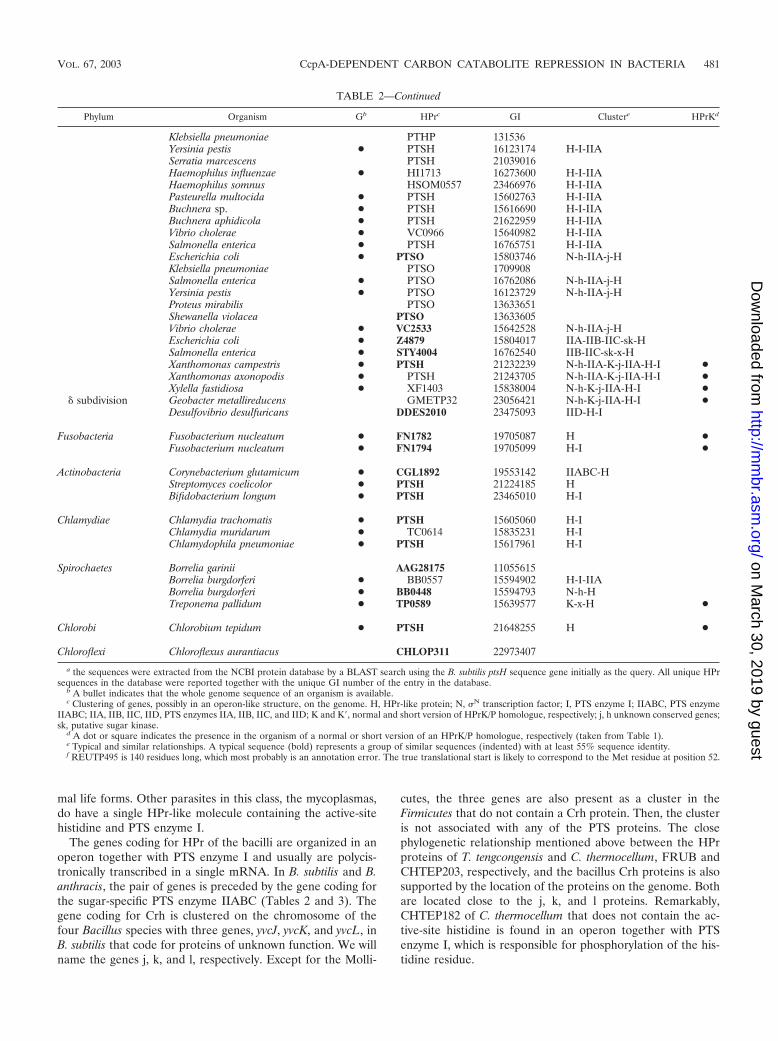
TABLE 2. The family of HPr-like proteinsa
Phylum Organism Gb HPre GI Clusterc HPrKd
FirmicutesBacilli Bacillus subtilis ● PTSH 16078454 IIABC-H-I ●
Bacillus anthracis ● BA4727 21402102 IIABC-H-I ●Bacillus megaterium PTHP 13633375Bacillus halodurans ● PTSH 15615636 H-I ●Geobacillus stearothermophilus PTHP 1172717 H-IStaphylococcus carnosus PTHP 131538Staphylococcus xylosus PTSH 11596363 ●Staphylococcus aureus ● PTSH 15924073 H-I ●Lactobacillus casei PTHP 13633570 ●Lactobacillus sakei PTHP 3122641Lactobacillus gasseri LGASP108 23003237 H-I ●Lactobacillus brevis PTSH 14133565 ●Leuconostoc mesenteroides LMESP115 23024451 H-I ●Lactococcus lactis ● PTHP 13633652 H-I ●Enterococcus faecalis PTHP 1172718 ●Streptococcus mutans PTHP 1172720 ●Streptococcus salivarius PTHP 1172721 ●Streptococcus bovis PTHP 13633635 ●Streptococcus pyogenes ● PTSH 15675305 H-I ●Streptococcus pneumoniae ● SP1177 15901042 H-I ●Streptococcus thermophilus PTSH 17978653Streptococcus agalactiae ● PTSH 22536985 H-I ●Oceanobacillus iheyensis ● OB2344 23099799 H ●Oenococcus oeni OOENP127 23038069 ●Listeria innocua ● PTSH 16800070 H-I ●Bacillus subtilis ● CRH 16080527 j-k-l-H ●Bacillus halodurans ● CHR 15616128 j-k-l-H ●Bacillus anthracis ● BA0239 21397614 j-k-l-H ●Oceanobacillus iheyensis ● OB2465 23099920 j-k-l-H ●
Clostridia Thermoanaerobacter tengcongensis ● FRUB 20808237 j-k-x10-l-H ●Clostridium thermocellum CHTEP203 23020129 j-k-x-l-H ●Thermoanaerobacter tengcongensis ● TTE0115 20806644 H ●Clostridium thermocellum CHTEP180 23020106 H ●Clostridium thermocellum CHTEP182 23021750 H-I ●Clostridium acetobutylicum ● CAC1820 15895096 H ●Clostridium perfringens ● HPR 18310651 H ●
Mollicutes Mycoplasma capricolum PTHP 1172719Mycoplasma genitalium ● PTSH 12044891 H ●Mycoplasma pneumoniae ● PTSH 13507792 H ●Mycoplasma pulmonis ● MYPU6030 15829074 H-I ●Ureaplasma urealyticum ● PTSH 13358152 H ●
Proteobacteria� subdivision Mesorhizobium loti ● MSL5292 13474414 H-I ●
Mesorhizobium loti ● MSL5090 13474245 K�-IIA-H fSinorhizobium meliloti ● SMC02754 15963793 K�-IIA-H fAgrobacterium tumefaciens ● AGRC49 15887388 K�-IIA-H fBrucella melitensis ● BMEI2031 17988314 K�-?-IIA-H fRhodopseudomonas palustris RPALP163 22962348 K�-IIA-H fCaulobacter crescentus ● CC0241 16124496 K�-IIA-H fRhodobacter sphaeroides RSPHP157 22957984 K�-j-IIA-H fMagnetospirillum magnetotacticum MAGNP479 23011831 fNovosphingobium aromaticivorans SAROP34 23110205 K�-j-IIA-H fRhodospirillum rubrum RRUBP475 22965880 K�-j-IIA-H-I fMagnetospirillum magnetotacticum MAGNP76 23015045 K�-j-IIA-H-I f
� subdivision Ralstonia eutropha PTHP 131532Ralstonia solanacearum ● PSTH 17545066 IIA-H-I ●Ralstonia metallidurans REUTP495f 22980242 IIA-H-I ●Nitrosomonas europaea NEURP196 22955983 - ●Burkholderia fungorum BCEPP510 22987169 IIA-H-I ●Neisseria meningitidis ● NMB2045 15677867 IIA-H-I ●
� subdivision Pseudomonas putida PTHP 13633649Pseudomonas aeruginosa ● PA4466 15599662 N-h-IIA-j-HPseudomonas fluorescens PFLUP387 23061779 N-h-IIA-j-HPseudomonas stutzeri PTSO 22138783Microbulbifer degradans MDEGP161 23027798 N-IIA-j-HAzotobacter vinelandii AVINP175 23103593 N-h-IIA-j-HEscherichia coli ● PTSH 15802948 H-I-IIA
Continued on following page
480 WARNER AND LOLKEMA MICROBIOL. MOL. BIOL. REV.
on March 30, 2019 by guest
http://mm
br.asm.org/
Dow
nloaded from
mal life forms. Other parasites in this class, the mycoplasmas,do have a single HPr-like molecule containing the active-sitehistidine and PTS enzyme I.
The genes coding for HPr of the bacilli are organized in anoperon together with PTS enzyme I and usually are polycis-tronically transcribed in a single mRNA. In B. subtilis and B.anthracis, the pair of genes is preceded by the gene coding forthe sugar-specific PTS enzyme IIABC (Tables 2 and 3). Thegene coding for Crh is clustered on the chromosome of thefour Bacillus species with three genes, yvcJ, yvcK, and yvcL, inB. subtilis that code for proteins of unknown function. We willname the genes j, k, and l, respectively. Except for the Molli-
cutes, the three genes are also present as a cluster in theFirmicutes that do not contain a Crh protein. Then, the clusteris not associated with any of the PTS proteins. The closephylogenetic relationship mentioned above between the HPrproteins of T. tengcongensis and C. thermocellum, FRUB andCHTEP203, respectively, and the bacillus Crh proteins is alsosupported by the location of the proteins on the genome. Bothare located close to the j, k, and l proteins. Remarkably,CHTEP182 of C. thermocellum that does not contain the ac-tive-site histidine is found in an operon together with PTSenzyme I, which is responsible for phosphorylation of the his-tidine residue.
TABLE 2—Continued
Phylum Organism Gb HPrc GI Clustere HPrKd
Klebsiella pneumoniae PTHP 131536Yersinia pestis ● PTSH 16123174 H-I-IIASerratia marcescens PTSH 21039016Haemophilus influenzae ● HI1713 16273600 H-I-IIAHaemophilus somnus HSOM0557 23466976 H-I-IIAPasteurella multocida ● PTSH 15602763 H-I-IIABuchnera sp. ● PTSH 15616690 H-I-IIABuchnera aphidicola ● PTSH 21622959 H-I-IIAVibrio cholerae ● VC0966 15640982 H-I-IIASalmonella enterica ● PTSH 16765751 H-I-IIAEscherichia coli ● PTSO 15803746 N-h-IIA-j-HKlebsiella pneumoniae PTSO 1709908Salmonella enterica ● PTSO 16762086 N-h-IIA-j-HYersinia pestis ● PTSO 16123729 N-h-IIA-j-HProteus mirabilis PTSO 13633651Shewanella violacea PTSO 13633605Vibrio cholerae ● VC2533 15642528 N-h-IIA-j-HEscherichia coli ● Z4879 15804017 IIA-IIB-IIC-sk-HSalmonella enterica ● STY4004 16762540 IIB-IIC-sk-x-HXanthomonas campestris ● PTSH 21232239 N-h-IIA-K-j-IIA-H-I ●Xanthomonas axonopodis ● PTSH 21243705 N-h-IIA-K-j-IIA-H-I ●Xylella fastidiosa ● XF1403 15838004 N-h-K-j-IIA-H-I ●
� subdivision Geobacter metallireducens GMETP32 23056421 N-h-K-j-IIA-H-I ●Desulfovibrio desulfuricans DDES2010 23475093 IID-H-I
Fusobacteria Fusobacterium nucleatum ● FN1782 19705087 H ●Fusobacterium nucleatum ● FN1794 19705099 H-I ●
Actinobacteria Corynebacterium glutamicum ● CGL1892 19553142 IIABC-HStreptomyces coelicolor ● PTSH 21224185 HBifidobacterium longum ● PTSH 23465010 H-I
Chlamydiae Chlamydia trachomatis ● PTSH 15605060 H-IChlamydia muridarum ● TC0614 15835231 H-IChlamydophila pneumoniae ● PTSH 15617961 H-I
Spirochaetes Borrelia garinii AAG28175 11055615Borrelia burgdorferi ● BB0557 15594902 H-I-IIABorrelia burgdorferi ● BB0448 15594793 N-h-HTreponema pallidum ● TP0589 15639577 K-x-H ●
Chlorobi Chlorobium tepidum ● PTSH 21648255 H ●
Chloroflexi Chloroflexus aurantiacus CHLOP311 22973407
a the sequences were extracted from the NCBI protein database by a BLAST search using the B. subtilis ptsH sequence gene initially as the query. All unique HPrsequences in the database were reported together with the unique GI number of the entry in the database.
b A bullet indicates that the whole genome sequence of an organism is available.c Clustering of genes, possibly in an operon-like structure, on the genome. H, HPr-like protein; N, N transcription factor; I, PTS enzyme I; IIABC, PTS enzyme
IIABC; IIA, IIB, IIC, IID, PTS enzymes IIA, IIB, IIC, and IID; K and K�, normal and short version of HPrK/P homologue, respectively; j, h unknown conserved genes;sk, putative sugar kinase.
d A dot or square indicates the presence in the organism of a normal or short version of an HPrK/P homologue, respectively (taken from Table 1).e Typical and similar relationships. A typical sequence (bold) represents a group of similar sequences (indented) with at least 55% sequence identity.f REUTP495 is 140 residues long, which most probably is an annotation error. The true translational start is likely to correspond to the Met residue at position 52.
VOL. 67, 2003 CcpA-DEPENDENT CARBON CATABOLITE REPRESSION IN BACTERIA 481
on March 30, 2019 by guest
http://mm
br.asm.org/
Dow
nloaded from
Proteobacteria
All HPr-like molecules found in the phylum Proteobacteriacontain the active-site histidine. Most HPr-like molecules arefound in the � subdivision, which contains the typical gram-negative bacteria such as Escherichia coli (Table 2). A numberof these, Klebsiella pneumoniae, E. coli, Yersinia pestis, Vibriocholerae, and Salmonella enterica, possess more than one HPr-like molecule but no HPrK/P. These organisms contain, in
addition to HPr, a homologue termed NPr, which is a compo-nent of the Ntr regulatory system involved in the regulation ofnitrogen metabolism, among others (26). The NPrs form aseparate cluster in the phylogenetic analysis of the HPr-likemolecules (the ptsO genes [Fig. 3]). HPr and NPr moleculesare distinguished by the clustering of the coding genes withother genes on the chromosome (Tables 2 and 3). The genecoding for HPr (ptsH) is organized together with genes codingfor the PTS enzymes EI and glucose-specific IIAGlc in the pts
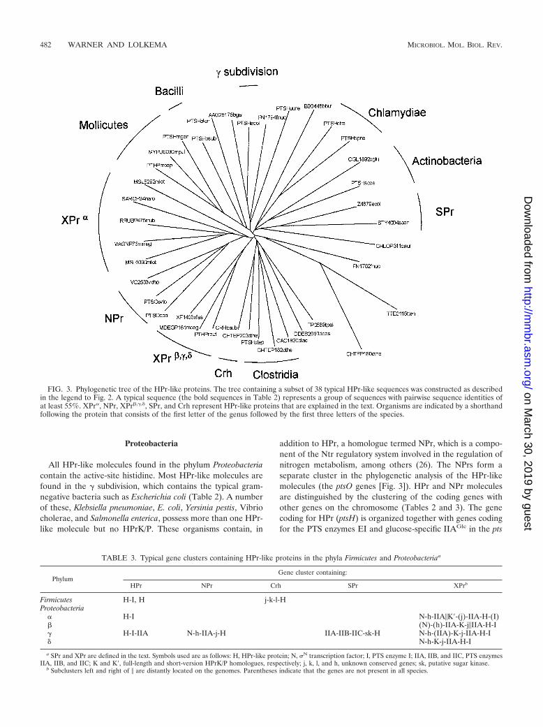
FIG. 3. Phylogenetic tree of the HPr-like proteins. The tree containing a subset of 38 typical HPr-like sequences was constructed as describedin the legend to Fig. 2. A typical sequence (the bold sequences in Table 2) represents a group of sequences with pairwise sequence identities ofat least 55%. XPr�, NPr, XPr�,�,�, SPr, and Crh represent HPr-like proteins that are explained in the text. Organisms are indicated by a shorthandfollowing the protein that consists of the first letter of the genus followed by the first three letters of the species.
TABLE 3. Typical gene clusters containing HPr-like proteins in the phyla Firmicutes and Proteobacteriaa
PhylumGene cluster containing:
HPr NPr Crh SPr XPrb
Firmicutes H-I, H j-k-l-HProteobacteria
� H-I N-h-IIA�K�-(j)-IIA-H-(I)� (N)-(h)-IIA-K-j�IIA-H-I� H-I-IIA N-h-IIA-j-H IIA-IIB-IIC-sk-H N-h-(IIA)-K-j-IIA-H-I� N-h-K-j-IIA-H-I
a SPr and XPr are defined in the text. Symbols used are as follows: H, HPr-like protein; N, N transcription factor; I, PTS enzyme I; IIA, IIB, and IIC, PTS enzymesIIA, IIB, and IIC; K and K�, full-length and short-version HPrK/P homologues, respectively; j, k, l, and h, unknown conserved genes; sk, putative sugar kinase.
b Subclusters left and right of � are distantly located on the genomes. Parentheses indicate that the genes are not present in all species.
482 WARNER AND LOLKEMA MICROBIOL. MOL. BIOL. REV.
on March 30, 2019 by guest
http://mm
br.asm.org/
Dow
nloaded from
operon in the order H-I-IIA. The three proteins are the PTScomponents involved in CRP-dependent carbon catabolite re-pression. The gene coding for NPr (ptsO) is located elsewhereon the chromosome in the Ntr cluster, together with four othergenes that are involved in the Ntr regulatory pathway (N-h-IIA-j-H). The cluster starts with an RNA polymerase 54 factor(N) followed by a putative 54 modulation protein (h), a PTSIIA homologue termed IIANtr, a P-loop-containing protein (j),and, finally, the gene coding for NPr. The P-loop-containingprotein j is homologous to the j protein in the j-k-l-Crh clusterfound in the Firmicutes (Table 3). The IIANtr protein and NPr,together with a third protein termed INtr, form an independentphosphoryl group transfer chain that uses phosphoenolpyru-vate as the donor (Fig. 1C). INtr is a multidomain proteinconsisting of a homologue of the PTS EI and a domain alsofound in NifA activator proteins (30).
The whole-genome sequences of Haemophilus influenzae,Pasteurella multocida, Buchnera sp., and Buchnera aphidicola inthe � subdivision contain a single HPr-like molecule embeddedin an HPr-like gene cluster (Table 2), suggesting that theseorganisms do not use NPr-mediated regulation. For sequencesimilarity reasons, the same is likely to be true for Serratiamarcescens and Haemophilus somnus. In contrast, the genomeof Pseudomonas aeruginosa contains a single HPr-like mole-cule embedded in an NPr-like gene cluster that, therefore, islikely to be an NPr species. The strictly aerobic Pseudomonasand also the Azotobacter species were thought not to use thePTS for uptake of sugars, except for fructose (32). Analysis ofthe P. aeruginosa genome revealed two complete sugar-specificPTSs, the one for fructose and another one for N-acetylglu-cosamine, in which HPr moieties are present as components ofmultidomain proteins (29). These HPr domains are not likelyto play a role in sugar uptake as general PTS components. ThePseudomonas and Azotobacter genera (and the same may betrue for the Shewanella, Proteus, and Microbulbifer genera [Ta-ble 2]) use an HPr-like protein only for regulatory purposes.
E. coli and S. enterica (and also S. enterica serovar Typhi-murium) both have, in addition to HPr and NPr, a third HPr-like molecule, Z4879 and STY4004, respectively. In E. coli, theprotein is present only in the enterohemorrhagic strainsOD157:H7 and OD157:H7 EDL933 and not in the K-12 strain.The proteins in the Escherichia and Salmonella strains areclosely related (Fig. 3) and are located in a similar gene clusteron the genome (Table 2). Upstream of the gene coding for theHPr-like protein, a gene annotated as a sugar kinase (sk) anda complete set of PTS enzyme II proteins (IIA-IIB-IIC) arelocated. The HPr-like protein in the cluster may represent asugar-specific HPr (SPr, for “sugar-specific HPr” [Table 3]).
Three bacteria in the �-subdivision contain an HPrK homo-logue: Xanthomonas campestris, Xanthomonas axonopodis, andXylella fastidiosa (Table 2). They also contain a single HPr-likeprotein; remarkably, both are in the same gene cluster thatconsists of seven or eight genes (N-h-IIA-K-j-IIA-H-I). We willterm this cluster and the single HPr-like protein the X-clusterand XPr, respectively (for “Xanthomonas cluster” and “Xan-thomonas HPr”). The first genes in the X-cluster form an Ntrgene cluster from which the gene coding for NPr is missing andin which the gene coding for HPrK is inserted upstream of thej gene (Table 3). The IIA protein encoded in this part of thecluster is of the IIANtr type and is not found in the X. fastidiosa
cluster (Table 2). A second IIA gene and the genes coding forXPr and PTS enzyme I follow the Ntr-like cluster. The secondIIA protein is of the IIAMan type, homologues of the IIAdomain of the mannose-specific IIABMan that is part of themannose uptake system in E. coli (34). The two Xanthomonasspecies and X. fastidiosa lack both NPr and INtr; i.e., it is notclear how the IIANtr protein encoded in the X-cluster is phos-phorylated. An exact copy of the X-cluster found in X. fastid-iosa, without IIANtr, is also found in Geobacter metallireducensin the � subdivision of the Proteobacteria.
With the exceptions of Mesorhizobium loti and Magnetospi-rillum magnetotacticum in the � subdivision, the bacteria in the� and � subdivisions of the Proteobacteria contain a singleHPr-like molecule (Table 2). The complete genome sequenceand the data from unfinished genomes suggest that the pres-ence of HPrK in both subdivisions is common. The HPrKs ofthe � subdivision are all of the short-version type (see above)(14), and the HPr-like proteins are in the same gene clusterthat, in addition, contains at least a IIA molecule of the IIAMan
type. The cluster resembles the last part of the X-cluster foundin the � and � subdivisions, but the genes coding for protein jand enzyme I are not always present (Table 3). The Rhodospi-rillum rubrum and M. magnetotacticum clusters are an exactmatch of this part of the X-cluster (Table 2). The genes in theremaining part of the X-cluster, N-h-IINtr, are also found clus-tered on the genome of the bacteria in the � subdivision butare located distantly from the XPr/HPrK part of the cluster(Table 3). All bacteria in the � subdivision contain the genecoding for INtr, while only M. loti, M. magnetotacticum, and R.rubrum contain the classical enzyme I. The presence of enzymeI correlates with the presence of a second HPr-like protein inM. loti and M. magnetotacticum.
The situation observed in the � subdivision is similar, but theX-cluster is broken up in different parts (Table 3). The singlegene coding for the HPr-like molecule (XPr) clusters with thegenes coding for enzyme I and IIAMan in the same order as inthe X-cluster of the � and � subdivisions. The gene coding forHPrK clusters elsewhere on the genome, together with IIANtr
and protein j in the order IIA-K-j. In the complete genomesequences of Ralstonia solanacearum and Neisseria meningiti-dis, the three genes are preceded only in the latter organism bythe N and h genes (Tables 2 and 3). A difference with the �subdivision is that in the � subdivision, all bacteria contain thePTS enzyme I but not INtr.
Summarizing, HPrK is found in all four subdivisions of theProteobacteria and seems to cluster on the genomes togetherwith the genes coding for components involved in Ntr-type ofgene regulation. The X-cluster in the Xanthomonas species inthe � subdivision represents the most complete cluster, whilefission of the cluster is observed in the � and � subdivisions.
Other Phyla
In the phyla other than Firmicutes and Proteobacteria, mostorganisms have a single HPr-like molecule that clusters withthe gene coding for enzyme I (H-I), as found in the Firmicutes(Table 2). Three bacteria contain an HPrK homologue, Fuso-bacterium nucleatum, Treponema pallidum, and Chlorobiumtepidum. F. nucleatum contains two HPr-like molecules, one ofwhich, FN1794, is adjacent to the gene coding for PTS EI,indicating that FN1794 is HPr. The second gene, FN1782, is
VOL. 67, 2003 CcpA-DEPENDENT CARBON CATABOLITE REPRESSION IN BACTERIA 483
on March 30, 2019 by guest
http://mm
br.asm.org/
Dow
nloaded from

located a couple of genes upstream of this pair in no apparentoperon structure. The genome contains genes coding forIIANtr and the j protein, but, like HPrK, these do not all clustertogether on the genome. No homologues are found for IIAMan
or INtr. On the genome of T. pallidum, the genes coding for theHPrK homologue and the HPr-like protein are separated by asingle gene, which is not related to any of the genes found inthe X-cluster in the Proteobacteria. Both T. pallidum and C.tepidum contain IIANtr encoded elsewhere on the genome, butthey contain no IIAMan, j protein, or INtr. The HPr-like mol-ecules of the bacteria in this category that do contain HPrK donot seem to be closely related to the HPr-like molecules foundin the X-cluster of the Proteobacteria (Fig. 3).
SEQUENCE ANALYSES
Full-Length and Short-Version HPr Kinases
The genes coding for the short-version HPrK homologues inthe � subdivision are organized, together with the HPr-likeprotein XPr, in a similar gene cluster (the X-cluster) to that ofthe full-length versions in the �, �, and � subdivisions of theProteobacteria, strongly suggesting that they serve the samefunction, putatively as HPr kinases/phosphatases. HPrK isknown to consist of two domains. The three-dimensional struc-ture of HPrK of Staphylococcus xylosus resolved at 1.95 Åshows a hexameric arrangement (a dimer of trimers) in whichthe N-terminal and C-terminal domains are well separated,with no apparent intramolecular contacts (20). While the func-tion of the N-terminal domain is not clear, the C-terminaldomain is the catalytic domain. Deletion of the N-terminal 127residues of Lactobacillus casei HPrK, more or less correspond-ing to the N-terminal domain, yielded an active entity whosethree-dimensional structure was resolved separately at 2.8 Å(8). The structures of the C-terminal catalytic domains (Fig.4A) from the two organisms closely matched each other. Mul-tiple sequence alignment of the full-length and short-versionHPrK homologues suggests that the latter corresponds to thecatalytic domain; the beginning of the short versions correlatesmore or less with the beginning of the C-terminal domain ofthe full-length HPrKs. However, the length of the short ver-sions and the C-terminal domains of the full-length proteinsdiffer by roughly 50 residues; they are about 150 and 200residues, respectively. The C-terminal domain of the full-length HPrK contains four conserved sequence motifs (A, B,C, and D in Fig. 4B), the first of which contains the so-calledWalker A motif typical for the binding of the phosphate groupsof ATP (43). Motifs A, B, and C are also found in the short-version homologues, but motif D is missing (Fig. 4B and C). Infact, the sequence similarity of the two versions covers onlyapproximately the first 100 residues of the short version, thepart that contains sequence motifs A, B, and C. The C-terminal50 residues of the short versions do not seem to be related tothe corresponding area in the full-length proteins. It followsthat the short versions correspond to the top part in the struc-ture depicted in Fig. 4A, up to strand �J. The C-terminal partof the full-length proteins, consisting of �J, �K, �3, and �4,would be missing. In the crystal structures (8, 20), the loopbetween �K and �3 in conserved domain D (the K3 loop) andthe two �-helices �3 and �4 are involved in intimate contactswithin two pairs of trimers that form the overall hexameric
structure of the complex. The short versions may not form amultimeric structure and may instead exist as monomers.
The structure of the catalytic domain of the L. casei HPrK/Pin complex with B. subtilis HPr was resolved at 2.8 Å (7). Thehexameric HPrK/P complex bound six HPr molecules, each atthe interface of two monomers. The HPr molecules bound attwo separate interfaces, one at each monomer. The catalyticinteraction, positioning the regulatory-site serine of HPr closeto the Walker A motif of HPrK, involves strand �A and con-served motifs A and B on one of the monomers (Fig. 4A). Itfollows that this interaction is still possible in the short-versionHPrKs. The second interface involves C-terminal helix �4 onthe neighboring monomer. This interaction is absent in theshort versions. The analysis supports the conclusion of thegenetic analysis, i.e., that the short-version HPrK homologuesactually may function as HPrK/phosphatases.
Phylogenetic Relationships of HPr-Like Proteins
Phylogenetic analysis of the HPr-like proteins shows that theHPrs from the gram-positive (lacto)bacilli are closely relatedwhile those from the clostridia are more distant (Fig. 3). Thefour Crh and four HPr proteins of B. subtilis, B. halodurans, B.anthracis, and O. iheyensis cluster in different branches of thetree. The Crh proteins are more closely related to the HPrproteins of the clostridia than to the HPr proteins of the bacilli.Pairwise sequence analysis revealed a higher sequence identitybetween the HPr sequences of the different species and theCrh sequences of the different species than between HPr andCrh of the same species (Table 4). If Crh had originated froman HPr gene duplication during evolution, this must have hap-pened before the primordial Bacillus diverged into the differ-ent species. A similar situation exists for the HPr and NPrproteins in gram-negative bacteria (Fig. 3).
The putative Crh proteins found in the clostridia, TTE0115of the thermophile T. tengcongensis and CHTEP180 andCHTEP182 of C. thermocellum, and PTSH of the mollicute U.urealyticum are not in the same cluster as the bacillus Crhs.Closest is CHTEP182 in the clostridium cluster. TTE0115 andCHTEP180 are the most distant relatives in the HPr-like pro-tein family, and PTSH of U. urealyticum clusters loosely withHPr-like proteins from the phyla Fusobacteria and Spirochaetesand HPrs from the � subdivision of the Proteobacteria (Fig. 3).Consistent with the phylogenetic distance between Crh andHPr of the bacillus species, PTSH of U. urealyticum is quitedistant from HPr of the related mollicutes and TTE0115 andCHTEP180 are distant from the clostridium branch. The con-servation that is observed in the region around the mutatedactive-site histidine residue in the bacillus Crh proteins is com-pletely absent in the four putative Crh proteins (see Table 5).Apart from the mutated active-site histidine, the latter do notseem to have much in common with the former. The gram-negative bacteria of the �, �, and � subdivisions of the Pro-teobacteria are likely to have CcpA-dependent CCR since theypossess HPrK. The HPr-like proteins of the � subdivision(XPr�) all cluster in one branch of the tree (Fig. 3). TheHPr-like proteins of the � and � subdivisions are on the samebranch as the XPr proteins of the bacteria in the � subdivisionof the Proteobacteria that possess HPrK (Xanthomonas andXylella species; XPr�,�,�). The branch is distant from HPr of the
484 WARNER AND LOLKEMA MICROBIOL. MOL. BIOL. REV.
on March 30, 2019 by guest
http://mm
br.asm.org/
Dow
nloaded from

gram-negative bacteria in the � subdivision that do not containHPrK (e.g., Escherichia and Klebsiella species). Both the XPr�
and XPr�,�,� proteins are distant from the gram-positive HPrproteins. Importantly, the XPr�,�,� proteins and, especially, theXPr� proteins are loosely associated with the NPr proteinsfrom the � subdivision. Moreover, the XPr proteins of the �, �,and � subdivisions have 55% sequence identity to the HPr-like proteins of the Pseudomonas species in the � subdivisionthat are likely to function in Ntr-type regulation (see above).
Sequence Motifs in HPr-Like Proteins
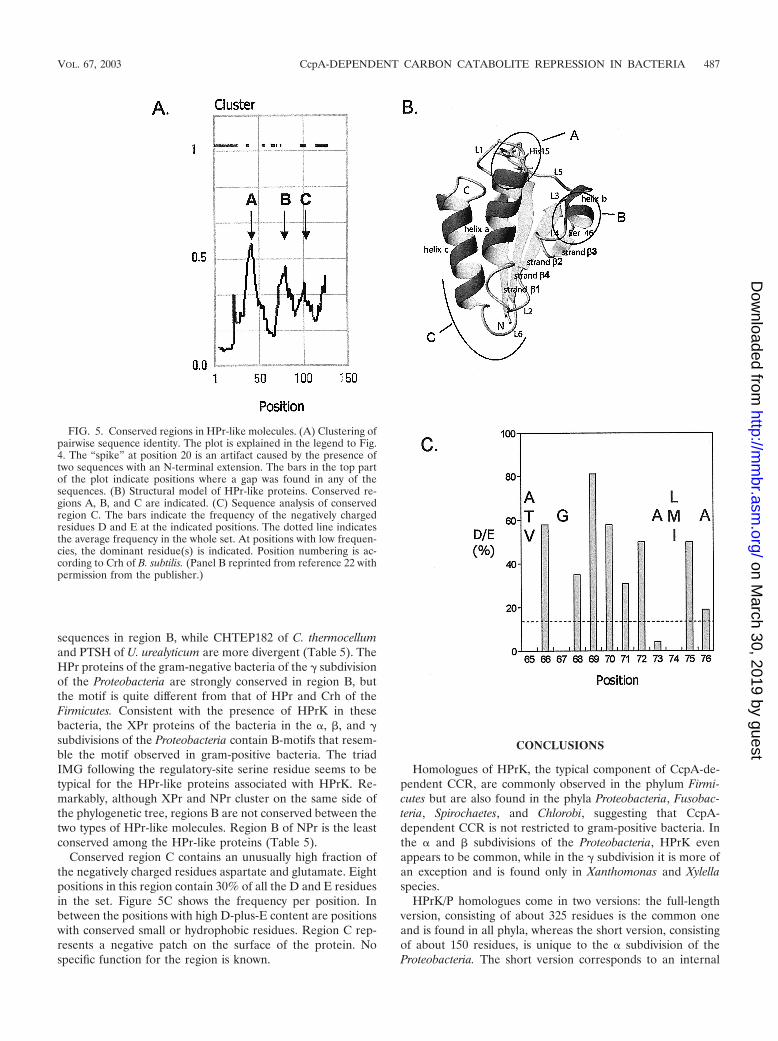
None of the residues in a multiple sequence alignment of theHPr-like family of proteins was completely conserved (data not
shown). An analysis of an all-against-all pairwise sequencealignment reveals the presence of three well-conserved re-gions: region A around position 16 (B. subtilis Crh numbering),region B around position 46, and region C around position 70in the C terminus (Fig. 5A). The solution structures of a num-ber of HPr proteins originating from different species and ofCrh of B. subtilis have been solved by nuclear magnetic reso-nance spectroscopy, and all of them show a similar overallfolding (5, 16, 22, 42). The HPr polypeptide folds as an open-faced �-sandwich, with three �-helices (a, b, and c [Fig. 5B]) ontop of a four-stranded �-sheet (strands �1 to �4 [Fig. 5B]). Ina linear representation, the order of the secondary-structureelements would be �1a�2�3b�4c. Conserved region A contains
FIG. 4. Sequence signatures in full-length and short-version HPrK homologues. (A) Structural model of the C-terminal domain of thefull-length HPrK/P. Ovals indicate conserved regions A, B, C, and D. (B) Pairwise sequence identity in the multiple sequence alignments of theshort-version (top) and full-length (bottom) HPr kinases. The “cluster” function gives the fraction of identities at each position in the alignmentin an all-against-all comparison. The scores were averaged over a sliding window of nine positions. The plot of the short-version HPrKs was“aligned” with the plot of the full-length versions based on the multiple sequence alignment. The set of full-length sequences used in the alignmentis described in the legend to Fig. 2, and the set of short versions is taken from Table 2 (Proteobacteria, � subdivision). The bars in the top half ofthe plots indicate positions where a gap occurs in any of the sequences in the alignment. The four conserved regions in the full-length HPrKhomologues are indicated as A to D. (C) Sequence motifs corresponding to regions A to D in panels A and B. The top and bottom sequences inthe boxes correspond to the full-length and short-version HPr kinases, respectively. Conserved motif D is not present in the short-version HPrkinases. (Panel A reprinted from reference 8 with permission from the publisher.)
VOL. 67, 2003 CcpA-DEPENDENT CARBON CATABOLITE REPRESSION IN BACTERIA 485
on March 30, 2019 by guest
http://mm
br.asm.org/
Dow
nloaded from

the active-site histidine, which is positioned just in front of�-helix a. The region comprises the interface of the loop pre-ceding helix a and helix a. The region is involved in the inter-action of HPr and the PTS enzymes EI and IIA. Region Bcontains the regulatory-site serine, which is positioned at theinterface of the loop between �3 and �-helix b. The regioncovers most of helix b, which is only two turns long. Region Ccomprises the loop between �4 and �-helix c plus the first turnat the N-terminal end of helix c. Region C is quite distant fromthe other two conserved regions on the surface of the protein.The HPr-like molecules from the Actinobacteria membersCorynebacterium glutamicum and Streptomyces coelicolor, aswell as from Chloroflexus aurantiacus, contain an insertion of 4residues in the loop connecting strands �2 and �3, where theyare not likely to disturb the folding of the protein (not shown).
With few exceptions, conserved region A in the HPr, Crh,NPr, and XPr proteins contains the consensus sequence G(LI)H(AT)R(PA)(AS), with the active-site histidine in bold (Table5). It was noted before that in the bacillus Crhs, the motif isretained with the exception of the histidine-to-glutamine mu-tation, suggesting that Crh might still interact with the enzymesenzyme I and IIA (5). In contrast, in the putative Crh proteinsfrom the clostridia and from U. urealyticum, TTE0115,CHTEP180, CHTEP182, and PTSH, the motif is completelylost during evolution, suggesting that these proteins have lostthe interaction with the PTS uptake system.
The regulatory-site serine in conserved region B is the best-
conserved residue in the whole family. Only three HPr-likemolecules in the family do not contain a serine at this position:STY4004 of Salmonella enterica, CHLOP311 of Chloroflexusaurantiacus, and PTSH of Bifidobacterium longum (Table 2).None of these three organisms contains an HPrK homologue.The missing regulatory-site serine in the S. enterica protein ismost probably an alignment artifact since the sequence SILG,which is similar to the consensus sequence (see below), islocated 4 residues downstream. In contrast, the serine residuein B. longum is replaced by arginine while the surroundingresidues are still conserved. In the C. aurantiacus protein, theserine residue is deleted and the adjacent residues are notconserved. The high conservation of the serine residuethroughout the whole set makes it difficult to accept that theserine, or at least the conserved region, has no function inthose bacteria that lack HPr kinase.
Conserved region B shows more clustering of the sequencesfollowing the phylogeny of the bacteria (Tables 1 and 2) thanmay be detected in the case of conserved region A (Table 5).Even though the HPrs and Crhs of (lacto)bacilli are in separatebranches of the phylogenetic tree (Fig. 3), the regions aroundthe regulatory-site serines are almost identical. The consensussequence is VN(AL)KSIMG(VL)MSL(AG), in which the reg-ulatory-site serine is shown in bold. The HPr and Crh se-quences differ only at the third, ninth, and last positions, asindicated. The putative Crh proteins TTE0115 of T. tengcon-gensis and CHTEP180 of C. thermocellum have very similar
TABLE 4. Pairwise sequence identity of the HPr and Crh sequences of Bacillus speciesa
Sequence% Identity to:
HPRbsub HPRbhal HPRbant HPRoihe CRHbsub CRHbhal CRHbant CRHoihe
HPRbsub 64 72 61HPRbhal 61 70HPRbant 55HPRoiheCRHbsub 45 42 39 41 73 71 65CRHbhal 42 45 40 42 73 65CRHbant 48 41 44 39 60CRHoihe 42 43 38 36
a bsub, B. subtilis; bhal, B. halodurans; bant, B. anthracis; oihe, O. iheyensis.
TABLE 5. Consensus sequence around the active-site histidine (region A) and the regulatory-site serine (region B) in the HPr-like proteins
Phylum HPr-likeproteina Region Ab Region Bb
Firmicutes HPr G (ILV) H (AT) R P A N (AL) K S (IL) (MIL) G (VIL) M S LCrh G L Q A R P A N A K S I M G L M S L
TTE0115 I E S V D A K S I M G L F S LCHTEP180 I T D V D A K S I M G I F S LCHTEP182 G W E A K L A N A K S L L G V L S LPTSHuure S A F I K A I D A K S I I N I M A L
Proteobacteria� HPr G L H T R P A (NS) A K S L F K (IL) Q T L� NPr G (LM) H A R (PA) A A X S V X (AG) (ML) L M L� XPr G L H A R A (AS) (AG) X S I M G L M M L� XPr G L H A R A S (ND) (AG) K S I M G (VIL) M (MT) L� XPr G L H A R A (AT) (ND) (AG) K S I M G (VL) M L L
a XPr is defined in the text.b parentheses represent single positions where the indicated residues are found in different species. The active-site histidine and regulatory-site serine are shown in
bold type.
486 WARNER AND LOLKEMA MICROBIOL. MOL. BIOL. REV.
on March 30, 2019 by guest
http://mm
br.asm.org/
Dow
nloaded from
sequences in region B, while CHTEP182 of C. thermocellumand PTSH of U. urealyticum are more divergent (Table 5). TheHPr proteins of the gram-negative bacteria of the � subdivisionof the Proteobacteria are strongly conserved in region B, butthe motif is quite different from that of HPr and Crh of theFirmicutes. Consistent with the presence of HPrK in thesebacteria, the XPr proteins of the bacteria in the �, �, and �subdivisions of the Proteobacteria contain B-motifs that resem-ble the motif observed in gram-positive bacteria. The triadIMG following the regulatory-site serine residue seems to betypical for the HPr-like proteins associated with HPrK. Re-markably, although XPr and NPr cluster on the same side ofthe phylogenetic tree, regions B are not conserved between thetwo types of HPr-like molecules. Region B of NPr is the leastconserved among the HPr-like proteins (Table 5).
Conserved region C contains an unusually high fraction ofthe negatively charged residues aspartate and glutamate. Eightpositions in this region contain 30% of all the D and E residuesin the set. Figure 5C shows the frequency per position. Inbetween the positions with high D-plus-E content are positionswith conserved small or hydrophobic residues. Region C rep-resents a negative patch on the surface of the protein. Nospecific function for the region is known.
CONCLUSIONS
Homologues of HPrK, the typical component of CcpA-de-pendent CCR, are commonly observed in the phylum Firmi-cutes but are also found in the phyla Proteobacteria, Fusobac-teria, Spirochaetes, and Chlorobi, suggesting that CcpA-dependent CCR is not restricted to gram-positive bacteria. Inthe � and � subdivisions of the Proteobacteria, HPrK evenappears to be common, while in the � subdivision it is more ofan exception and is found only in Xanthomonas and Xylellaspecies.
HPrK/P homologues come in two versions: the full-lengthversion, consisting of about 325 residues is the common oneand is found in all phyla, whereas the short version, consistingof about 150 residues, is unique to the � subdivision of theProteobacteria. The short version corresponds to an internal
FIG. 5. Conserved regions in HPr-like molecules. (A) Clustering ofpairwise sequence identity. The plot is explained in the legend to Fig.4. The “spike” at position 20 is an artifact caused by the presence oftwo sequences with an N-terminal extension. The bars in the top partof the plot indicate positions where a gap was found in any of thesequences. (B) Structural model of HPr-like proteins. Conserved re-gions A, B, and C are indicated. (C) Sequence analysis of conservedregion C. The bars indicate the frequency of the negatively chargedresidues D and E at the indicated positions. The dotted line indicatesthe average frequency in the whole set. At positions with low frequen-cies, the dominant residue(s) is indicated. Position numbering is ac-cording to Crh of B. subtilis. (Panel B reprinted from reference 22 withpermission from the publisher.)
VOL. 67, 2003 CcpA-DEPENDENT CARBON CATABOLITE REPRESSION IN BACTERIA 487
on March 30, 2019 by guest
http://mm
br.asm.org/
Dow
nloaded from

fragment of the full version. The full-length HPrKs may consistof three rather than two domains, an N-terminal domain ofunknown function, a catalytic domain consisting of the next100 residues, and a C-terminal domain that is responsible forthe association of the protein into a multimeric structure. Theshort version corresponds to the internal catalytic domain.
Three lines of evidence suggest that the HPrK homologuesin the phyla other than the Firmicutes function as HPrKs: (i)the interaction site with HPr on the catalytic domain is con-served (motifs A and B [Fig. 4]); (ii) the genes coding for theHPrK homologues and the HPr-like proteins of the same or-ganisms (XPrs) are clustered on the genome (the X cluster[Table 3]); and (iii) the amino acid sequence around the reg-ulatory-site serine in XPr (region B) is similar to that observedin the HPr-like proteins (HPr and Crh) of the Firmicutes thatare phosphorylated by HPrK and different from the HPr-likeproteins (HPr and NPr) from organisms that do not containHPrK (Table 5). Overall, the XPr proteins do not cluster onthe same branch of the phylogenetic tree as the HPr and Crhproteins of the Firmicutes (Fig. 3). A number of organisms inthe � subdivision of the Proteobacteria do contain an HPrKhomologue but no enzyme I, the PTS enzyme responsible forthe phosphorylation of HPr at the active-site histidine (e.g., R.sphaeroides). XPr of these organisms seems to function only inCcpA-dependent gene regulation and not in PTS-mediateduptake, a situation similar to that observed in the strictly aer-obic bacteria of the Pseudomonas species with respect to theNtr regulatory system.
Crh-mediated signal transduction is observed only in thephylum Firmicutes. Uncoupling of the PTS transport functionand the CCR function by mutation of the active-site histidine,yielding Crh proteins, allows the coexistence of independentuptake and regulatory functions in these bacteria. Again, aparallel may be drawn to the Ntr regulatory system involvingNPr in the � subdivision of the Proteobacteria, which is alsothought to operate independently of the PTS uptake system.Close homologues of Crh of B. subtilis are found only in otherbacilli. The putative Crh proteins of the Clostridia and U.urealyticum differ from the bacillius Crh in that the amino acidsequence of conserved region A containing the mutated active-site histidine is not conserved. Regions B of C. thermocellumCHTEP180 and of T. tengcongensis TTE0115 resemble theserine phosphorylation motif in the Firmicutes HPr and Crhproteins and therefore are the best candidates for a Crh func-tion (Table 5). The presence of a second putative Crh proteinin C. thermocellum with a divergent region B suggests a moredifferentiated regulatory system in this Clostridium species.
The most remarkable finding of the database searches pre-sented here is a possible relationship between CcpA-depen-dent gene regulation and Ntr found in the � subdivision of theProteobacteria. The relationship is suggested by (i) the cluster-ing of CCR and Ntr components on the genomes of membersof the Proteobacteria (the X-cluster), (ii) the phylogenetic re-lationship between XPr and NPr (Fig. 3), and (iii) the presenceof the j protein in the Ntr cluster and Crh cluster of theFirmicutes. The X-cluster in the Xanthomonas species in the �subdivision represents the most complete cluster of genes. Itcontains the genes for IIANtr, HPrK, IIAMan, XPr, and enzymeI (Table 3). Since NPr and INtr are missing in the Xanthomonasspecies, their role may have been taken over by XPr and
enzyme I, respectively. This would place XPr at the center of acomplex regulatory network; it would be phosphorylated byHPrK in CcpA-dependent CCR and would be intermediatebetween IIANtr and enzyme I in the Ntr system. The X-clustersof X. fastidiosa and G. metallireducens in the � subdivision lackthe gene for IIANtr. If the N, h, and j genes are indicative of Ntrregulation, the function of IIANtr may have been taken over byIIAMan. In the � and � subdivision of the Proteobacteria, thegenes of the X-cluster are organized in two subclusters. In the� subdivision, the gene coding for HPrK is found together withthe genes for IIAMan and XPr, while in the � subdivision, theHPrK gene is clustered with the Ntr-associated genes. In bothsubdivisions, IIANtr and IIAMan are present. Most of the bac-teria from the � subdivision do not possess enzyme I, but all ofthem contain INtr. The components in a bacterium like Cau-lobacter crescentus in the � subdivision provide the best evi-dence for XPr being an intermediate in both CcpA-dependentCCR and Ntr type of regulation. It contains the putative phos-photransfer chain INtr XPr IIANtr, and XPr contains theserine phosphorylation motif (region B), which allows phos-phorylation by (the short version of) HPrK (Fig. 6). The mech-anism could serve as a coupling between carbon and nitrogenmetabolism of the cell.
PROSPECTS
Since the first description of the PTS in E. coli by Kundigand Roseman in 1964 (18), our knowledge of the physiologicalrelevance of the system, which turned out to be a typical bac-terial system, has grown enormously. The many sugar-specificcomponents that were discovered defined the PTS as one ofthe transporter classes next to the primary, secondary, andATP binding cassette-type transporters. The roles of the PTSin regulation of the expression of metabolic genes showed thatthe system was one of the important regulatory networks con-trolling metabolism in the bacterial cell. The many bacterialgenome sequences that are available to date provide an inven-tory of the “hardware” of the PTS in different organism. Thehardware comprises the genes coding for PTS proteins andtheir clustering on the genome. Only experiments can revealthe physiological function of the proteins by showing expres-sion of the genes, control on other genes, interaction with
FIG. 6. Putative signal transduction network in the � subdivision ofProteobacteria. HPr-like protein XPr would connect two signal trans-duction pathways, CcpA-mediated gene regulation on the left and theNtr-regulation system on the right.
488 WARNER AND LOLKEMA MICROBIOL. MOL. BIOL. REV.
on March 30, 2019 by guest
http://mm
br.asm.org/
Dow
nloaded from

other proteins, interaction with sites on the genome, phosphor-ylation states of the proteins, etc. Already it is clear that thehardware differs significantly among different bacteria. Onlyexperiments will be able to tell how this translates to differ-ences in the physiology of the organisms. Most of the analysispresented here is based purely on data mining, and none of theconclusions should be considered proven as such. For manyorganisms, we do not know if the open reading frames pre-sented here as genes are actually expressed, nor do we know ifthe clusters of genes that play an important role in the analysisrepresent real operon structures that are transcribed in singlemessengers. Nevertheless, the analysis provides importantclues that can and should be tested experimentally. If it showsone thing, it is that we cannot get a complete overview of acomplex system like the PTS by studying one or two modelorganisms like E. coli and B. subtilis. To find the roots of thePTS (at least the regulatory part), more exotic bacteria in the�, �, �, and � subdivisions of the phylum Proteobacteria mayhave to be studied. For each question asked, we may have tofind the best organism to study. With the ongoing genome-sequencing projects, this may soon become reality.
ACKNOWLEDGMENTS
This work was supported by grants from the Ministry of EconomicAffairs of The Netherlands, in the framework of “IOP Milieutechnolo-gie/Zware Metalen,” project IZW97404.
REFERENCES
1. Altschul, S. F., and E. V. Koonin. 1998. Iterated profile searches with PSI-BLAST—a tool for discovery in protein databases. Trends Biochem. Sci.23:444–447.
2. Darbon, E., A. Galinier, D. Le Coq, and J. Deutscher. 2001. Phosphotransferfunctions mutated Bacillus subtilis HPr-like protein Crh carrying a histidinein the active site. J. Mol. Microbiol. Biotechnol. 3:439–444.
3. Deutscher, J., E. Kuster, U. Bergstedt, V. Charrier, and W. Hillen. 1995.Protein kinase-dependent HPr/CcpA interaction links glycolytic activity tocarbon catabolite repression in gram-positive bacteria. Mol. Microbiol. 15:1049–1053.
4. Deutscher, J., and M. H. Saier, Jr. 1983. ATP-dependent protein kinase-catalyzed phosphorylation of a seryl residue in HPr, a phosphate carrierprotein of the phosphotransferase system in Streptococcus pyogenes. Proc.Natl. Acad. Sci. USA 80:6790–6794.
5. Favier, A., B. Brutscher, M. Blackledge, A. Galinier, J. Deutscher, F. Penin,and D. Marion. 2002. Solution structure and dynamics of Crh, the Bacillussubtilis catabolite repression HPr. J. Mol. Biol. 317:131–144.
6. Felsenstein, J. 2002. PHYLIP (Phylogeny Inference Package), version 3.6a3.Department of Genome Sciences, University of Washington, Seattle.
7. Fieulaine, S., S. Morera, S. Poncet, I. Mijakovic, A. Galinier, J. Janin, J.Deutscher, and S. Nessler. 2002. X-ray structure of a bifunctional proteinkinase in complex with its protein substrate HPr. Proc. Natl. Acad. Sci. USA99:13437–13441.
8. Fieulaine, S., S. Morera, S. Poncet, V. Monedero, V. Gueguen-Chaignon, A.Galinier, J. Janin, J. Deutscher, and S. Nessler. 2001. X-ray structure of HPrkinase: a bacterial protein kinase with a P-loop nucleotide-binding domain.EMBO J. 20:3917–3927.
9. Galinier, A., J. Haiech, M. C. Kilhoffer, M. Jaquinod, J. Stulke, J. Deutscher,and I. Martin-Verstraete. 1997. The Bacillus subtilis crh gene encodes aHPr-like protein involved in carbon catabolite repression. Proc. Natl. Acad.Sci. USA 94:8439–8444.
10. Galinier, A., M. Kravanja, R. Engelmann, W. Hengstenberg, M. C. Kilhoffer,J. Deutscher, and J. Haiech. 1998. New protein kinase and protein phos-phatase families mediate signal transduction in bacterial catabolite repres-sion. Proc. Natl. Acad. Sci. USA 95:1823–1828.
11. Glass, J. I., E. J. Lefkowitz, J. S. Glass, C. R. Heiner, E. Y. Chen, and G. H.Cassell. 2000. The complete sequence of the mucosal pathogen Ureaplasmaurealyticum. Nature 407:757–762.
12. Guedon, E., E. Jamet, and P. Renault. 2002. Gene regulation in Lactococcuslactis: the gap between predicted and characterized regulators. AntonieLeeuwenhoek 82:93–112.
13. Henkin, T. M. 1996. The role of CcpA transcriptional regulator in carbonmetabolism in Bacillus subtilis. FEMS Microbiol. Lett. 135:9–15.
14. Hu, K. Y., and M. H. Saier, Jr. 2002. Phylogeny of phosphoryl transfer
proteins of the phosphoenolpyruvate-dependent sugar-transporting phos-photransferase system. Res. Microbiol. 153:405–415.
15. Jault, J. M., S. Fieulaine, S. Nessler, P. Gonzalo, A. Di Pietro, J. Deutscher,and A. Galinier. 2000. The HPr kinase from Bacillus subtilis is a homo-oligomeric enzyme which exhibits strong positive cooperativity for nucleotideand fructose 1,6-bisphosphate binding. J. Biol. Chem. 275:1773–1780.
16. Jia, Z., M. Vandonselaar, W. Hengstenberg, J. W. Quail, and L. T. Delbaere.1994. The 1.6 Å structure of histidine-containing phosphotransfer proteinHPr from Streptococcus faecalis. J. Mol. Biol. 236:1341–1355.
17. Kravanja, M., R. Engelmann, V. Dossonnet, M. Bluggel, H. E. Meyer, R.Frank, A. Galinier, J. Deutscher, N. Schnell, and W. Hengstenberg. 1999.The hprK gene of Enterococcus faecalis encodes a novel bifunctional enzyme:the HPr kinase/phosphatase. Mol.Microbiol. 31:59–66.
18. Kundig, W., S. Gosh, and S. Roseman. 1964. Phosphate bound to histidinein a protein as an intermediate in a novel phosphotransferase system. Proc.Natl. Acad Sci. USA 52:1067–1074.
19. Kunst, F., N. Ogasawara, I. Moszer, A. M. Albertini, G. Alloni, V. Azevedo,M. G. Bertero, P. Bessieres, A. Bolotin, S. Borchert, R. Borriss, L. Boursier,A. Brans, M. Braun, S. C. Brignell, S. Bron, S. Brouillet, C. V. Bruschi, B.Caldwell, V. Capuano, N. M. Carter, S. K. Choi, J. J. Codani, I. F. Conner-ton, and A. Danchin. 1997. The complete genome sequence of the Gram-positive bacterium Bacillus subtilis. Nature 390:249–256.
20. Marquez, J. A., S. Hasenbein, B. Koch, S. Fieulaine, S. Nessler, R. B.Russell, W. Hengstenberg, and K. Scheffzek. 2002. Structure of the full-length HPr kinase/phosphatase from Staphylococcus xylosus at 1.95 A reso-lution: mimicking the product/substrate of the phospho transfer reactions.Proc. Natl. Acad Sci. USA 99:3458–3463.
21. Martin-Verstraete, I., A. Galinier, E. Darbon, Y. Quentin, M. C. Kilhoffer, V.Charrier, J. Haiech, G. Rapoport, and J. Deutscher. 1999. The Q15H mu-tation enables Crh, a Bacillus subtilis HPr-like protein, to carry out someregulatory HPr functions, but does not make it an effective phosphocarrierfor sugar transport. Microbiology 145:3195–3204.
22. Maurer, T., R. Doker, A. Gorler, W. Hengstenberg, and H. R. Kalbitzer.2001. Three-dimensional structure of the histidine-containing phosphocar-rier protein (HPr) from Enterococcus faecalis in solution. Eur. J. Biochem.268:635–644.
23. Michiels, J., T. Van Soom, I. D’hooghe, B. Dombrecht, T. Benhassine, P. deWilde, and J. Vanderleyden. 1998. The Rhizobium etli rpoN locus: DNAsequence analysis and phenotypical characterization of rpoN, ptsN, and ptsAmutants. J. Bacteriol. 180:1729–1740.
24. Mijakovic, I., S. Poncet, A. Galinier, V. Monedero, S. Fieulaine, J. Janin, S.Nessler, J. A. Marquez, K. Scheffzek, S. Hasenbein, W. Hengstenberg, and J.Deutscher. 2002. Pyrophosphate-producing protein dephosphorylation byHPr kinase/phosphorylase: a relic of early life? Proc. Natl. Acad. Sci. USA99:13442–13447.
25. Postma, P. W., J. W. Lengeler, and G. R. Jacobson. 1993. Phosphoenolpyru-vate:carbohydrate phosphotransferase systems of bacteria. Microbiol. Rev.57:543–594.
26. Powell, B. S., D. L. Court, T. Inada, Y. Nakamura, V. Michotey, X. Cui, A.Reizer, M. H. Saier, Jr., and J. Reizer. 1995. Novel proteins of the phos-photransferase system encoded within the rpoN operon of Escherichia coli.Enzyme IIANtr affects growth on organic nitrogen and the conditional le-thality of an erats mutant. J. Biol. Chem. 270:4822–4839.
27. Reizer, J., U. Bergstedt, A. Galinier, E. Kuster, M. H. Saier, Jr., W. Hillen,M. Steinmetz, and J. Deutscher. 1996. Catabolite repression resistance of gntoperon expression in Bacillus subtilis conferred by mutation of His-15, thesite of phosphoenolpyruvate-dependent phosphorylation of the phosphocar-rier protein HPr. J. Bacteriol. 178:5480–5486.
28. Reizer, J., C. Hoischen, F. Titgemeyer, C. Rivolta, R. Rabus, J. Stulke, D.Karamata, M. H. Saier, Jr., and W. Hillen. 1998. A novel protein kinase thatcontrols carbon catabolite repression in bacteria. Mol. Microbiol. 27:1157–1169.
29. Reizer, J., A. Reizer, M. J. Lagrou, K. R. Folger, C. K. Stover, and M. H.Saier, Jr. 1999. Novel phosphotransferase systems revealed by bacterialgenome analysis: the complete repertoire of pts genes in Pseudomonasaeruginosa. J. Mol. Microbiol. Biotechnol. 1:289–293.
30. Reizer, J., A. Reizer, and M. H. Saier, Jr. 1995. Novel phosphotransferasesystem genes revealed by bacterial genome analysis—a gene cluster encodinga unique Enzyme I and the proteins of a fructose-like permease system.Microbiology 141:961–971.
31. Reizer, J., and M. H. Saier, Jr. 1997. Modular multidomain phosphoryltransfer proteins of bacteria. Curr. Opin. Struct. Biol. 7:407–415.
32. Romano, A. H., S. J. Eberhard, S. L. Dingle, and T. D. McDowell. 1970.Distribution of the phosphoenolpyruvate: glucose phosphotransferase sys-tem in bacteria. J. Bacteriol. 104:808–813.
33. Segura, D., and G. Espın. 1998. Mutational inactivation of a gene homolo-gous to Escherichia coli ptsP affects poly-�-hydroxybutyrate accumulationand nitrogen fixation in Azotobacter vinelandii. J. Bacteriol. 180:4790–4798.
34. Stolz, B., M. Huber, Z. Markovic-Housley, and B. Erni. 1993. The mannosetransporter of Escherichia coli. Structure and function of the IIABMan sub-unit. J. Biol. Chem. 268:27094–27099.
35. Stulke, J., M. Arnaud, G. Rapoport, and I. Martin-Verstraete. 1998.
VOL. 67, 2003 CcpA-DEPENDENT CARBON CATABOLITE REPRESSION IN BACTERIA 489
on March 30, 2019 by guest
http://mm
br.asm.org/
Dow
nloaded from

PRD—a protein domain involved in PTS-dependent induction and carboncatabolite repression of catabolic operons in bacteria. Mol. Microbiol. 28:865–874.
36. Stulke, J., and W. Hillen. 1998. Coupling physiology and gene regulation inbacteria: the phosphotransferase sugar uptake system delivers the signals.Naturwissenschaften 85:583–592.
37. Stulke, J., and W. Hillen. 2000. Regulation of carbon catabolism in Bacillusspecies. Annu. Rev. Microbiol. 54:849–880.
38. Tan, M. W., L. G. Rahme, J. A. Sternberg, R. G. Tompkins, and F. M.Ausubel. 1999. Pseudomonas aeruginosa killing of Caenorhabditis elegansused to identify P. aeruginosa virulence factors. Proc. Natl. Acad Sci. USA96:2408–2413.
39. Tchieu, J. H., V. Norris, J. S. Edwards, and M. H. Saier, Jr. 2001. Thecomplete phosphotransferase system in Escherichia coli. J. Mol. Microbiol.Biotechnol. 3:329–346.
40. Thompson, J. D., T. J. Gibson, F. Plewniak, F. Jeanmougin, and D. G.
Higgins. 1997. The CLUSTAL_X windows interface: flexible strategies formultiple sequence alignment aided by quality analysis tools. Nucleic AcidsRes. 25:4876–4882.
41. Titgemeyer, F., and W. Hillen. 2002. Global control of sugar metabolism: aGram-positive solution. Antonie Leeuwenhoek 82:59–71.
42. van Nuland, N. A., J. Grotzinger, K. Dijkstra, R. M. Scheek, and G. T.Robillard. 1992. Determination of the three-dimensional solution structureof the histidine-containing phosphocarrier protein HPr from Escherichia coliusing multidimensional NMR spectroscopy. Eur.J.Biochem. 210:881–891.
43. Walker, J. E. 1994. The regulation of catalysis in ATP synthase. Curr.Opin.Struct.Biol. 4:912–918.
44. Warner, J. B., B. P. Krom, C. Magni, W. N. Konings, and J.S. Lolkema. 2000.Catabolite repression and induction of the Mg2�-citrate transporter CitM ofBacillus subtilis. J. Bacteriol. 182:6099–6105.
45. Warner, J. B., and J. S. Lolkema. 2003. A Crh specific function in carboncatabolite repression in Bacillus subtilis. FEMS Microbiol. Lett. 220:277–280.
490 WARNER AND LOLKEMA MICROBIOL. MOL. BIOL. REV.
on March 30, 2019 by guest
http://mm
br.asm.org/
Dow
nloaded from