cclement asms st louis may31st-june 4th 2015 talk
TRANSCRIPT
LYMPH-CARRIED SELF- PEPTIDOME DERIVES FROM A VARIETY OF PROCESSING ENZYMES
AND CONTRIBUTES TO THE DENDRITIC CELLS MHC II PEPTIDOME
Cristina C. Clement Aniuska Becerra Scott Shafer Lawrence J. Stern and Laura Santambrogio
June 4th 2015
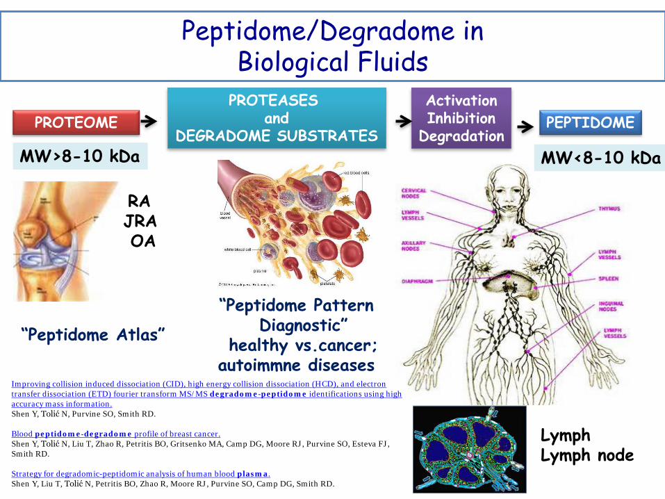
“Peptidome Pattern Diagnostic”
healthy vs.cancer; autoimmne diseases
Peptidome/Degradome in Biological Fluids
“Peptidome Atlas”
RA JRA OA
PROTEOME PROTEASES
and DEGRADOME SUBSTRATES
Activation Inhibition
Degradation PEPTIDOME
MW>8-10 kDa MW<8-10 kDa
Lymph Lymph node
Improving collision induced dissociation (CID), high energy collision dissociation (HCD), and electron transfer dissociation (ETD) fourier transform MS/MS degradome-peptidome identifications using high accuracy mass information. Shen Y, Tolić N, Purvine SO, Smith RD.
Blood peptidome-degradome profile of breast cancer. Shen Y, Tolić N, Liu T, Zhao R, Petritis BO, Gritsenko MA, Camp DG, Moore RJ, Purvine SO, Esteva FJ, Smith RD. Strategy for degradomic-peptidomic analysis of human blood plasma. Shen Y, Liu T, Tolić N, Petritis BO, Zhao R, Moore RJ, Purvine SO, Camp DG, Smith RD.

Lymph Biological fluid rich in biomarkers
Prenodal lymph is generated from the interstitial fluid that surrounds organs, and thus contains products of organ metabolism and catabolism. New proteomic analyses of lymph have
identified proteins and peptides that are derived from capillary extravasation and tissue-specific proteins.
Proteins and processed peptides are filtered from the lymph by circulating immature dendritic cells (DCs) or non-activated nodal antigen-presenting cells (APCs) (macrophages, B cells and immature DCs.
Organ-specific self-antigens are displayed to circulating and nodal APCs, thus contributing to the maintenance of peripheral tolerance.
When the tolerance to self-antigens is broken autoimmune diseases
PEPTIDOME OF HUMAN LYMPH
Endogenously Peptides MW<8-10 kDa NEW BIOMARKERS
The lymph as a pool of self-antigens. Clement CC, Rotzschke O, Santambrogio L. Trends Immunol. 2011 Jan;32(1):6-11
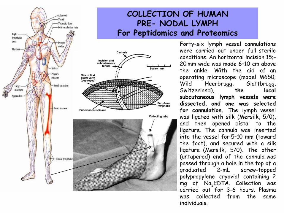
COLLECTION OF HUMAN PRE- NODAL LYMPH
For Peptidomics and Proteomics Forty-six lymph vessel cannulations were carried out under full sterile conditions. An horizontal incision 15;–20 mm wide was made 6–10 cm above the ankle. With the aid of an operating microscope (model M650; Wild Heerbrugg, Glattbrugg, Switzerland), the local subcutaneous lymph vessels were dissected, and one was selected for cannulation. The lymph vessel was ligated with silk (Mersilk, 5/0), and then opened distal to the ligature. The cannula was inserted into the vessel for 5–10 mm (toward the foot), and secured with a silk ligature (Mersilk, 5/0). The other (untapered) end of the cannula was passed through a hole in the top of a graduated 2-mL screw-topped
polypropylene cryovial containing 2 mg of Na2EDTA. Collection was carried out for 3-6 hours. Plasma was collected from the same individuals..
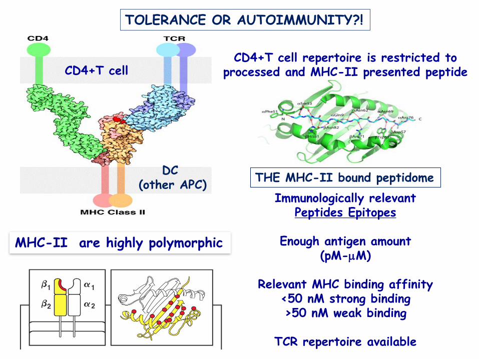
MHC-II are highly polymorphic
CD4+T cell repertoire is restricted to processed and MHC-II presented peptide
Immunologically relevant Peptides Epitopes
Enough antigen amount
(pM-µM)
Relevant MHC binding affinity <50 nM strong binding >50 nM weak binding
TCR repertoire available
TOLERANCE OR AUTOIMMUNITY?!
THE MHC-II bound peptidome DC (other APC)
CD4+T cell

Protein Autoimmune disease
Collagen I anterior uveitis, scleritis, thromboangiitis obliterans, thyroid-associated ophthalmopathy
Collagen II RA, otosclerosis, auricular chondritis Collagen III , thromboangiitis obliterans, thyroid-associated ophthalmopathy Collagen IV Goodpasture's (alpha3 chain), GN, systemic sclerosis, Alport syndrome (alpha5
chain), thyroid-associated ophthalmopathy Collagen V thyroid-associated ophthalmopathy Collagen VI Cogan's syndrome (high in neural CT) Collagen VII epidermolysis bullosa acquisita + bullous systemic lupus erythematosus Collagen IX otosclerosis Collagen XI RA Collagen XVII Pemphigoid gestationis, mucus membrane pemphigoid, bullous pemphigus Collagen XVIII Cleaved to endostatin, involved in regulating proliferation laminin thyroid-associated ophthalmopathy, SLE (alpha 1), diabetic microangiopathy Fibrosin Fxn = modulates expression of myofibroblasts (wound heal, fibrosis)
Alcohol-induced fibrosis, scleroderma Cartilage intermediate layer protein
OA, RA, lumbar disc disease
Junctophilin I disease, hypertrophic cardiomyopathy Cartilage derived morphogenic protein I
OA
Adlecan ECM remodeling in chronic CAD Mucin Asthma, allograph rejection Aggrecan RA, juvenile idiopathic arthritis Desmoglein 1 pemphigus foliaccus (Pemphigus vulgaris = desmoglein 3) Matrix GIA protein Dental caries (prevents mineralization)
Vit K dependent prevents vascular Ca2+ Fibronectin thyroid-associated ophthalmopathy, SLE, glomerular diseases, vasculitis
TOLERANCE OR AUTOIMMUNITY?!
DR1-haplotype

Lymph-carried self- peptidome Extraction, fractionation and top-down sequencing by nLC MS/MS on Orbitrap. Analysis of the molecular function and cellular pathways associated with the self-peptidome carried by the human lymph. Degradome analysis: antigen processing pathways. Prediction and functional validation of the immunological significance for the human lymph peptidome by measuring the affinity of lymph-derived endogeneous peptides for the MHC-II DR1 molecules (ELISA/fluorescence polarization assay).
Two PEPTIDOMES
Dendritic cells
The peptidome displayed by MHC-II DR1 Elution of peptides from MHC-II-DR1, fractionation and top-down sequencing by nLC MS/MS on Orbitrap/Q-exactive. Analysis of the molecular function and cellular pathways characterizing the self-peptidome displayed by MHC-II DR-1. Degradome analysis: antigen processing pathways for the peptidome displayed by MHC-II DR-1 molecules. Functional validation of the immunological relevant peptidome displayed by MHC-II by assessing its affinity for MHC-II DR1-molecules.
Clement CC et al. L. Santambrogio lab Aniuska Becerra Lawrence J. Stern lab

Lymph Self-Peptidome Sequenced at low resolution by nLC-LTQ ion trap
Clement CC et al. PLoS One 2010
LII_8 #8633 RT: 50.32 AV: 1 NL: 1.31E6T: ITMS + c NSI Full ms [ 300.00-1800.00]
400 600 800 1000 1200 1400 1600 1800m/z
0
5
10
15
20
25
30
35
40
45
50
55
60
65
70
75
80
85
90
95
100
Rel
ativ
e A
bund
ance
585.02
602.30
679.86
548.18
311.34
741.29
875.54455.48
820.45
935.39368.10 1321.741033.601168.65 1458.64 1614.92 1749.44959.19
A (2
80 n
m) Peptidome
FPLC

Lymph Self-Peptidome Sequenced at low resolution by nLC-LTQ ion trap
Molecular and cellular functions
Clement et al. PLoS One 2010
Analysis of more then 300 sequences from the healthy human lymph identified self-peptides derived from both intracellular and extracellular proteins. The peptidome of the human lymph reflects the variety of catabolic products
transported by human lymph. Quantitative analysis established that at least some of these peptides are present in the circulating lymph in nanomolar concentration.

Empty HLA-DR1
Analysis of HLA-DR1 and HLA-DR4 binding affinity
peptide loaded HLA-DR1
competition ELISA displacement assays with recombinant MHC-II molecules
Immunologically relevant Peptides Epitopes
Enough antigen amount
Relevant MHC binding affinity
TCR repertoire
The peptidome, generated by physiological tissue catabolism and transported by the pre-nodal lymph, is in addition to the self-peptidome displayed by MHC-II, generated in endosomal compartment. The peptidome carried by the lymph expands the tissue-specific self-repertoire available for the maintenance of immunological tolerance.
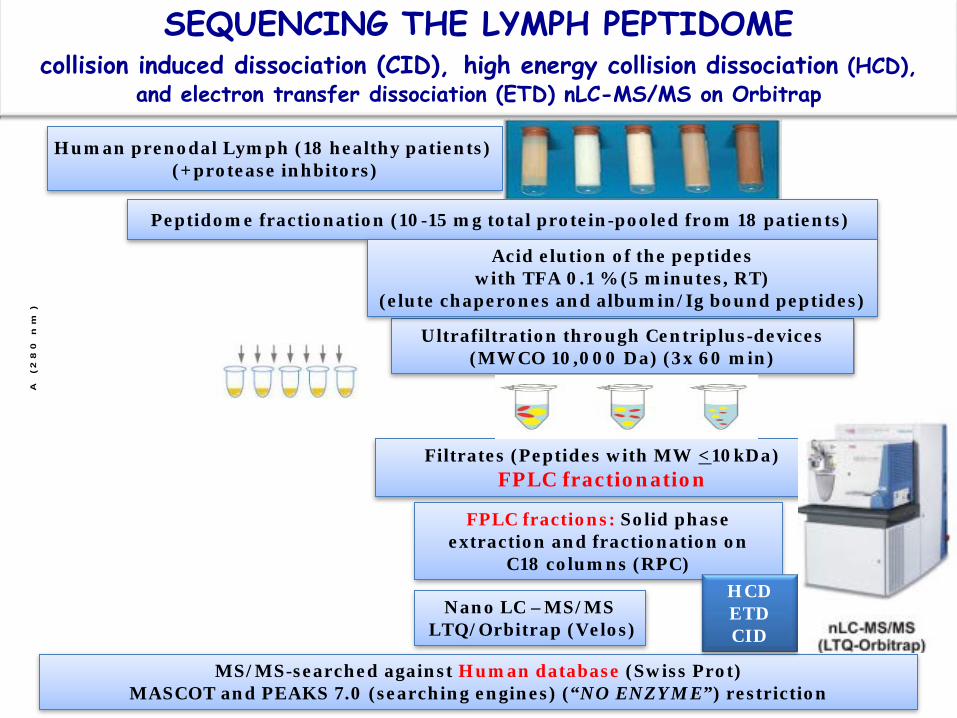
SEQUENCING THE LYMPH PEPTIDOME collision induced dissociation (CID), high energy collision dissociation (HCD),
and electron transfer dissociation (ETD) nLC-MS/MS on Orbitrap
Human prenodal Lymph (18 healthy patients) (+protease inhbitors)
Peptidome fractionation (10-15 mg total protein-pooled from 18 patients)
Ultrafiltration through Centriplus-devices (MWCO 10,000 Da) (3x 60 min)
Filtrates (Peptides with MW <10kDa) FPLC fractionation
Acid elution of the peptides with TFA 0.1 % (5 minutes, RT)
(elute chaperones and albumin/Ig bound peptides)
FPLC fractions: Solid phase extraction and fractionation on
C18 columns (RPC)
Nano LC –MS/MS LTQ/Orbitrap (Velos)
MS/MS-searched against Human database (Swiss Prot) MASCOT and PEAKS 7.0 (searching engines) (“NO ENZYME”) restriction
HCD ETD CID
A (
28
0 n
m)

Mice (DR1+)
Transgenic mice (humanized) for MHC-II-DR-1
Expand the number of conventional DC (cDC: CD11c+)
HLA-DR1 molecules purification by immuno-affinity chromatography using anti-MHC-II (DR1) antibodies.
Protein A/G
Purified human MHC-II-DR1 with mouse displayed peptides
Acid elution of the peptides with TFA 0.1 % (5 minutes, RT)
Nano LC –MS/MS LTQ/Orbitrap/Q exactive
MS/MS-searched against Mouse database (Swiss Prot) MASCOT and PEAKS 7.0 (searching engines)
“NO ENZYME” restriction
Human MHC-II DR1
SEQUENCING THE MHC-II PEPTIDOME high energy collision dissociation (HCD), nLC-MS/MS on Orbitrap/Qexactive
0 5 10 15 20 25 30 35 40 45 50 55 60 65 70 75 80 8Time (min)
64.13
60.47
72.22
59.78
59.06
45.86 67.55
41.9557.84
53.68
73.2041.5148.96 76.0738.75
80.2935.990.49 13.48 26.3022.5519.6811.51 28.547.07
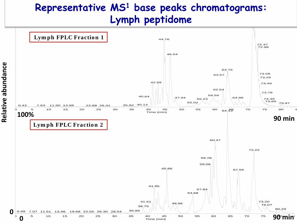
Lymph FPLC Fraction 2
0 5 10 15 20 25 30 35 40 45 50 55 60 65 70 75 80 8Time (min)
71.80
71.91
44.76
72.3772.98
46.54
63.76
73.0863.5773.29
42.99 73.49
62.5473.78
59.5940.64 47.94 64.99 74.4656.2374.8955.02 79.97
40.1435.9211.900.43 13.987.63 22.88 26.31
Lymph FPLC Fraction 1
0 90 min
Rela
tive
abun
danc
e
0
100%
100%
Representative MS1 base peaks chromatograms: Lymph peptidome
90 min

Representative MS1 base peaks chromatograms: eluted peptidome from MHC-II DR1
minutes 0 190

DE novo sequencing and database search analysis of the lymph and MHC-II eluted peptidomes
The PEAKS Studio workflow including de novo sequencing of endogenous peptides

Representative 2D-map m/z vs time for a lymph peptidome fraction
identified features (m/z vs RT) PEAKS DB identified MS/MS
m/z
RT
(m
in)
Representative 2D-map m/z vs time for MHC-II eluted peptidome fraction
60
50
40
30
200
150
100
50
0 200 1500 200 1500 m/z
Novel peptide? PTM? Mutations?
Novel peptide? PTM? Mutations?

FDR <0.5%
Representative PEAKS 7.0 output for a FPLC-lymph peptidome fraction
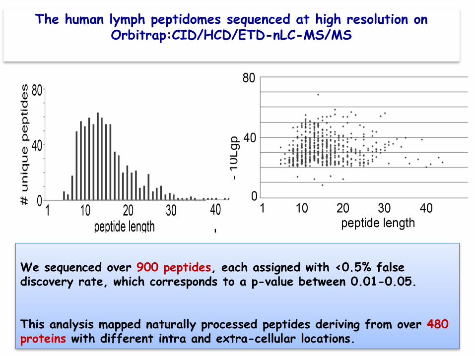
We sequenced over 900 peptides, each assigned with <0.5% false discovery rate, which corresponds to a p-value between 0.01-0.05. This analysis mapped naturally processed peptides deriving from over 480 proteins with different intra and extra-cellular locations.
The human lymph peptidomes sequenced at high resolution on Orbitrap:CID/HCD/ETD-nLC-MS/MS
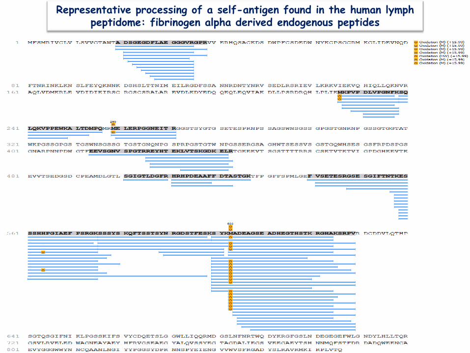
Representative processing of a self-antigen found in the human lymph peptidome: fibrinogen alpha derived endogenous peptides
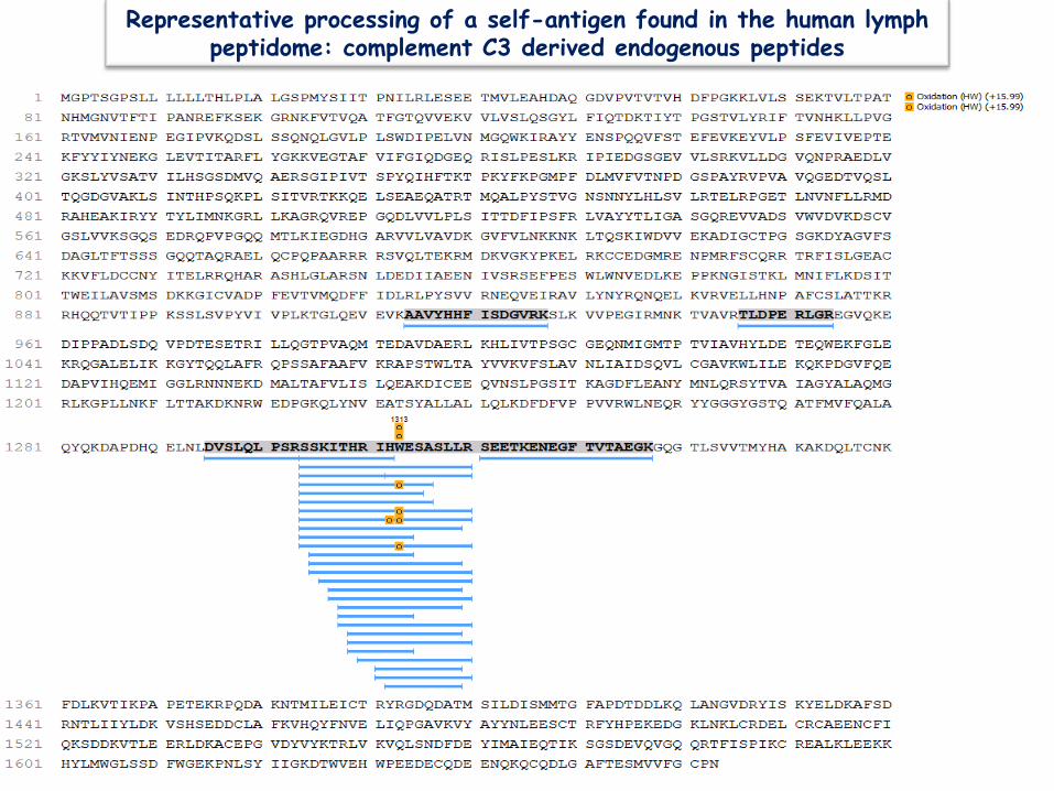
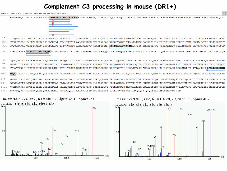
Representative processing of a self-antigen found in the human lymph peptidome: complement C3 derived endogenous peptides
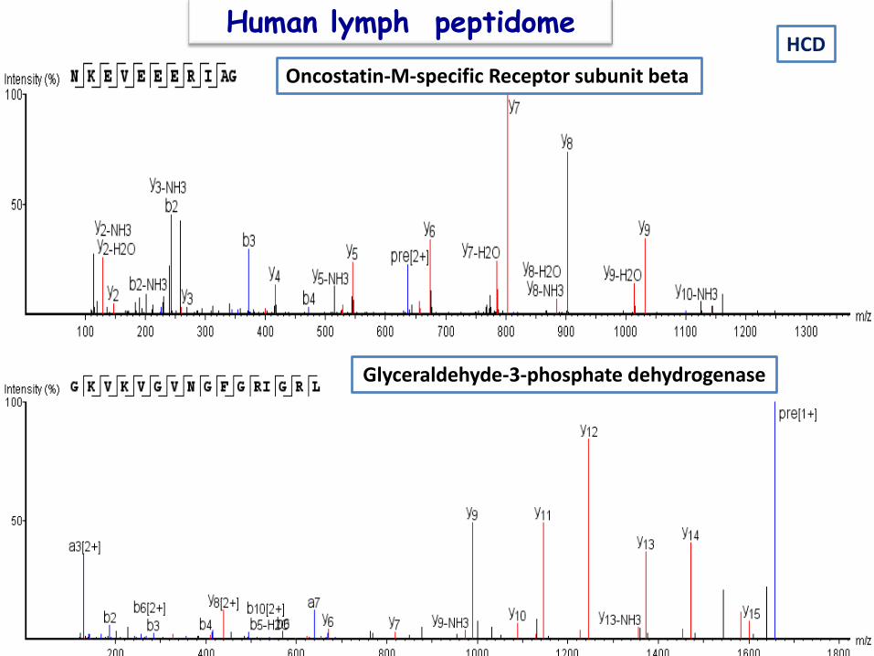
Oncostatin-M-specific Receptor subunit beta
Glyceraldehyde-3-phosphate dehydrogenase
HCD Human lymph peptidome
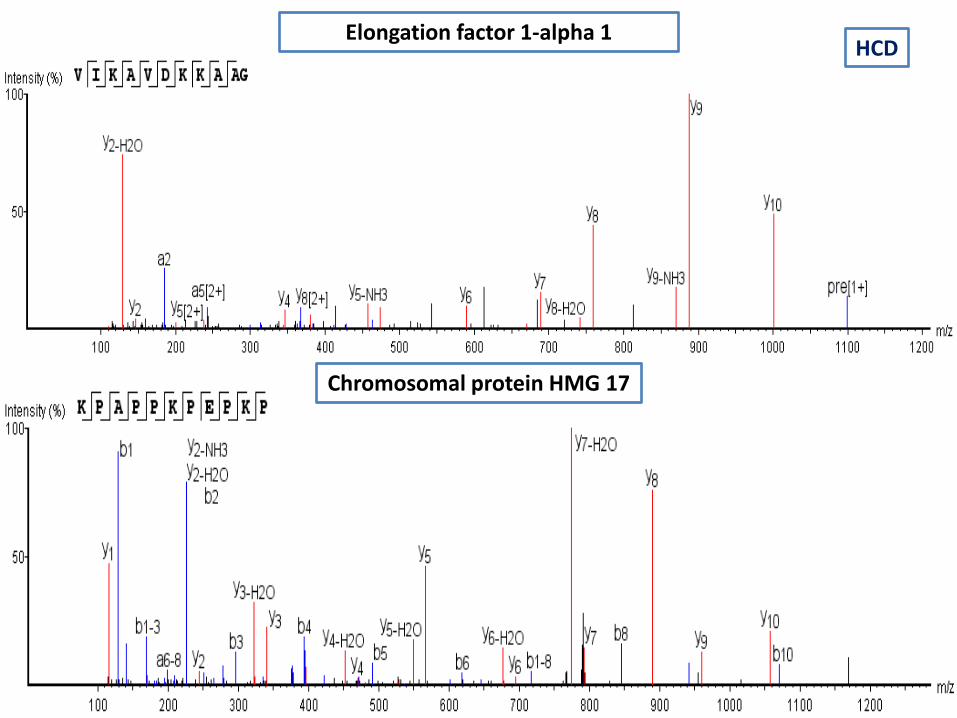
Elongation factor 1-alpha 1 HCD
Chromosomal protein HMG 17
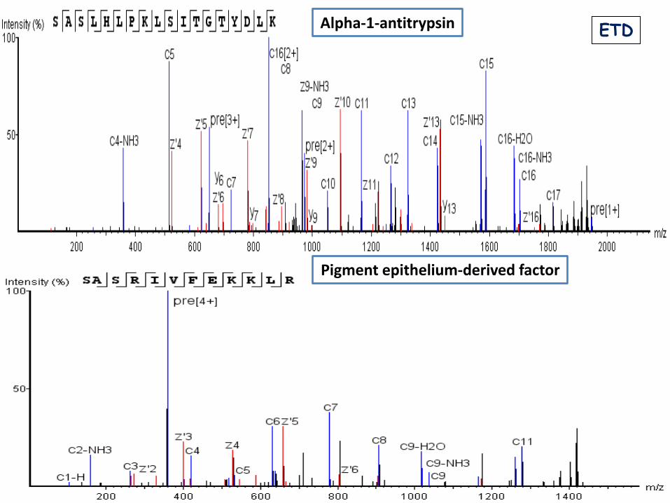
Alpha-1-antitrypsin
Pigment epithelium-derived factor
ETD
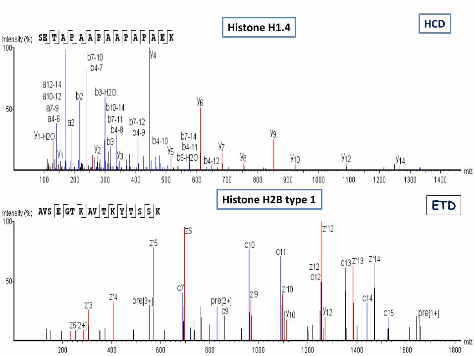
Histone H1.4
Histone H2B type 1
HCD
ETD
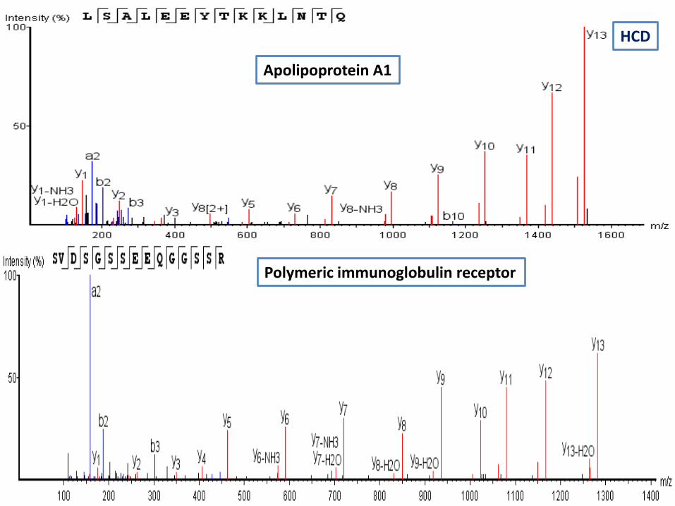
Polymeric immunoglobulin receptor
Apolipoprotein A1
HCD
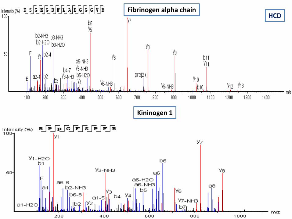
Fibrinogen alpha chain
Kininogen 1
HCD
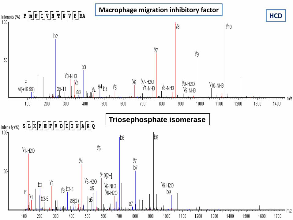
Macrophage migration inhibitory factor
Triosephosphate isomerase
HCD
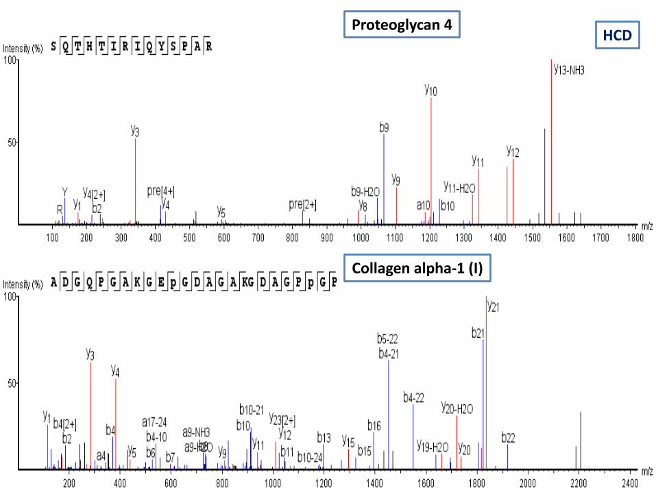
Proteoglycan 4
Collagen alpha-1 (I)
HCD

Cytochrome c oxidase subunit 5A
Malate dehydrogenase, mitochondrial
HCD

Splicing factor 3A subunit-2
Solute carrier organic anion transporter family member 6A1
HCD
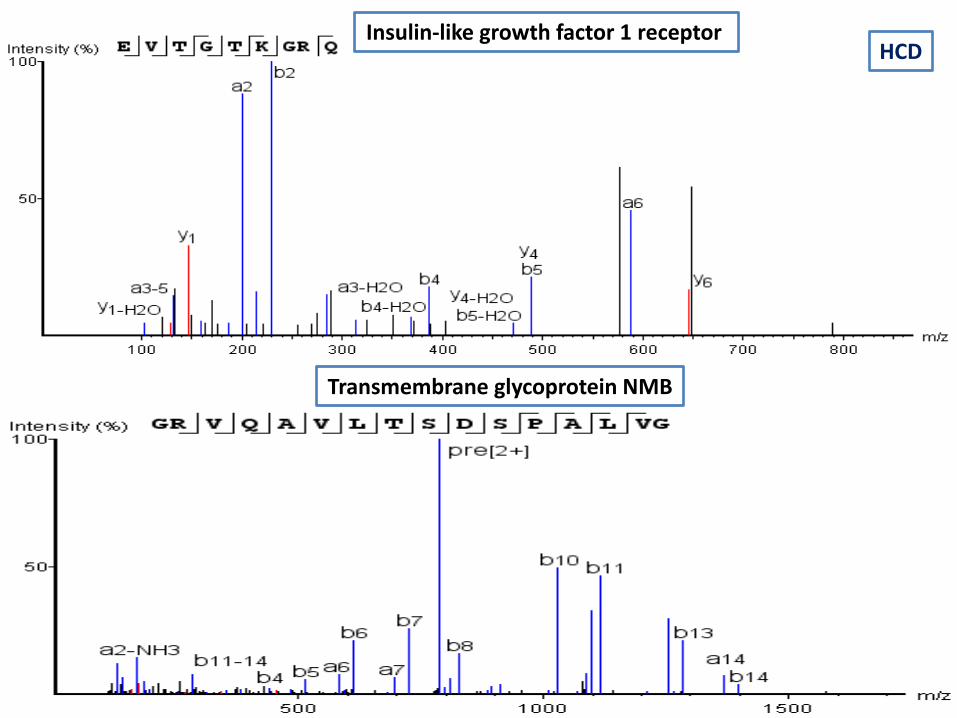
Insulin-like growth factor 1 receptor
Transmembrane glycoprotein NMB
HCD
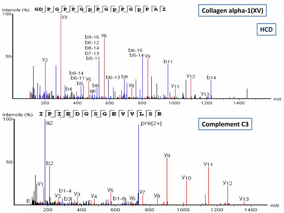
Collagen alpha-1(XV)
Complement C3
HCD
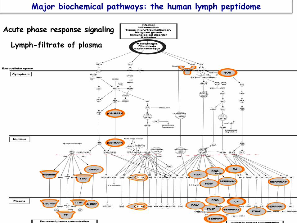
Acute phase response signaling
Major biochemical pathways: the human lymph peptidome
Lymph-filtrate of plasma
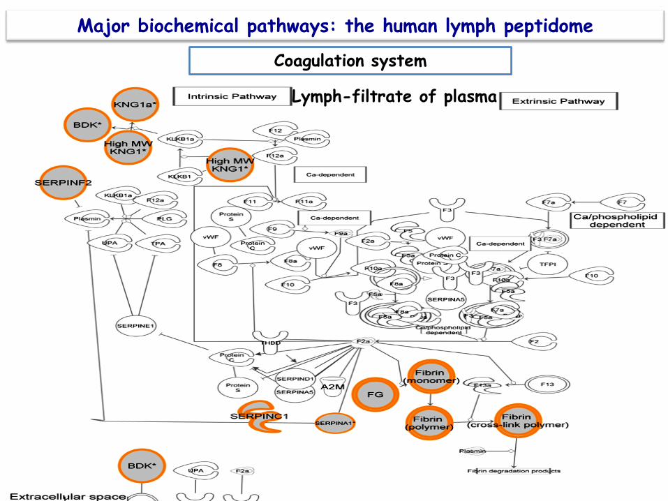
Coagulation system
Major biochemical pathways: the human lymph peptidome
Lymph-filtrate of plasma

Molecular functions and cellular distribution of the human lymph peptidome
The endogenous peptides from human lymph derived from mitochondria, ER, Golgi enzymes, cytosolic proteins, nuclear proteins and peptides from extracellular matrix proteins; playing important roles as chaperones and in innate immune responses. Several additional naturally-processed peptides derived from plasma membrane proteins and surface receptors, in addition to the proteins found in the filtrate of the plasma (fibrinogen and other acute-phase response proteins).
Lymph-is not only a filtrate of plasma

Validation of the proteins generating the human lymph peptidome A proteomics approach
Protein expression profiles of human lymph and plasma mapped by 2D-DIGE and 1D SDS-PAGE coupled with nanoLC-ESI-MS/MS bottom-up proteomics. Clement CC, Aphkhazava D, Nieves E, Callaway M, Olszewski W, Rotzschke O, Santambrogio L. J Proteomics. 2013 Jan 14;78:172-87.

MHC-II are highly polymorphic
CD4+T cell repertoire is restricted to processed and MHC-II presented peptide
Immunologically relevant Peptides Epitopes
Enough antigen amount
(pM-µM)
Relevant MHC binding affinity <50 nM strong binding >50 nM weak binding
TCR repertoire available
TOLERANCE OR AUTOIMMUNITY?!
THE MHC-II bound peptidome DC (other APC)
CD4+T cell
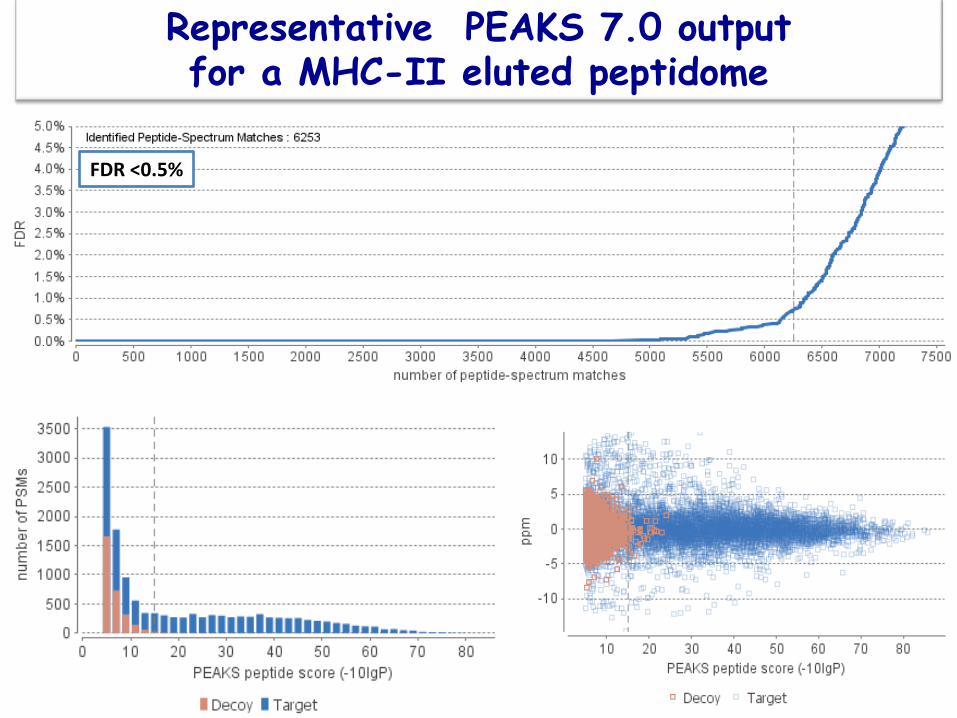
Representative PEAKS 7.0 output for a MHC-II eluted peptidome
FDR <0.5%

Representative processing of a mouse self-antigen displayed by the human MHC-II DR1 molecule
Deoxyribonuclease gamma processing in the mice DR1+
m/z=758.9308, z=2, RT=114.28, -lgP=33.69, ppm=-0.7 m/z=766.9274, z=2, RT=100.52, -lgP=32.31, ppm=-2.0
Complement C3 processing in mouse (DR1+)

m/z=705.3583, z=2, RT=109.95 -lgP=33.60, ppm=2.4 m/z=631.8220, z=2, RT=92.23 -lgP=27.81, ppm=-0.6
MHC-II antigen: alpha chain processing in mouse (DR1 (+)

cDC MHC-II Eluted Peptidome
3216 peptides, from 688 proteins FDR <0. 1% p 0.004
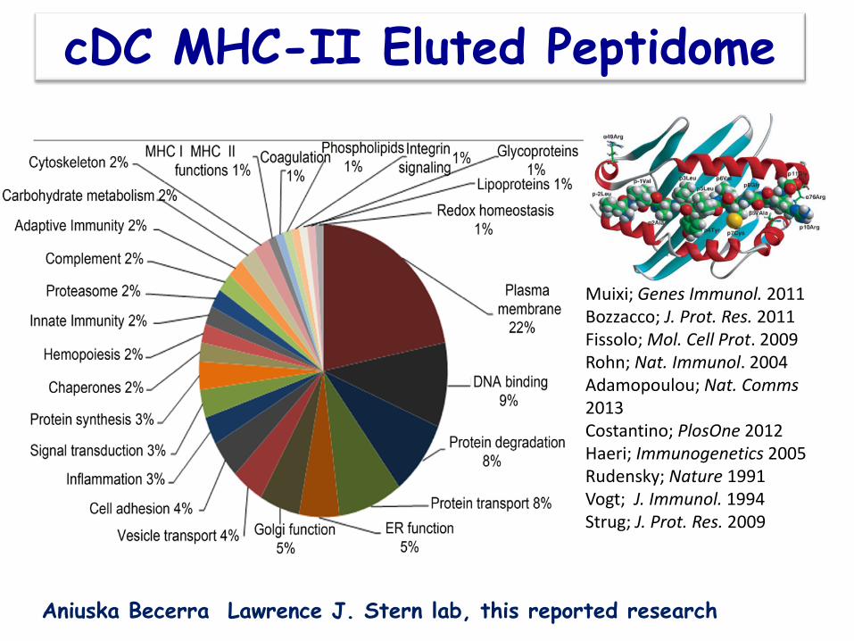
Muixi; Genes Immunol. 2011 Bozzacco; J. Prot. Res. 2011 Fissolo; Mol. Cell Prot. 2009 Rohn; Nat. Immunol. 2004 Adamopoulou; Nat. Comms 2013 Costantino; PlosOne 2012 Haeri; Immunogenetics 2005 Rudensky; Nature 1991 Vogt; J. Immunol. 1994 Strug; J. Prot. Res. 2009
cDC MHC-II Eluted Peptidome
Aniuska Becerra Lawrence J. Stern lab, this reported research

Complement system
Major biochemical pathways associated with the eluted peptidome from MHC-II-DR1 molecules
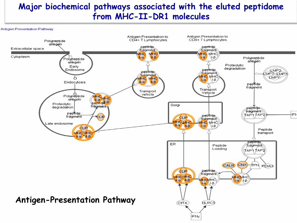
Major biochemical pathways associated with the eluted peptidome from MHC-II-DR1 molecules
Antigen-Presentation Pathway

Clathrin-mediated endocytosis signaling
Major biochemical pathways associated with the eluted peptidome from MHC-II-DR1 molecules

Processing Pathways of Lymph and HLA-DR1-eluted peptidome
(The degradomes)
Experimentally-based databases
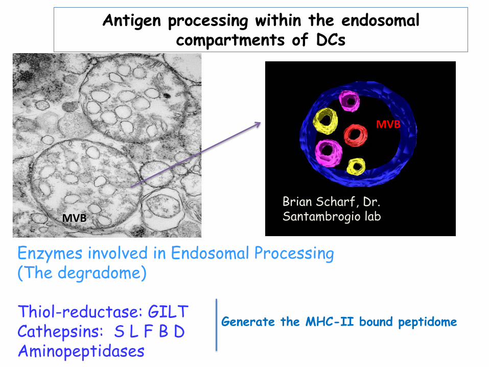
Enzymes involved in Endosomal Processing (The degradome) Thiol-reductase: GILT Cathepsins: S L F B D Aminopeptidases
Antigen processing within the endosomal compartments of DCs
MVB
MVB
Brian Scharf, Dr. Santambrogio lab
Generate the MHC-II bound peptidome

Human Lymph Peptidome/degradome
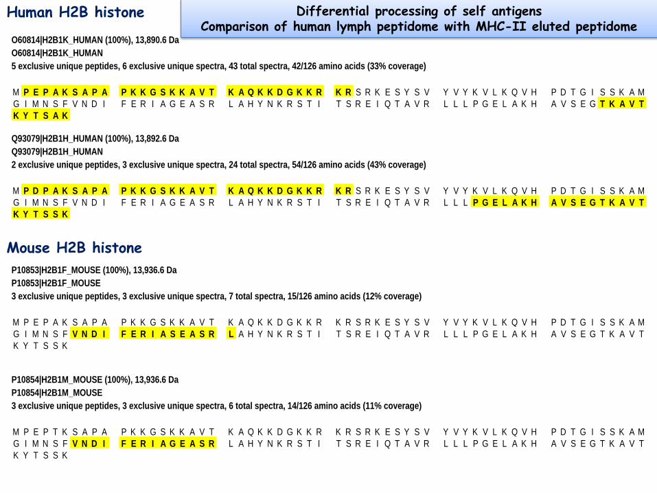
O60814|H2B1K_HUMAN (100%), 13,890.6 DaO60814|H2B1K_HUMAN5 exclusive unique peptides, 6 exclusive unique spectra, 43 total spectra, 42/126 amino acids (33% coverage)
M P E P A K S A P A P K K G S K K A V T K A Q K K D G K K R K R S R K E S Y S V Y V Y K V L K Q V H P D T G I S S K A MG I M N S F V N D I F E R I A G E A S R L A H Y N K R S T I T S R E I Q T A V R L L L P G E L A K H A V S E G T K A V TK Y T S A K
Q93079|H2B1H_HUMAN (100%), 13,892.6 DaQ93079|H2B1H_HUMAN2 exclusive unique peptides, 3 exclusive unique spectra, 24 total spectra, 54/126 amino acids (43% coverage)
M P D P A K S A P A P K K G S K K A V T K A Q K K D G K K R K R S R K E S Y S V Y V Y K V L K Q V H P D T G I S S K A MG I M N S F V N D I F E R I A G E A S R L A H Y N K R S T I T S R E I Q T A V R L L L P G E L A K H A V S E G T K A V TK Y T S S K
P10853|H2B1F_MOUSE (100%), 13,936.6 DaP10853|H2B1F_MOUSE3 exclusive unique peptides, 3 exclusive unique spectra, 7 total spectra, 15/126 amino acids (12% coverage)
M P E P A K S A P A P K K G S K K A V T K A Q K K D G K K R K R S R K E S Y S V Y V Y K V L K Q V H P D T G I S S K A MG I M N S F V N D I F E R I A S E A S R L A H Y N K R S T I T S R E I Q T A V R L L L P G E L A K H A V S E G T K A V TK Y T S S K
P10854|H2B1M_MOUSE (100%), 13,936.6 DaP10854|H2B1M_MOUSE3 exclusive unique peptides, 3 exclusive unique spectra, 6 total spectra, 14/126 amino acids (11% coverage)
M P E P T K S A P A P K K G S K K A V T K A Q K K D G K K R K R S R K E S Y S V Y V Y K V L K Q V H P D T G I S S K A MG I M N S F V N D I F E R I A G E A S R L A H Y N K R S T I T S R E I Q T A V R L L L P G E L A K H A V S E G T K A V TK Y T S S K
Human H2B histone
Mouse H2B histone
Differential processing of self antigens Comparison of human lymph peptidome with MHC-II eluted peptidome

P02647|APOA1_HUMAN (100%), 30,778.5 DaP02647|APOA1_HUMAN12 exclusive unique peptides, 18 exclusive unique spectra, 29 total spectra, 18/267 amino acids (7% coverage)
M K A A V L T L A V L F L T G S Q A R H F W Q Q D E P P Q S P W D R V K D L A T V Y V D V L K D S G R D Y V S Q F E G SA L G K Q L N L K L L D N W D S V T S T F S K L R E Q L G P V T Q E F W D N L E K E T E G L R Q E M S K D L E E V K A KV Q P Y L D D F Q K K W Q E E M E L Y R Q K V E P L R A E L Q E G A R Q K L H E L Q E K L S P L G E E M R D R A R A H VD A L R T H L A P Y S D E L R Q R L A A R L E A L K E N G G A R L A E Y H A K A T E H L S T L S E K A K P A L E D L R QG L L P V L E S F K V S F L S A L E E Y T K K L N T Q
Q00623|APOA1_MOUSE (100%), 30,587.4 DaQ00623|APOA1_MOUSE7 exclusive unique peptides, 7 exclusive unique spectra, 11 total spectra, 36/264 amino acids (14% coverage)
M K A V V L A V A L V F L T G S Q A W H V W Q Q D E P Q S Q W D K V K D F A N V Y V D A V K D S G R D Y V S Q F E S S SL G Q Q L N L N L L E N W D T L G S T V S Q L Q E R L G P L T R D F W D N L E K E T D W V R Q E M N K D L E E V K Q K VQ P Y L D E F Q K K W K E D V E L Y R Q K V A P L G A E L Q E S A R Q K L Q E L Q G R L S P V A E E F R D R M R T H V DS L R T Q L A P H S E Q M R E S L A Q R L A E L K S N P T L N E Y H T R A K T H L K T L G E K A R P A L E D L R H S L MP M L E T L K T K A Q S V I D K A S E T L T A Q
Human Apolipoprotein A1
Mouse Apolipoprotein A1
Differential processing of self antigens Comparison of human lymph peptidome with MHC-II eluted peptidome
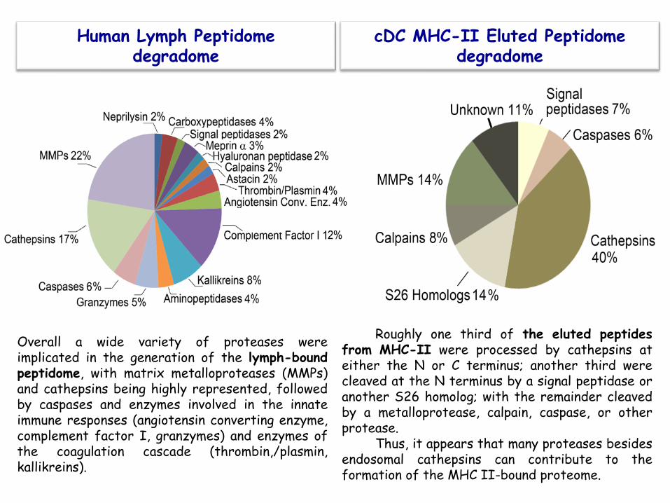
cDC MHC-II Eluted Peptidome degradome
Human Lymph Peptidome degradome
Overall a wide variety of proteases were implicated in the generation of the lymph-bound peptidome, with matrix metalloproteases (MMPs) and cathepsins being highly represented, followed by caspases and enzymes involved in the innate immune responses (angiotensin converting enzyme, complement factor I, granzymes) and enzymes of the coagulation cascade (thrombin,/plasmin, kallikreins).
Roughly one third of the eluted peptides from MHC-II were processed by cathepsins at either the N or C terminus; another third were cleaved at the N terminus by a signal peptidase or another S26 homolog; with the remainder cleaved by a metalloprotease, calpain, caspase, or other protease.
Thus, it appears that many proteases besides endosomal cathepsins can contribute to the formation of the MHC II-bound proteome.
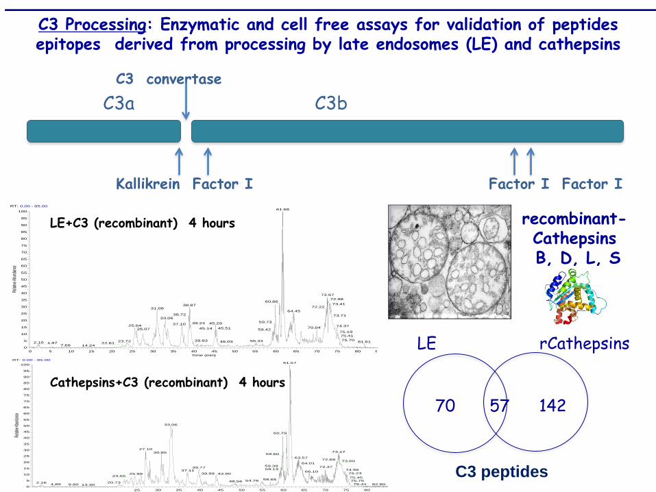
C3a C3b C3 convertase Kallikrein Factor I Factor I Factor I
LE rCathepsins
70 57 142
recombinant-Cathepsins B, D, L, S
C3 Processing: Enzymatic and cell free assays for validation of peptides epitopes derived from processing by late endosomes (LE) and cathepsins
RT: 0.00 - 85.00
0 5 10 15 20 25 30 35 40 45 50 55 60 65 70 75 80 8Time (min)
0
5
10
15
20
25
30
35
40
45
50
55
60
65
70
75
80
85
90
95
100
Relati
ve Ab
undanc
e
61.68
72.6772.98
60.8673.4138.87 72.2231.08
64.4538.72 73.71
33.0659.7339.24 45.2937.1025.64 74.3770.0445.5145.1426.07 59.42
75.1975.4175.7039.83 55.3323.72 46.03 81.612.10 1.97 22.61
7.68 14.24
LE+C3 (recombinant) 4 hours
RT: 0.00 - 85.00
0 5 10 15 20 25 30 35 40 45 50 55 60 65 70 75 80
0
5
10
15
20
25
30
35
40
45
50
55
60
65
70
75
80
85
90
95
100
Relativ
e Abun
dance
61.57
33.06
60.79
27.1073.1730.85 59.60
63.5772.69 73.60
64.0159.39 72.3739.77 59.13 74.5637.11 66.10
75.2339.99 43.9025.9923.55 75.45
58.6554.76 75.7548.5620.732.16 76.41 82.804.89 9.80 13.30
Cathepsins+C3 (recombinant) 4 hours
C3 peptides

Comparison between the cDC, MHC II-eluted, and lymph peptidome
,B, D
P9
scor
e
nM
DC-Eluted
P9
scor
e
nM
Lymph
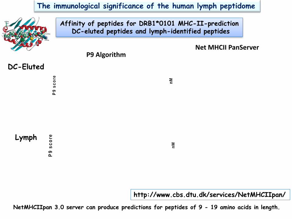
Affinity of peptides for DRB1*0101 MHC-II-prediction DC-eluted peptides and lymph-identified peptides
P9 Algorithm Net MHCII PanServer
The immunological significance of the human lymph peptidome
NetMHCIIpan 3.0 server can produce predictions for peptides of 9 - 19 amino acids in length.
http://www.cbs.dtu.dk/services/NetMHCIIpan/

Empty HLA-DR1
Analysis of HLA-DR1 and HLA-DR4 binding affinity
peptide loaded HLA-DR1
Competition ELISA displacement assays with recombinant MHC-II molecules
Immunologically relevant Peptides Epitopes
Enough antigen amount
Relevant MHC binding affinity
TCR repertoire
IPMYSIITPNVLRL
∆IC50= 0 IC50= 0.03 ± 0.01 µM
SSKITHRIHWESASLLR
∆IC50= 0.06 µM IC50= 0.07 ± 0.01 µM
EEFGRFASFEAQGALA
∆IC50= 0.07 µM IC50= 0.14 ± 0.04 µM
ASFEAQGALANIAVDKA
∆IC50= 3.40 µM IC50= 1.71 ± 0.28 µM
Complement Protein C3 MHC-II a chain Lymph MHC-II-eluted Lymph MHC-II-eluted

Dr. Lawrence Stern, UMASS Clement CC et al. PLoS One 2010
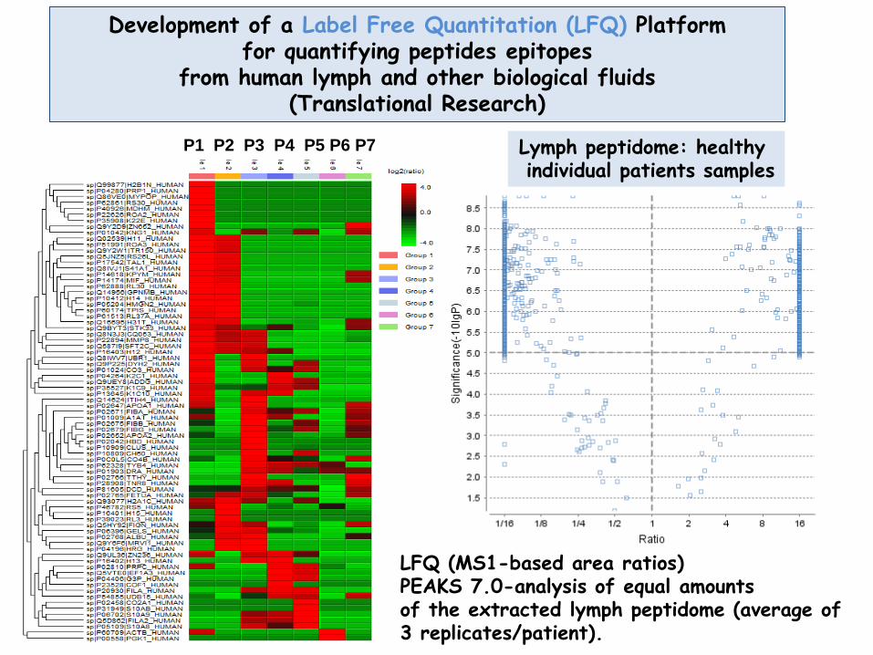
Development of a Label Free Quantitation (LFQ) Platform for quantifying peptides epitopes
from human lymph and other biological fluids (Translational Research)
P1 P2 P3 P4 P5 P6 P7 Lymph peptidome: healthy individual patients samples
LFQ (MS1-based area ratios) PEAKS 7.0-analysis of equal amounts of the extracted lymph peptidome (average of 3 replicates/patient).
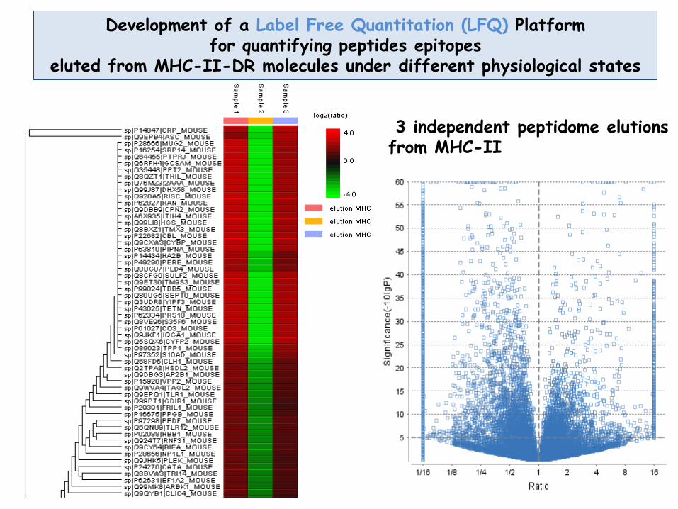
Development of a Label Free Quantitation (LFQ) Platform for quantifying peptides epitopes
eluted from MHC-II-DR molecules under different physiological states
3 independent peptidome elutions from MHC-II
CONCLUSIONS
Selected peptides from the human lymph were shown to have low nM binding affinity for MHC-II DR1 molecules.
Extracellular protein processing can contribute to the overall MHC II presented peptidome, expanding the peptide repertoire available for the induction of self-tolerance.
The first correlated analysis of the human lymph and MHC-II peptidomes employing nanoLC Orbitrap-ESI-MS/MS and bioinformatic analysis of corresponding degradomes.
Immunological significance of the self-peptidome carried by the human lymph
This analysis enabled the characterization of human lymph peptidome at high resolution and reveals new peptides epitopes never reproted before this study.
The bioinformatics analysis of the reported peptidomes highlights the diverse processing pathways involved in the generation of the MHC II peptidome.
The first reported degradome analysis: Several categories of protease/peptidase in additional to those present in endosomal/lysosomal compartments are involved in the formation of the lymph peptidome.
More than 3000 MHC-II bound mouse peptides with over 1000 core epitopes identified. And many proteases from various non-lysosomal intracellular and extracellular compartments
contribute to the shaping of the MHC II peptidome.
Acknowledgements Albert Einstein College of Medicine (AECOM) Laura Santambrogio lab Cristina C. Clement Valerio Zolla Lawrence Stern Lab UMASS Aniuska Becerra Scott Shafer
$$$ NIH
Mass spectrometry Collection of data and “Raw files”
Edward Nieves (AECOM) Fa-Yun Che (AECOM)
MS Bioworks