cath procedure: coronary angiography - sickkids · cath procedure: coronary angiography ......
TRANSCRIPT
CATHETERIZATION PROTOCOL Prepared by Ra Han (2005-12-30)
Edited by K-J Lee Cath Procedure: Coronary Angiography Indications: Kawasaki disease with significant coronary aneurysms1
Post-heart transplant surveillance for vasculopathy2
Postoperative assessment of coronary anatomosis e.g. TGA s/p coronary reimplantation3, coronary bypass graft
Preoperative assessment of coronary artery course e.g. prior to RV-PA conduit revision
Hypertrophic cardiomyopathy with possible myocardial bridging4
Suspected coronary artery abnormalities5 (abnormal origins, course, fistulae) not adequately assessed by noninvasive testing
Chest pain, syncope, or aborted sudden death with high-risk findings on noninvasive testing
Hospitalization Requirement: Same day admission or outpatient unit Blood on hold : No (unless intervention planned) Pre-Cath Preparation: CXR, ECG within 6 weeks prior to cath CBC if cyanotic, lytes if on diuretics or digoxin Sickle cell test if African or Caribbean descent Cardiac Catheterization: Access: • Femoral artery for aortic root angiogram or selective coronary angiography • Venous access and right heart cath if relationship to RV-PA conduit required or assessing
RV-coronaries fistulae in PA-IVS Catheters: • Pigtail (retrograde/antegrade) or Berman angio
catheter (antegrade) for aortic root angiogram • Judkins coronary catheters (JR, JL) for selective
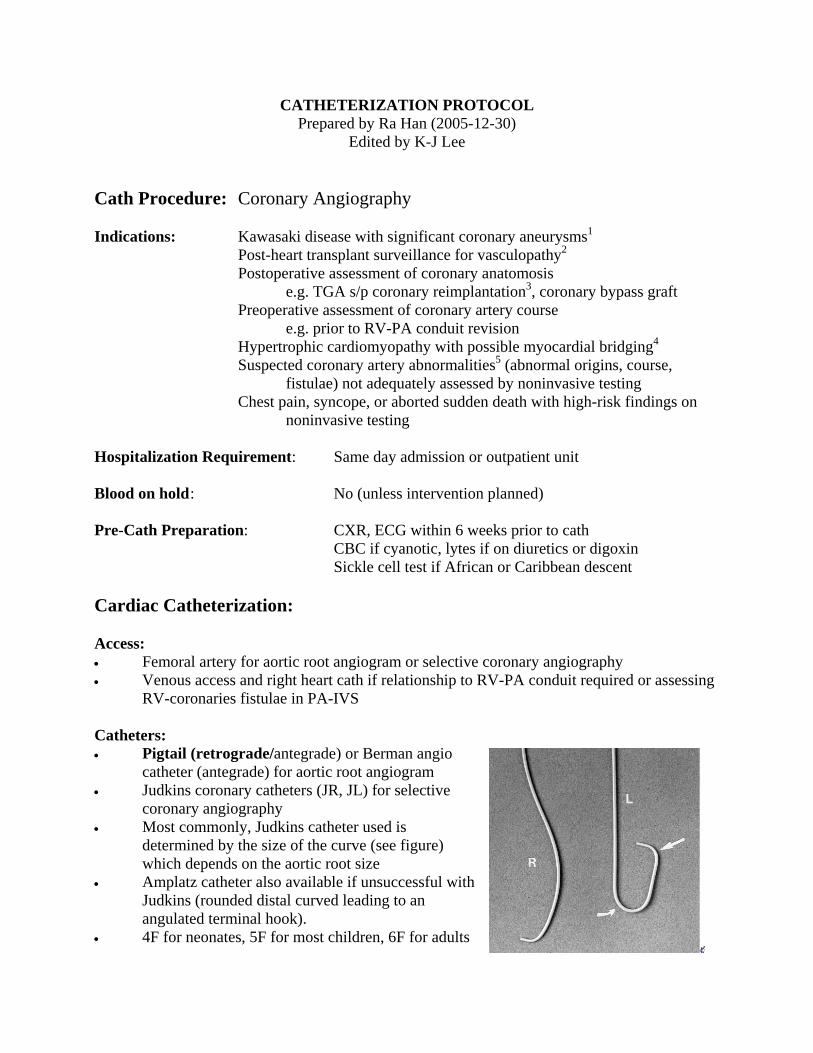
coronary angiography • Most commonly, Judkins catheter used is
determined by the size of the curve (see figure) which depends on the aortic root size
• Amplatz catheter also available if unsuccessful with Judkins (rounded distal curved leading to an angulated terminal hook).
• 4F for neonates, 5F for most children, 6F for adults

Types of coronary catheters
Angiography: • Set up manifold with contrast agent bottle, contrast injection syringe, discard syringe, and
pressure manometer attached to set up a closed system (for selective coronary angiogram). This allows for continuous source of contrast without disconnecting.
• Obtain access via femoral artery with modified Seldinger technique • Give heparin 50-150 U/kg (staff-variable) once access obtained • Insert catheter with wire in situ to straighten the catheter tip • Advance catheter to the ascending aorta • Remove guidewire and aspirate blood then attach to manifold while flushing with saline
to remove any air • Aortic root injection the catheter tip should be in the ascending aorta distal to the
sinotubular junction. Inject ~1 cc/kg over 2 cardiac cycles in the AP and lateral projections or a “down-the-barrel” projection (30º RAO, 30º LAO 45º Caudal)
• Selective LCA catheterization6 gently advance the JL catheter in the ascending aorta, the catheter tip should naturally fall into the ostium of the LCA; if not, apply the “push-pull” technique: advance the catheter until the tip forms an acute angle which enters the left sinus of Valsalva then pull up until the LCA ostium is engaged (see figure below)

• Selective RCA catheterization6 a bit trickier, once the JR catheter tip is at the level of
•
•
Angiog u• ws7 although should be modified to obtain the
ts’ anatomy
B Plane
the sinuses, rotate the catheter clockwise until the tip points anteriorly and should engage the RCA ostium; a small jog of contrast in the right coronary sinus will help to show to location of the ostium Once the coronary ostium is engaged, check pressure tracing to make sure that the catheter tip is not wedged and give a contrast injection jog to confirm position Use the minimum amount of contrast to fill the entire coronary artery for 2 cardia cycles, approximately 4-8 cc for LCA and 3-6 cc for RCA in adults
raphic Views: (see also in cath lab manThese are the common angiographic viebest information for the individual patien
c
al for projections)
View A Plane Selective LCA #1 30º RAO 60º LAO Selective LCA #2 30º RAO 30º Cranial 60º LAO 25º Cranial Selective LCA #2 30º RAO 30º Caudal 90º LAO Selective RCA #1 30º RAO 60º LAO Selective RCA #2 30º LAO 30º Cranial parked

Selective LCA injections6 Selective RCA injections6
• Watch the ECG and coronary
pressures carefully with each injection. Disengage the catheter tip from the ostium if any concerns.
• After angiograms are completed, remove the catheter with wire in situ and apply pressure
• Consider vascular closure device (e.g. perclose) if older patient. May allow for earlier ambulation
Single plane views: LCA: 4 injections 1. LAO 30°, Cranial 25-30°°
2. RAO 30°, Cranial 25-30° 3. LAO 30°, Caudal 25-30° 4. RAO 30°, Caudal 25-30°

RCA: 2 injections
ost-Cath Management: Bedrest for 4-6 hours
omplications (from adult registry) :
farction 0.05% %
.43%
ons 0.26%
70%
eferences: r JW et al. Diagnosis, treatment, and long-term management of Kawasaki Disease.
2. me of transplant coronary artery disease in a pediatric -
3. dre A et al. Coronary events after arterial switch operation for transposition of the
4. ertrophic cardiomyopathy – a risk
5. art 2005;91:1240-1245.
l
7. Freedom RM. Angiography. Pediatric Cardiovascular Medicine, Moller
1. RAO 30°, cranial 25-30° 2. LAO 30°, cranial 25-30°
P Discharge home same day unless complications
6C• Death 0.11% • Myocardial in• Cerebrovascular accident 0.07• Significant arrhythmias 0.38% • Major vascular complications 0• Contrast reaction 0.37% • Hemodynamic complicati• Perforation of heart chamber 0.03% • Other complications 0.28% • Total major complications 1. R1. Newburge
Circulation 2004;110:2747-2771. Pahl E et al. The impact and outcopopulation: a 9-year multi-institutional study. J Heart Lung Transplantation 2005;24:645651. Legengreat arteries. Circulation 2003;108[suppl II]:II186-II190. Yetman AT et al. Myocardial bridging in children with hypfactor for sudden death. N Engl J Med 1998;339:1201-1209. Hauser M. Congenital anomalies of the coronary arteries. He
6. Popma JJ. Chapter 18: Coronary angiography and intravascular ultrasound imaging. Braunwald’s Heart Disease: a textbook of cardiovascular medicine, 7th Ed. Zipes et a(Editors), 2005. Nykanen DG andand Hoffman (Editors), 2000