catalytic “triangles”: binding of iron in task … · supplementary material to the article:...
TRANSCRIPT
SUPPLEMENTARY MATERIAL TO THE ARTICLE:
Catalytic “Triangles”: Binding of Iron in Task-Spec ific Ionic Liquids
Denis Prodius, Fliur Macaev, Eugenia Stingaci, Vsevolod Pogrebnoi, Valeriu Mereacre, Ghenadie Novitchi, George E. Kostakis, Christopher E. Anson, and Annie K. Powell
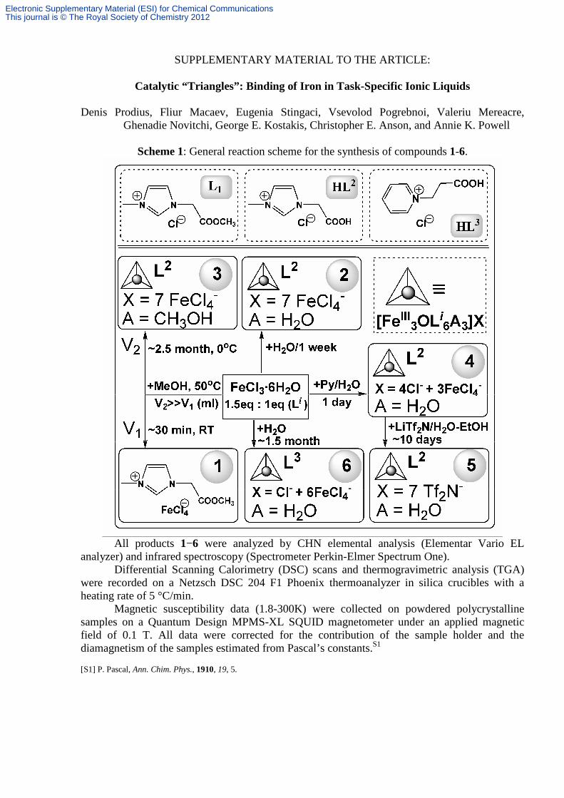
Scheme 1: General reaction scheme for the synthesis of compounds 1-6.
All products 1−6 were analyzed by CHN elemental analysis (Elementar Vario EL
analyzer) and infrared spectroscopy (Spectrometer Perkin-Elmer Spectrum One). Differential Scanning Calorimetry (DSC) scans and thermogravimetric analysis (TGA)
were recorded on a Netzsch DSC 204 F1 Phoenix thermoanalyzer in silica crucibles with a heating rate of 5 °C/min.
Magnetic susceptibility data (1.8-300K) were collected on powdered polycrystalline samples on a Quantum Design MPMS-XL SQUID magnetometer under an applied magnetic field of 0.1 T. All data were corrected for the contribution of the sample holder and the diamagnetism of the samples estimated from Pascal’s constants.S1
[S1] P. Pascal, Ann. Chim. Phys., 1910, 19, 5.
Electronic Supplementary Material (ESI) for Chemical CommunicationsThis journal is © The Royal Society of Chemistry 2012

Preparation of ligands L1, HL2 and HL3.
NNH3C i iiNN
H3CCOOCH3
Cl
NNH3C
COOH
Cl
L1 HL 2
Scheme S1: Synthetic route to ligands L1 ( and HL2 (cmmimH). Reagents and conditions: i) 1 equiv ClCH2COOCH3, RT, 1.5 h; ii) 37% aqueous HCl solution, 100°C, 2h.
N
i NCOOH
Cl
HL 3
Scheme S2: Synthetic route to ligand HL3 (cepyH). Reagents and conditions: i) 1 equiv ClCH2CH2COOH, 80°C, 3 h, EtOAc.
The crystalline products L 1, HL 2 and HL 3 were synthesized according to the literature methods [R1] (imidazolium salts) and [R2] (pyridinium salts) with slightly modificated synthetical procedures (Schemes S1 and S2). [R1] Z. Fei, T. J. Geldbach, D. Zhao, P. J. Dyson, Chem. Eur. J. 2006, 12, 2122–2130. [R2] M. Szafran, M. Jaskolski, I. Kowalczyk, Z. Dega-Szafran, J. Mol. Struct. 1998, 448, 77–89. Preparation of [C1ImC1CO2CH3]FeCl4 (1):
Freshly prepared methyl 2-(3-methyl-1H-imidazol-1-yl)acetate chloride (L 1) (1.91 g, 10 mmol) was added to a methanolic solution (7 mL) of FeCl3⋅6H2O (4.06 g, 15 mmol) with heating and stirring at 50°C (maximum 10 min). Crystals appear immediately or during the next 30 min at room temperature. Monocrystals were washed by methanol and air dried (Fig. SI-1a). Calc. for C7H11N2O2Cl4Fe (1): C 23.83; H 3.14; N 7.94%. Found: C 23.95; H 3.17; N 7.95%. Yield: ~1.25 g (~24% based on Fe). Selected IR data for compound 1 (KBr/cm-1): 3158 (w), 3124 (w), 1758 (s, sh), 1613 (mw, sh), 1580 (w), 1569 (w, sh), 1439 (w), 1425 (mw, sh), 1386 (w), 1361 (mw), 1244 (sm), 1197 (mw, sh), 1183 (m, sh), 1109 (w), 996 (mw, sh), 966 (w), 829 (mw, sh), 770 (w, sh), 753 (mw, sh), 619 (m, sh), 565 (w). Preparation of [Fe3O(C1ImC1CO2)6(H2O)3](FeCl4)7 ⋅5H2O (2):
To a solution of L 1 (1.91 g, 10 mmol) in methanol (20 mL) was added FeCl3⋅6H2O (4.06 g, 15 mmol). The mixture was stirred at room temperature for 30 min. The resulting solution was left for crystallization (Petri dish ∅110). Next day the brown microcrystalline compound was separated from some drops of orange oil and dissolved in 15-20 mL of boiling water. Recrystallization process was repeated several times. After about 1 week, good quality crystals for x-ray measurement were collected and air dried. Calc. for C36H64N12O21Cl28Fe10 (2): C 16.94; H 2.53; N 6.59%. Found: C 17.07; H 2.56; N 6.68%. Yield: ~1.5 g (~40% based on Fe). Selected IR data for compound 2 (KBr/cm-1): 3147 (m, vb), 1755 (w), 1648 (s), 1587 (mw), 1453 (s, sh), 1402 (vs, sh), 1344 (mw), 1317 (m, sh), 1170 (s, sh), 1105 (w), 1087 (w), 1035 (w), 972 (mw, sh), 842 (mw), 795 (mw, sh), 735 (mw, sh), 702 (ms, sh).
Electronic Supplementary Material (ESI) for Chemical CommunicationsThis journal is © The Royal Society of Chemistry 2012

Preparation of [Fe3O(C1ImC1CO2)6(CH3OH)3](FeCl4)7 ⋅H2O⋅2CH3OH (3):
To a solution of L 1 (1.91 g, 10 mmol) in methanol (25 mL) was added FeCl3⋅6H2O (4.06 g, 15 mmol). The mixture was stirred at room temperature for 30 min. The solution was devided in two portions for slow evaporation at RT (solution A) and at ~0˚C (solution B). Dark brown single crystals of 3 were formed over two (solution A) and five (solution B) months. Crystals (Fig. SI-1b) were separated from orange-brown oil and air dried. Calc. for C41H70N12O19Cl28Fe10 (3): C 19.04; H 2.73; N 6.50%. Found: C 19.19; H 2.74; N 6.57%. Yield: 120 mg (~3% based on Fe). Selected IR data for compound 3 (KBr/cm-1): 3427 (mw, vb), 3118 (m, b), 2995 (w), 2958 (w), 1755 (mw), 1642 (vs), 1602 (ms), 1455 (s, sh), 1398 (vs, sh), 1346 (mw), 1317 (ms), 1284 (w), 1233 (w, b), 1173 (s, sh), 1107 (w), 1088 (w), 1036 (w), 1007 (w), 970 (w, sh), 947 (w), 835 (mw), 795 (mw, sh), 742 (m), 702 (ms). Preparation of [Fe3O(C1ImC1CO2)6(H2O)3](FeCl4)3Cl4 ⋅7H2O (4):
1-carboxymethyl-3-methylimidazolium chloride (HL 2) (1.766 g, 10 mmol) and pyridine (1.21 mL, 15 mmol) were added to an aqueous solution (25 mL) of FeCl3⋅6H2O (4.06 g, 15 mmol) with heating and stirring at 70°C during 30 min. The color of solution changed from red to deep black-brown one. After adding (with stirring) of 20 mL of CH3CN, the brown amorphous precipitate has been formed. This precipitate was carefully separated from the mother solution and dissolved in small quantities of water (maximum 10 mL). The red monocrystals were found on the next day. Yield: ~800 mg (~17% based on Fe). Calc. for C36H68N12O23Cl16Fe6 (4): C 22.30; H 3.53; N 8.67%. Found: C 22.37; H 3.54; N 8.77%. Selected IR data for compound 4 (KBr/cm-1): 3250 (m, vb), 1648 (s), 1588 (mw), 1453 (s, sh), 1403 (vs, sh), 1344 (mw), 1317 (m, sh), 1170 (s, sh), 1035 (w), 973 (mw, sh), 949 (w), 844 (mw), 795 (mw, sh), 736 (mw, sh), 704 (m, sh). Preparation of [Fe3O(C1ImC1CO2)6(H2O)3](Tf2N)7 ⋅2.43H2O (5):
To a solution of LiTf2N (623 mg, 2.17 mmol) in water (20 mL) was added solid 4 (600 mg, 0.31 mmol). The mixture was stirred at room temperature for 30 min and non dissolved product was filtered off. After that the 5 mL of water and some volume of ethanol were added to non dissolved orange compound until total dissolving. The red monocrystals were found after about 10 days. Yield: ~160 mg (~16% based on Fe). Calc. for C50H58.86N19O46.63F42Fe3S14 (5): C 19.48; H 1.92; N 8.63%. Found: C 19.54; H 2.04; N 8.68%. Selected IR data for compound 5 (KBr/cm-1): 3392 (w, b), 3167 (w), 1646 (m), 1581 (w), 1457 (m, sh), 1409 (ms, sh), 1343 (s), 1323 (s), 1176 (vs), 1129 (vs), 1054 (vs), 974 (sh, mw), 948 (w), 850 (mw), 792 (m, sh), 765 (w, sh), 739 (m, sh), 704 (m, sh), 654 (w). Preparation of [Fe3O(PyC2CO2)6(H2O)3](FeCl4)6 Cl⋅4H2O (6):
The synthetic procedure for 2 was repeated by using HL 3 instead of HL 2. Only yellow crystalline powder, not suitable for x-ray experiment, was formed during one week. After about 1.5 month some quantities of red crystals were found among yellow crystalline mass. These crystals (Figure SI-1c) were separated mechanically and recrystallized in small quantities of water. Current yield: ~30-40 mg. After additional 6 month, the yellow mass with some red inclusions (Figure SI-1c) was re-dissolved in water and left for crystallization. After two days the mixture of yellow mass and red crystals were observed. The red crystals were separated mechanically and recrystallized in water again (2-3 mL). After 1 day the good quality crystals were collected and analyzed. Yield (total): ~80 mg (~2% based on Fe). Calc. for C48H68N6O20Cl25Fe9 (6): C 23.65; H 2.81; N 3.45%. Found: C 22.87; H 2.84; N 3.52%. Selected IR data for compound 6 (KBr/cm-1): 3376 (m, vb), 1734 (m), 1639 (sm), 1605 (m), 1488 (m), 1452 (m), 1443 (m), 1396 (mw), 1323 (mw), 1302 (w), 1249 (w), 1199 (w), 1175 (m), 1111 (mw), 1055 (w), 1029 (w), 999 (mw), 940 (m, sh), 878 (w), 780 (m, sh), 681 (s, sh), 611 (sm, sh), 591 (m), 528 (w).
Electronic Supplementary Material (ESI) for Chemical CommunicationsThis journal is © The Royal Society of Chemistry 2012
a) b)
c)
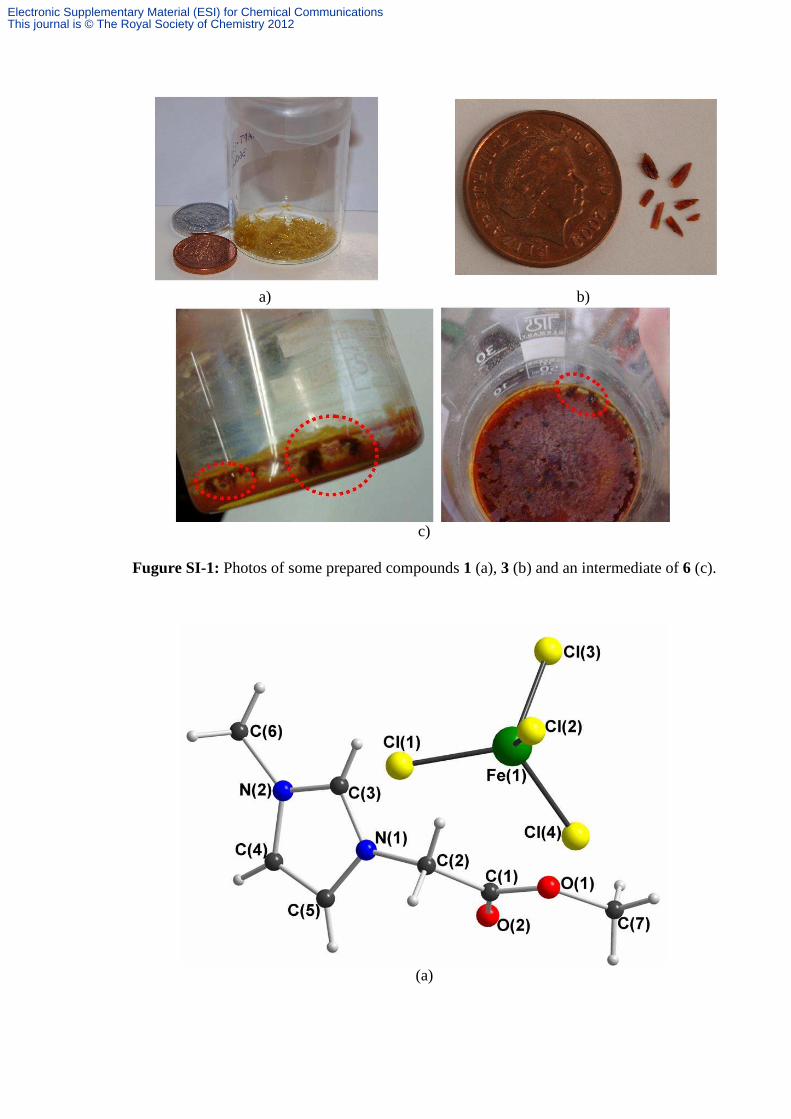
Fugure SI-1: Photos of some prepared compounds 1 (a), 3 (b) and an intermediate of 6 (c).
(a)
Electronic Supplementary Material (ESI) for Chemical CommunicationsThis journal is © The Royal Society of Chemistry 2012
(b)
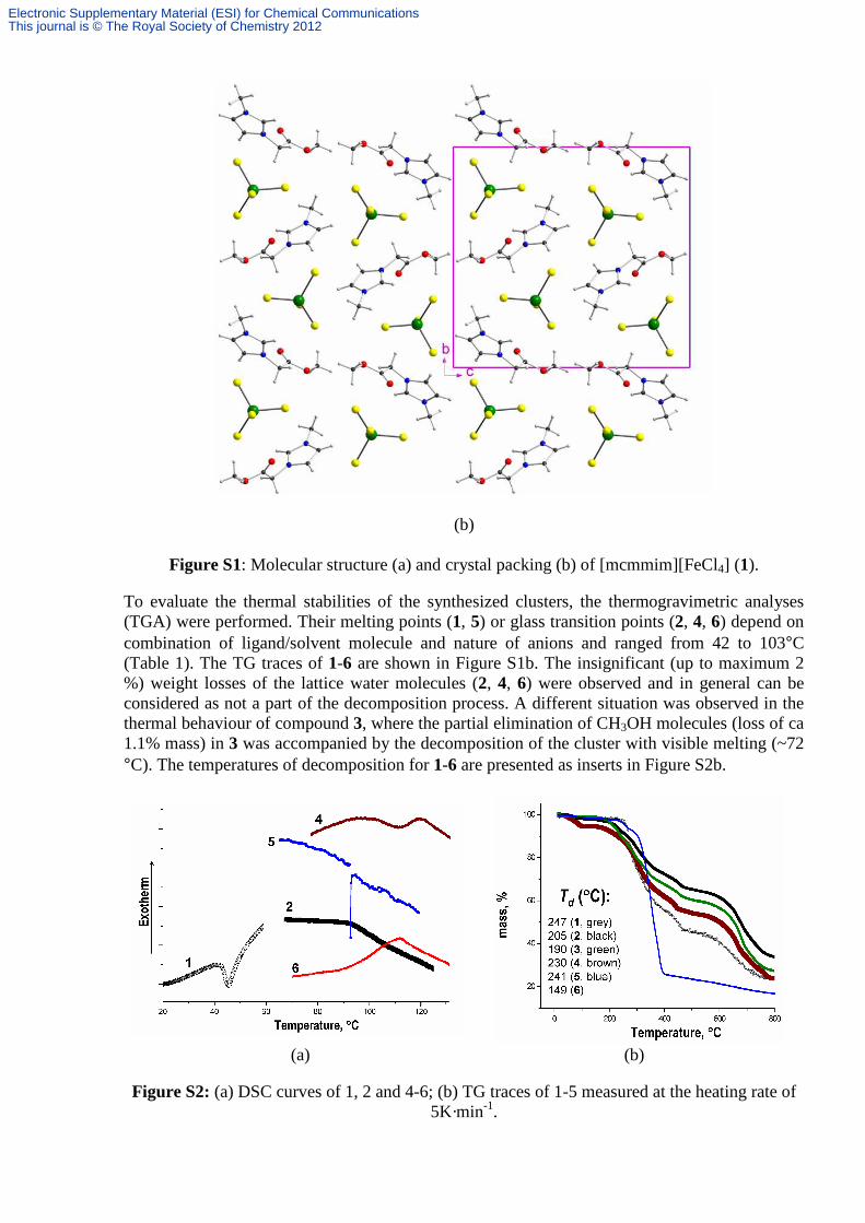
Figure S1: Molecular structure (a) and crystal packing (b) of [mcmmim][FeCl4] (1).
To evaluate the thermal stabilities of the synthesized clusters, the thermogravimetric analyses (TGA) were performed. Their melting points (1, 5) or glass transition points (2, 4, 6) depend on combination of ligand/solvent molecule and nature of anions and ranged from 42 to 103°C (Table 1). The TG traces of 1-6 are shown in Figure S1b. The insignificant (up to maximum 2 %) weight losses of the lattice water molecules (2, 4, 6) were observed and in general can be considered as not a part of the decomposition process. A different situation was observed in the thermal behaviour of compound 3, where the partial elimination of CH3OH molecules (loss of ca 1.1% mass) in 3 was accompanied by the decomposition of the cluster with visible melting (~72 °C). The temperatures of decomposition for 1-6 are presented as inserts in Figure S2b.
(a) (b)
Figure S2: (a) DSC curves of 1, 2 and 4-6; (b) TG traces of 1-5 measured at the heating rate of
5K·min-1.
Electronic Supplementary Material (ESI) for Chemical CommunicationsThis journal is © The Royal Society of Chemistry 2012
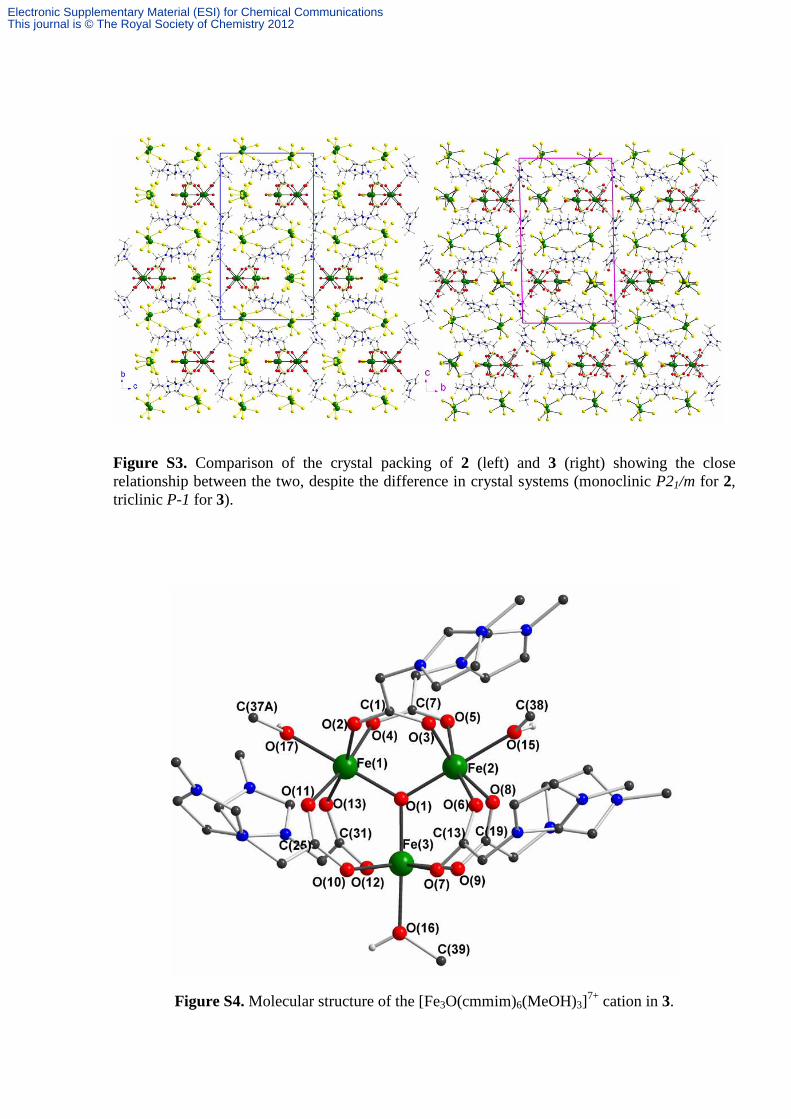
Figure S3. Comparison of the crystal packing of 2 (left) and 3 (right) showing the close relationship between the two, despite the difference in crystal systems (monoclinic P21/m for 2, triclinic P-1 for 3).
Figure S4. Molecular structure of the [Fe3O(cmmim)6(MeOH)3]7+ cation in 3.
Electronic Supplementary Material (ESI) for Chemical CommunicationsThis journal is © The Royal Society of Chemistry 2012
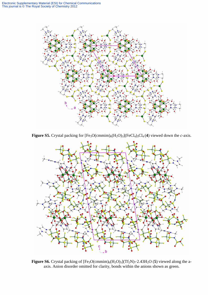
Figure S5. Crystal packing for [Fe3O(cmmim)6(H2O)3](FeCl4)3Cl4 (4) viewed down the c-axis.
Figure S6. Crystal packing of [Fe3O(cmmim)6(H2O)3](Tf2N)7·2.43H2O (5) viewed along the a-axis. Anion disorder omitted for clarity, bonds within the anions shown as green.
Electronic Supplementary Material (ESI) for Chemical CommunicationsThis journal is © The Royal Society of Chemistry 2012

(a) (b)
Figure S7. (a) Molecular structure of the trinuclear [Fe3O(cepy)6(H2O)3]7+ complex in 6 (organic
H-atoms omitted for clarity - the metal complex has crystallographically-imposed twofold rotational symmetry and the prime character in the atom labels indicates that these atoms are at the equivalent position {-x+1, y, -z+½}); (b) Crystal packing in [Fe3O(cepy)6(H2O)3][FeCl4]6Cl·4H2O (6) (anion disorder omitted for clarity.
Crystallography: Data were measured on Stoe IPDS 2 (2, 3, 5, 6) or IPDS 1 (1, 4)
diffractometers using graphite-monochromated Mo-Kα radiation (λ = 0.71073 Å). Details of the
crystal structures, data collection and structure refinement are summarised in Table S1. Structure
solution (direct methods) and full-matrix least-squares refinement against all data were carried
out with the SHELXTL 6.14 software package. [G.M. Sheldrick, Acta Cryst. 2008, A64, 112-
122]. In general, all ordered non-H atoms were assigned anisotropic temperature factors, and H-
atoms bonded to carbon were placed in calculated positions. Comments to specific structures are
as follows:
2: One FeCl4 anion (Fe(8)) was disordered either side of the mirror plane, with the two mirror-
related components overlapping. The half-occupancy anion was refined with similarity restraints
applied both to the Fe-Cl and Cl..Cl distances, and to the anisotropic thermal parameters (DELU)
of the five atoms. It proved not possible to refine the hydrogen atoms of the aquo ligands,
because of the disorder of the anions to whicjh they make hydrogen bonds. Lattice waters were
disordered between the (FeCl4)- anions, and proved difficult to model in the presence of the
heavy atoms of the anions. They were handled using the SQUEEZE option within PLATON
[A.L. Spek, J. Appl. Cryst. 2003, 36, 7-13].
Electronic Supplementary Material (ESI) for Chemical CommunicationsThis journal is © The Royal Society of Chemistry 2012

3: The crystal structure is closely related to the monoclinic P21/m structure of complex 2, and is
derived from it by a slight change of the angles α and γ from 90 degrees, followed by the
necessary axis permutation to give the conventional triclinic cell. The unit cell contents of 3 also
approximate to that of 2 (Fig. S3), and the resulting pseudo-symmetry hampered refinement.
Not unexpectedly, this pseudo-symmetry also resulted in pseudo-merohedral twinning by 180
degree rotation about the triclinic c-axis (corresponding to monoclinic b in 2). This was not
noticeable during data collection (ca. 14% for the minor domain), so a datafile in HKLF 5 format
was created using the TWINROTMAT option within PLATON Refinement was then relatively
straightforward. One of the (FeCl4)- anions was disordered: the Fe of the minor component
refined to a site occupancy of 0.16. The chlorines of this minor component were then further
disordered, and could not be refined satisfactorily. One MeOH ligand, and the lattice water to
which it hydrogen bonds, were twofold disordered either side of the pseudo-mirror plane.
4: Geometrical restraints were applied to positions of water H atoms. A chloride anion and a
lattice water were 50:50 disordered; a similarity restraint was applied to the anisotropic thermal
parameters of the closely separated atoms.
5: O-H bond lengths were restrained to 0.92(4) Ang. Three of the (Tf2N)- anions were
extensively disordered. Refinement (with disordered partial-occupancy atoms isotropic) was
successful with S-N, S-O, S-C and C-F bond lengths restrained to common values, as were S..S
and F..F 1-3 distances where necessary. A (partial occupancy) lattice water, O(63B), was only
consistent with the minor component of one of the Tf2N anions; no corresponding major
component appeared to be present, resulting in the total of 2.43 lattice waters per formula unit.
6: The Fe and two Cl atoms of a (FeCl4)- anion were twofold disordered, while three Cl atoms
of another anion were similarly disordered. Within each anion, similarity restraints were applied
to Fe-Cl distances. Geometrical restraints were applied to O-H bonds.
Crystallographic data (excluding structure factors) for the structures in this paper have been
deposited with the Cambridge Crystallographic Data Centre as supplementary publication nos.
CCDC 896157 (1), 896158 (2), 896159 (3), 896160 (4), 896161 (5), 896162 (6). Copies of the
data can be obtained, free of charge, on application to CCDC, 12 Union Road, Cambridge CB2
1EZ, UK: http://www.ccdc.cam.ac.uk/cgi-bin/catreq.cgi, e-mail: [email protected],
or fax: +44 1223 336033.
Electronic Supplementary Material (ESI) for Chemical CommunicationsThis journal is © The Royal Society of Chemistry 2012

Table S1. Crystallographic and structure refinement data for compounds 1-6. 1 2 3 4 5 6 Formula C7H11Cl4FeN2O2 C36H64Cl28Fe10N12O21 C41H70Cl28Fe10N12O19 C36H68Cl16Fe6N12O23 C50H58.86F42Fe3N19O46.43S14 C48H68Cl25Fe9N6O20
Mr 352.83 2552.09 2586.19 1939.32 3083.29 2437.98
Crystal System Monoclinic Monoclinic Triclinic Hexagonal Triclinic Orthorhombic
Space group P21/c P21/m P-1 P-62c P-1 Pbcn
T (K) 253(2) 150(2) 180(2) 200(2) 180(2) 150(2)
a (Å) 6.3649(5) 12.6816(9) 12.7495(4) 13.3201(6) 14.9606(4) 24.8363(12)
b (Å) 14.6411(11) 26.2169(14) 15.5320(5) 13.3201(6) 15.5623(4) 13.7847(7)
c (Å) 15.9618(12) 15.5404(10) 26.1211(7) 24.7380(13) 27.4325(9) 27.4404(19)
α (°) 90 90 90.846(2) 90 93.562(2) 90
β (°) 101.004(9) 109.243(5) 91.244(2) 90 101.381(2) 90
γ (°) 90 90 108.346(2) 120 115.135(2) 90
V (Å3) 1460.12(19) 4878.1(5) 4907.3(3) 3801.1(3) 5591.2(3) 9394.5(9)
Ζ 4 2 2 2 2 4
Dcalc (g cm-3) 1.605 1.738 1.750 1.694 1.831 1.724
F(000) 708 2536 2576 1960 3087 4868
(Mo-K) (mm-1) 1.7532 2.265 2.252 1.748 0.807 2.117
Reflections collected 11346 67061 61617 29754 37919 82847
Unique reflections 2832 9461 17953 2588 20945 8600
Rint 0.0261 0.0669 0.1154 0.0869 0.0472 0.0833
Parameters 147 511 980 171 1724 527
R1 (I > 2σ(I)) 0.0350 0.0596 0.0701 0.0457 0.0579 0.0768
wR2 (all data) 0.0910 0.1726 0.2169 0.1226 0.1603 0.2165
S (all data) 1.073 0.999 1.060 1.066 0.993 1.032
Electronic Supplementary Material (ESI) for Chemical CommunicationsThis journal is © The Royal Society of Chemistry 2012

0
5
10
15
20
25
30
35
0 100 200 300
χ M(c
m3 m
ol-1
)
χ MT
(cm
3 mo
l-1K
)
T(K)
χ
χT
fit-(χ)
fit -(χT)
Figure S8. Magnetic susceptibility χ and χT versus T plots data for compound 3. The black solid lines correspond to the best fit according the model and parameters indicated in text.
The isomer shifts are comparable to those of chloride- or oxo-bridged [S2] high-spin
ferric complexes. The isomer shifts of ca. 0.33 mm/s for 7[FeCl4]- are in good agreement
with the reported values for tetrahedral FeIII .[S3] Although the quadrupole splitting of high-spin FeIII in tetrahedral environments is usually negligible or even zero, small values of quadrupole splitting are conceivable in distorted surroundings. In some cases, e.g. NMe4FeCl4 or NEt4FeCl4, quadrupole splitting was zero, while in AsPh4FeCl4 a quadrupole splitting of 0.2 mm/s was reported.[S3a] These features are regarded as owing to the regular tetrahedron in some of the reported examples and the slightly distorted ones for 7[FeCl4]
- in the compound 3. An increase in the isomer shift upon cooling is attributed to a second-order Doppler effect.
Figure S9. The 57Fe Mössbauer spectra of 3 at indicated temperatures.
[S2] (a) A. L. Feig, S. J. Lippard, Chem. Rev. 1994, 94, 759-805; (b) Y. Zang, G. Pan, L. Jr. Que, B. G. Fox, E. Münck, J. Am. Chem. Soc. 1994, 116, 3653-3654; (c) Y. Zang, Y. Dong, L. Jr. Que, K. Kauffmann, E. Münck, J. Am. Chem. Soc. 1995, 117, 1169-1170.
[S3] (a) P. R. Edwards, C. E. Johnson, J. Chem. Phys. 1968, 49, 211-216; (b) S. Kitao, M. Seto, Y. Maeda, T. Matsuyama, S. Masubuchi, S. Kazama, J. Phys. Soc. Jpn. 1997, 66, 1195-1200.
Electronic Supplementary Material (ESI) for Chemical CommunicationsThis journal is © The Royal Society of Chemistry 2012

Table S2: Mössbauer parameters for 3 at indicated temperatures.
T, K Site δ[a], mm/s ∆EQ, mm/s Γ, mm/s Fe3O 0.431(6) 0.60(1) 0.40(1)
150 7xFeCl4 0.320(3) 0.17(1) 0.45(1)
Fe3O 0.435(1) 0.621(3) 0.33(1) 3
7xFeCl4 0.337(1) 0.197(2) 0.41(1) [a] Relative to α-Fe at room temperature. The statistical errors are given in parentheses. δ is the isomer shift, ∆EQ - quadrupolar splitting, Γ - average width of the doublet peaks.
Table S3: The recycle of catalysts in synthesis of ethyl 2-methyl-4-(2-oxo-2,3-dihydro-1H-3-indolyl)-5-phenyl-1H-3-pyrrolecarboxylate (7a).
Number of cycles/Isolated yield [%] Catalyst
1 2 3 4 5 6 7 8 9 10 1 86.2 85.1 82.7 78.3 65.0 31.0 - - - - 2 94.5 90.2 86.2 70.5 69.0 45.0 - - - - 3 - - - - - - - - - - 4 98.0 96.5 95.1 93.8 90.0 89.6 88.6 88.3 85.0 62.0 5 98.0 96.5 93.6 92.4 92.0 92.0 89.6 86.2 84.7 73.8 6 96.0 94.1 93.2 91.6 89.6 88.2 88.0 84.1 76.1 73.2
Procedure for synthesis of ethyl 2-methyl-4-(2-oxo-2,3-dihydro-1H-3-indolyl)-5-phenyl-1H-3-pyrrolecarboxylate (7a) The mixture of 3-(2-oxo-2-phenylethylidene)indolin-2-one (0.001 mol), ethyl 3-oxobutanoate (0.001 mol), catalyst (10 mg) and ammonium acetate (0.0025 mol) was stirred at reflux in dry EtOAc (4 ml) for 1.5-2 hour (monitoring by TLC), filtered. After removal of ethyl 2-methyl-4-(2-oxo-2,3-dihydro-1H-3-indolyl)-5-phenyl-1H-3-pyrrolecarboxylate 7a, the rest was directly recycled in subsequent runs. Analytical sample with m.p. 317-318 oC has been obtained by recrystallization from EtOH. IR (KBr), /cm-1: 3347, 3208, 1702, 1667, 1466, 1101, 698; Anal. calc. for C22H20N2O3 (7a): C, 73.32; H, 5.59; N, 7.77. Found: C, 73.25; H, 5.57; N, 7.73. Table S4: The recycling of catalysts in synthesis of 3,3'-((4-methoxyphenyl)methylene)bis(1H-indole) (8a).
Number of cycles Isolated yield [%]/Time [hours]
Catalyst
1 2 3 4 5 1 82/5 72/5 56/15 - - 2 92/5 89/5 78/10 75/10 70/15 3 - - - - - 4 85/5 78/10 75/10 72/15 - 5 70/5 65/10 - - - 6 89/5 74/10 69/10 60/10 56/15
Procedure for synthesis of 3,3'-((4-methoxyphenyl)methylene)bis(1H-indole) (8a). A mixture of indole (0.5 mmol), 4-methoxybenzaldehyde (0.25 mmol), and catalyst (3 mg) in benzene (2.5 ml) was refluxed for the appropriate time (Table S4). After completion of the reaction as indicated by TLC, the catalyst was separate by decantation, and used for next run with out additional purification. The benzene was concentrated and crude compound purified by column chromatography (EtOAc/benzene) to afford the pure 3,3'-((4-methoxyphenyl)methylene)bis(1H-indole) 8a. M.p. 187-189 oC; IR (KBr), /cm-1: 1218, 1245, 1453, 1509, 1611, 2925, 3412; Anal. calc. for C24H20N2O (8a): C, 81.79; H, 5.72; N, 7.95. Found: C, 81.73; H, 5.75; N, 7.91.
Electronic Supplementary Material (ESI) for Chemical CommunicationsThis journal is © The Royal Society of Chemistry 2012