casos especviales de toraxz
TRANSCRIPT


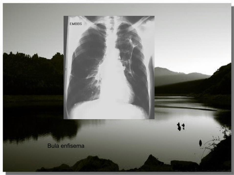
Bula enfisema

neumomediuastino


LUL pneumonia with volume lossNote the difference when there are only two lobes
Loss of heart borders/silhouetting Note anterior displacement of fissure on lateral
view Dx: pneumococcal pneumonia; Incomplete collapse
raises question of space-occupying abnormality such as pneumonia vs. incomplete atelectasis

enfisema
Flattening of diaphragms/increased lung volumes Enlarged left pulmonary artery
Attenuation of vessels Diffuse hyperlucency

No lung markings above pneumo thin line of pleural surface parallel to chest wall
Neumotorax apical izq


Pulmonary artery enlargement and cardiomegaly fron pulmonary arteryLarge pulmonary arteries and pruning
Large heart Median sternotomy
Differential diagnosis includes atrial septal defect


