cases%from%dermatology%vts%workshp%day% dermatology… day/case studies from... · 2)...
TRANSCRIPT
CASES FROM DERMATOLOGY VTS WORKSHP DAY
Dermatology Case 1 18/10/11
A 17 year old girl comes into your surgery very upset and embarrassed about the spots on her face. On further questioning she tells you she has had spots for 2-‐3 years and tried all the over the counter medications to little effect. It is now affecting her social life as she is afraid to go out and socialise due to being self conscious. She has stopped eating fatty foods and chocolate as she has heard this may be causing it.
She is otherwise well. She is not in a relationship at present and is not on contraception.
On examination you notice she has multiple small comedomes and some larger pustules on both cheeks and across the forehead. You can see no evidence of scarring.
How would you grade her acne?
What types of lesions can be present in acne?
What would be you management plan?
What advice would you give with regards to diet?
She come back to see you after 6 months of treatments you instigated. The acne initially improved, but has not settled and if anything got worse again recently.
On re examination today you note her acne is still pustular with large lesions and now you can see some small scars present on the cheeks.
What types of scarring do you know about in acne?
What management might you consider now?
How would you council her for this and how to do you instigate treatment?
Dermatology Case 1 ACNE VULGARIS. NOTES FOR GROUP LEAD
A 17 year old girl comes into your surgery very upset and embarrassed about the spots on her face. On further questioning she tells you she has had spots for 2-‐3 years and tried all the over the counter medications to little effect. It is now affecting her social life as she is afraid to go out and socialise due to being self conscious. She has stopped eating fatty foods and chocolate as she has heard this may be causing it.
She is otherwise well. She is not in a relationship at present and is not on contraception.
On examination you notice she has multiple small comedomes and some larger pustules on both cheeks and across the forehead. You can see no evidence of scarring.
1) How would you grade her acne?
There are over 25 recognised grading scales, the most common being Mild, Moderate and Severe. (See chart)
Mild - Open and closed comedomes
Moderate - Papules and pustules present
Severe - Papules pustules, cysts and nodules present. Evidence of scarring
The Leeds Revised Acne grading scale (enclosed)
Grading from 1- 12 in each area when compared to photographs. Agreed by panel of experts. Cumbersome and essentially does not alter management
She thereofre has Moderate acne.
2) What types of lesions can be present in acne?
Important to understand the types of lesion as can affect management strategy used.
Open Comedomes - (blackheads) wider opening follicles containing slough appear black
Closed comedomes - (whitehead) smaller opening comedomes therefore blocked and appear white
Sand paper comedomes Multiple small comedomes giving skin a rough texture
Papules Superficial inflammation
Pustules Superfical inflammation containing pus
Nodules and cysts Deep inflammation
Occasionally nodules can combine to cause sinuses in severe cases
3) What would be you management plan?
Consider psychosocial affect. Reassurance, counselling and depression screening may be appropriate.
Specific treatments:
See enclosed algorithm.
Topical retinoids – Adapalene (Differin), Tretinoin (retin-A)
Good first line. Warn re: local reaction, erythema and peeling plus risks with sun exposure. Avoid on broken skin.
Benzoyl peroxide Good for inflamed lesions. Can dry skin. Azelaic acid has antimicrobial properties also
Topical antimicrobials (zineryt, Dalacin T).
Combined therapies (DUAC – Benzyl peroxide and clindamycin)
Oral antibx Most common Lymecycline as once daily dose 408mg od.
Also can use oxy tetracycline, doxycycline minocycline and erythromycin
Takes 3-4 weeks for benefit to be apparent. Use 3-4 month courses then consider stopping or changing. Warn re riks with pregnancy and teratogenicity
COCP Dianette to high DVT risk. ‘Yasmin’/Marvalon considered. Normal COCP if used also for contraception may be of benefit.
?spironolactone
?laser treatments
AVOID steroids, moisturisiers, hair gels, emollients, progesterones, picking spots (acne excoriee)
4) What advice would you give with regards to diet?
Dispel myths about diet. However, recent studies have pointed toward the fact that diet may have an causative effect with regards to acne.
Health balanced diet advice is recommended.
She come back to see you after 6 months of treatments you instigated. The acne initially improved, but has not settled and if anything got worse again recently.
On re examination today you note her acne is still pustular with large deep lesions and now you can see some scars present on the cheeks.

5) What types of scarring do you know about in acne?
'Ice pick scars' on the cheeks (initially presents in purple colour and later fades becoming white) (1)
'Tissue paper scars' across the shoulders
Keloid scars on the middle chest and shoulders
6) What management might you consider now?
Referral for oral isotretinoin
7) How would you council her for this and how to do you instigate treatment?
Specialist initiation and management only.
Isotretinoin (a derivative of vitamin A) is available on hospital-only prescription because of its potential toxicity and teratogenicity. It is used in the treatment of acne, reducing sebum production and surface propionibacterium acnes.
Since it may be obvious early on which patients are going to be referred, it may be worth starting the patient on antibiotics at the time of referral.
Isotretinoin is contraindicated in:
• hypervitaminosis A • uncontrolled hyperlipidaemia • during pregnancy or lactation (1)
if the patient is female then it is important that a negative pregnancy test is confirmed before therapy is commenced. Also female patients must use effective contraception for at least four weeks before treatment, during the treatment period and for at least four weeks following its cessation
Oral isotretinoin indications include
• nodulocystic acne • scarring • frequent relapsing acne (4) • patients with mild-to-moderate acne that has failed to respond to at least
two courses of antibiotics
Side-effects include:
• mucocutaneous dryness causing: o cracked lips o nose bleeds o colonization of S. aureus leading to complications like abscesses,
conjunctivitis, impetigo, cellulitis, and folliculitis (1) • headaches • myalgia • hyperlipidaemia, hypercholesterolaemia and disordered liver function
tests: o lipid levels and liver function tests are monitored during treatment
• teratogenesis: o the rate of fetal abnormality is high so contraception must be
reliable and termination is usually recommended if pregnancy did occur
o ear defects together with CNS defects or cardiovascular defects or both can be seen (1)
• psychiatric disorders e.g. depression, suicide
Cumulative dose is important for a long term response
• 85% of patients respond to a full dose for 4 months. More patients will respond to longer courses of treatment.
• 40% of the patients are free of acne after one course of treatment • 40% relapse requiring medications • 20% will need to repeat isotretinoin
Urticaria Case Study
Through this case we aim to cover:
15.10
• Manage primary contact with patients with angio-‐oedema/urticaria • Appreciate the importance of the social and psychological impact of urticaria/angio-‐
oedema on the patients QOL including sleep. Recognise how disfigurement and cosmetic skin changes can affect patients’ confidence, mood and interpersonal relationships
• Identify health beliefs and either reinforce, modify or challenge these beliefs as appropriate
• Advised on lifestyle interventions • Coordinate care with other professionals, dermatologists and other specialists • Intervene urgently when patients present with emergency skin problems such as angio-‐
oedema with airways involvement.
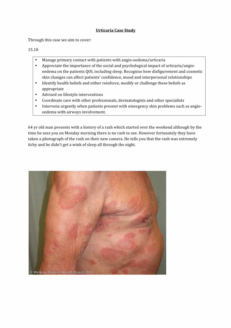
64 yr old man presents with a history of a rash which started over the weekend although by the time he sees you on Monday morning there is no rash to see. However fortunately they have taken a photograph of the rash on their new camera. He tells you that the rash was extremely itchy and he didn’t get a wink of sleep all through the night.

To aid discussion consider discussing:
What further information do you want from the history?
What tests/investigations would you do?
What types of urticaria do you know?
What treatment would you try?
What would you do differently if the rash had been present for >6 weeks?
What foods/drugs can cause urticaria?
Difference with angio-‐oedema?

Facilitators notes History
Detail on any triggers, duration of Wheals, previous episodes, changes to diet/medicines
Dermographism?
Atopy? Autoimmune conditions?
Associated joint pains, unwell systemically may indicate vasculitic urticaria
Investigations
If chronic urticaria test FBC, LFT, ESR and TFT. Consider C3 and C4, Autoimmune screen.
Skin prick testing rarely yields useful information
Consider exclusion diet if food suspected trigger.
Types of Urticaria
Ordinary – may be triggered by stress or viral infections • Acute (up to 6 weeks of continuous activity) • Chronic (6 weeks or more of continuous activity) • Episodic (acute intermittent or recurrent activity)
Physical urticarias (reproducibly induced by the same physical stimulus) Mechanical
• Delayed pressure urticaria • Symptomatic dermographism • Vibratory angio-‐oedema
Thermal
• Cholinergic urticaria • Cold contact urticaria • Localized heat urticaria
Other
• Aquagenic urticaria • Solar urticaria • Exercise-‐induced anaphylaxis
Angio-‐oedema without weals
• Idiopathic

• Drug-‐induced • C1 esterase inhibitor deficiency
Contact urticaria (contact with allergens or chemicals) Urticarial vasculitis (defined by vasculitis on skin biopsy) Autoinflammatory syndromes
• Hereditary o Cryopyrin-‐associated periodic syndromes (CIAS1 mutations)
• Acquired o Schnitzler syndrome, SLE
Treatment
Avoid triggers – heat, pressure, stop ACEi, stress, alcohol
Avoiding scratching – nails short, rubbing with palm of hand
Natural clothing fabrics e.g cotton
Calamine lotion or cool baths to soothe, 1% menthol cream can be helpful
Non sedating H1 blocker during day time – up to three times licensed dose often used e.g cetirizine 10mg tds
What would you do differently if the rash had been present for >6 weeks?
This would be defined as Chronic Urticaria. Management pharmacologically can be the same as for acute.
May also consider:
• ??mast cell stabiliser: Monteleukast. – unlicensed and response variable • Sedating antihistamine at night – e.g chlormphanamine • Consider addition of H2 blocker – ranitidine -‐ unlicensed • Can consider prednisolone 40mg for 3-‐5 days, if relapse occurs after stopping
seek specialist advice.
All treatment may need to be continued for up to 1 yr – by which point most cases will have “burnt out”
Can be a considerable cause of poor QOL – poor sleep, self image, social isolation – constant hypervigilance e.g foods can eat, preservatives, heat, cold, stress
Interpersonal relationships can be affected, 14% develop depression
What foods/drugs can cause urticaria?
Foods high in salicylate:
Strawberry, Kiwi, grape, dates, all dried fruit, apricot, cranberry, coffee, tea, processed meats, salami and other seasoned/preserved meats, almond, peanuts, brazil nuts....to name but a few!
Drugs: NSAID, aspirin, opiods, ACEi, radiocontrast media, antipsychotics
Angio-oedema
Swelling in the dermis, subcutaneous tissues and submucosal tissues. 1:10 patients presenting will present with lone angio-‐oedema without urticaria. 4:10 present with a combination.
Swelling less well defined, can oddur anywhere but commonly, lips, eyes, hands, feet genitalia.
If anaphylaxis – follow RC guidelines for management.
If any history of airway compromise patient should be referred to hospital.
Otherwise treat with combination of antihistamine and prednisolone 40mg OD for 5 days
C1 esterase inhibitor suggestive if C4 level < 30% of expected levels.

Infections
Aim:
• Manage primary care contact with a patient with skin problem such as fungal, yeast and bacterial infections
• Work with patients to empower them to look after their own health and take responsibility for managing skin problems
• Appreciate the importance of the psychological impact of skin problems • Demonstrate a reasoned approach to the diagnosis using history, examination,
incremental investigations and appropriate referrals • Be able to describe the side effects of medications • Be able to take specimens for mycology from hair, skin and nails
Case Study A 10 year old boy comes with his mother complaining of a rash to his scalp which has been present for 4 months. The scalp is now very flaky and itchy. He has been off school a few times complaining of abdominal pains, but his mum suspects that he may be being bullied. She heard one of them call him “Daniel Druff” in the playground. What would your initial approach be with this case? Assuming that he hasn’t got an acute abdomen outline questions from the history that you would want to answer with regard to the scalp condition? Below is the appearance of his scalp -‐ What is the likely diagnosis and differential?
What investigations would you do?
What treatment would you consider? What about school? When do you stop treatment? Where are the following infections of? For each decide on an appropriate treatment regime? Tinea pedis Tinea capitis Tinea corporis Tinea unguim Tinea cruris Tinea Manum Tinea barbae During the consultation you also note that the child has sores either side of his mouth, which appear as erythematous fissures. What are these and what treatment would you prescribe? Would you do any investigations?

Facilitators notes Background Most common dermatophyte infections are Trychophyton rubrum, Trychophyton mentagrophytes, and Trychophyton tonsurans. Infection can spread from person, animal or soil. Tinea pedis is common in adults whilst tinea capitis is common in children. What would your initial approach be with this case?
Dont get blinkered! Take full history and examination regarding the tummy pains to acknowledge this and show you are taking him seriously and gaining trust. Establish that no underlying cause for abdo pain. Good opportunity to explain about “worry tummy” which may allow you to ask if anything is worrying him.
Need to take holistic approach encompassing mum also – most significantly discussion about the psychological effects this is having on him.
Be aware that conditions such as DM, pregnancy, immunocompromise, corticosteroid use and cushings disease can predispose to dermatophyte infections, so a quick check such as urine dip will help to exclude DM, and also UTI for abdo pain. Outline questions from the history that you would want to answer? Duration, itch, boggy swelling, alopecia, other skin conditions/areas affected, family members, contact at school ,atopic history, treatments applied. What is the likely diagnosis and differential? Tinea capitis – commonly caused by Trychophyton tonsurans. Consider eczema, seborrheic dermatitis, contact dermatitis, psoriasis. What investigations would you do? Scalp brushings – disposable toothbrush easiest way to collect samples – validated method, the negative charge generated from rubbing the bristles on the scalp will collect the scale. Alternatively may send hair samples or scalp scrapings – use blunt edge of scalpel. Samples can take 4-‐6 weeks for results from lab. Other infections may be visible as glowing blue/green under wood’s lamp What treatment would you consider? T. tonsurans penetrates hair shaft therefore topical treatments do not work. Terbinafine is used – off licence in < 12. Dose 62.5mg day < 20kg, 125mg 20-‐40kg, > 40kg 250mg /day. Ususally tablets need to be crushed and concealed in yoghurt or jam. Treat for 4-‐6 weeks. Baseline bloods not required. Se’s headache, nasopharyngitis, rash and GI effects.

Alternatively Griseofulvin can be used – is licensed. Dose per body weight -‐ > 12 yrs 500mg /day for 8-‐10 weeks. Selenium sulphide – selsun shampoo twice weekly will not treat alone but may prevent transmission and decrease the fungal load. Children should not be excluded form school once treatment commenced. Recommended to repeat sample at end of recommended treatment time – treatment success is based on mycology results. Where are the following infections of? For each decide on an appropriate treatment regime? Tinea pedis – foot. Terbinafine cream or tablets – tablets more effective. 250mg daily for 4 weeks Tinea capitis – scalp. as above Tinea corporis – body. Imidazole creams such as miconazole, clotrimazole for 2-‐4 weeks – cont for 2 weeks once cleared. If extensive, or topical failed – terbinafine for 4 weeks Tinea unguim – Nails. terbinafine 250mg OD for Approx 3 mo for toe nails and 6-‐8 weeks in finger. Tinea cruris – groin. Imidazole cream, or if fails, terbinafine 250mg OD for 2-‐4 weeks. Tinea Manum – hand. imidazole cream Tinea barbae – Beard. Try topical but may need terbinafine Angular Cheilitis Candida infection. Candida causes vulvovaginal candidiasis, napkin dermatitis, balanitis, intertrigo and oral thrush. Presents in angular cheilitis as erythema, soreness and fissuring to angles of the mouth. Can have superinfection with staph aureus. Responds well to topical miconazole or nystatin. May be worth check B12 and anaemia.
Dermatology Case 7 ACNE ROSACEA NOTES FOR GROUP LEAD
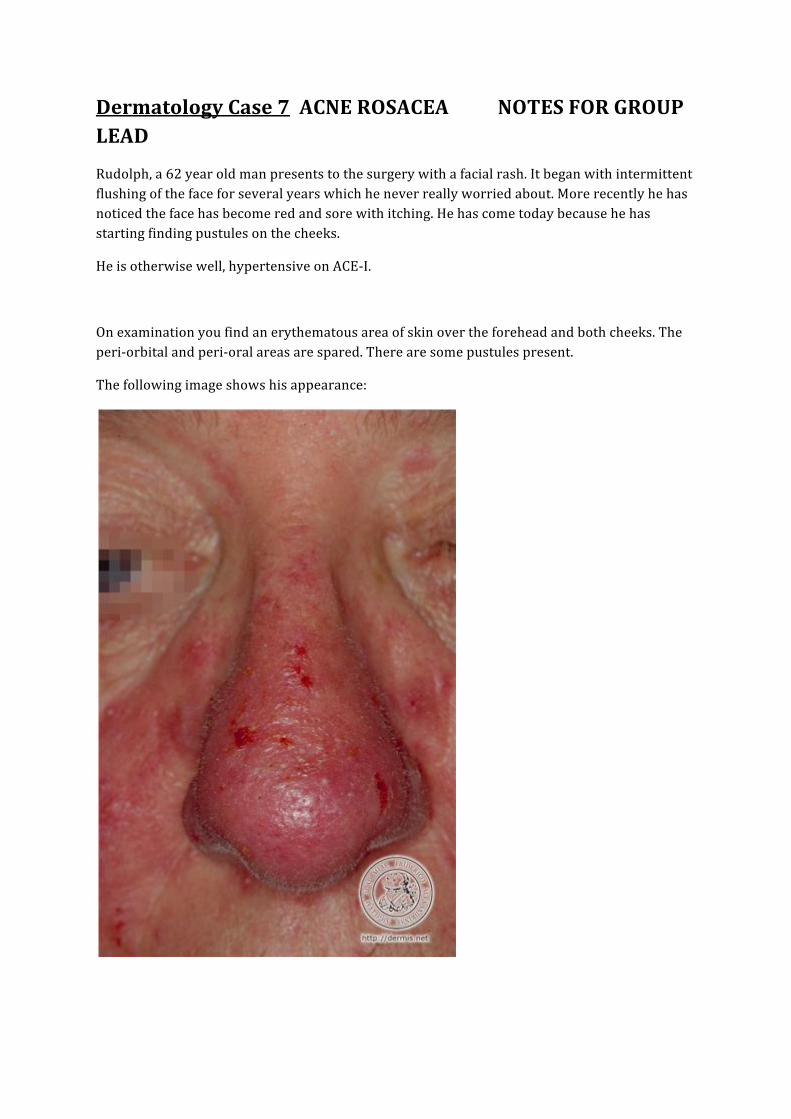
Rudolph, a 62 year old man presents to the surgery with a facial rash. It began with intermittent flushing of the face for several years which he never really worried about. More recently he has noticed the face has become red and sore with itching. He has come today because he has starting finding pustules on the cheeks.
He is otherwise well, hypertensive on ACE-‐I.
On examination you find an erythematous area of skin over the forehead and both cheeks. The peri-‐orbital and peri-‐oral areas are spared. There are some pustules present.
The following image shows his appearance:

1) What is the diagnosis?
Acne Rosacea
2) Who gets this and why does it occur?
This is a disorder of fair-skinned people in which there is fixed or recurrent erythema, telangiectasia, oedema, papules and pustules affecting the forehead, cheeks, nose and often the chin.
It is relatively common, accounting for 1% of dermatological out-patients in Britain. It occurs in the second half of life and is associated with facial flushing.
Rosacea is more common in women, but it tends to be more severe in men.
Ocular changes occur in more than 50% of the patients.
Aetiology is unknown
Emotion, sun, hot drinks, alcohol and the incorrect use of topical corticosteroids are important precipitating factors suggesting a lack of normal homeostatic control of the blood vessels supplying the pilosebaceous follicle
The mite Demodex folliculorum can be found on the face in large numbers but this association is speculative. Similarly rosacea has been anecdotally associated with seborrhoeic dermatitis, and in women with migraine headaches
3) What is your initially management plan?
Mild to moderate rosacea:
For people with few papules or pustules and mild to moderate persistent erythema, topical treatment is recommended
Topical metronidazole (Metronidazole gel (0.75%) applied twice a day, or cream (1%) applied once a day) is the preferred topical treatment -‐ note that gel preparations that contain alcohol may be more irritating to the skin

topical azelaic acid (15% gel, applied twice a day) may be considered for people who are intolerant of or not responding to topical metronidazole
These may cause a mild burning or stinging sensation when initially applied to the skin
(Some dermatologists also consider the use of other topical antibiotics or topical retinoids -‐ these treatments are not licensed for this condition)
Patients who do not respond to topical treatment and/or have severe rosacea (i.e. extensive papules, pustules, or plaques):
These patients are treated with systemic antibiotics -‐ oral tetracycline or oxytetracycline are recommended first-‐choice systemic treatments for rosacea; erythromycin is an alternative (e.g. pregnancy, reactions to sunlight)
(tetracycline or oxytetracycline, 500mg twice a day on an empty stomach)
full doses are given initially but gradually reduced once the condition has been controlled -‐ usually after 1-‐3 months. Antibiotics should not be stopped suddenly because this may result in rebound rosacea
There is evidence of benefit for the use of low dose isotretinoin (10mg per day) in rosacea -‐ the use of isotretinoin in rosacea should only be initiated by a specialist
After 12 weeks of your excellent treatment he is still no better. If anything things are worse with a large number of pustular lesions on the cheeks and nose. He is becoming quite distressed by it.
4) What would you do now?
Consider referral to dermatologist
Routine referral to dermatologist is advised for people with:
Flushing, persistent erythema, telangiectasia, or phymatous rosacea that is causing psychological or social distress
Papulopustular rosacea that has not responded to 12 weeks of oral plus topical treatment.
An uncertain diagnosis

He comes back again to see you (becoming a heartsink patient now) with eye symptoms. He has had mild blepharitis for years but over the past few days his vision has become blurred and he has bilateral eye pain plus photophobia.
5) What complication is this? What should you do?
KERATITIS/CORNEAL ULCER
Patients with ocular symptoms should be referred to an ophthalmologist:
Urgent referral- if keratitis is suspected (eye pain, blurred vision, sensitivity to light)
Routine referral- if ocular symptoms are severe or resistant to maximal treatment in primary care
Mild eye symptoms are usually treated with a combination of eyelid hygiene measures, ocular lubricants (for dry eye symptoms), and oral tetracyclines
A year goes by and you think you have cracked it. However, Rudolph presents once more (you are beginning to feel sorry for this extremely unfortunate chap) with eyes all better but now complains of the following:
( Can you see why I called Rudolph now?)

6) What he now developed? What should you do?
Routine referral to a plastic surgeon is advised for those people with:
Severe phymatous disease (e.g. prominent rhinophyma)
You Trawl the internet to show him what the condition is and find this picture. You opt not to show him it!
SEVERE RHINOPHYMA
Dermatology Case 11 ECZEMA HERPETICUM AND ERYTHEMA MULTIFORME.
NOTES FOR GROUP LEAD
It has been a long Friday in surgery. 18:11, running late as usual. You yearn to be at home with the Chinese takeaway and bottle of Pinot Grigio your partner had promised you whilst watching ‘Strictly Come Dancing’. Just 2 more patients left on your list to see. Come on you can do it.
You call in your next patient. He is a five year old boy with a Hx of mild eczema. Should be easy, lets go.
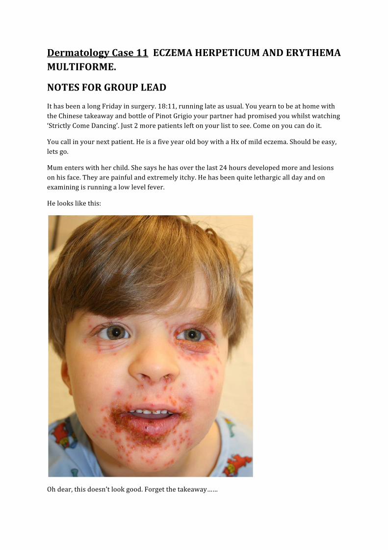
Mum enters with her child. She says he has over the last 24 hours developed more and lesions on his face. They are painful and extremely itchy. He has been quite lethargic all day and on examining is running a low level fever.
He looks like this:
Oh dear, this doesn’t look good. Forget the takeaway……
1) Describe the lesions
areas of rapidly worsening, painful eczema
clustered blisters consistent with early-‐stage cold sores
punched-‐out erosions (circular, depressed, ulcerated lesions) usually 1-‐3 mm that are uniform in appearance (these may coalesce to form larger areas of erosion with crusting)
2) What is the diagnosis and what is it caused by?
ECZEMA HERPETICUM
SEVERE PRIMARY HERPES INFECTION
3) How are you going to manage this?
ADMIT THEM!
Intravenous and topical acyclovir
Broad spectrum antibiotics are added in to treat or prevent superinfection
Monitor fluid and electrolyte balance
Good skin care

Having safely negotiated this difficult and unusual case you feel shattered. Just one more case before you can go home… another child, great quick diagnosis URTI and then over to Bruce and Tess
Mum brings in the child, and 8 year old boy previously fit and well. He has developed a rash on his extremities and hands and feet plus some crusted lesion son his face. He is feeling very unwell and is lethargic.
He is unable to eat or drink as a result of his mouth lesions
You note your partner has seen him 3 days earlier for a suspected UTI and prescribed antibx.
You start off by looking at the arms:
This again doesn’t look good, but you remember back to the excellent dermatology study day you attended run by the caring, conscientious and good looking TpDs and remember what it is
4) Describe the lesions
The appearance of lesions is very variable -‐ the lesions may have central pallor with peripheral erythema or central erythema with peripheral pallor. Frank bullae may be present and represent epidermal necrosis
The lesions begins as numerous, sharply demarcated erythematous macules which then become papular.
These papules gradually evolve into plaques
CLASSIC TARGET LESIONS IN MINOR CASES NORMALLY

5) What is the diagnosis?
ERYTHEMA MULTIFORME
6) Which conditions are associated with this diagnosis?
Crohns disease
Tuberculosis
Sarcoidosis
Herpes simplex virus – most strongly associated
E. Staphylococcal infections
You then look at the face:
7) Describe what you see
Crusted erosions on his lips, erosions on his palate and buccal mucosa
8) What is the diagnosis?
STEVEN JOHNSON SYNDROME
9) What is you management?
ADMIT if BLISTERING PRESENT. BRAVE NOT TO ADMIT ON FRIDAY NIGHT
Involves 2 or more mucous membranes
Commonly caused by sulphonamides, NSAIDS and anticonvulsants
If blisters are present, refer
Systemic antibiotics are indicated if there are any features of infection There may be a role for systemic steroids (controversial) -‐ discuss with a dermatologist. Benefits of intravenous immunoglobulin treatment in Stevens-‐Johnson syndrome have been documented in some studies
10) Which antibiotic is strongly associated with this condition?
Co-trimoxazole
Also sulphonylureas
11) What causative organisim of pneumonia is most associated with this condition?
Mycoplasma
Having safely negotiated both these cases you are free to go home safe in the knowledge it is a job well done and you have saved lives. Its 19:05 and you are just leaving the building when the phone rings….
Forget it, Out of Hours have taken over now, you’re going home.
Missed the start of Strictly, but thank goodness for Sky+.
Leg ulcer case
Aims
• Statement 15.1 Understand and utilise venous dopplers and ankle brachial pressure index (ABPI) measurement
• Be able to manage PVD – venous and arterial • Recognise the impact CV problems have on ability to work and disability • • 15.10 Appreciate the importance of the social adn psychological impact of skin problems
on the patients QOL • Recognise the impact of skin problems on fitness to work • Recognise how skin changes fundamentally affect the patients’ confidence, mood and
interpersonal relationships.
72 yr old smoker is referred to you for the HCA as he mentioned during his BP check that he had a small ulcer to his left leg. His last BP on the system was 164/92. He is on bendroflumethaizide 2.5mg, ramipril 5mg, aspirin 75mg OD, allopurinol 300mg OD, quinine sulphate 200mg ON and paracetamol 1g qds.
What further history would you want from him? What will you look for on examination?
What are the indicators of venous and arterial disease?
You ask the practice nurse to do some Doppler studies. He comes back the next week for you to review the results:
Left leg: ABPI 0.9
Right leg: ABPI 0.7
What does this mean? What would your management plan be?
A year later he has another ulcer, this time the pain is keeping him up at night and the ulcer is on the right foot, near to his 3rd and 4th toes.
You ask for repeat dopplers:
Lt leg : ABPI – 0.7
Rt leg: ABPI – 0.4
What would you do on this occasion?

Facilitators’ notes
• Definition: Loss of skin below the knee on the leg or foot which takes more than six weeks to heal
• About 120,000 people are suffering from leg ulcers today in the UK
• About 500,000 people get recurrent leg ulcers in the UK
• A third of patients develop their ulcer before the age of 50
• 2% of those over 80 years of age are thought to suffer with this condition
• Recurrence rates are high: 26% after one year and 31% after 18 months of healing for venous
ulcer patients
• The cost burden to the NHS has been estimated at around 400 million per annum
• The causes of leg ulcers can be put down to 3 main categories
1] Venous Causes (veins not working) - about 80% of leg ulcers
2] Arterial Causes (arteries not working) - about 15% of leg ulcers
3] Other Causes - about 5% of leg ulcers
What further information do you require from the history?
Assess cardiovascular risk – smoking, obesity, diabetes, DVT
Assess other risk factors such as immobility and co-morbidities such as rheumatoid arthritis
or vasculitis.
Wound history – previous ulcers, duration, treatment, Surgical history – varicose veins,
sclerotherapy
Ask about pain (site, nature, severity), odour, and discharge, length of history of ulcer.
Social history – including occupation and standing, dietary history and assessment of
malnutrition
Obstetric history – multiple pregnancies
Family history of venous disease.
Assess the impact on QOL –e.g, can they move around and carry out normal activities of
daily living such as shopping, housework, or employment
What do you look for on examination?
• Assess skin condition: lipodermatosclerosis( induration or changes in skin texture as fat is
replaced by fibrous tissue, haemosiderin deposition (brown staining), venous eczema,
atrophie blanche (avascular areas of white tissue within the gaiter area)
• Look for signs of an infected leg ulcer such as:
o Enlarging ulcer.
o Increased exudate or pain.
o Pyrexia.
o Foul odour.
o Cellulitis — surrounding skin is red, hot, and non-scaling.
• Assess:
o Size and depth — a photograph may be helpful. Examine to assess the depth of
the ulcer.
o Wound bed — look for granulation, and fibrous or necrotic tissue which may need
to be removed to allow healing. Look for exudate to help determine which dressing
is needed.
o The ulcer edge often give a good indication of progress and should be carefully
documented (for example shallow, epithelialising, punched out).
o The position of the ulcer(s) should be clearly described.
What are the indicators of venous and arterial disease?
VENOUS ARTERIAL
Site: Gaiter/malleoli Foot / below ankle
Large and irregular size Punched out and circular
Shallow base Deep base
Family history IHD
Hypertension TIA/CVA history
Varicose veins Diabetes
Proven DVT in affected leg PVD
Phlebitis Intermittent claudication
Surgery/leg fracture Smoker
Warm, pink, well perfused leg Rheumatoid arthritis
Examination Findings
Good foot pulses Poor foot pulses
Varicose eczema Cool, pale foot
Brown gaiter staining – haemosiderin Thickened toe nails
Lipodermatosclerosis Ischaemic rest pain
Hairless limb
You ask the practice nurse to do some Doppler studies. He comes back the next week for you to review the results:
Left leg: ABPI 0.9
Right leg: ABPI 0.7
What does this mean?
Venous ulcer in left leg, with normal arterial supply.
Arterial insufficiency in the right leg – although no ulcer is high risk for developing arterial ulcer. Advise re lifestyle re arterial disease – exercise, diet, smoking cessation
What would your management plan be?
ABPI TYPE OF ULCER COMPRESSION HOISERY COMMENT 0.8-1.2 Venous (normal
arterial flow) Aim for 40mmHg compression at ankle:
K2 (two layer)
K4 (four layer)
Class 3 :
e.g Scholl
25-35mmHg
Compression kit designed to be worn for 7 days – cost approx £10.
Stockings cost approx £8/pair
0.7-0.8 Mixed 23mmHg compression e.g Profore lite
Class 1 (17mmHg)
Class 2 18-24mmHg)
Remove if any arterial compromise
0.6-0.7 Arterial 17mmHg Toe to knee e.g crepe spiral
n/a ?? vascular referral

0.5-0.6 Arterial NO COMPRESSION
n/a Ref for vascular assessment
< 0.5 Arterial Tubifast only to secure dressings
n/a Urgent ref to vascular
• An ankle brachial pressure index (ABPI) involves the measurement of a person's
systolic blood pressure at their ankle and arm (brachial) using a Doppler machine.
• To calculate = highest pressure from ankle – post tibial artery/ highest brachial
pressure
o Less than 0.5 indicates severe arterial insufficiency and compression treatment is
contraindicated. Refer urgently to a specialist vascular clinic for further
assessment.
o Between 0.5 and 0.8 indicates that the person has arterial disease. Refer to a
specialist vascular clinic for further assessment. Compression bandaging should
generally be avoided. However, reduced compression can be used under strict
supervision (assess progress daily) if the ulcer is clinically venous and the
healthcare professional has sufficient experience.
o Greater than 0.8 indicates that graduated compression bandages may be safely
applied.
• People with diabetes mellitus, atherosclerotic disease, rheumatoid arthritis, and
systemic vasculitis may give falsely high (and misleading) ABPI readings due to
calcification of the blood vessels.
A year later he has another ulcer, this time the pain is keeping him up at night and the ulcer is on the right foot, near to his 3rd and 4th toes.
You ask for repeat dopplers:
Lt leg : ABPI – 0.7
Rt leg: ABPI – 0.4
What would you do on this occasion?
Tubifast/light spiral dressing only and refer URGENTLY to vascular team for angiogram.
Skin cancer case study
• Promote skin wellbeing by applying health promotion and disease prevention strategies appropriately including sun protection
• Make timely appropriate referrals on behalf of patients to specialist services, especially to rapid-‐access pigmented lesion (sometimes called skin cancer, mole or melanoma) clinics.
• Recognise risk factors and red flag signs in suspected skin cancer
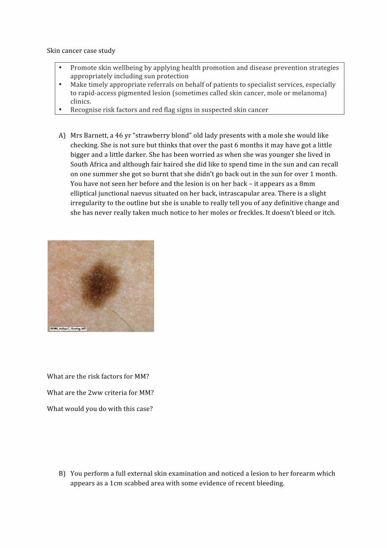
A) Mrs Barnett, a 46 yr “strawberry blond” old lady presents with a mole she would like checking. She is not sure but thinks that over the past 6 months it may have got a little bigger and a little darker. She has been worried as when she was younger she lived in South Africa and although fair haired she did like to spend time in the sun and can recall on one summer she got so burnt that she didn’t go back out in the sun for over 1 month. You have not seen her before and the lesion is on her back – it appears as a 8mm elliptical junctional naevus situated on her back, intrascapular area. There is a slight irregularity to the outline but she is unable to really tell you of any definitive change and she has never really taken much notice to her moles or freckles. It doesn’t bleed or itch.
What are the risk factors for MM?
What are the 2ww criteria for MM?
What would you do with this case?
B) You perform a full external skin examination and noticed a lesion to her forearm which appears as a 1cm scabbed area with some evidence of recent bleeding.

She tells you that it is nothing, she thinks that she may have had an insect bite there about 3 months ago, and she has never had particularly good healing skin and it has never healed up?
What might you consider as a differential diagnosis?
What are the red flags?

Factilitators notes
Current UK lifetime risk is about 1:150 for men and 1:120 for women. Mean age is 50, but around a fifth occur in younger adults. About 50% of MM appear as de novo moles A)Risk factors include
• Skin types 1 and 2 • Exposure to sunburn in childhood/excessive sun -‐ occupation, living abroad • Large number of naevi and tendency to freckle -‐ > 100 moles inc risk by 6% • Family history menlanoma • People at risk of skin cancer should protect their skin from the sun by avoidance and
clothing primarily. • They should also use a sun protection factor (SPF) of 20 to 30, and five star ultraviolet A
(UVA) protection as an adjunct. • Base-‐line photography is a useful aid to monitoring moles.
Skin types and effect of exposure to sun
Type Description
1 White skin, never tans,
always burns
2 White skin, burns initially,
tans with difficulty
3 White skin, tans easily,
burns rarely
4 White skin, never burns,
always tans,
Mediterranean type
5 Brown skin -‐ for example,
people from India
6 Black Afro-‐Caribbean skin
Red flags for MM
• Major signs: Change in size, Change in shape-‐irregular, Change in colour-‐ irregular(score 2)
• Minor signs: Inflammation, Crusting/bleeding, Sensory change, Diameter >=7 mm(score 1)
• Score of 3 or more = suspicious lesion

• A = Asymmetry • B = Borders – Irregular, blurred, jagged • C=Colour – uneven colour showing > 1 shade • D=Diameter -‐ >6mm • E = Evolution – changing features of lesion
BAD recommend referring URGENTLY: A new mole appearing after the onset of puberty which is changing in shape, colour or size • A long-‐standing mole which is changing in shape, colour or size • Any mole which has three or more colours or has lost its symmetry • A mole which is itching or bleeding • Any new persistent skin lesion especially if growing, if pigmented or vascular in appearance, and if the diagnosis is not clear • A new pigmented line in a nail especially where there is associated damage to the nail • A lesion growing under a nail So in this case........ It would be reasonable to refer under 2ww – irregular borders, asymmetrical and > 6mm diameter.
B ) You perform a full external skin examination and noticed a lesion to her forearm which appears as a 1cm scabbed area with some evidence of recent bleeding.
Differential:
SCC
BCC
Keratoacanthoma
Dermatofibroma
Pyogenic granuloma
2ww Squamous Cell Carcinoma:
• Non-‐healing keratinizing or crusted tumours larger than 1 cm with significant induration on palpation, with documented expansion over 8 weeks.
• Patients who have had an organ transplant and have developed new or growing cutaneous lesions.
• Histological diagnosis of SCC