case study 1 - jccnb.net · nccn/jccnb seminar in japan april 15, 2012 rie ... stereo-guided vacuum...
TRANSCRIPT

Case study 1
NCCN/JCCNB Seminar in Japan
April 15, 2012
Rie Horii, M.D., Ph.D.
Division of Pathology
Cancer Institute Hospital,
Japanese Foundation for Cancer Research
Present illness:
A 50 y.o. premenopausal woman was found abnormal
mammogram results and referred to Cancer Institute
Hospital.
Physical findings:
No mass palpable in either bilateral breasts nor No mass palpable in either bilateral breasts nor
bilateral axillary fosses. No nipple discharge.
Family history of breast and ovarian cancer: none
Past history of breast disease: none
Gravitates: three times, Parturition: twice
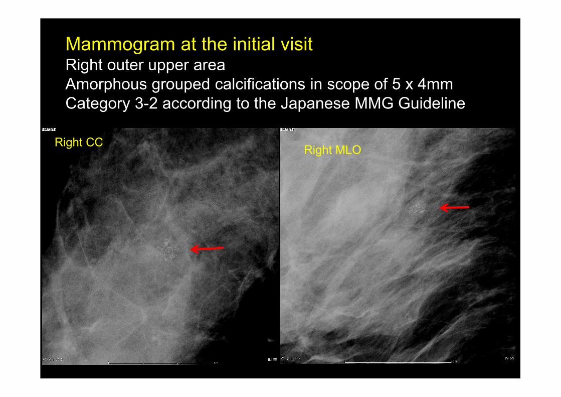
Mammogram at the initial visitRight outer upper area
Amorphous grouped calcifications in scope of 5 x 4mm
Category 3-2 according to the Japanese MMG Guideline
Right CCRight MLO
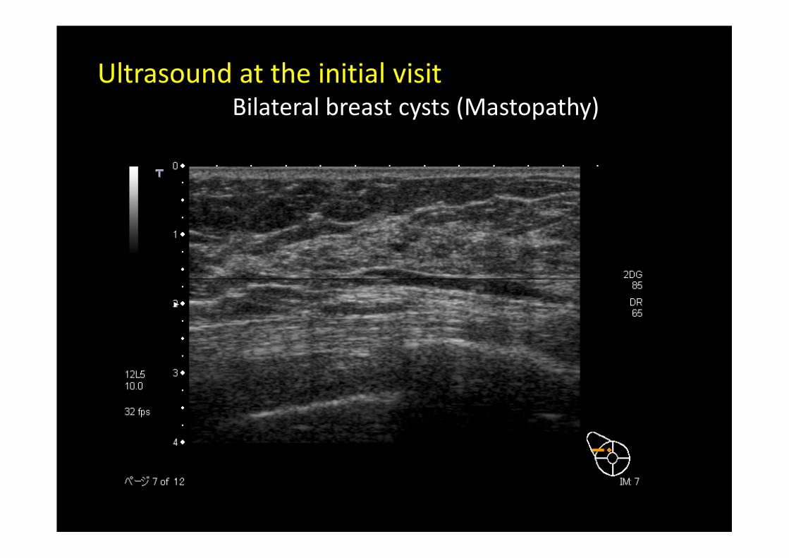
Ultrasound at the initial visitBilateral breast cysts (Mastopathy)

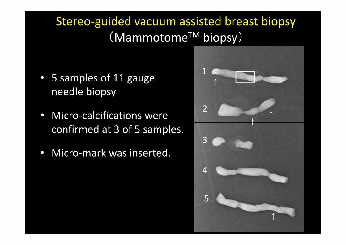
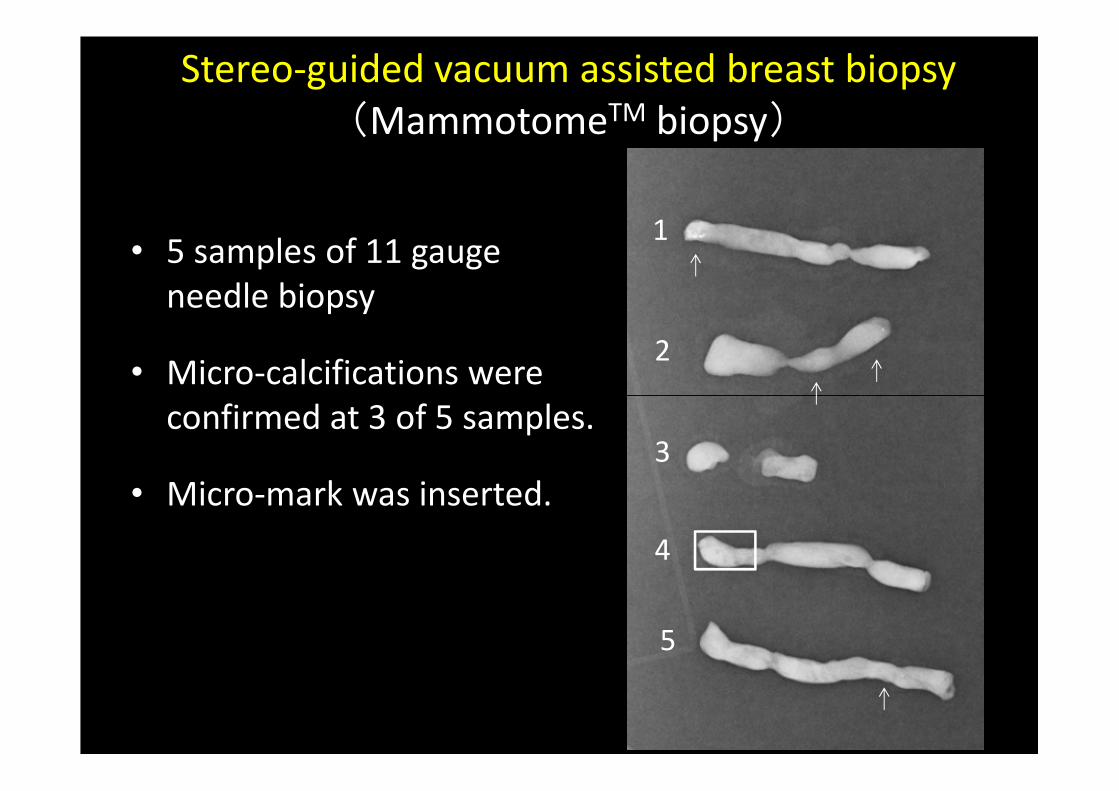
Stereo-guided vacuum assisted breast biopsy
(MammotomeTM biopsy)
• 5 samples of 11 gauge
needle biopsy
• Micro-calcifications were
confirmed at 3 of 5 samples.
1
2
confirmed at 3 of 5 samples.
• Micro-mark was inserted.
3
4
5
Pathological findings
of
biopsy specimensbiopsy specimens

Stereo-guided vacuum assisted breast biopsy
(MammotomeTM biopsy)
1
2
• 5 samples of 11 gauge
needle biopsy
• Micro-calcifications were
confirmed at 3 of 5 samples. 3
4
5
confirmed at 3 of 5 samples.
• Micro-mark was inserted.
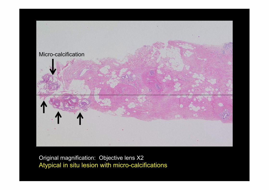
Micro-calcification
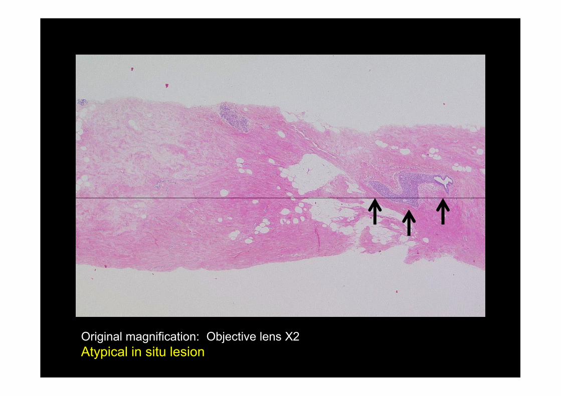
Original magnification: Objective lens X2
Atypical in situ lesion with micro-calcifications
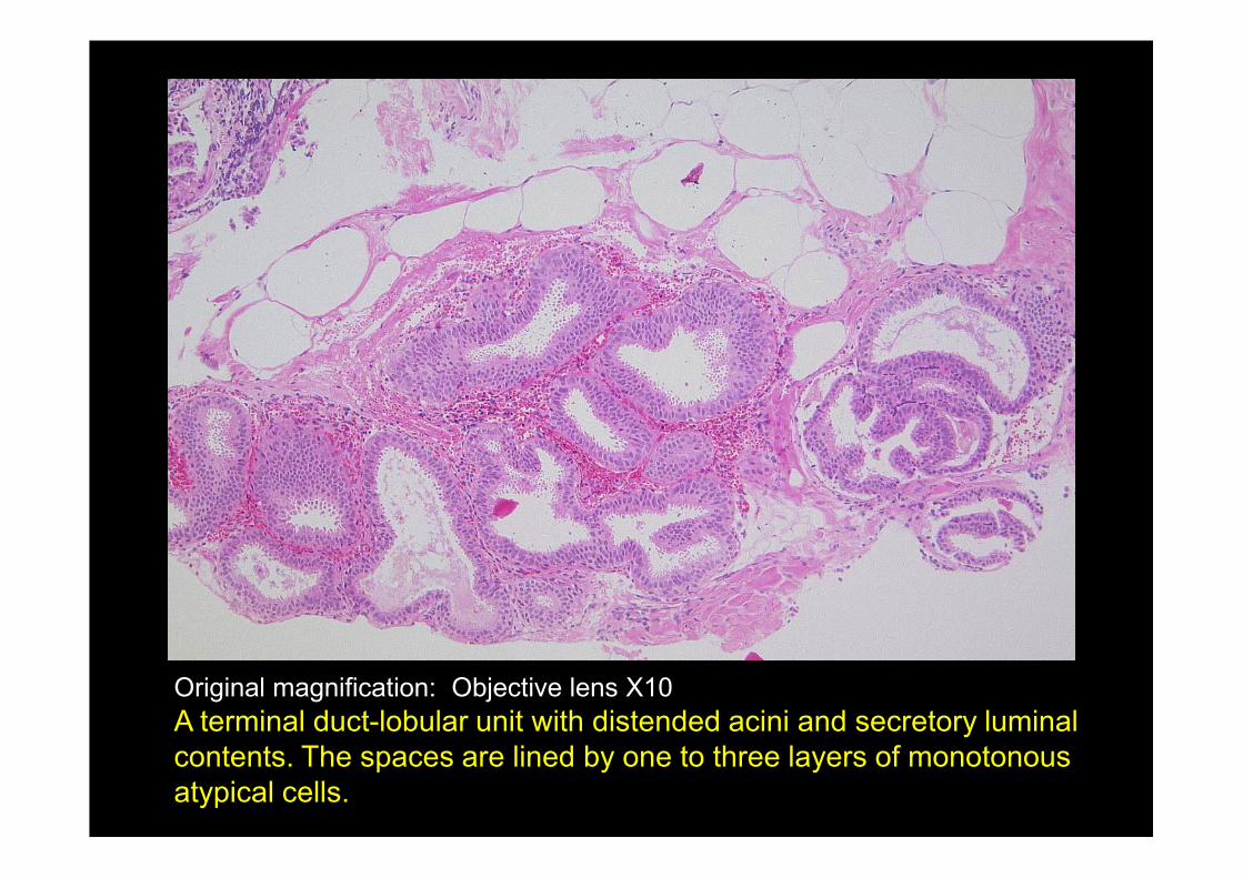
Original magnification: Objective lens X10
A terminal duct-lobular unit with distended acini and secretory luminal
contents. The spaces are lined by one to three layers of monotonous
atypical cells.
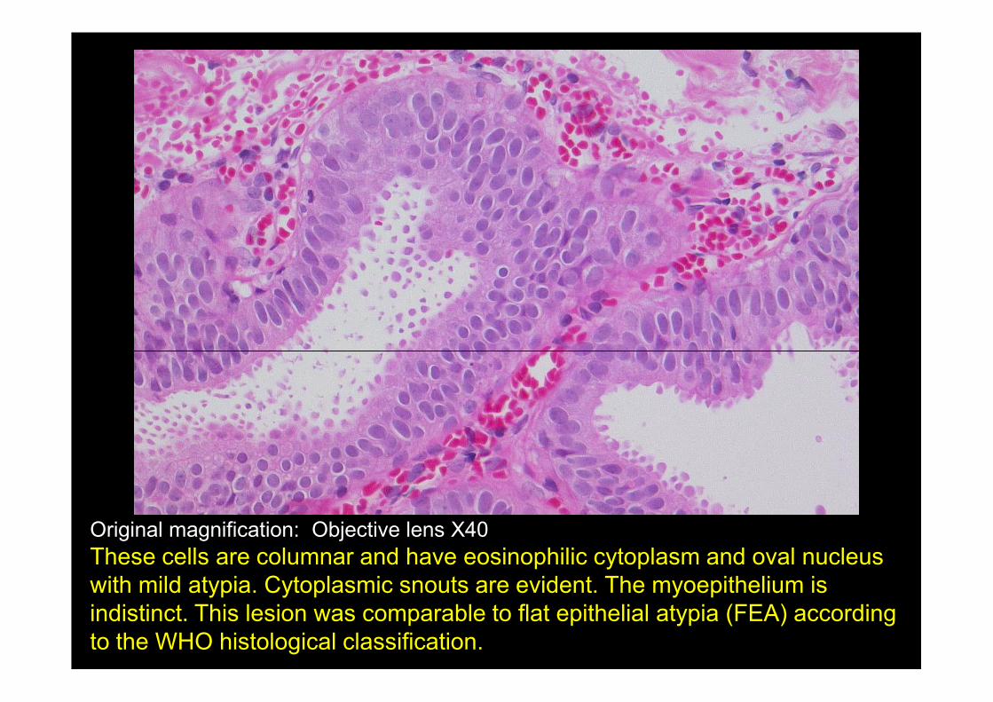
Original magnification: Objective lens X40
These cells are columnar and have eosinophilic cytoplasm and oval nucleus
with mild atypia. Cytoplasmic snouts are evident. The myoepithelium is
indistinct. This lesion was comparable to flat epithelial atypia (FEA) according
to the WHO histological classification.
Stereo-guided vacuum assisted breast biopsy
(MammotomeTM biopsy)
1
2
• 5 samples of 11 gauge
needle biopsy
• Micro-calcifications were
confirmed at 3 of 5 samples.3
4
5
confirmed at 3 of 5 samples.
• Micro-mark was inserted.
Original magnification: Objective lens X2
Atypical in situ lesion

Original magnification: Objective lens X10
A solid growth of monotonous atypical cells fills the duct lumen.
A few micro-lumens are evident.
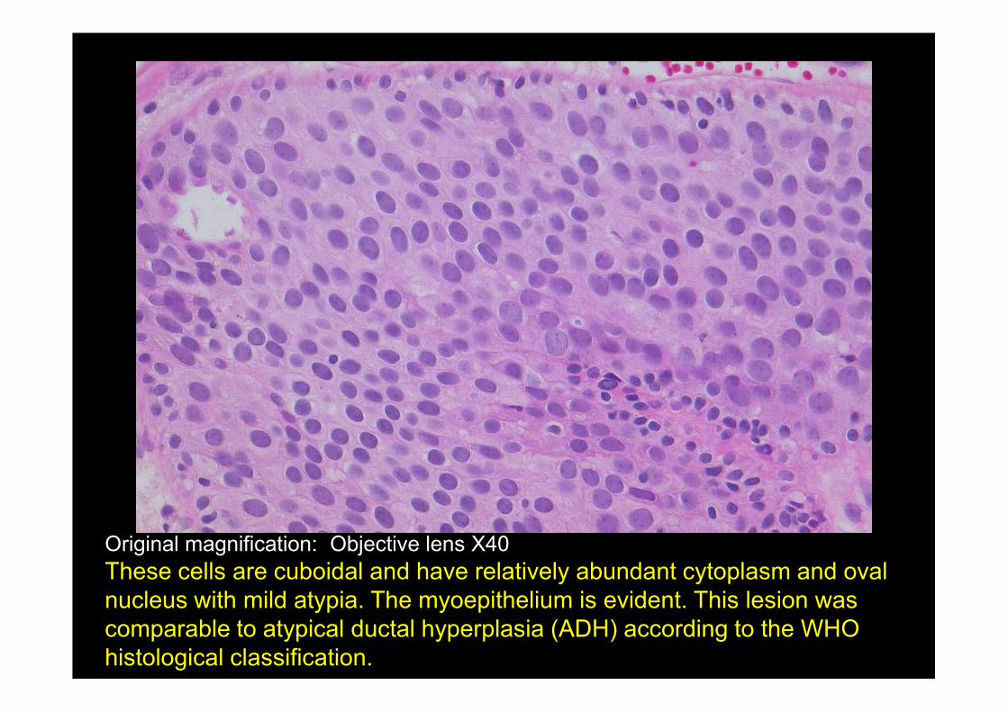
Original magnification: Objective lens X40
These cells are cuboidal and have relatively abundant cytoplasm and oval
nucleus with mild atypia. The myoepithelium is evident. This lesion was
comparable to atypical ductal hyperplasia (ADH) according to the WHO
histological classification.
Stereo-guided vacuum assisted breast biopsy
(MammotomeTM biopsy)
1
2
• 5 samples of 11 gauge
needle biopsy
• Micro-calcifications were
confirmed at 3 of 5 samples.3
4
5
confirmed at 3 of 5 samples.
• Micro-mark was inserted.
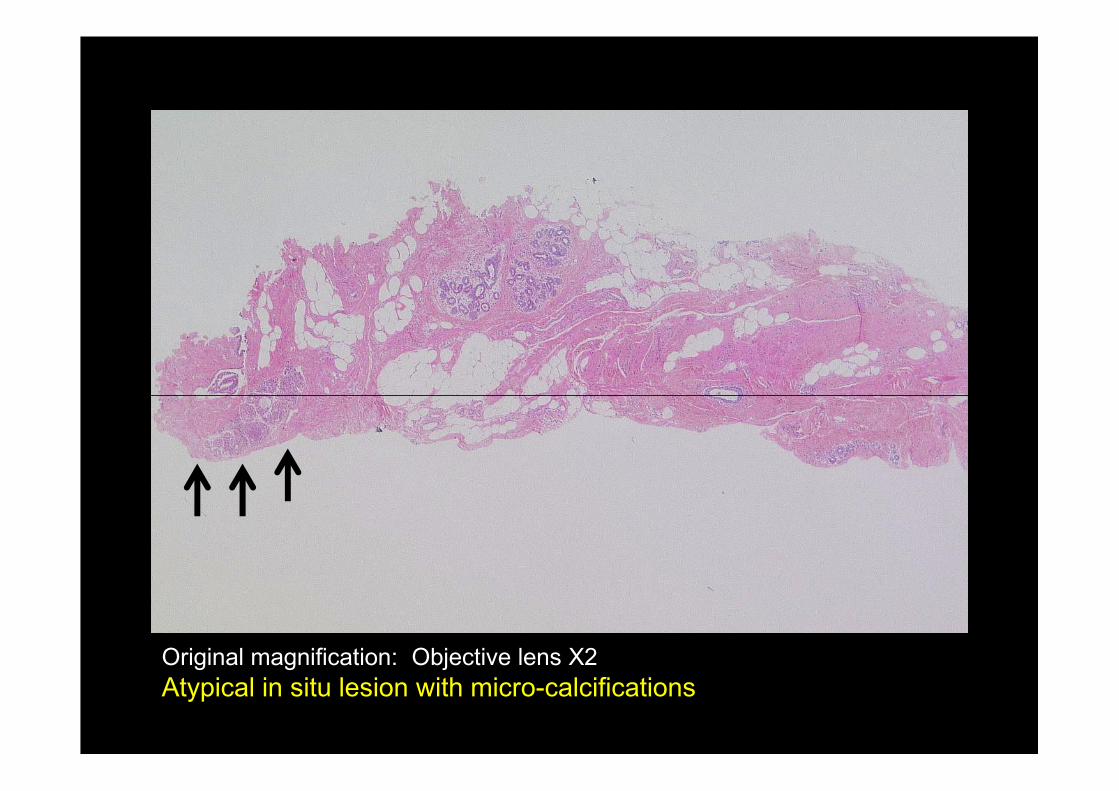
Original magnification: Objective lens X2
Atypical in situ lesion with micro-calcifications

非病変部
病変部
Original magnification: Objective lens X10
A solid growth of atypical cells expands acini of a lobule.

Original magnification: Objective lens X40
These cells show mild atypia and loss of cohesion. This lesion was
comparable to atypical lobular hyperplasia (ALH) according to the WHO
histological classification.

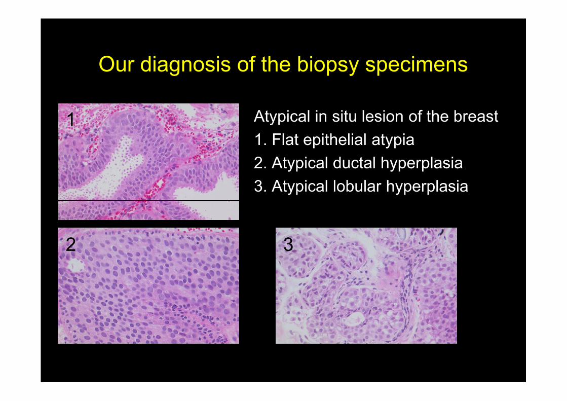
Our diagnosis of the biopsy specimens
Atypical in situ lesion of the breast.
WHO histological classification
- Flat epithelial atypia (FEA)
- Atypical ductal hyperplasia (ADH)- Atypical ductal hyperplasia (ADH)
- Atypical lobular hyperplasia (ALH)
Comments:
Benignity or malignancy cannot be determined.
Periodic follow up is needed.

1 year follow up
MMG: No change
US: Right outer upper area
Low echoic lesion, 5mm in diameter
Suspicious of concentrate cyst
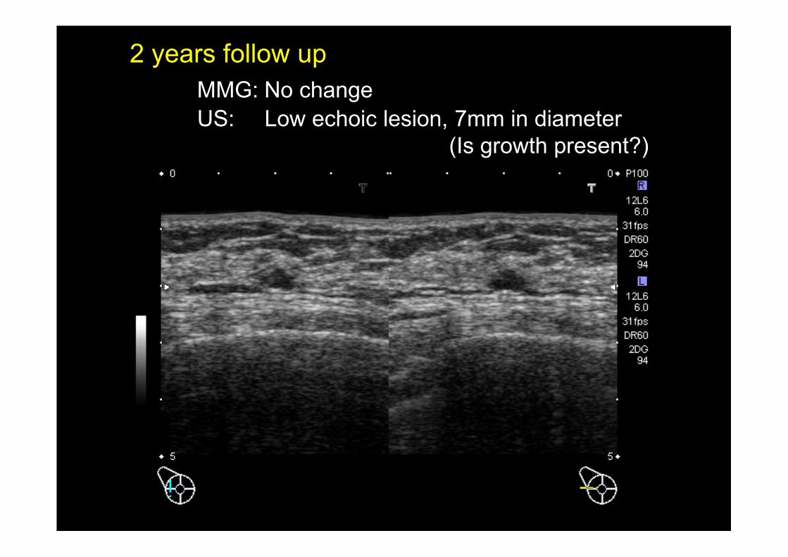
2 years follow up
MMG: No change
US: Low echoic lesion, 7mm in diameter
(Is growth present?)
2 years follow up
US-guided aspiration biopsy cytology was
performed to the low echoic lesion in the upper
outer area of the right breast.
Cytological diagnosis is suspicious of malignancy.
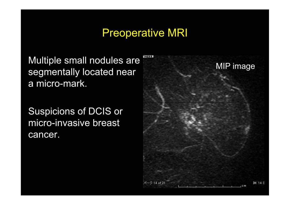
Preoperative MRI
MIP imageMultiple small nodules are
segmentally located near
a micro-mark.
Suspicions of DCIS or
micro-invasive breast
cancer.

Operation
Right partial mastectomy with sentinel lymph node
biopsy was performed.
Sentinel lymph nodes showed negative for cancer
by frozen sections during the surgery.by frozen sections during the surgery.
Axillary lymph nodes dissection was omitted.

Specimen mammography of the partial
mastectomy material
Peripheral site
Micro-mark
Micro-calcifications
Nipple site
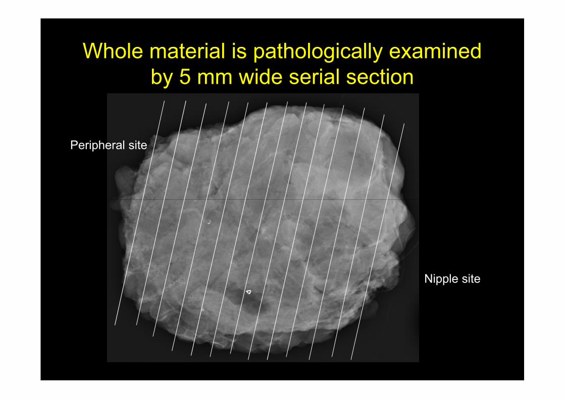
Whole material is pathologically examined
by 5 mm wide serial section
Peripheral site
Nipple site
Pathological findings
of
surgical materialssurgical materials

Our diagnosis of the surgical materials
Noninvasive mixed ductal and lobular carcinoma of
the breast.
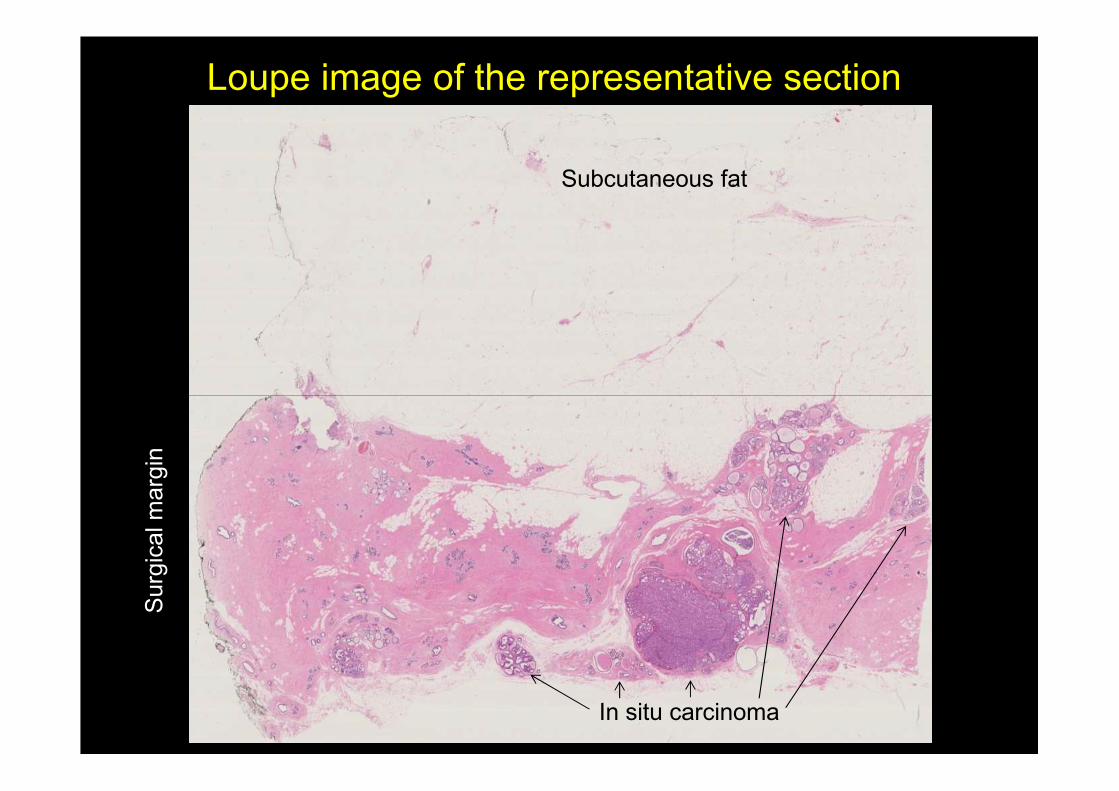
Loupe image of the representative section
Subcutaneous fatSurgical margin
In situ carcinoma
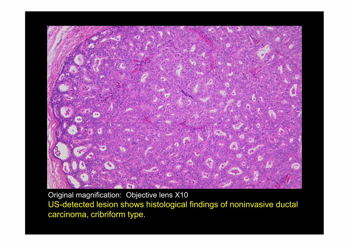
Original magnification: Objective lens X10
US-detected lesion shows histological findings of noninvasive ductal
carcinoma, cribriform type.

Original magnification: Objective lens X40
Cancer cells show the cribriform structure.

Loupe image of the representative section
Subcutaneous fatSurgical margin
Carcinoma in situ

Noninvasive ductal carcinoma
Lobular carcinoma in situ
Original magnification: Objective lens x 4
Two distinct morphological patterns are seen in this area:
noninvasive ductal carcinoma on the left and lobular carcinoma in situ
on the right. Transitional zone between ductal and lobular carcinoma is
seen on the center.
Transitional zone
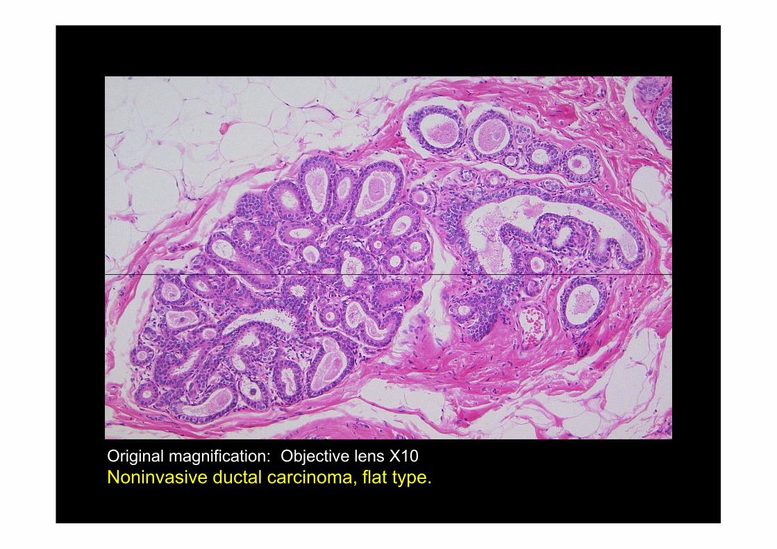
Original magnification: Objective lens X10
Noninvasive ductal carcinoma, flat type.
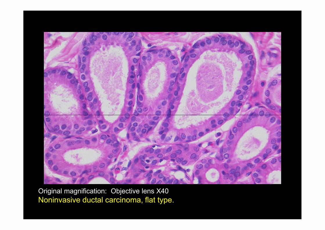
Original magnification: Objective lens X40
Noninvasive ductal carcinoma, flat type.

Noninvasive ductal carcinoma
Flat type Cribriform type
Original magnification: Objective lens X40
Cancer cells have relatively abundant cytoplasm, round to oval shaped
nucleus and show tubular formations.
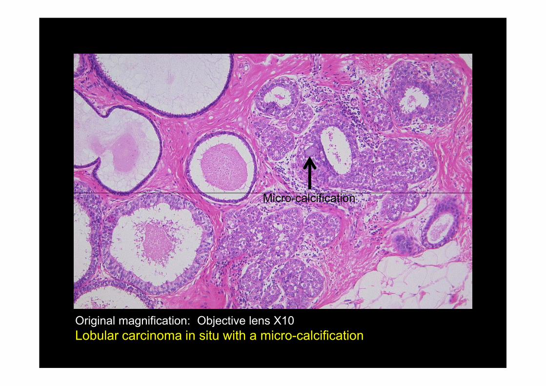
Micro-calcification
Original magnification: Objective lens X10
Lobular carcinoma in situ with a micro-calcification
Micro-calcification
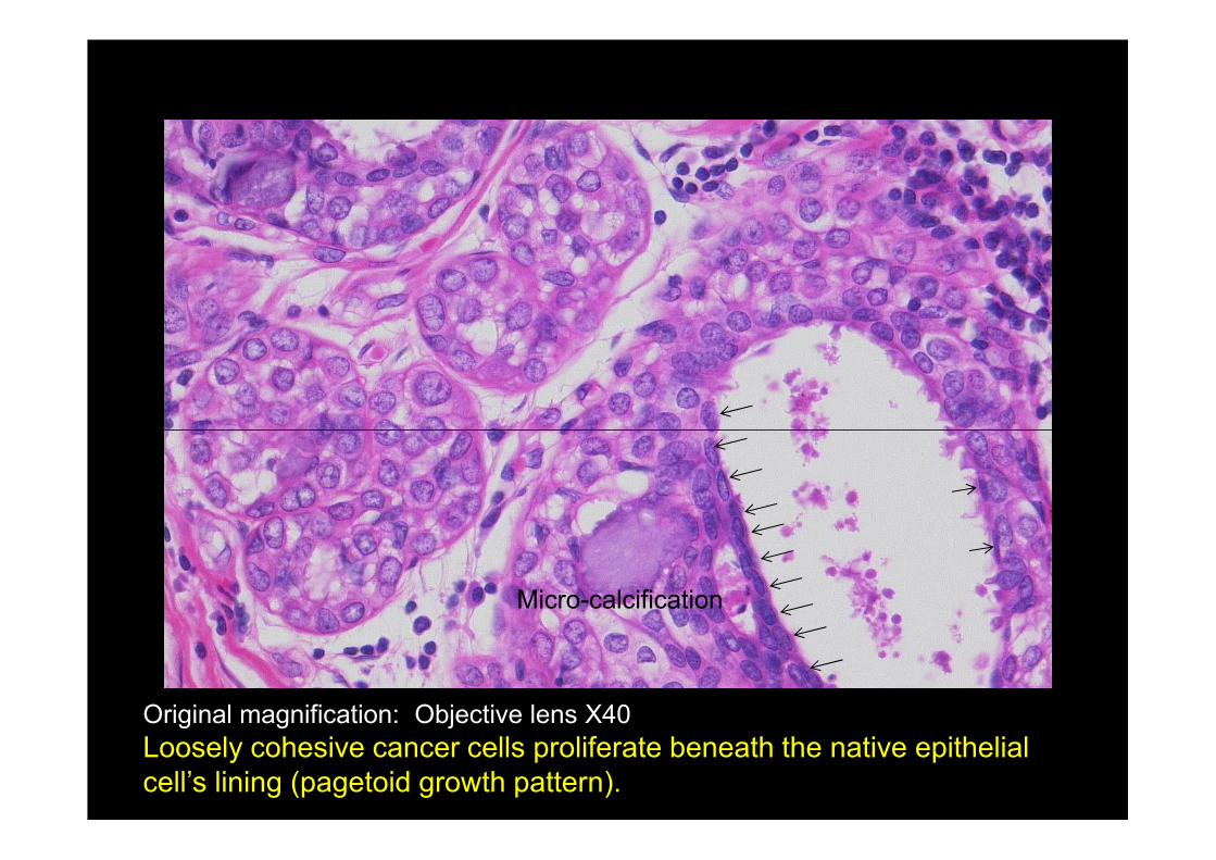
Micro-calcification
Original magnification: Objective lens X40
Loosely cohesive cancer cells proliferate beneath the native epithelial
cell’s lining (pagetoid growth pattern).
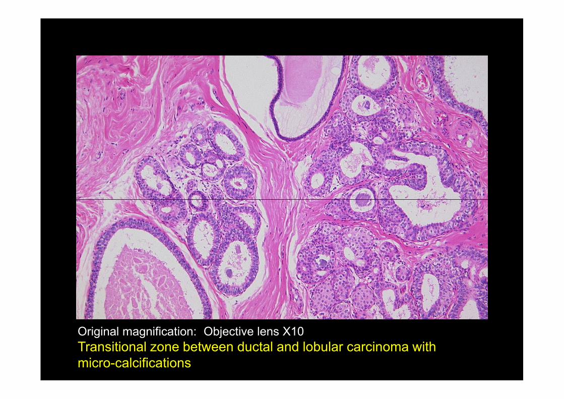
Original magnification: Objective lens X10
Transitional zone between ductal and lobular carcinoma with
micro-calcifications

Original magnification: Objective lens X40
Histological structure of this area resembles those of lobular carcinoma in
situ, but cancer cells appear more adherent than typical lobular
carcinoma in situ.

Noninvasive mixed ductal and lobular carcinoma
HE E-Cadherin
Transitional zone
Lobular carcinoma
in situ
Original magnification: Objective lens X20
Two distinct morphological patterns are seen in this area:
lobular carcinoma in situ on the right and transitional zone on the left.
Lobular carcinoma cells show negative staining for E-Cadherinin.
in situ

Surgical margin: negative for cancer.
No lymph node metastasis
No adjuvant therapy
The patient is now alive without any recurrence 4 The patient is now alive without any recurrence 4
years after the surgery.

Review of pathological findings
of
biopsy specimensbiopsy specimens

Our diagnosis of the biopsy specimens
Atypical in situ lesion of the breast
1. Flat epithelial atypia
2. Atypical ductal hyperplasia
3. Atypical lobular hyperplasia
1
2 3

Biopsy specimen
Flat epithelial atypia
Surgical specimen
Noninvasive ductal
carcinoma, flat type
Original magnification: Objective lens X40
Pathological findings of FEA resemble those of noninvasive ductal
carcinoma, flat type.
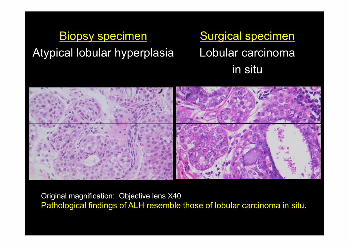
Biopsy specimen
Atypical lobular hyperplasia
Surgical specimen
Lobular carcinoma
in situ
Original magnification: Objective lens X40
Pathological findings of ALH resemble those of lobular carcinoma in situ.
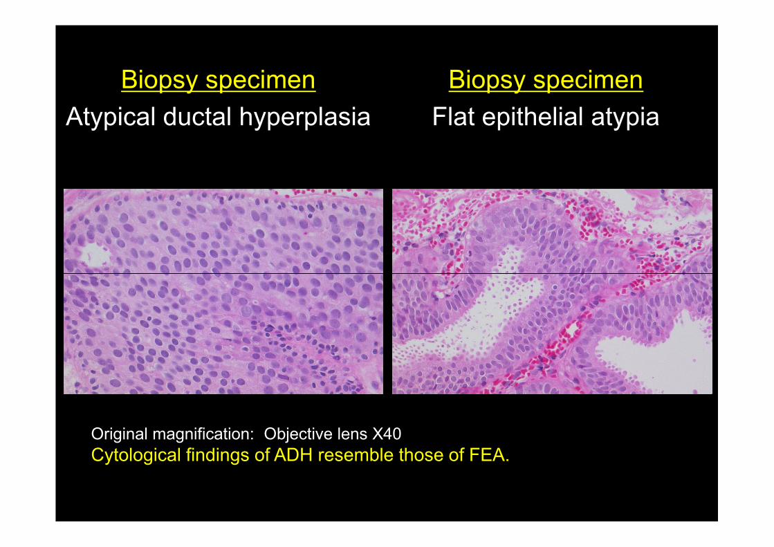
Biopsy specimen
Atypical ductal hyperplasia
Biopsy specimen
Flat epithelial atypia
Original magnification: Objective lens X40
Cytological findings of ADH resemble those of FEA.
Review of pathological findings
of biopsy specimens
Lesions which were diagnosed as atypical in
situ lesion in the biopsy specimens could be
parts of noninvasive carcinoma.parts of noninvasive carcinoma.

Discussion points for the case
(A) Pathological diagnosis of this case
- Surgical specimen
- Biopsy specimen- Biopsy specimen
(B) Management of this case after needle biopsy
Our diagnosis of the surgical materials
Noninvasive mixed ductal and lobular carcinoma of
the breast.
Discussion (A): Surgical specimen
• What would be your diagnosis of the surgical
specimen?
• What would be your terminology of noninvasive • What would be your terminology of noninvasive
carcinoma which have both ductal and lobular
components?
Our diagnosis of the biopsy specimens
Atypical in situ lesion of the breast
1. Flat epithelial atypia
2. Atypical ductal hyperplasia
3. Atypical lobular hyperplasia
1
2 3

Discussion (A) : Biopsy specimen
• What would be your diagnosis of the biopsy
specimen?
• After seeing the surgical specimen, did you • After seeing the surgical specimen, did you
change your diagnosis of the biopsy specimen?
• What would be your terminology of these
lesions?
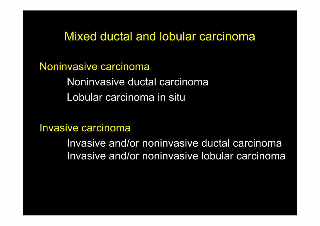
Mixed ductal and lobular carcinoma
Noninvasive carcinoma
Noninvasive ductal carcinoma
Lobular carcinoma in situ
Invasive carcinoma
Invasive and/or noninvasive ductal carcinoma
Invasive and/or noninvasive lobular carcinoma

Mixed ductal and lobular carcinoma
Mixed ductal and lobular carcinoma often contains noninvasive ductal carcinoma, flat type and lobular carcinoma in situ.
When only these components are sampled by a When only these components are sampled by a needle biopsy, pathological diagnosis of mixed ductal and lobular carcinoma is difficult.
Mixed ductal and lobular carcinoma should be considered, when both FEA and ALH are seen in a needle biopsy specimen.
Discussion (B):
Management of this case after needle biopsy
When atypical in situ lesion was found in needle
biopsy specimen
• Excision?• Excision?
• Re-needle biopsy?
• Follow up?
by MMG or US?
Interval?
Indication for re-biopsy?