case review #3: 27 year old female with adolescent scoliosis, pseudoarthrosis, and flatback
TRANSCRIPT

Case Review:
Adolescent Idiopathic Scoliosis Revision Surgery for Psuedoarthrosis and Flatback Syndrome
Robert S Pashman, MDScoliosis and Spinal Deformity Surgerywww.eSpine.com
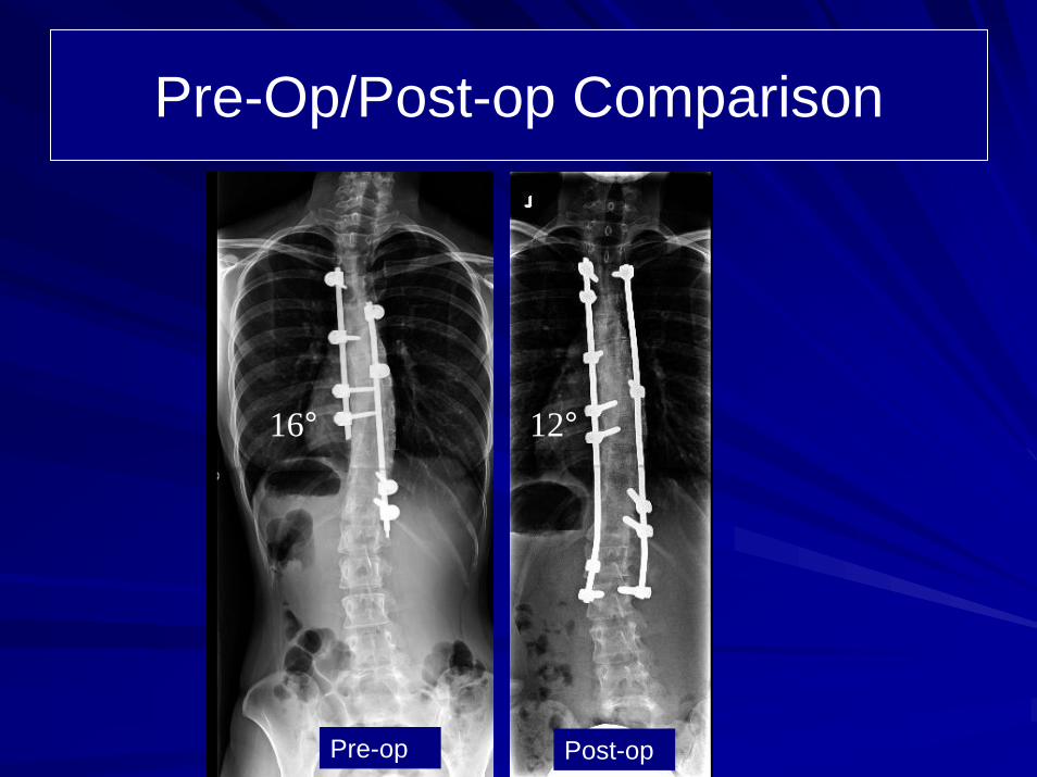
16°

Patient History27 year old female from Dubai, United Arab Emirates Adolescent Idiopathic ScoliosisStatus post posterior instrumented fusion in 2000 for a 45° right Lenke-type II curve which resulted in significant correction from 45° to 16°. The patient then had instrumentation from T4 to T12 and was fused short from T6 to T12 on the right-hand side. The patient awoke from surgery with seering left-sided pain and ultimately had revision for removal of some instrumentation distally which improved the patient's symptoms somewhat, but she continues to have left-sided flank pain. CT scan and bone scan indicated that the patient's pseudoarthrosis of the plate type centered approximately around T10, 11, 12 areas. The patient had increasing rotation and because she was fused to T12 it was my impression that the patient had subjacent "adding on" phenomenon with a short fusion at T12 which had caused a spinal imbalance and a significant disability from the subjacent curve. Moreover, the patient had some imbalance of her shoulders. This was due to a fusion short proximally, and although the patient had been followed conservatively, she had increasing pain. The only solution as far as I was concerned was to remove the hardware, explore the fusion mass to fuse her in a supra- and subjacent way to stabilize the residual curve.

Pre-op Films
16°

Indications for Surgery
Status post posterior instrumented fusion for Adolescent Idiopathic Scoliosis. Status post revision instrumentation for Adolescent Idiopathic Scoliosis. Now with "adding on" subjacent degeneration and increased curve with rotation, thoracic spine. Increasing pain/instrumentation, status post posterior instrumentation for adolescent idiopathic scoliosis. Shoulder imbalance, status post posterior instrumented fusion. Presumed pseudoarthrosis, thoracic spine, status post posterior instrumented fusion.

Surgical StrategyThoracic 3 to lumbar 2 segmental spinal instrumentation with CD Legacy stainless steel 5.5 pedicle screw-rod contrast.
Posterior spinal fusion, thoracic 3 to lumbar 2, using combination of locally harvested autogenous bone and recombinant human bone morphogenicprotein.
Interlaminar decompression under the microscope, placement of screws and nerve root decompression, thoracic 12-lumbar 1 and thoracic 11-thoracic 12 on the right-hand side.
Repair pseudoarthrosis, thoracic 10-11 and thoracic 10-12.
Spinal osteotomy, thoracic 4-5 and thoracic 5-6, for recorrection of proximal curvature.
Removal of retained hardware.
Intraoperative motor evoked potentials.
Intraoperative fluoroscopy.

Post-Op Films
At the time of operation, significant residual subjacent rotation and curvature was found. The joints were somewhat arthritic subjacent to T12-L1, L1, and pseudoarthrosis was also found which was repaired.
She has minimal back pain on the left. This is a radiculardistribution. This is probably from ongoing irritation or from healing. Instrumentation looks good. Her posture is excellent.
12°
Pre-Op/Post-op Comparison
Pre-op Post-op
12°16°
Pre-Op/Post-op Comparison
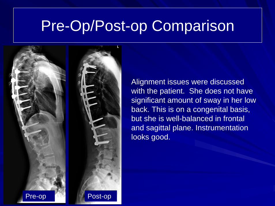
Alignment issues were discussed with the patient. She does not have significant amount of sway in her low back. This is on a congenital basis, but she is well-balanced in frontal and sagittal plane. Instrumentation looks good.
Pre-op Post-op