case reports tbc1d24 mutations in a sibship with … reports tbc1d24 mutations in a sibship with...
TRANSCRIPT

Case Reports
TBC1D24 Mutations in a Sibship with Multifocal Polymyoclonus
Adeline Ngoh1,2{
, Jose Bras3,4{
, Rita Guerreiro3,4{
, Amy McTague1,2
, Joanne Ng1,2
, Esther Meyer1, W. Kling Chong
5, Stewart Boyd
6,
Linda MacLellan7, Martin Kirkpatrick
8& Manju A. Kurian
1,2*
1 Neurosciences Unit, University College London, Institute of Child Health, London, UK, 2 Department of Neurology, Great Ormond Street Hospital for
Children NHS Foundation Trust, London, UK, 3 Department of Molecular Neuroscience, UCL Institute of Neurology, London, UK, 4 Department of Medical
Sciences and Institute of Biomedicine – iBiMED, University of Aveiro, Aveiro, Portugal, 5 Department of Neuroradiology, Great Ormond Street Hospital for
Children NHS Foundation Trust, London, UK, 6 Department of Neurophysiology, Great Ormond Street Hospital for Children NHS Foundation Trust,
London, UK, 7 NHS Highland, UK, 8 Tayside Children’s Hospital, Dundee, UK
Abstract
Background: Advances in molecular genetic technologies have improved our understanding of genetic causes of rare neurological disorders with features of
myoclonus.
Case Report: A family with two affected siblings, presenting with multifocal polymyoclonus and neurodevelopmental delay, was recruited for whole-exome
sequencing following unyielding diagnostic neurometabolic investigations. Compound heterozygous mutations in TBC1D24, a gene previously associated with
various epilepsy phenotypes and hearing loss, were identified in both siblings. The mutations included a missense change c.457G.A (p.Glu157Lys), and a novel
frameshift mutation c.545del (p.Thr182Serfs*6).
Discussion: We propose that TBC1D24-related diseases should be in the differential diagnosis for children with polymyoclonus.
Keywords: TBC1D24, myoclonus
Citation: Ngoh A, Bras J, Guerreiro R, et al. TBC1D24 mutations in a sibship with multifocal polymyoclonus. Tremor Other Hyperkinet Mov. 2017; 7.
doi: 10.7916/D8Q52VBV
* To whom correspondence should be addressed. E-mail: [email protected]
{These authors contributed equally to the manuscript.
Editor: Elan D. Louis, Yale University, USA
Received: January 25, 2017 Accepted: March 16, 2017 Published: April 13, 2017
Copyright: ’ 2017 Ngoh et al. This is an open-access article distributed under the terms of the Creative Commons Attribution–Noncommercial–No Derivatives License, which permits
the user to copy, distribute, and transmit the work provided that the original authors and source are credited; that no commercial use is made of the work; and that the work is not altered
or transformed.
Funding: A.N. is funded by a Guarantors of Brain Entry Fellowship and an Action Medical Research Training Fellowship. J.B. and R.G.’s work is supported by Fellowships from the
Alzheimer’s Society. A.M. and J.N. are funded by Medical Research Council Clinical Research Training Fellowships. A.M. also receives funding from Medical Research Foundation,
Child Brain Research and Young Epilepsy; and J.N. is also supported by Great Ormond Street Hospital Children’s Charities. M.A.K. is funded by a Wellcome Trust Intermediate
Clinical Fellowship and receives funding from the Rosetrees Trust, Gracious Heart Charity Foundation Great Ormond Street Hospital Children’s Charities (GOSHCC) Child Brain
Research and Rachel Marie Trafford Trust.
Financial Disclosures: None.
Conflict of Interest: The authors report no conflict of interest.
Ethics Statement: All patients that appear on video have provided written informed consent; authorization for the videotaping and for publication of the videotape was provided.
Introduction
Myoclonus is defined as a sudden, brief (less than 100 ms), shock-like
muscle contraction involving agonist and antagonist muscles, leading
to a sudden jerky movement.1 The lifetime prevalence of myoclonus is
estimated to be 8.6 cases per 100,000 population.2 Physiological myoc-
lonus, in the form of hiccoughs or hypnic jerks, is easily recognized.
Pathological myoclonus has a wide range of etiologies including acquired/
hypoxic neurological injury, as well as neoplastic, infectious, post-
infectious, metabolic, and genetic causes. There are a number of ways
in which myoclonus is classified, including by anatomical/physiological
origin (cortical, subcortical, spinal, peripheral), or by etiology. In the
approach to pathological myoclonus, it is useful to consider the dis-
tribution of myoclonus, age of onset, insidious/acute onset, triggering/
alleviating factors and associated signs or symptoms (Supplementary
Table 1).
Recently, advances in molecular genetic technologies have enabled
accelerated gene discovery, adding to our understanding of rare neuro-
logical disorders, including myoclonus (Supplementary Table 1).
Freely available online
Tremor and Other Hyperkinetic Movementshttp://www.tremorjournal.org
The Center for Digital Research and ScholarshipColumbia University Libraries/Information Services1

We describe two siblings with infantile-onset multifocal polymyo-
clonus at rest who were found on whole-exome sequencing to have
mutations in TBC1D24, a gene previously associated with various
epilepsy phenotypes (including familial infantile myoclonic epilepsy,
migrating partial seizures of infancy),3–13 hearing impairment,14–17
and DOORS (deafness, onychodystrophy, osteodystrophy, mental
retardation, and seizures) syndrome.18 Prolonged electroencephalo-
grams (EEGs) revealed no epileptiform features; furthermore, multiple
EEGs captured episodes of polymyoclonus and demonstrated no EEG
correlation.
To our knowledge, this is the first report of TBC1D24 mutations in a
phenotype with prolonged, multifocal polymyoclonus as the main
presenting feature with no discernible features of epilepsy.
Case report
A family with two affected children was ascertained and recruited
for molecular genetic analysis. The participating family gave written
informed consent and the study was performed in accordance with the
Declaration of Helsinki. Genomic DNA from the affected individuals,
and both parents was extracted from peripheral lymphocytes by
standard techniques.
Whole-exome sequencing was carried out in both affected indi-
viduals using Illumina’s TruSeq Exome Enrichment kit (Illumina, Inc
San Diego, California, USA), according to manufacturer’s recom-
mendations. Sequencing was performed on Illumina HiSeq2000 using
100-bp paired-end reads. Data were analyzed following Genome
Analysis Toolkit’s (GATK) Best Practices (PMIDs, 20644199,
21478889, 25431634). To confirm the identified TBC1D24 vari-
ants direct Sanger sequencing was performed. The appropriate exon
amplified by polymerase chain reaction (PCR) (primer sequences and
PCR conditions on request) was directly sequenced by the Big Dye
Terminator Cycle Sequencing System (Applied Biosystems Inc.) on an
ABI PRISM 3730 DNA Analyzer (Applied Biosystems Inc.) and
analyzed using Chromas (http://www.technelysium.com.au/chromas.
html).
We present two siblings born to non-consanguineous parents of
Polish descent. There is no significant family history.
The older sibling, A1, was diagnosed with cardiac arrhythmia
antenatally, and was born via elective caesarean section at term. At the
age of 5 weeks, his mother noted he had intermittent mouth-twitching
movements while breastfeeding. This progressed to paroxysmal myo-
clonic twitching and jerking movements involving varying muscle
groups, including those of his eyelids, face, lips, abdomen, and limbs.
Myoclonic episodes ranged from short, spontaneously resolving
tolerated events with small amplitude twitches to prolonged distressing
events with larger amplitude jerks (Video 1A,B). He had preserved
awareness with most episodes, but some prolonged episodes resulted in
autonomic disturbance such as pallor, sweating, and reduced respon-
siveness. In casualty, prolonged episodes of myoclonus were often
Video 1. (A) Patient A1 at various ages. The video demonstrates orolingual and facial myoclonus. (B) Patient A1 at various ages. The video demonstrates
limb myoclonus, abdominal myoclonus and widespread polymyoclonus. Towards the end of the video distal choreoathetoid movements are also present in the upper
limbs. (C) Patient A2 at approximately 3 months of age. The video demonstrates myoclonus in the right arm and orolingual myoclonus.
Ngoh A, Bras J, Guerreiro R, et al. TBC1D24 and Multifocal Polymyoclonus
Tremor and Other Hyperkinetic Movementshttp://www.tremorjournal.org
The Center for Digital Research and ScholarshipColumbia University Libraries/Information Services2

treated as status epilepticus. Benzodiazepines and rectal paraldehyde
were reported to terminate some events. Although his symptoms were
initially paroxysmal, by the age of 2 years he had almost constant
myoclonus involving varying muscle groups. These symptoms were
exacerbated by fatigue and abolished by sleep.
At the age of 10 months, A1 received a pacemaker for third-degree
atrioventricular block. His development was delayed. He started
walking unsteadily at 3 years and said his first word at 3 years of age.
At 16 months of age, he was diagnosed with extreme hypermetropia.
An early hearing screen was normal, but at the age of 5 years,
he developed profound sensorineural hearing loss. On examination, he
had dysplastic ears, downslanting palpebral fissures, silvery pigmenta-
tion to his hair, and a full philtrum. His weight was on the 25th centile
and his head circumference was between the 9th and 25th centile. He
had bilateral rotatory nystagmus. He had truncal hypotonia but
increased peripheral tone, with a dynamic component, in all four
limbs. Deep tendon reflexes were brisk in his lower limbs, with flexor
plantar responses bilaterally. Asynchronous migratory myoclonus
involving various muscle groups was easily observed throughout the
examination.
Patient A2 had an unremarkable perinatal history. She started
having myoclonic twitching and jerking in her limbs at the age of
6 weeks. Like her brother, this progressed to paroxysmal episodes of
myoclonus, most often involving the abdomen or face. As she grew
older, her myoclonus also became more widespread and constant.
A2 did not have cardiac symptoms. There have been no concerns
regarding her vision or hearing. Currently, at the age of 3 years, she
has poor balance and coordination in addition to speech delay. She
does not have abnormal eye movements. She has normal tone and
deep tendon reflexes.
Extensive neurometabolic investigations performed on blood, cere-
brospinal fluid (CSF) and urine for both siblings revealed nothing of
note apart from a mildly reduced CSF glucose on one occasion for
A1 (1.9 mmol/l, plasma glucose 4.4 mmol/l) and two occasions for A2
(1.7 mmol/l, plasma 4.5 mmol/l; and 1.9 mmol/l, plasma 4.7 mmol/l).
For both individuals, these normalized on repeat testing.
A1 had a normal male karyotype and normal microarray results.
Testing for mutations in candidate genes for GLUT1 deficiency
syndrome and mitochondrial disorders, including SLC2A1, POLG, and
DGOUK, yielded negative results.
Repeated EEGs including prolonged EEGs encompassing sleep for
both siblings showed a mild excess of slow activity, but no evidence of
epileptiform activity. Polygraphic electromyogram (EMG) recordings
of right and left deltoids, biceps, forearm, and quadriceps muscles
demonstrated myoclonias of approximately 80–110 ms duration in all
the EMG channels without any evidence of spread from one to another.
A few myoclonias were more hypersynchronous, approximately 40–60 ms
in duration (Supplementary Figure 2). Back-averaging did not reveal
any preceding EEG change in either child. Nerve conduction studies
and somatosensory evoked potential tests were normal.
A1 had serial cranial magnetic resonance imaging (MRI) scans from
the age of 2 months to 19 months. These demonstrated hypoplasia of
the frontal and temporal lobes. The deep grey matter appeared normal
with no evidence of cortical dysplasia (Figure 1A). A computed tomo-
graphy (CT) head scan showed tiny non-specific foci of calcification in
the basal ganglia (Figure 1B).
Cranial MRI and MR spectroscopy performed at 6 weeks of age for
A2 were normal. However, a repeat MRI scan at 3 years 10 months
showed atrophy of the lateral aspects of the cerebellar hemispheres and
symmetrical signaling abnormalities (Figure 1C).
Figure 1. Magnetic Resonance Imaging Features in Sibship with TBC1D24 Mutations. (A) MRI brain scan (Axial T2-weighted) of A1 age 19 months
showing underdevelopment of the frontal and temporal lobes. (B) CT head scan of A1 20 months showing small non-specific foci of calcification within the basal
ganglia. (C) MRI head scan (Coronal T2-weighted) of A2 at 3 years 10 months showing symmetrical signal abnormalities and atrophy of the lateral aspects of the
cerebellar hemispheres.
TBC1D24 and Multifocal Polymyoclonus Ngoh A, Bras J, Guerreiro R, et al.
Tremor and Other Hyperkinetic Movementshttp://www.tremorjournal.org
The Center for Digital Research and ScholarshipColumbia University Libraries/Information Services3

A1 received trials of treatment with an extensive range of medication
including anti-epileptics (valproate, carbamazepine, nitrazepam, levetir-
acetam, clobazam), metabolic supplements (coenzyme Q10, biotin,
riboflavin, thiamine), levodopa, and piracetam. None of these achieved
sustained benefit, although levetiracetam and clobazam each controlled
myoclonus briefly. Immunotherapy had no significant effect.
Levetiracetam exacerbated A2’s symptoms and pyridoxine was
ineffective. She did not tolerate the ketogenic diet. Bromocriptine
resulted in dyskinetic movements, which terminated with discontinuation
of the medication. Carbamazepine afforded her some symptomatic relief.
For both siblings as symptoms were abolished by sleep, the best
therapeutic strategy seemed to be chloral hydrate used, as required, to
induce sleep during periods of distress.
Variants identified through whole-exome sequencing were filtered
by the following criteria: 1) very low frequency in control populations
(exclusion of variants with a minor allele frequency . 0.1% in the
established databases and 2,000 in-house analyzed exomes); 2) pre-
sentation of homozygous or compound heterozygous changes considering
an autosomal recessive inheritance pattern; and 3) prediction of puta-
tive pathogenicity based on mutation type or in silico prediction of
effects on protein function and/or structure. Using these criteria, two
heterozygous variants confirmed by Sanger sequencing were identi-
fied in TBC1D24 (Supplementary Figure 1): 1) a previously reported
missense change c.457G.A (p.Glu153Lys; rs376712059)6,13,15 and
2) a previously unreported frameshift mutation, c.545del (p.Thr
182Serfs*6) predicted to cause nonsense-mediated RNA decay or result
Table 1. Phenotypes Associated with TBC1D24 Mutations
Clinical
Phenotype
Familial Infantile
Myoclonic Epilepsy
(OMIM 605021)
Focal Epilepsy with
Cerebrocerebellar
Malformation
MMPSI
(OMIM 615338)
Progressive Myoclonic
Epilepsy with Dystonia
(OMIM 615338)
EOEE and Hearing
Loss
References Zara et al.3,de Falco et al.4,Falace et al.5, Poulat et al.6
Corbett et al.7,Afawi et al.8
Milh et al.9 Duru et al.10,Guven and Tolun11
Strazisar et al.12
Reportedmutations/genotype
c.439G.C (p.Asp147His)c.1526C.T (p.Ala509Val)c.457G.A (p.Glu157Lys)
c.751C.T (p.Phe251Leu) c.468C.A (p.Cys156*)c.686T.A (p.Phe229Ser)
c.969_970delGT(p.Ser324Thrfs*3)
c.32A.G (p.Asp11Gly)c.1008delT(p.His336GInfs*12)
Inheritance AR AR AR AR AR
Clinical features Seizures Normalpsychomotor developmentand neurologicalexamination to moderateintellectual andpsychomotor impairmentBulbous nose and flat nasalroot
SeizuresMyoclonusModerate intellectualdisabilityAtaxic with cerebellarsignsDystoniaDysarthria
SeizuresPsychomotor regressionAxial hypotoniaLoss of visual contact
SeizuresPost-ictal hemiparesisDystonic episodesMyoclonus with startleresponses to auditory andtactile stimuliAxial hypotoniaPyramidal signsSevere neurodevelopmentalimpairmentVulnerability to infectionBilateral optic atrophy,macular degeneration andvisual impairment in oneindividual
SeizuresProfound sensorineuraldeafnessMyoclonic jerksAcquired microcephalyDyskinetic movementsAxial hypotoniaPoor visual contact
Types of seizures GTC Myoclonic: triggersensitive
Focal seizures with aurasTonic–clonicMyoclonic
Focal prolonged migratingclonic seizures
Focal/unilateralClonic seizuresMyoclonicTonic
Clonic seizuresTonic seizures
EEG/EMGfindings
Preserved background.1 individual with slowbackground activity inoccipital region. Interictalmultiple diffuse spikes andslow waves. Ictal EEGwith low amplitude spikesat vertex Jerk-locked backaveraging confirmedcortical myoclonus
Slow background rhythms.No epileptiformdischarges. Ictal EEG notavailable
Focal migrating EEGdischarges during seizuresInterictal EEG:disorganized
Slow background in EEGMultifocal or bilateralgeneralized multiple spikes andspike waves in EEG associatedwith myoclonias.
Generalized spike-wavedischarges with frontocentralpredominance during seizuresNo clear EEG correlate formyoclonic jerks
Imaging findings Normal 1 individual withnodular periventricularheterotopia 1 individualhad MRI abnormalities inlentiform nuclei,ventricular dilatation andwhite matter changespost-cardiac arrest. Anearlier MRI was normal
Selective atrophy andsignal abnormality incerebellumCerebral corticalthickening most marked incingulate regions andoccipital poles
Global cerebral atrophysparing the posterior fossa
Thin corpus callosumDelayed myelinationDiffuse cerebral atrophy(asymmetrical for one patient)Cerebellar atrophy
Prominent frontotemporalatrophy
Ngoh A, Bras J, Guerreiro R, et al. TBC1D24 and Multifocal Polymyoclonus
Tremor and Other Hyperkinetic Movementshttp://www.tremorjournal.org
The Center for Digital Research and ScholarshipColumbia University Libraries/Information Services4

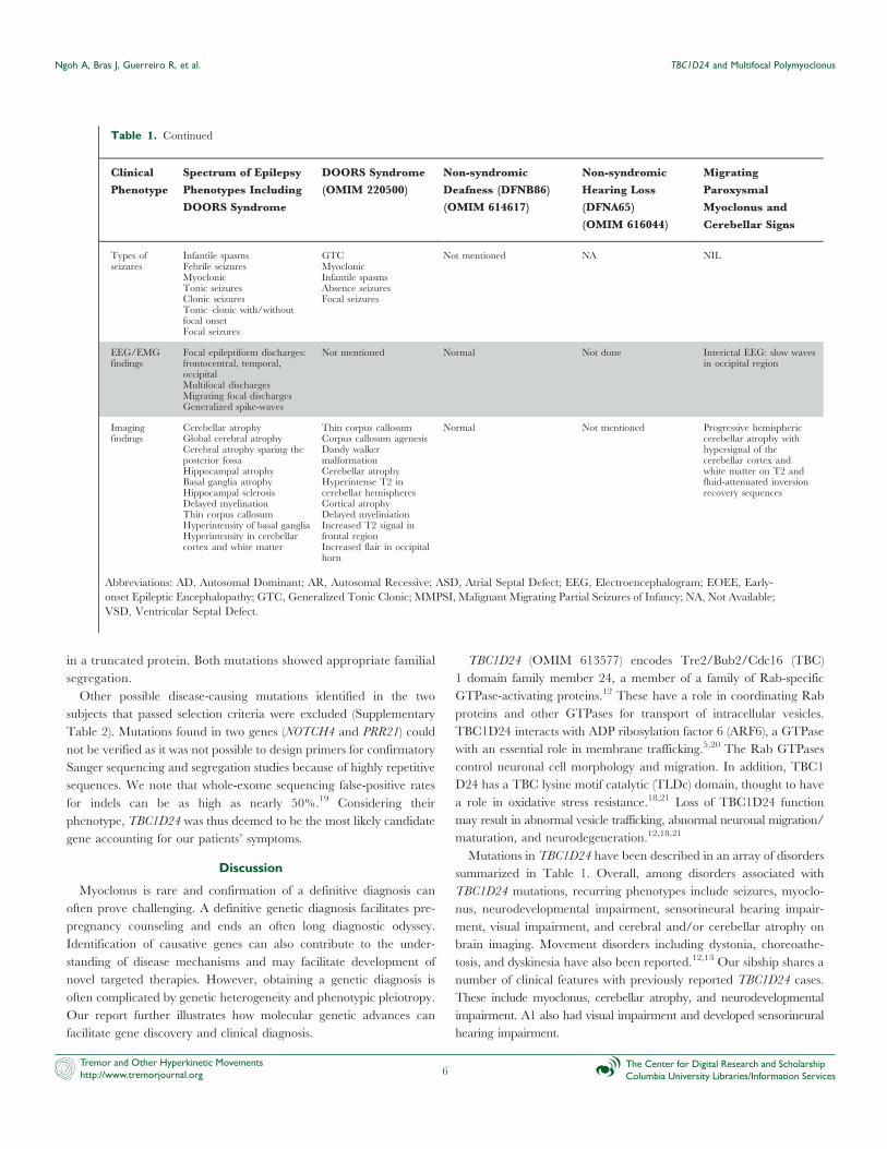
Table 1. Continued
Clinical
Phenotype
Spectrum of Epilepsy
Phenotypes Including
DOORS Syndrome
DOORS Syndrome
(OMIM 220500)
Non-syndromic
Deafness (DFNB86)
(OMIM 614617)
Non-syndromic
Hearing Loss
(DFNA65)
(OMIM 616044)
Migrating
Paroxysmal
Myoclonus and
Cerebellar Signs
References Balastrini et al.13 Campeau et al.18 Rehman et al.14,Bakhchane et al.15
Azaiez et al.16,Zhang et al.17
Doummar et al.22
Reportedmutations/genotype
c.32A.G (p.Asp11Gly)c.58C.T (p.Gln20*)c.115G.C (p.Ala39Pro)c.118C.T (p.Arg40Cys)c.119G.T (p.Arg40Leu)c.277C.T (p.Pro93Ser)c.313T.C (p.Cys105Arg)c.328G.A (p.Gly110Ser)c.439G.C (p.Asp147His)c.457G.A (p.Glu153Lys)c.468C.A (p.Cys156*)c.533C.G (p.Ser178Trp)c.619C.T (p.Gln207*)c.679C.T (p.Arg227Trp)c.680G.A (p.Arg227Gln)c.686T.C (p.Phe229Ser)c.724C.T (p.Arg242Cys)c.731C.T (p.Ala244Val)c.751T.C (p.Phe251Leu)c.809G.A (p.Arg270His)c.845C.G (p.Pro282Arg)c.919A.G (p.Asn307Asp)c.957G.C (p.Lys319Asn)c.969_970delGT(p.Ser324Thrfs*3)c.999G.T (p.Leu333Phe)c.1008delT (p.His336Glnfs*12)c.1126G.C (p.Gly376Arg)c.1384del (p.Glu462Serfs*61)c.1460dup (p.His487Glnfs*71)c.1079G.T (p.Arg360Leu)c.1499C.T (p.Ala500Val)c.1544C.T (p.Ala515Val)c.1661_1667del(p.Gln554Leufs*12)
c.724C.T (p.Arg242Cys)c.118C.T (p.Arg40Cys)c.119G.T (p.Arg40Leu)c.1008delT(p.His336GInfs*12)c.1206+5G.A (Splice site)c.58C.G (p.Gln20Glu)c.328G.A (p.Gly110Ser)c.999G.T (p.Leu333Phe)
c.208G.T (p.Asp70Tyr)c.878G.C (p.Arg293Pro)c.641G.A (p.Arg214His)c.1316insG (p.Val439Valfs*32)c.457G.A (p.Glu153Lys)c.798G.T (p.Lys266Asn)
c.533C.T (p.Ser178Leu) c.809G.A (p.Arg270His)
Inheritance AR AR AR AD AR
Clinicalfeatures
SeizuresIn some
Axial hypotoniaAcquired microcephalyPoor visual contact, corticalblindness, bilateral opticatrophy, macular degenerationSensorineural deafnessDysmorphia includingbulbous nose with flat nasalroot, thin or prominentphiltrum, synophrys, up ordown slanting palpebral fissuresAcral abnormalities:hypoplastic terminalphalanges, brachydactylySkeletal abnormalities tibialtorsion, scoliosis, etc.Movement disorders: dystonicepisodes, tremor, dyskinesiaAtaxiaFeeding difficultiesHeart defectsAutism spectrum disorderPsychosisHyperactivityPeripheral neuropathyRenal anomalies
SeizuresSensorineural deafnessSmall or absent nailsHypoplastic terminalphalanges2-Oxoglutaric aciduriaNeurodevelopmentalImpairmentBulbous nose with flatnasal rootIn some individuals:
Microcephaly inone-thirdOccasionalcraniosynostosisAutistic spectrumdisorderEyes: colobomas,visual impairmentHeart defects (ASD/VSD), double outletright ventricle)Kidneys, adrenal glands,and genitaliamalformations
Non-syndromic sensorineuraldeafnessOf 15 affected individualsassessed for epilepsy inRehman et al.16,1 individual had a historyof seizures: attributed tocoincidenceFamily history of epilepsy
Non-syndromic hearingloss with onset in the thirddecade
Paroxysmal migratingmyoclonus with preservedawarenessAtaxiaProgressive cognitivedecline
TBC1D24 and Multifocal Polymyoclonus Ngoh A, Bras J, Guerreiro R, et al.
Tremor and Other Hyperkinetic Movementshttp://www.tremorjournal.org
The Center for Digital Research and ScholarshipColumbia University Libraries/Information Services5
in a truncated protein. Both mutations showed appropriate familial
segregation.
Other possible disease-causing mutations identified in the two
subjects that passed selection criteria were excluded (Supplementary
Table 2). Mutations found in two genes (NOTCH4 and PRR21) could
not be verified as it was not possible to design primers for confirmatory
Sanger sequencing and segregation studies because of highly repetitive
sequences. We note that whole-exome sequencing false-positive rates
for indels can be as high as nearly 50%.19 Considering their
phenotype, TBC1D24 was thus deemed to be the most likely candidate
gene accounting for our patients’ symptoms.
Discussion
Myoclonus is rare and confirmation of a definitive diagnosis can
often prove challenging. A definitive genetic diagnosis facilitates pre-
pregnancy counseling and ends an often long diagnostic odyssey.
Identification of causative genes can also contribute to the under-
standing of disease mechanisms and may facilitate development of
novel targeted therapies. However, obtaining a genetic diagnosis is
often complicated by genetic heterogeneity and phenotypic pleiotropy.
Our report further illustrates how molecular genetic advances can
facilitate gene discovery and clinical diagnosis.
TBC1D24 (OMIM 613577) encodes Tre2/Bub2/Cdc16 (TBC)
1 domain family member 24, a member of a family of Rab-specific
GTPase-activating proteins.12 These have a role in coordinating Rab
proteins and other GTPases for transport of intracellular vesicles.
TBC1D24 interacts with ADP ribosylation factor 6 (ARF6), a GTPase
with an essential role in membrane trafficking.5,20 The Rab GTPases
control neuronal cell morphology and migration. In addition, TBC1
D24 has a TBC lysine motif catalytic (TLDc) domain, thought to have
a role in oxidative stress resistance.18,21 Loss of TBC1D24 function
may result in abnormal vesicle trafficking, abnormal neuronal migration/
maturation, and neurodegeneration.12,18,21
Mutations in TBC1D24 have been described in an array of disorders
summarized in Table 1. Overall, among disorders associated with
TBC1D24 mutations, recurring phenotypes include seizures, myoclo-
nus, neurodevelopmental impairment, sensorineural hearing impair-
ment, visual impairment, and cerebral and/or cerebellar atrophy on
brain imaging. Movement disorders including dystonia, choreoathe-
tosis, and dyskinesia have also been reported.12,13 Our sibship shares a
number of clinical features with previously reported TBC1D24 cases.
These include myoclonus, cerebellar atrophy, and neurodevelopmental
impairment. A1 also had visual impairment and developed sensorineural
hearing impairment.
Table 1. Continued
Clinical
Phenotype
Spectrum of Epilepsy
Phenotypes Including
DOORS Syndrome
DOORS Syndrome
(OMIM 220500)
Non-syndromic
Deafness (DFNB86)
(OMIM 614617)
Non-syndromic
Hearing Loss
(DFNA65)
(OMIM 616044)
Migrating
Paroxysmal
Myoclonus and
Cerebellar Signs
Types ofseizures
Infantile spasmsFebrile seizuresMyoclonicTonic seizuresClonic seizuresTonic–clonic with/withoutfocal onsetFocal seizures
GTCMyoclonicInfantile spasmsAbsence seizuresFocal seizures
Not mentioned NA NIL
EEG/EMGfindings
Focal epileptiform discharges:frontocentral, temporal,occipitalMultifocal dischargesMigrating focal dischargesGeneralized spike-waves
Not mentioned Normal Not done Interictal EEG: slow wavesin occipital region
Imagingfindings
Cerebellar atrophyGlobal cerebral atrophyCerebral atrophy sparing theposterior fossaHippocampal atrophyBasal ganglia atrophyHippocampal sclerosisDelayed myelinationThin corpus callosumHyperintensity of basal gangliaHyperintensity in cerebellarcortex and white matter
Thin corpus callosumCorpus callosum agenesisDandy walkermalformationCerebellar atrophyHyperintense T2 incerebellar hemispheresCortical atrophyDelayed myeliniationIncreased T2 signal infrontal regionIncreased flair in occipitalhorn
Normal Not mentioned Progressive hemisphericcerebellar atrophy withhypersignal of thecerebellar cortex andwhite matter on T2 andfluid-attenuated inversionrecovery sequences
Abbreviations: AD, Autosomal Dominant; AR, Autosomal Recessive; ASD, Atrial Septal Defect; EEG, Electroencephalogram; EOEE, Early-
onset Epileptic Encephalopathy; GTC, Generalized Tonic Clonic; MMPSI, Malignant Migrating Partial Seizures of Infancy; NA, Not Available;
VSD, Ventricular Septal Defect.
Ngoh A, Bras J, Guerreiro R, et al. TBC1D24 and Multifocal Polymyoclonus
Tremor and Other Hyperkinetic Movementshttp://www.tremorjournal.org
The Center for Digital Research and ScholarshipColumbia University Libraries/Information Services6

To our knowledge, TBC1D24 mutations have not been reported in a
phenotype involving prolonged, almost continuous multifocal myoclo-
nus as the main presenting feature with no discernible epileptiform
features on repeated interictal and ictal EEG recordings. Doummar
et al.22 reported a case with similar multifocal myoclonus and homo-
zygous c.809G.A (p.Arg270His) TBC1D24 mutations. However,
unlike our sibship, interictal EEGs of this case presented paroxysmal
epileptiform abnormalities and there was evidence suggesting his
myoclonus had cortical origin. The myoclonia our sibship present with
are more variable, asynchronous, and isolated, with a multifocal seg-
mental pattern, than Doummar’s case, who had synchronous, gener-
alized bursts of myoclonia. Interestingly, both siblings also had frequent
abdominal myoclonus, which is rare and associated with myoclonus of
spinal origin.
In conclusion, our report expands the TBC1D24 mutation spectrum
by describing a novel mutation and extends the gene’s phenotypic
spectrum. It suggests TBC1D24 should be considered amongst candi-
date genes in children with myoclonus and neurodevelopmental impair-
ment even in the absence of clear epileptiform features.
Supplementary Material
All supplementary data referenced in this article is available here:
http://dx.doi.org/10.7916/D8QN6CZG.
References
1. Lozsadi D. Myoclonus: a pragmatic approach. Pract Neurol 2012;12:215–
224. doi: 10.1136/practneurol-2011-000107
2. Caviness JN, Alving LI, Maraganore DM, Black RA, McDonnell SK,
Rocca WA. The incidence and prevalence of myoclonus in Olmsted County,
Minnesota. Mayo Clin Proc 1999;74:565–569. doi: 10.4065/74.6.565
3. Zara F, Gennearo E, Stabile M, et al. Mapping of a locus for a familial
autosomal recessive idiopathic recessive myoclonic epilepsy of infancy to
chromosome 16p13. Am J Hum Genet 2000;66:1552–1557. doi: 10.1086/302876
4. de Falco FA, Majello L, Santangelo R, Stabile M, Bricarelli FD, Zara F.
Familial infantile myoclonic epilepsy: clinical features in a large kindred with
autosomal recessive inheritance. Epilepsia 2001;42:1541–1548. doi: 10.1046/
j.1528-1157.2001.26701.x
5. Falace A, Filipello F, La Padula V, et al. TBC1D24, an ARF6-interacting
protein, is mutated in familial infantile myoclonic epilepsy. Am J Hum Genet
2010;87:365–370. doi: 10.1016/j.ajhg.2010.07.020
6. Poulat AL, Ville D, de Bellescize J, et al. Homozygous TBC1D24
mutation in two siblings with familial infantile myoclonic epilepsy (FIME) and
moderate intellectual disability. Epilepsy Res 2015;111:72–77. doi: 10.1016/
j.eplepsyres.2015.01.008
7. Corbett MA, Bahlo M, Jolly L, et al. A focal epilepsy and intellectual
disability syndrome is due to a mutation in TBC1D24. Am J Hum Genet 2010;87:
371–375. doi: 10.1016/j.ajhg.2010.08.001
8. Afawi Z, Mandelstam S, Korczyn AD, et al. TBC1D24 mutation
associated with focal epilepsy, cognitive impairment and a distinctive cerebro-
cerebellar malformation. Epilepsy Res 2013;105: 240–244. doi: 10.1016/
j.eplepsyres.2013.02.005
9. Milh M, Falace A, Villeneuve N, et al. Novel compound heterozygous
mutations in TBC1D24 cause familial malignant migrating partial seizures of
infancy. Hum Mutat 2013;34:869–872. doi: 10.1002/humu.22318
10. Duru N, Iseri SA, Selcuk N, Tolun A. Early-onset progressive myoclonic
epilepsy with dystonia mapping to 16pter-p13.3. J Neurogenet 2010;24:207–215.
doi: 10.3109/01677063.2010.514368
11. Guven A, Tolun A. TBC1D24 truncating mutation resulting in severe
neurodegeneration. J Med Genet 2013;50:199–202. doi: 10.1136/jmedgenet-
2012-101313
12. Strazisar BG, Neubauer D, Paro Panjan D, Writzl K. Early-onset
epileptic encephalopathy with hearing loss in two siblings with TBC1D24
recessive mutations. Eur J Paediatr Neurol 2015;19:251–256. doi: 10.1016/j.ejpn.
2014.12.011
13. Balestrini S, Milh M, Castiglioni C, et al. TBC1D24 genotype-phenotype
correlation: Epilepsies and other neurologic features Neurology 2016;87:77–85.
doi: 10.1212/WNL.0000000000002807
14. Rehman AU, Santos-Cortez RL, Morell RJ, et al. Mutations in
TBC1D24, a gene associated with epilepsy, also cause nonsyndromic deafness
DFNB86. Am J Hum Genet 2014;94:144–152. doi: 10.1016/j.ajhg.2013.12.004
15. Bakhchane A, Charif M, Salime S, et al. Recessive TBC1D24 mutations
are frequent in Moroccan non-syndromic hearing loss pedigrees. PLoS ONE
2015;10:e0138072. doi: 10.1371/journal.pone.0138072
16. Azaiez H, Booth KT, Bu F, et al. TBC1D24 mutation causes autosomal-
dominant nonsyndromic hearing loss. Hum Mutat 2014;35:819–823. doi: 10.1002/
humu.22557
17. Zhang L, Hu L, Chai Y, Pang X, Yang T, Wu H. A dominant mutation
in the stereocilia-expressing gene TBC1D24 is a probable cause for non-
syndromic hearing impairment. Hum Mutat 2014;35:814–818. doi: 10.1002/
humu.22558
18. Campeau PM, Kasperaviciute D, Lu JT, et al. The genetic basis of
DOORS syndrome: an exome-sequencing study. Lancet Neurol 2014;13:44–58.
doi: 10.1016/S1474-4422(13)70265-5
19. Belkadi A, Bolze A, Itan Y, et al. Whole-genome sequencing is more
powerful than whole-exome sequencing for detecting exome variants. Proc Natl
Acad Sci USA 2015;112:5473–5478. doi: 10.1073/pnas.1418631112
20. Falace A, Buhler E, Fadda M, et al. TBC1D24 regulates neuronal
migration and maturation through modulation of the ARF-6 dependent
pathway. Proc Natl Acad Sci USA 2014;111:2337–2342. doi: 10.1073/pnas.
1316294111
21. Finelli MJ, Sanchez-Pulido L, Liu KX, et al. The evolutionarily
conserved Tre2/Bub2/Cdc16 (TBC), lysin motif (LysM), domain catalytic
(TLDc) domain is neuroprotective against oxidative stress. J Biol Chem 2016;
291:2751–2763. doi: 10.1074/jbc.M115.685222
22. Doummar D, Mignot C, Apartis E, et al. A novel homozygous
TBC1D24 mutation causing multifocal myoclonus with cerebellar involvement.
Mov Disord 2015;30:1431–1432. doi: 10.1002/mds.26303
TBC1D24 and Multifocal Polymyoclonus Ngoh A, Bras J, Guerreiro R, et al.
Tremor and Other Hyperkinetic Movementshttp://www.tremorjournal.org
The Center for Digital Research and ScholarshipColumbia University Libraries/Information Services7