case report transformation of follicular lymphoma to...
TRANSCRIPT

Case ReportTransformation of Follicular Lymphoma toDouble Hit B-Cell Lymphoma Causing Hypercalcemia ina 69-Year-Old Female: A Case Report and Review ofthe Literature
Sakshi Kapur1 and Miles B. Levin2
1 Department of Internal Medicine, Overlook Medical Center, 99 Beauvoir Avenue, Summit, NJ 07902, USA2Division of Pathology, Overlook Medical Center, 99 Beauvoir Avenue, Summit, NJ 07902, USA
Correspondence should be addressed to Sakshi Kapur; [email protected]
Received 13 March 2014; Accepted 8 July 2014; Published 4 August 2014
Academic Editor: Salah Aref
Copyright © 2014 S. Kapur and M. B. Levin. This is an open access article distributed under the Creative Commons AttributionLicense, which permits unrestricted use, distribution, and reproduction in any medium, provided the original work is properlycited.
Double hit B-cell lymphomas are rare tumors that are defined by a chromosomal breakpoint affecting the MYC/8q24 locusin combination with another recurrent breakpoint, mainly a t(14;18)(q32;q21) involving BCL2. These tumors mostly occur inadults and carry a very poor prognosis. Double hit lymphomas can occur de novo, or arise from transformation of follicularlymphoma. We report a case of a 69-year-old female with abdominal distention and progressively worsening weakness over sixmonths. Patient presented with severe hypercalcemia and multiple intra-abdominal/pelvic masses. Histopathology results of theabdominal mass were compatible with a double hit B-cell lymphoma. However, bone marrow biopsy results showed a low gradefollicular lymphoma, thus suggesting peripheral transformation of follicular lymphoma to double hit B-cell lymphoma. Patient wastransferred to a tertiary care center and was started on combination chemotherapy (EPOCH: doxorubicin, etoposide, vincristine,cyclophosphamide, and prednisone). Our paper highlights not only transformation of follicular lymphoma to double hit B-celllymphoma and the challenges encountered in diagnosing and treating these aggressive tumors, but also the association of newonset/worsening hypercalcemia in such patients.
1. Introduction
Double hit B-cell lymphomas are rare tumors that are definedby a chromosomal breakpoint affecting the MYC/8q24 locusin combination with another recurrent breakpoint, mainly at(14;18)(q32;q21) involving BCL2. The partner of BCL2/18q21breakpoint mostly is the IGH locus at 14q32, and in somecases a t(8;14;18) may be present. Double hit B-cell lym-phomas can arise either de novo or from transformation offollicular lymphoma. More than half of the patients presentwith widespread, often extranodal disease. Patients usuallypresent with poor prognostic factors such as elevated LDH,bone marrow/CNS involvement, and a high internationalprognostic index score. Double hit B-cell lymphomas usuallyshow a poor response to standard chemotherapy regimens,otherwise used for treating B-cell lymphomas.
2. Case Report
A 69-year-old Caucasian female presented to our hospi-tal with complaints of progressively worsening abdominaldistention over one month. Patient also complained ofworsening confusion and weakness over the last few days.Review of systems was positive for loss of appetite and 20 IBweight loss over the last six months. Physical examinationrevealed an average sized female with no acute distress.Vital signs were as follows: temp.: 98.4 F, pulse: 110 beats perminute, blood pressure: 110/74mm of Hg, and a respiratoryrate of 16 per minute. Head and neck exam revealed drymucous membranes; however no thyromegaly or lymphnode enlargement was noted. The abdomen appeared to bedistended on exam, and faint bowel sounds were presentin all the four quadrants. Multiple masses were palpated
Hindawi Publishing CorporationCase Reports in HematologyVolume 2014, Article ID 619760, 8 pageshttp://dx.doi.org/10.1155/2014/619760
2 Case Reports in Hematology
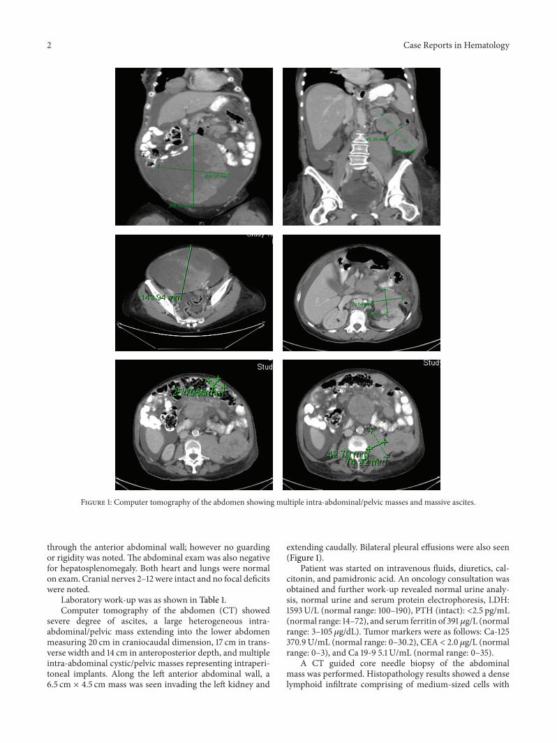
Figure 1: Computer tomography of the abdomen showing multiple intra-abdominal/pelvic masses and massive ascites.
through the anterior abdominal wall; however no guardingor rigidity was noted. The abdominal exam was also negativefor hepatosplenomegaly. Both heart and lungs were normalon exam. Cranial nerves 2–12 were intact and no focal deficitswere noted.
Laboratory work-up was as shown in Table 1.Computer tomography of the abdomen (CT) showed
severe degree of ascites, a large heterogeneous intra-abdominal/pelvic mass extending into the lower abdomenmeasuring 20 cm in craniocaudal dimension, 17 cm in trans-verse width and 14 cm in anteroposterior depth, and multipleintra-abdominal cystic/pelvic masses representing intraperi-toneal implants. Along the left anterior abdominal wall, a6.5 cm × 4.5 cm mass was seen invading the left kidney and
extending caudally. Bilateral pleural effusions were also seen(Figure 1).
Patient was started on intravenous fluids, diuretics, cal-citonin, and pamidronic acid. An oncology consultation wasobtained and further work-up revealed normal urine analy-sis, normal urine and serum protein electrophoresis, LDH:1593U/L (normal range: 100–190), PTH (intact): <2.5 pg/mL(normal range: 14–72), and serum ferritin of 391𝜇g/L (normalrange: 3–105 𝜇g/dL). Tumor markers were as follows: Ca-125370.9U/mL (normal range: 0–30.2), CEA < 2.0 𝜇g/L (normalrange: 0–3), and Ca 19-9 5.1 U/mL (normal range: 0–35).
A CT guided core needle biopsy of the abdominalmass was performed. Histopathology results showed a denselymphoid infiltrate comprising of medium-sized cells with

Case Reports in Hematology 3
(a) (b)
(c)
Figure 2: (a) 100x H&E image shows a monomorphic infiltrate of small round blue cells with adjacent necrosis, (b) 400x H&E high powerdemonstrates medium sized cells with admixed apoptotic bodies and mitotic figures reminiscent of Burkitt or a Burkitt-like lymphoma, and(c) Ki-67: high proliferation rate over 90% supports the morphologic impression and is indicative of an aggressive lymphoma.
increased mitotic figures and apoptotic bodies with adjacentgeographic necrosis. Immunohistochemical stains on neo-plastic cells were positive for CD20, CD45, CD10, BCL2,BCL6, and PAX8 and negative for cyclinD1, CD5, and weaklypositive expression for MUM1. The ki-67 proliferation ratewas 95% (Figure 2), findings consistent with a high gradelymphoma such as Burkitt’s or a B-cell lymphoma, unclassifi-able with features intermediate between diffuse large diffuseB-cell lymphoma and Burkitt lymphoma. Flow cytometryof the abdominal mass was also suggestive of an aggressiveB-cell lymphoma (Figure 3). Subsequent FISH testing waspositive for t(8;14), c-myc-IgH translocation, and a t(14;18)IgH–BCL2 translocation (Figure 4). These findings werediagnostic of the so-called “Double hit B-cell lymphoma,”WHO recognized category of B-cell lymphoma, unclassifi-able with features intermediate between diffuse large B-celllymphoma and Burkitt lymphoma.
A whole body PET-CT revealed large hypermetabolicconfluent nodal masses involving the left perirenal andperipancreatic regions with extension into the abdominalmesentery, a very large hypermetabolic mass in the anteriorabdominopelvic midline extending into the left anteriorabdominal wall, hypermetabolic soft tissue masses implantedalong the diaphragm bilaterally, and focal hypermetabolicactivity within the lateral wall of the left ventricle of theheart.Multiple foci of hypermetabolic activity throughout thebones, suspicious for osseous metastatic disease, were alsonoted (Figure 5).
However, bone marrow biopsy and aspirate showed asprinkling of small B-cells comprising 10% of marrow cel-lularity without significant aggregate formation. The corre-sponding flow cytometry (Figure 6) demonstrated clonalitywith a lambda restricted CD20+/CD10+ population consis-tent with a low grade B-cell lymphomawith a germinal centerphenotype.Thus, these findings weremost consistent with anunderlying follicular lymphoma, with subsequent peripheraltransformation into an aggressive nodal-based “double hit”lymphoma.
Following hydration, diuretics, calcitonin, and pamid-ronic acid, patient’s serum calcium started trending down,and her kidney function showed improvement.
As per recommendation by oncology, patient was trans-ferred to a tertiary care center. She was started on combi-nation chemotherapy comprising of EPOCH (doxorubicin10mg/m2 plus etoposide 50mg/m2 plus vincristine 0.4mg/m2 by continuous IV infusion on days 2–4 plus cyclophos-phamide 750mg/m2 on day 6 plus prednisone 60mg/m2on days 1–6, every 21 d). Patient also received intrathecalmethotrexate for CNS prophylaxis.
3. Discussion
Double hit lymphomas (DHLs) are defined by a chromoso-mal breakpoint affecting theMYC/8q24 locus in combinationwith another recurrent breakpoint, mainly a t(14;18)(q32;q21)

4 Case Reports in Hematology
CD45
APC
-H7
-A
SSC-A (×1,000)50 100 150 200 250
105
104
103
102
(a)
105
104
103
102
0
105
104
103
102
0
−114
−52
CD20
FITC
-A
CD10 PE-A
(b)
105
104
103
102
0
105
104
103
102
0
−114
−52
sLA
MBD
A P
E-A
sKAPPA FITC-A
(c)
Figure 3: (a) Flow cytometry of the ungated specimen of the abdominal mass showing a Cd45 bright population, compatible with alymphoproliferative process (rather than a carcinoma, melanoma, etc.), (b) gating only the CD45 bright/low side scatter population shows apredominant CD20+/CD10+ population, consistent with a B-cell population of germinal center origin, and (c) light chain expression showslambda light chain restriction, diagnostic of a B-cell lymphoma.
involving BCL2. The partner of BCL2/18q21 breakpointmostly is the IGH locus at 14q32, and in some cases at(8;14;18) may be present [1–4]. Many DHLs arise in patientswith prior follicular lymphoma often with known BCL2translocations, though de novo lymphomas are also knownto occur [5, 6]. Lymphomas with double hit genotype includeBurkitt or Burkitt-like lymphoma, diffuse large B-cell lym-phoblastic lymphoma, TdT+B-cell lymphoblastic lymphoma,low grade follicular lymphoma, and plasmablastic lymphoma[1, 2]. DHLs are relatively infrequent and mainly occur inadults.
More than half of the patients present with widespread,often extranodal disease. Unlike Burkitt lymphoma, there isno preferential localization in the ileocecal region or jaws.Magro et al. reported three patientswith an aggressive formof
B-cell lymphoma secondarily involving the skin. Twopatientshad an antecedent and/or concurrent history of follicularlymphoma, and one patient developed a de novo lymphoma.In each case there was a c-MYC and BCL2/IGH rearrange-ment, diagnostic of DHL [7]. Kaplan et al. reported anothercase of a 53-year-old male who presented with abdominalpain, shortness of breath, night sweats, ascites, and extensivelymphadenopathy. Cytologic examination of the peritonealfluid showed two distinct populations of neoplastic cells,findings compatible with a B-cell double hit lymphoblasticlymphoma [8].
Patients usually present with lymphadenopathy and/ormass lesions in extranodal sites. Some patients may have aleukemic presentation. DHLs show frequent involvement ofthe bone marrow, peripheral blood, and CNS and are usually

Case Reports in Hematology 5
LSI myc
LSI IgH
LSI myc con LSI IgH
(a)
LSI IgH con LSI BCL 2LSI BCL2LSI IgH
(b)
Figure 4: FISH analysis using Abbott Tri-color Dual fusion Translocation Probe hybridized to a nucleus revealing, (a) an IgH/c-mycrearrangement by the juxtaposition of the red (c-myc) and green (IgH) yielding the yellow fusion. One native c-myc (red) and native IgH(green) are also present; (b) a BCL-2/IgH rearrangement by the juxtaposition of the red (BCL-2) and green (IgH) yielding the yellow fusion.One native BCL-2 (red) and native IgH (green) are also present.
Figure 5: WHOLE BODY PET-CT showing multiple areas of hypermetabolic activity in the abdomen with hypermetabolic bony lesionssuspicious of extensive metastatic disease.
associated with a very poor prognosis. Patients present withpoor prognostic factors such as elevated LDH, bone mar-row/CNS involvement, and a high international prognosticindex score [9]. Most studies on larger series of patientssuggest a poor prognosis, also if treated with R-CHOP(rituximab, cyclophosphamide, doxorubicin, vincristine, andprednisolone) or high intensity treatment modalities [10].
Transformation of follicular lymphoma to a more aggres-sive non-Hodgkin lymphoma is a pivotal event in the naturalhistory of follicular lymphoma [11–16]. Follicular lymphomasare mainly known to transform to diffuse large B-cell lym-phoma, to B-cell unclassifiable lymphoma (DHLs), and veryrarely to lymphoblastic lymphoma and acute lymphoblasticleukemia. Al-Tourah et al. established a clinical diagnosis oftransformation based on the presence of at least one of thefollowing: sudden rise in LDH, rapid discordant localizednodal growth, new involvement of unusual extranodal sites,new B symptoms, and new hypercalcemia [17]. However, in
some patients these symptomsmay occur with progression offollicular lymphoma andnot necessarilywith transformation.Several studies have reported higher risk of transformation inpatients with advanced disease stage, presence of B symptomsand bulky disease, high 𝛽2 microglobulin and low albuminlevels, and higher scores of follicular lymphoma internationalprognostic index [18, 19]. Also, early initiation of follicularlymphoma treatment does not decrease the risk of transfor-mation. The median time from diagnosis to transformationin the reported series ranges from 40 to 66 months [16].
The optimal treatment for these lymphomas remainsundefined. DHL responds poorly to R-CHOP regimens,CODOX-M/IVAC regimens, and Hyper-CVAD chemo-therapy [20]. Most patients following transformation aretreated with standard doxorubicin containing combinationchemotherapy regimens and a complete remission up to40% has been reported [21]. Radiation, either alone or incombination with chemotherapy, has also been used in

6 Case Reports in Hematology
CD45
APC
-H7
-A
SSC-A (×1,000)50 100 150 200 250
105
104
103
102
(a)
105
104
103
102
0
105
104
103
102
0
−118
−45
CD20
FITC
-A
CD10 PE-A
(b)
−45
105
104
103
102
0
105
104
103
102
0
sKAPPA FITC-A
−147
CD19
PerC
P-Cy
5-5
-A
(c)
−118
−147
sLAMBDA PE-A
105
104
103
102
0
105
104
103
102
0
CD19
PerC
P-Cy
5-5
-A
(d)
Figure 6: (a) Flow cytometry of the ungated bone marrow aspirate shows 16% cellularity in the CD45 bright/low side scatter gate, (b) gatingonly the CD45 bright/low side scatter gate shows a population of CD10+/CD20+ lymphocytes representing 7% of overall cellularity, and (c)this population shows dim surface lambda light chain restriction, all findings consistent with a low grade lymphoma of germinal center origin.
patients with limited disease, and a complete remission rateof 70% has been reported [21]. Radioimmunotherapy usingradioactive nucleotide labeled antibodies such as yttriumY90 ibritumomab and iodine I131 tositumomab has shownto exhibit antilymphoma activity in a small number ofpatients with transformed follicular lymphoma [22]. Highdose chemotherapy and autologous stem cell transplantationhave also been evaluated for the treatment of patients withtransformation of follicular lymphoma [23]. Parker et al.reported two cases of DHL successfully treated with aggres-sive immunochemotherapy followed by autologous stemcell transplantation and radiation therapy [24]. However,more research is needed before these treatment modalitiescan be widely accepted to treat patients with these rarelymphomas.
Hypercalcemia ofmalignancy occurs in 20–30%of cancerpatients [25]. It occurs in patients with solid tumors aswell as hematological malignancies. Various mechanismsare known to cause hypercalcemia in malignancy; theseinclude osteolytic metastasis, tumor secretion of parathyroidhormone related protein (PTHrP), and tumor productionof 1, 25-dihydroxyvitamin D (calcitriol). The most commoncause of hypercalcemia in patients with nonmetastatic solidtumors and in some patients with non-Hodgkin lymphomais secretion of PTHrP, a condition called humoral hypercal-cemia of malignancy [26]. However, excessive productionof calcitriol is the most common cause of hypercalcemia inpatients with Hodgkin lymphoma and approximately one-third of patients with non-Hodgkin lymphoma [27]. Newonset/worsening hypercalcemia is a very rare manifestation
Case Reports in Hematology 7
Table 1: Laboratory work-up.
Hb/Hct 9.5 gm/dL/30.1% ↓WBC 6.34/nL NPlatelet count 321/nL NBUN 21mg/dL ↑Creatinine 1.4mg/dL ↑Calcium 16.3mg/dL ↑Ionized calcium 7.87mg/dL ↑Phosphorus 2.6mg/dL ↓Magnesium 1.5mg/dL ↓Potassium 3.6mmol/L NSodium 140mmol/L NUric acid 9.1mg/dL ↑Glomerular filtration rate 40mL/min/1.73m2
↓
Albumin 3.8 gm/dL ↓N: normal.
of follicular lymphoma transformation to double hit B-celllymphoma [17].
Our paper highlights not only peripheral transformationof follicular lymphoma to B-cell DHL and the challengesencountered in diagnosing and treating these aggressivetumors, but also the association of new onset/worseninghypercalcemia in such patients.
4. Conclusion
Double hit B-cell lymphomas are rare tumors, which canarise either de novo or following transformation of follicularlymphoma. These lymphomas usually occur in adults andoften carry a very poor prognosis. Our paper highlights acase of a 69-year-old female who presented with abdominaldistention and weakness over six months. Although biopsyof the abdominal mass revealed findings compatible with adouble hit lymphoma, bone marrow biopsy results showeda low grade follicular lymphoma, thus suggesting peripheraltransformation of follicular lymphoma to double hit B-celllymphoma.
Conflict of Interests
The authors declare that they have no conflict of interests.
References
[1] A. M. Perry, D. Crockett, B. J. Dave et al., “B-cell lymphoma,unclassifiable, with features intermediate between diffuse largeB-cell lymphoma and burkitt lymphoma: study of 39 cases,”British Journal of Haematology, vol. 162, no. 1, pp. 40–49, 2013.
[2] Y. Y. Song, Y. H. Tan, Y. Yuan, W. Guo, Z. Y. Pan, and O. Bai,“Current perspectives in genetics of “Double-Hit” lymphomawith possible clinical implications,” Cell Biochemistry and Bio-physics, vol. 69, no. 2, pp. 203–208, 2014.
[3] D. L. Luo, Y. H. Liu, F. Zhang et al., “B-cell lymphomaswith concurrent myc and bcl-2/IgH or bcl-6 translocations,”Zhonghua Bing Li Xue Za Zhi, vol. 42, no. 9, pp. 584–588, 2013.
[4] S. M. Aukema, R. Siebert, E. Schuuring et al., “Double-hit B-celllymphomas,” Blood, vol. 117, no. 8, pp. 2319–2331, 2011.
[5] X. Xu, L. Zhang, Y. Wang, Q. Zhang, B. Sun, and Y. Zhang,“Double-hit and triple-hit lymphomas arising from follicularlymphoma following acquisition of MYC: report of two casesand literature review,” International Journal of Clinical andExperimental Pathology, vol. 6, no. 4, pp. 788–794, 2013.
[6] W. Kishimoto, T. Shirase, D. Chihara et al., “Double-hit lym-phoma with a feature of follicular lymphoma concurrent withclonally related B lymphoblastic leukemia: a preference oftransformation for the bone marrow,” Journal of Clinical andExperimental Hematopathology, vol. 52, no. 2, pp. 113–119, 2012.
[7] C. M. Magro, X. Wang, S. Subramaniyam, N. Darras, and S.Mathew, “Cutaneous double-Hit B-cell lymphoma: an aggres-sive form of B-cell lymphoma with a propensity for cutaneousdissemination,”TheAmerican Journal of Dermatopathology, vol.36, no. 4, pp. 303–310, 2014.
[8] A. Kaplan, A. Samad, M. M. Dolan et al., “Follicular lymphomatransformed to “double-hit” B lymphoblastic lymphoma pre-senting in the peritoneal fluid,” Diagnostic Cytopathology, vol.41, no. 11, pp. 986–990, 2013.
[9] S. M. Hubbard, B. A. Chabner, V. T. DeVita Jr. et al., “Histologicprogression in non-Hodgkin’s lymphoma,” Blood, vol. 59, no. 2,pp. 258–264, 1982.
[10] T. Kobayashi, Y. Tsutsumi, N. Sakamoto et al., “Double-hitlymphomas constitute a highly aggressive subgroup in diffuselarge B-cell lymphomas in the era of rituximab,” JapaneseJournal of Clinical Oncology, vol. 42, no. 11, pp. 1035–1042, 2012.
[11] E. B. Hicks, H. Rappaport, and W. J. Winter, “Follicularlymphoma; a re-evaluation of its position in the scheme ofmalignant lymphoma, based on a survey of 253 cases,” Cancer,vol. 9, no. 4, pp. 792–821, 1956.
[12] R. Qazi, A. C. Aisenberg, and J. C. Long, “The natural history ofnodular lymphoma,” Cancer, vol. 37, no. 4, pp. 1923–1927, 1976.
[13] M. H. Cullen, T. A. Lister, R. I. Brearley, W. S. Shand, andA. G. Stansfeld, “Histological transformation of non-Hodgkin’slymphoma: a prospective study,” Cancer, vol. 44, no. 2, pp. 645–651, 1979.
[14] A. J. Garvin, R.M. Simon, C. K.Osborne, J.Merrill, R. C. Young,andC.W. Berard, “An autopsy study of histologic progression innon-Hodgkin’s lymphomas. 192 cases from the national cancerinstitute,” Cancer, vol. 52, no. 3, pp. 393–398, 1983.
[15] D. L. Oviatt, J. B. Cousar, R. D. Collins, J. M. Flexner, andR. S. Stein, “Malignant lymphomas of follicular center cellorigin in humans. V. Incidence, clinical features, and prognosticimplications of transformation of small cleaved cell nodularlymphoma,” Cancer, vol. 53, no. 5, pp. 1109–1114, 1984.
[16] Y. Bastion, C. Sebban, F. Berger et al., “Incidence, predictivefactors, and outcome of lymphoma transformation in follicularlymphoma patients,” Journal of Clinical Oncology, vol. 15, no. 4,pp. 1587–1594, 1997.
[17] A. J. Al-Tourah, K. K. Gill, M. Chhanabhai et al., “Population-based analysis of incidence and outcome of transformed non-Hodgkin’s lymphoma,” Journal of Clinical Oncology, vol. 26, no.32, pp. 5165–5169, 2008.
[18] S. Montoto, A. J. Davies, J. Matthews et al., “Risk and clinicalimplications of transformation of follicular lymphoma to dif-fuse large B-cell lymphoma,” Journal of Clinical Oncology, vol.25, no. 17, pp. 2426–2433, 2007.
[19] E. Gine, S. Montoto, F. Bosch et al., “The Follicular LymphomaInternational Prognostic Index (FLIPI) and the histological

8 Case Reports in Hematology
subtype are the most important factors to predict histologicaltransformation in follicular lymphoma,” Annals of Oncology,vol. 17, no. 10, pp. 1539–1545, 2006.
[20] J. Munoz, M. Vekaria, A. Hanbali, and N. Janakiraman, “Pro-gression of double-hit lymphoma in the midst of R-hyperCVAD,” American Journal of Hematology, vol. 88, no. 1, pp. 87–88, 2013.
[21] A. R. Yuen, O. W. Kamel, J. Halpern, and S. J. Horning,“Long-term survival after histologic transformation of low-grade follicular lymphoma,” Journal of Clinical Oncology, vol. 13,no. 7, pp. 1726–1733, 1995.
[22] M. S. Kaminski, A. D. Zelenetz, O. W. Press et al., “Pivotalstudy of iodine I 131 tositumomab for chemotherapy-refractorylow-grade or transformed low-grade B-cell non-Hodgkin’s lym-phomas,” Journal of Clinical Oncology, vol. 19, no. 19, pp. 3918–3928, 2001.
[23] H. C. Schouten, P. J. Bierman, W. P. Vaughan et al., “Autologousbone marrow transplantation in follicular non-Hodgkin’s lym-phoma before and after histologic transformation,” Blood, vol.74, no. 7, pp. 2579–2584, 1989.
[24] S. M. Parker, H. Olteanu, P. Vantuinen et al., “Follicularlymphoma transformation to dual translocated Burkitt-likelymphoma: improved disease control associated with radiationtherapy,” International Journal of Hematology, vol. 90, no. 5, pp.616–622, 2009.
[25] A. F. Stewart, “Clinical practice. Hypercalcemia associated withcancer,”TheNewEngland Journal ofMedicine, vol. 352, no. 4, pp.373–379, 2005.
[26] W. A. Ratcliffe, A. C. J. Hutchesson, N. J. Bundred, and J.G. Ratcliffe, “Role of assays for parathyroid-hormone-relatedprotein in investigation of hypercalcaemia,”TheLancet, vol. 339,no. 8786, pp. 164–167, 1991.
[27] J. F. Seymour and R. F. Gagel, “Calcitriol: the major humoralmediator of hypercalcemia in Hodgkin's disease and non-Hodgkin's lymphomas,” Blood, vol. 82, no. 5, pp. 1383–1394,1993.

Submit your manuscripts athttp://www.hindawi.com
Stem CellsInternational
Hindawi Publishing Corporationhttp://www.hindawi.com Volume 2014
Hindawi Publishing Corporationhttp://www.hindawi.com Volume 2014
MEDIATORSINFLAMMATION
of
Hindawi Publishing Corporationhttp://www.hindawi.com Volume 2014
Behavioural Neurology
EndocrinologyInternational Journal of
Hindawi Publishing Corporationhttp://www.hindawi.com Volume 2014
Hindawi Publishing Corporationhttp://www.hindawi.com Volume 2014
Disease Markers
Hindawi Publishing Corporationhttp://www.hindawi.com Volume 2014
BioMed Research International
OncologyJournal of
Hindawi Publishing Corporationhttp://www.hindawi.com Volume 2014
Hindawi Publishing Corporationhttp://www.hindawi.com Volume 2014
Oxidative Medicine and Cellular Longevity
Hindawi Publishing Corporationhttp://www.hindawi.com Volume 2014
PPAR Research
The Scientific World JournalHindawi Publishing Corporation http://www.hindawi.com Volume 2014
Immunology ResearchHindawi Publishing Corporationhttp://www.hindawi.com Volume 2014
Journal of
ObesityJournal of
Hindawi Publishing Corporationhttp://www.hindawi.com Volume 2014
Hindawi Publishing Corporationhttp://www.hindawi.com Volume 2014
Computational and Mathematical Methods in Medicine
OphthalmologyJournal of
Hindawi Publishing Corporationhttp://www.hindawi.com Volume 2014
Diabetes ResearchJournal of
Hindawi Publishing Corporationhttp://www.hindawi.com Volume 2014
Hindawi Publishing Corporationhttp://www.hindawi.com Volume 2014
Research and TreatmentAIDS
Hindawi Publishing Corporationhttp://www.hindawi.com Volume 2014
Gastroenterology Research and Practice
Hindawi Publishing Corporationhttp://www.hindawi.com Volume 2014
Parkinson’s Disease
Evidence-Based Complementary and Alternative Medicine
Volume 2014Hindawi Publishing Corporationhttp://www.hindawi.com