case report simplified technique for sealing corneal
TRANSCRIPT

Case ReportSimplified Technique for Sealing Corneal Perforations Usinga Fibrin Glue-Assisted Amniotic Membrane Transplant-Plug
Selcuk Kara, Sedat Arikan, Ismail Ersan, and Arzu Taskiran Comez
Department of Ophthalmology, Canakkale Onsekiz Mart University, School of Medicine, Canakkale, Turkey
Correspondence should be addressed to Selcuk Kara; [email protected]
Received 2 March 2014; Accepted 8 June 2014; Published 18 June 2014
Academic Editor: Alexander A. Bialasiewicz
Copyright © 2014 Selcuk Kara et al. This is an open access article distributed under the Creative Commons Attribution License,which permits unrestricted use, distribution, and reproduction in any medium, provided the original work is properly cited.
Purpose. To describe a surgical technique using amniotic membrane transplant (AMT) with fibrin glue (FG) for treating smallercorneal perforations more practically and appropriately filling the defect. Method. A patient with noninfectious central cornealperforation, in 1mm in diameter, was treated with FG-assisted AMT-plug. An AMT was folded in on itself twice by using FG thena small piece of this FG-AMT mixture was cut to maintain an appropriate plug for the site of the corneal perforation. The FG-assisted AMT-plug was placed in the perforation area by using FG. An amniotic membrane patch was placed over the plug, whichwas then secured by a bandage contact lens. Result. Surgery to restore corneal stromal thickness without recurrence of perforation.Conclusion. The FG-assisted AMT-plug allowed a successful repair of 1mm in diameter corneal perforation. This technique waseasily performed, thus seeming to be a good alternative to treat corneal perforations with restoring corneal thickness.
1. Introduction
Corneal perforations presenting as a result of infection,inflammation, or trauma are ophthalmic emergencies thaturgently require appropriate intervention. Management ofthe perforation depends on the size of the defect, the under-lying disease, the surgeon’s experience, and the presenceof amniotic membrane transplant (AMT) or donor cornea.Medical treatment alone is effective for small perforations(<1mm); bandage contact lenses can also be used successfullyfor perforations of this size [1, 2]. However, larger defects(>2mm in diameter) require surgical procedures, includingtissue adhesives, AMT, and corneal transplantation [3–5].Although the tissue adhesives cyanoacrylate and fibrin glueare widely used in smaller corneal perforations, they cannotbe sustained in the defect site as a filling material [6]. Thesuccess rate decreases to 37% with cyanoacrylate applicationalone in corneal perforations associated with herpetic kerati-tis [7].
AMT, which includes growth factors, neurotrophins, andcytokines, has been useful in the treatment of ulcerativecorneal diseases [8]. Furthermore, multilayer application ofAMT has also been used to restore corneal stromal thicknessof perforations up to 3mm in diameter, though Rodrıguez-Ares et al. reported that the success rate decreased to 40%
in perforations of 1.5mm or larger [4, 9]. Kim and Parkreported on a way to eliminate the limitations of multilayerAMT [10]. They maintained a thick single piece of 5- or 7-plyAMT by using fibrin glue (FG) between the sheets, a methodcalled “augmented AMT,” and successfully treated cornealperforations in a range of 2–5mm in diameter. Althoughthis technique is very useful for larger corneal perforations,it is relatively difficult to prepare FG-assisted multilayerAMT and there is no need for the manipulation of separatepieces of AMT in small perforations. We aimed to modifythis technique to treat smaller corneal perforations morepractically and appropriately fill the defect by providing amechanical scaffold.
In the current case, we present an intervention of FG-assisted AMT-plug for a corneal perforation of 1mm indiameter and evaluate the corneal stroma stability untilkeratoplasty.
2. Case
A 76-year-old male patient was referred to our clinic withepiphora and vision decrease in his left eye. He had previouslyundergone a cataract surgery and had received an intraocularlens two years earlier. His visual acuity had been handmotionfor two months. On examination, a 1mm in diameter central
Hindawi Publishing CorporationCase Reports in Ophthalmological MedicineVolume 2014, Article ID 351534, 3 pageshttp://dx.doi.org/10.1155/2014/351534
2 Case Reports in Ophthalmological Medicine
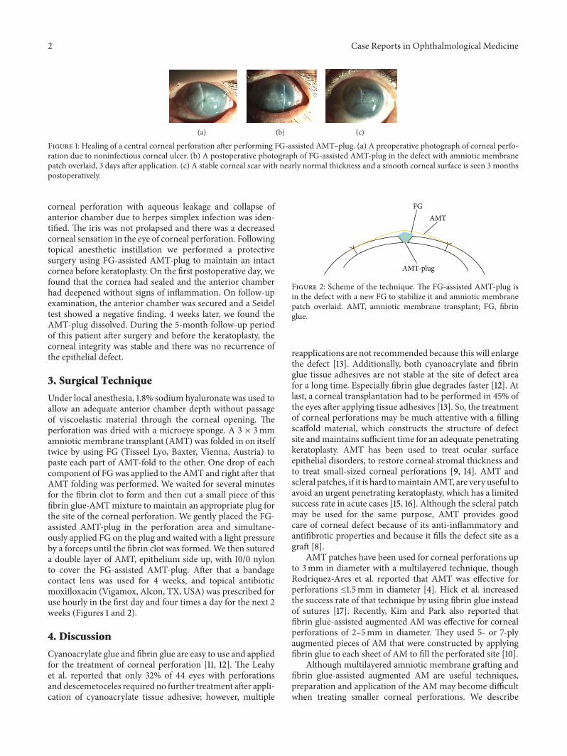
(a) (b) (c)
Figure 1: Healing of a central corneal perforation after performing FG-assisted AMT–plug. (a) A preoperative photograph of corneal perfo-ration due to noninfectious corneal ulcer. (b) A postoperative photograph of FG-assisted AMT-plug in the defect with amniotic membranepatch overlaid, 3 days after application. (c) A stable corneal scar with nearly normal thickness and a smooth corneal surface is seen 3 monthspostoperatively.
corneal perforation with aqueous leakage and collapse ofanterior chamber due to herpes simplex infection was iden-tified. The iris was not prolapsed and there was a decreasedcorneal sensation in the eye of corneal perforation. Followingtopical anesthetic instillation we performed a protectivesurgery using FG-assisted AMT-plug to maintain an intactcornea before keratoplasty. On the first postoperative day, wefound that the cornea had sealed and the anterior chamberhad deepened without signs of inflammation. On follow-upexamination, the anterior chamber was secured and a Seideltest showed a negative finding. 4 weeks later, we found theAMT-plug dissolved. During the 5-month follow-up periodof this patient after surgery and before the keratoplasty, thecorneal integrity was stable and there was no recurrence ofthe epithelial defect.
3. Surgical Technique
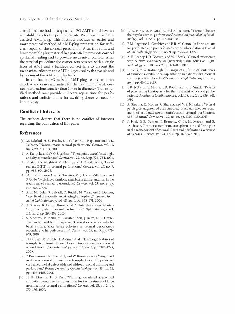
Under local anesthesia, 1.8% sodium hyaluronate was used toallow an adequate anterior chamber depth without passageof viscoelastic material through the corneal opening. Theperforation was dried with a microeye sponge. A 3 × 3mmamniotic membrane transplant (AMT) was folded in on itselftwice by using FG (Tisseel Lyo, Baxter, Vienna, Austria) topaste each part of AMT-fold to the other. One drop of eachcomponent of FGwas applied to the AMT and right after thatAMT folding was performed. We waited for several minutesfor the fibrin clot to form and then cut a small piece of thisfibrin glue-AMTmixture to maintain an appropriate plug forthe site of the corneal perforation. We gently placed the FG-assisted AMT-plug in the perforation area and simultane-ously applied FG on the plug and waited with a light pressureby a forceps until the fibrin clot was formed.We then sutureda double layer of AMT, epithelium side up, with 10/0 nylonto cover the FG-assisted AMT-plug. After that a bandagecontact lens was used for 4 weeks, and topical antibioticmoxifloxacin (Vigamox, Alcon, TX, USA) was prescribed foruse hourly in the first day and four times a day for the next 2weeks (Figures 1 and 2).
4. Discussion
Cyanoacrylate glue and fibrin glue are easy to use and appliedfor the treatment of corneal perforation [11, 12]. The Leahyet al. reported that only 32% of 44 eyes with perforationsand descemetoceles required no further treatment after appli-cation of cyanoacrylate tissue adhesive; however, multiple
FGAMT
AMT-plug
Figure 2: Scheme of the technique. The FG-assisted AMT-plug isin the defect with a new FG to stabilize it and amniotic membranepatch overlaid. AMT, amniotic membrane transplant; FG, fibringlue.
reapplications are not recommended because this will enlargethe defect [13]. Additionally, both cyanoacrylate and fibringlue tissue adhesives are not stable at the site of defect areafor a long time. Especially fibrin glue degrades faster [12]. Atlast, a corneal transplantation had to be performed in 45% ofthe eyes after applying tissue adhesives [13]. So, the treatmentof corneal perforations may be much attentive with a fillingscaffold material, which constructs the structure of defectsite andmaintains sufficient time for an adequate penetratingkeratoplasty. AMT has been used to treat ocular surfaceepithelial disorders, to restore corneal stromal thickness andto treat small-sized corneal perforations [9, 14]. AMT andscleral patches, if it is hard tomaintainAMT, are very useful toavoid an urgent penetrating keratoplasty, which has a limitedsuccess rate in acute cases [15, 16]. Although the scleral patchmay be used for the same purpose, AMT provides goodcare of corneal defect because of its anti-inflammatory andantifibrotic properties and because it fills the defect site as agraft [8].
AMT patches have been used for corneal perforations upto 3mm in diameter with a multilayered technique, thoughRodrıquez-Ares et al. reported that AMT was effective forperforations ≤1.5mm in diameter [4]. Hick et al. increasedthe success rate of that technique by using fibrin glue insteadof sutures [17]. Recently, Kim and Park also reported thatfibrin glue-assisted augmented AM was effective for cornealperforations of 2–5mm in diameter. They used 5- or 7-plyaugmented pieces of AM that were constructed by applyingfibrin glue to each sheet of AM to fill the perforated site [10].
Although multilayered amniotic membrane grafting andfibrin glue-assisted augmented AM are useful techniques,preparation and application of the AM may become difficultwhen treating smaller corneal perforations. We describe
Case Reports in Ophthalmological Medicine 3
a modified method of augmented FG-AMT to achieve anadjustable plug for the perforation site. We termed it an “FG-assisted AMT-plug.” This method provides an easier andmore practical method of AMT-plug preparation for suffi-cient repair of the corneal perforation. Also, this solid andbiocompatible plugmaterial has potential to promote cornealepithelial healing and to provide mechanical scaffold. Afterthe surgical procedure the cornea was covered with a singlelayer of AMT and a bandage contact lens to prevent themechanical effect on the AMT-plug caused by the eyelids andhydration of the AMT-plug by tears.
In conclusion, FG-assisted AMT-plug seems to be aneffective and easier alternative for the treatment of acute cor-neal perforations smaller than 3mm in diameter. This mod-ified method may provide a shorter repair time for perfo-rations and sufficient time for awaiting donor corneas forkeratoplasty.
Conflict of Interests
The authors declare that there is no conflict of interestsregarding the publication of this paper.
References
[1] M. Lekskul, H. U. Fracht, E. J. Cohen, C. J. Rapuano, and P. R.Laibson, “Nontraumatic corneal perforation,” Cornea, vol. 19,no. 3, pp. 313–319, 2000.
[2] A. Kanpolat and O. O. Ucakhan, “Therapeutic use of focus nightandday contact lenses,”Cornea, vol. 22, no. 8, pp. 726–734, 2003.
[3] H. Siatiri, S. Moghimi, M. Malihi, and A. Khodabande, “Use ofsealant (HFG) in corneal perforations,” Cornea, vol. 27, no. 9,pp. 988–991, 2008.
[4] M. T. Rodrıguez-Ares, R. Tourino, M. J. Lopez-Valladares, andF. Gude, “Multilayer amniotic membrane transplantation in thetreatment of corneal perforations,” Cornea, vol. 23, no. 6, pp.577–583, 2004.
[5] A. B. Nurozler, S. Salvarli, K. Budak, M. Onat, and S. Duman,“Results of therapeutic penetrating keratoplasty,” Japanese Jour-nal of Ophthalmology, vol. 48, no. 4, pp. 368–371, 2004.
[6] A. Sharma, R. Kaur, S. Kumar et al., “Fibrin glue versusN-butyl-2-cyanoacrylate in corneal perforations,” Ophthalmology, vol.110, no. 2, pp. 291–298, 2003.
[7] S. Moorthy, V. Jhanji, M. Constantinou, J. Beltz, E. O. Graue-Hernandez, and R. B. Vajpayee, “Clinical experience with N-butyl cyanoacrylate tissue adhesive in corneal perforationssecondary to herpetic keratitis,” Cornea, vol. 29, no. 9, pp. 971–975, 2010.
[8] D. G. Said, M. Nubile, T. Alomar et al., “Histologic features oftransplanted amniotic membrane: implications for cornealwound healing,” Ophthalmology, vol. 116, no. 7, pp. 1287–1295,2009.
[9] P. Prabhasawat, N. Tesavibul, andW. Komolsuradej, “Single andmultilayer amniotic membrane transplantation for persistentcorneal epithelial defect with and without stromal thinning andperforation,” British Journal of Ophthalmology, vol. 85, no. 12,pp. 1455–1463, 2001.
[10] H. K. Kim and H. S. Park, “Fibrin glue-assisted augmentedamniotic membrane transplantation for the treatment of largenoninfectious corneal perforations,” Cornea, vol. 28, no. 2, pp.170–176, 2009.
[11] L. W. Hirst, W. E. Smiddy, and E. De Juan, “Tissue adhesivetherapy for corneal perforations,”Australian Journal of Ophthal-mology, vol. 11, no. 2, pp. 113–118, 1983.
[12] F.M. Lagoutte, L. Gauthier, and P. R.M. Comte, “A fibrin sealantfor perforated and preperforated corneal ulcers,” British Journalof Ophthalmology, vol. 73, no. 9, pp. 757–761, 1989.
[13] A. B. Leahey, J. D. Gottsch, andW. J. Stark, “Clinical experiencewith N-butyl cyanoacrylate (nexacryl) tissue adhesive,” Oph-thalmology, vol. 100, no. 2, pp. 173–180, 1993.
[14] T. Celik, Y. A. Katircioglu, E. Singar et al., “Clinical outcomesof amniotic membrane transplantation in patients with cornealand conjunctival disorders,” Seminars inOphthalmology, vol. 28,no. 1, pp. 41–45, 2013.
[15] J. R. Nobe, B. T. Moura, J. B. Robin, and R. E. Smith, “Resultsof penetrating keratoplasty for the treatment of corneal perfo-rations,” Archives of Ophthalmology, vol. 108, no. 7, pp. 939–941,1990.
[16] A. Sharma, K. Mohan, R. Sharma, and V. S. Nirankari, “Scleralpatch graft augmented cyanoacrylate tissue adhesive for treat-ment of moderate-sized noninfectious corneal perforations(3.5–4.5 mm),” Cornea, vol. 32, no. 10, pp. 1326–1330, 2013.
[17] S. Hick, P. E. Demers, I. Brunette, C. La, M. Mabon, and B.Duchesne, “Amnioticmembrane transplantation andfibrin gluein the management of corneal ulcers and perforations: a reviewof 33 cases,” Cornea, vol. 24, no. 4, pp. 369–377, 2005.

Submit your manuscripts athttp://www.hindawi.com
Stem CellsInternational
Hindawi Publishing Corporationhttp://www.hindawi.com Volume 2014
Hindawi Publishing Corporationhttp://www.hindawi.com Volume 2014
MEDIATORSINFLAMMATION
of
Hindawi Publishing Corporationhttp://www.hindawi.com Volume 2014
Behavioural Neurology
EndocrinologyInternational Journal of
Hindawi Publishing Corporationhttp://www.hindawi.com Volume 2014
Hindawi Publishing Corporationhttp://www.hindawi.com Volume 2014
Disease Markers
Hindawi Publishing Corporationhttp://www.hindawi.com Volume 2014
BioMed Research International
OncologyJournal of
Hindawi Publishing Corporationhttp://www.hindawi.com Volume 2014
Hindawi Publishing Corporationhttp://www.hindawi.com Volume 2014
Oxidative Medicine and Cellular Longevity
Hindawi Publishing Corporationhttp://www.hindawi.com Volume 2014
PPAR Research
The Scientific World JournalHindawi Publishing Corporation http://www.hindawi.com Volume 2014
Immunology ResearchHindawi Publishing Corporationhttp://www.hindawi.com Volume 2014
Journal of
ObesityJournal of
Hindawi Publishing Corporationhttp://www.hindawi.com Volume 2014
Hindawi Publishing Corporationhttp://www.hindawi.com Volume 2014
Computational and Mathematical Methods in Medicine
OphthalmologyJournal of
Hindawi Publishing Corporationhttp://www.hindawi.com Volume 2014
Diabetes ResearchJournal of
Hindawi Publishing Corporationhttp://www.hindawi.com Volume 2014
Hindawi Publishing Corporationhttp://www.hindawi.com Volume 2014
Research and TreatmentAIDS
Hindawi Publishing Corporationhttp://www.hindawi.com Volume 2014
Gastroenterology Research and Practice
Hindawi Publishing Corporationhttp://www.hindawi.com Volume 2014
Parkinson’s Disease
Evidence-Based Complementary and Alternative Medicine
Volume 2014Hindawi Publishing Corporationhttp://www.hindawi.com