case report / ПРИКАЗ БОЛЕСНИКА … · sillar abscess. only sporadically, it is a...
TRANSCRIPT

460
DOI: https://doi.org/10.2298/SARH161103021T
UDC: 616.14-005.6; 616.322/.323-089.85-06
CASE REPORT / ПРИКАЗ БОЛЕСНИКА
Thrombophlebitis of the internal jugular vein after tonsillectomySaša Z. Tabaković1, Maja Đukić-Božović2, Goran Videnović1, Aleksandar Pavlović1,3, Jelena Todić1, Jasna Pavlović1, Brankica Martinović1
1University of Priština, Faculty of Medicine, Department of Dentistry, Kosovska Mitrovica, Serbia;2Dr. Dragiša Mišović University Hospital Center, Hospital of Otorhinolaryngology, Belgrade, Serbia;3University of Priština, Faculty of Medicine, Clinic for Surgery and Anesthesia, Kosovska Mitrovica, Serbia
SUMMARYIntroduction Thrombophlebitis of the internal jugular vein may appear as a rare complication of oropha-ryngeal infection and tonsillectomy procedure. Clinical features usually include an acute onset of inflam-mation with formation of venous thrombosis and secondary septic propagation (Lemierre’s syndrome).The aim of this work was to present a rare case of internal jugular vein thrombophlebitis as a late com-plication following tonsillectomy.Case outline We present an otherwise healthy 25-year-old male patient on whom tonsillectomy was performed due to chronic tonsillitis. Two weeks after surgery, the patient was rehospitalized for high temperature, diffuse swelling on the left side of the neck, fatigue, painful swallowing, and constrained mouth opening. Thrombophlebitis of the left internal jugular vein was diagnosed by the neck ultrasound. Complete recovery was achieved in three weeks’ time by the combination of antibiotics and anticoagu-lant/antithrombotic therapy. Conclusion Tonsillectomy is a risk factor for the internal jugular vein thrombosis in adults with chronic tonsillitis, especially if fibrous adhesions are expected or found during the surgical procedure.Keywords: Lemierre’s syndrome; tonsillectomy, complication; venous thrombosis, etiology, diagnosis, therapy
Received • Примљено: November 3, 2016
Revised • Ревизија: February 28, 2018
Accepted • Прихваћено: March 5, 2018
Online first: March 13, 2018
Correspondence to:Saša TABAKOVIĆFaculty of MedicineDepartment of DentistryAnri Dinana b. b.38220 Kosovska Mitrovica, [email protected]
INTRODUCTION
Thrombophlebitis of the internal jugular vein with septicemia accompanied by the presence of secondary abscesses refers to a rare compli-cation of oropharyngeal infection mostly due to tonsillopharyngitis complicated by periton-sillar abscess. Only sporadically, it is a repre-sentation of a post-tonsillectomy outcome, predominantly in adults. Similar clinical pic-ture may also develop secondary to mastoiditis, otitis, sinusitis, and odontogenic infection, as well as appendicitis, urinary infection, and sup-purative endometritis after delivery [1, 2, 3]. Because of the common clinical aspects, these septicemias with thrombus formation were grouped together by André Lemierre in 1936, and later on they were named after him [1].
Lemierre’s syndrome was a common com-plication of oropharyngeal infections before the discovery of antibiotics [4]. Although an increasing number of cases has been noticed by some authors since 1990, the incidence rate of 14.4 cases per million indicated a rare con-dition [5].
Lemierre’s syndrome occurs mostly in the young adult population at the end of winter or in early spring [6, 7, 8]. An equal sex distri-bution is reported by most authors, while the higher prevalence of males is seldom found [7].
The main causative agents of tonsillitis and peritonsillar abscesses are Streptococcus pyo-
genes, an anaerobic Gram-positive coccus, and Fusobacterium necrophorum, a non-spore-forming obligate anaerobic Gram-negative bac-terium, which belong to the physiological flora of the oropharynx. The latter is also the most responsible one for Lemierre’s syndrome.
Clinical manifestations vary depending on the organs affected by infection [9]. Fever and gastrointestinal discomforts are found in 82% of all cases [1]. Thromboembolic systemic complications may appear as bacteremia, septi-cemia, pneumonia, pericarditis, hepatospleno-megaly, meningitis, arthritis, and kidney and brain abscesses. Pulmonary septic thrombosis is the most common complication occurring in 95% of cases [10, 11]. Before the discovery of antibiotics, the mortality rate was over 90%, mostly due to the anaerobe postanginal septi-cemia [1, 4]. Today, Lemierre’s syndrome may have incomplete clinical forms because of bet-ter diagnosis and treatment, but it is a serious and potentially lethal condition nonetheless. Nowadays, mortality rate fluctuates between 4% and 22% [6, 7, 12].
Benzylpenicillin, third-generation cepha-losporins, carbapenems (imipenem, me-ropenem), clindamycin, chloramphenicol, and metronidazole achieved a high anti-microbial effect against obligate anaero-bic bacteria in vitro [13, 14]. Administra-tion of penicillin as a monotherapy is not recommended, due to the ability of some

461
Srp Arh Celok Lek. 2018 Jul-Aug;146(7-8):460-465 www.srpskiarhiv.rs
strains of fusobacteria to create beta-lactamase in 22.7% of the cases [15–18]. Metronidazole is generally considered the drug of choice for this type of infection. The weak resistance of anaerobes to this antibiotic, good resorp-tion, and bioavailability of the drug enable simple oral administration, and a rapid therapeutic effect [6, 15, 19, 20]. Antibiotic therapy in Lemierre’s syndrome is required within the range of three to six weeks, while intravenous administration of antibiotics may be replaced by the per os one when a patient becomes afebrile [6, 13, 18]. Adminis-tration of antibiotics in the period of six weeks represents the optimal period for curing due to difficult penetration of the drug in the clot. A period of curing shorter than two weeks may lead to a relapse [6]. The benefits and risks of the anticoagulation therapy in Lemierre’s syndrome still have to be clarified [6].
Surgical treatment of thrombophlebitis of the inter-nal jugular vein is needed in patients with a risk of septic thrombosis and embolic complications [19, 20, 21]. Sup-purative abscesses in parts of the neck, lungs, liver, joints, and muscles have to be drained [19, 20, 21]. Hyperbaric oxygen therapy as an addition to the surgical treatment may have beneficial results [22].
The aim of this work was to present a rare case of inter-nal jugular vein thrombophlebitis as a late complication following tonsillectomy.
CASE REPORT
Tonsillectomy was indicated in a 25-year-old male patient, due to the anamnesis and clinically demonstrated chronic tonsillitis with frequent exacerbations. Otherwise, the pa-tient was mostly healthy with no risk factors for throm-bosis and other presumed intraoperative or postoperative complications. Preoperative microbiological examination of oropharyngeal and nasal secretion demonstrated physi-ological bacterial microflora with no evidence of Candida albicans, while routine hematological and biochemical analyses showed results within reference ranges.
At the time of hospital admission, moderately hyper-trophic palatine tonsils with detritus within the crypts were present on clinical examination without symptoms and signs of exacerbated infection. Tonsillectomy was performed under general endotracheal anesthesia using Ultracision Harmonic SNGHK scalpel (Ethicon Endo-Surgery, LLC, Guaynabo, PR, USA) dissection in 30 min-utes time. Fibrous adhesions of the tonsillar capsule were dissected with minimal bleeding and with no injury to the tonsillar bed. The postoperative period during hospitaliza-tion was uneventful, the patient was physically active, and there was no need for antibiotic administration. On the fourth postoperative day, according to our hospital policy, the patient was discharged from the hospital with normal local postoperative findings and in good general health condition.
Due to an acute onset of subjective sensation of pres-sure and blunt pain during swallowing, followed by the appearance of swelling on the left side of the neck, the patient contacted a practicing physician on the fourth postoperative day and was prescribed peroral antibiotic therapy (amoxicillin-clavulanate, 625 mg three times dai-ly). In spite of treatment, the intensity of symptoms and signs persisted in the following days.
The patient under the therapy reported to the hospital again on the 14th postoperative day and was immediately rehospitalized. On readmission, the patient reported pain-ful swallowing, constrained opening of the mouth, fatigue, and nausea. Body temperature was elevated (38.5°C). Local examination of each tonsillar fossa demonstrated wound healing process in the stage of epithelization, without hem-orrhage and inflammation. Palpatory sensitive, 4 × 5 cm diffuse swelling was found on the left side of the subman-dibular region (Figure 1). Both a parapharyngeal abscess and a submandibular abscess were suspected. Hematologi-cal and biochemical blood analysis showed high erythro-cyte sedimentation rate (ESR 86 mm/hour), leucocytosis in white blood cell count (WBC 13.3 × 10⁹/L), increased level of a high-sensitivity C-reactive protein test result (hs-CRP 102.2 mg/L), moderately increased procalcitonin
Figure 1. Swelling of the left neck side Figure 2. Ultrasound imaging of thrombophlebitis of the left internal jugular vein
Thrombophlebitis of the internal jugular vein after tonsillectomy
462
Srp Arh Celok Lek. 2018 Jul-Aug;146(7-8):460-465
DOI: https://doi.org/10.2298/SARH161103021T
level (PCT 0.47 × 10¹²/L), reactive thrombocytosis in platelet count (PLT 508 × 109/L), and elevated D-dimer values (FDP 1.58 mg/L). Ultrasound imaging of the neck demonstrated thrombophlebitis of the left internal jugu-lar vein together with inflammation, edema, and reactive lymphadenitis in the surrounding soft tissue of the up-per third part of the neck. The internal jugular vein to the base of the neck was incompressible, with thickened walls without the presence of a CD signal, with the swell-ing of the perivascular structures (Figure 2). The surgi-cal area was swabbed, but microbiological findings were negative. Both intraoral examination and panoramic x-ray radiography imaging excluded odontogenic infection as a causative agent. Detailed clinical examination, chest X-ray, abdominal ultrasound imaging, and computed tomogra-phy imaging of the endocranium confirmed no existence of thromboembolism of the lungs, abdomen, joints, and central nervous system.
Based on these findings, we established the diagnosis of internal jugular vein thrombophlebitis with parapharyn-geal soft tissue inflammation on the left side of the neck in association with general infectious symptoms and signs but without further propagation of septic thrombi.
Antibiotic therapy was empirically administered on the day of the patient’s readmission: intravenously ceftri-axone, 2 g two times daily, and metronidazole, 500 mg three times daily, during a seven-day period. In spite of contradictoriness related to the therapeutic justifiability of anticoagulant treatment in Lemierre’s syndrome, and the body weight of the patient being 70 kg, we opted for its administration: nadroparin subcutaneous injection, 0.3 ml two times daily over ten days.
The patient responded well to the therapy. On the third day of the therapy, laboratory findings important for infection and blood clots showed declining values: ESR 68 mm/h, WBC 60 × 109/L, hs-CRP 51.9 mg/L, PCT 0.318 × 10¹²/L, PLT 482 × 109/L.
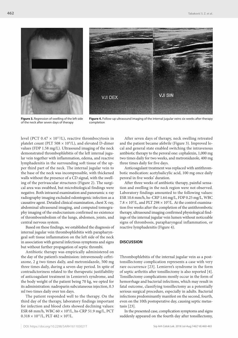
After seven days of therapy, neck swelling retreated and the patient became afebrile (Figure 3). Improved lo-cal and general state enabled switching the intravenous antibiotic therapy to the peroral one: cephalexin, 1,000 mg two times daily for two weeks, and metronidazole, 400 mg three times daily for five days.
Anticoagulant treatment was replaced with antithrom-botic medication: acetylsalicylic acid, 100 mg once daily peroral in five weeks’ duration.
After three weeks of antibiotic therapy, painful sensa-tion and swelling in the neck region were not observed. Laboratory findings amounted to the following values: ESR 10.6 mm/h, hs–CRP 1.64 mg/L, FDP 0.25 mg/L, WBC 7.8 × 10⁹/L, and PLT 298 × 109/L. At the control examina-tion five weeks after the completion of the antithrombotic therapy, ultrasound imaging confirmed physiological find-ings of the internal jugular vein lumen without noticeable signs of thrombosis, parapharyngeal inflammation, or reactive lymphadenitis (Figure 4).
DISCUSSION
Thrombophlebitis of the internal jugular vein as a post-tonsillectomy complication represents a case with very rare occurrence [23]. Lemierre’s syndrome in the form of septic arthritis after tonsillectomy is also reported [4]. Tonsillectomy complications mostly occur in the form of hemorrhage and bacterial infections, which may result in fatal outcome, classifying tonsillectomy as a potentially serious surgical procedure, especially in adults. Bacterial infections predominantly manifest on the second, fourth, even on the 10th postoperative day, causing septic metas-tasis [23].
In the presented case, complication symptoms and signs suddenly appeared on the fourth day after tonsillectomy,
Figure 3. Regression of swelling of the left side of the neck after seven days of therapy
Figure 4. Follow-up ultrasound imaging of the internal jugular veins six weeks after therapy completion
Tabaković S. Z. et al.

463
Srp Arh Celok Lek. 2018 Jul-Aug;146(7-8):460-465 www.srpskiarhiv.rs
while a diagnosis of septic thrombophlebitis without sec-ondary septic metastasis was established on the 14th post-operative day in the patient who was already under peroral antibiotic therapy.
Thrombophlebitis occurs from the spread of inflam-mation from the surrounding structures to the wall veins, while phlebothrombosis is characterized by non-specific inflammatory reaction with the formation of a thrombus fixed to the internal wall of the vein, in order to ultimate-ly develop fibrous organization. Thrombophlebitis most commonly occurs in superficial veins and the phlebo-thrombosis in deep veins.
Thromboses of the upper limbs and the neck are rare in comparison with those of the lower extremities. Internal jugular vein thrombosis is a serious event with a poten-tially fatal outcome. Complications include pulmonary embolism, sepsis with septic emboli to different organs and tissues, as well as intracranial propagation of the thrombus with cerebral edema. As any thrombosis, inter-nal jugular vein thrombosis is precipitated by Virchow’s triad: endothelial damage, alteration of blood flow, and hy-percoagulability. The history and examination of patients with internal jugular vein thrombosis may be vague and misleading. Patients may present with a painful swelling of the neck, but they also may be asymptomatic [24].
Interestingly, there were no signs of infection at the lo-cal clinical examination of the surgical region, pharyngeal space, and oral cavity. The wound healing process locally appeared quite normal at all times. In contrast, the patient’s febrile status, painful swallowing and the neck soft tissues’ unilateral swelling and tenderness with regional lymphad-enitis raised strong suspicion to postoperative infection, mainly contributing to bacterial causes. Additionally, thrombus of the left internal jugular vein was diagnosed by ultrasound neck imaging. Values of sedimentation, leuco-cytes, C-reactive protein, procalcitonin, and D-dimer were diagnostically significant in favor of either a serious local bacterial infection or a systemic one. Values of thrombo-cytes pointed to a reactive thrombocytosis, rather than to the primary one. These clinical and laboratory findings indicated a diagnosis of Lemierre’s syndrome.
We consider that in this rare case of Lemierre’s syn-drome, an infective agent spread either from chronically infected tonsillar crypts during tonsil dissection or from the colonization of the open surgical wound by oral cavity bacterial flora during the postoperative period. Infection propagated through the parapharyngeal space causing soft tissue inflammation and thrombophlebitis of the internal jugular vein. In addition, both tonsillectomy as a surgical procedure and postoperative infection may be risk factors for reactive thrombocytosis and blood hypercoagulability.
As mentioned above, microbiological findings of oro-pharyngeal mucus showed physiological flora preop-eratively. Microbial agents were not isolated at the time of rehospitalization when samples were taken from the patient under therapy. We consider that a possible fac-tor for negative microbiological finding was a course of beta-lactam with the beta-lactamase-inhibitor antibiotic combination, administered by the practicing physician.
This therapy, in our opinion, provided a certain degree of coverage of common microbes of the oral cavity, but it was insufficient for adequate infection control. Also, isolating anaerobes is a relatively long and difficult process which requires special conditions of sample cultivation.
In the presented case, we adhered to the widely spread conception that evidence for the beneficial use of anti-microbial prophylaxis in tonsillectomy in the otherwise healthy patient was insufficient, opposite to other clean-contaminated head and neck procedures. Antibiotics were also not administered to the patient during the postop-erative period in the hospital, for it was uneventful and in accordance with normal preoperative microbiological findings.
To treat the complication and to prevent secondary sep-tic thrombosis in our patient, antibiotic and anticoagu-lant therapy was administered. Broad-spectrum antibiotic therapy was selected empirically to cover a wide range of aerobic and anaerobic Gram-positive and Gram-negative bacteria. Switching to the infection-specific therapy was not done because of both negative microbiological culture and high efficacy of administered combination of ceftri-axone and metronidazole. As far as anticoagulant therapy administration in patients with Lemierre’s syndrome is concerned, opinions are divided. Some authors suggest anticoagulant therapy in all cases [25, 26]. Others consider anticoagulant therapy may be administered if thrombo-sis affects cerebral sinuses or if antibiotic therapy did not give satisfying results [5, 6, 18, 27, 28]. In our case, low-molecular-weight heparin was administered in a preven-tive dose, and the patient was switched to antithrombotic medication after 10 days due to the possibility of late post-tonsillectomy hemorrhage [29].
For low-molecular-weight heparin at a dose of 0.3 ml twice daily, we decided to avoid the possibility of com-plications after tonsillectomy in the form of secondary bleeding, which can be reported after seven days and two weeks. Tonsillectomy is made with an ultrasound scalpel, which in the area of the operational region produces a greater wound surface in the form of burns and, therefore, prolongs the epithelial period compared to the standard operating technique.
Even if the microbiological finding was preoperatively negative, we think that venous jugular thrombophlebitis was internal postoperatively as a result of the spread of inflammation from the operative region and the soft tissue structures. Laboratory results, swelling in the neck area, and febrile condition of the patient support this stance.
In our case, in addition to antibiotic therapy, a preven-tive dose of nadroparin was administered taking into ac-count the time past from the tonsillectomy, body weight of the patient, and thrombophlebitis internal jugular vein as a complication still in the initial phase without systemic complications.
The preventive dose of nadroparin with 0.3ml is given instead of curative dose because of the risk of bleeding in the operative area. The period of epithelialization of the surgical wound after tonsillectomy is known as potentially risky for postoperative bleeding. For the same reasons,
Thrombophlebitis of the internal jugular vein after tonsillectomy

464
Srp Arh Celok Lek. 2018 Jul-Aug;146(7-8):460-465
anticoagulant therapy is replaced with a low-dose anti-thrombotic medication after 10 days.
Aggravation of a patient’s general status and neck swell-ing following tonsillectomy, especially within young adult patients, should raise a suspicion of a complication devel-oping in a form of Lemierre’s syndrome. Doctors should have high levels of cautiousness in this situation, as it could
imperil the life of the patient, while adequate antibiotic and anticoagulant therapy may prevent metastatic septic thrombosis and lead to a complete recovery.
Tonsillectomy is a risk factor for the internal jugular vein thrombosis in adults with chronic tonsillitis, espe-cially if fibrous adhesions are expected or found during the surgical procedure.
REFERENCES
1. Lemierre A. On certain septicaemias due to anaerobic organisms. Lancet. 1936; 227(5874):701–3.
2. Ramirez S, Hild TG, Rudolph CN, Sty JR, Kehl SC, Havens P, et al. Increased diagnosis of Lemierre’s syndrome and other Fusobacterium necrophorum infections at a children’s hospital. Pediatrics. 2003; 112(5):380–5.
3. Chacko EM, Krilov LR, Patten W, Lee PJ. Lemierre’s and Lemierre’s-like syndromes in association with infectious mononucleosis. J Laryngol Otol. 2010; 124(12):1257–62.
4. Beldman TF, Teunisse HA, Schouten TJ. Septic arthritis of the hip by Fusobacterium necrophorum after tonsillectomy: a form of Lemierre syndrome? Eur J Pediatr. 1997; 156(11):856–7.
5. Hagelskjaer Kristensen L, Prag J. Lemierre’s syndrome and other disseminated Fusobacterium necrophorum infections in Denmark: a prospective epidemiological and clinical survey. Eur J Clin Microbiol Infect Dis. 2008; 27(9):779–89.
6. Riordan T. Human infection with Fusobacterium necrophorum (necrobacillosis), with a focus on Lemierre’s syndrome. Clin Microbiol Rev. 2007; 20(4):622–59.
7. Karkos PD, Asrani S, Karkos CD, Leong SC, Theochari EG, Alexopoulou TD, et al. Lemierre’s syndrome: a systematic review. Laryngoscope. 2009; 119(8):1552–9.
8. Venglarcik J. Lemierre’s syndrome. Pediatr Infect Dis J. 2003; 22:921–3.
9. Gunatilake SS, Yapa LG, Gallala M, Gamlath R, Rodrigo C, Wimalaratna H. Lemierre’s syndrome secondary to community-acquired methicillin-resistant Staphylococcus aureus infection presenting with cardiac tamponade, a rare disease with a life-threatening presentation: a case report. Int J Emerg Med. 2014; 7:39.
10. Golpe R, Marin B, Alonso M. Lemierre’s syndrome (necrobacillosis). Postgrad Med J. 1999; 75(881):141–4.
11. Moore B, Dekle C, Werkhaven J. Bilateral Lemierre’s syndrome: a case report and literature review. Ear Nose Throat J. 2002; 81:234–6, 238–40, 242.
12. Chirinos JA, Lichenstein DM, Garcia J, Tamariz LJ. The evolution of Lemierre syndrome: report of 2 cases and review of the literature. Medicine (Baltimore) 2002; 81(6):458–65.
13. Bondy P, Grant T. Lemierre’s syndrome: What are the roles of anticoagulation and long-term antibiotic therapy? Ann Otol Rhinol Laryngol. 2008; 117(9):679–83.
14. Kowalsky SF, Echols RM, McCormick EM. Comparative serum bactericidal activity of ceftizoxime/metronidazole, ceftizoxime, clindamycin, and imipenem against obligate anaerobic bacteria. J Antimicrob Chemother. 1990; 25(5):767–75.
15. Malis DD, Busaidy KF, Marchena JM. Lemierre syndrome and descending necrotizing mediastinitis following dental extraction. J Oral Maxillofac Surg. 2008; 66(8):1720–5.
16. Ahkee S, Srinatha L, Huang A, Raff MJ, Ramirez JA. Lemierre’s syndrome: postanginal sepsis due to anaerobic oropharyngeal infection. Ann Otol Rhinol Laryngol. 1994; 103(3):208–10.
17. Appelbaum PC, Spangler SK, Jacobs MR. Beta-lactamase production and susceptibilities to amoxicillin, amoxicillin-clavulanate, ticarcillin, ticarcillin-clavulanate, cefoxitin, imipenem, and metronidazole of 320 non-Bacteroides fragilis Bacteroides isolates and 129 fusobacteria from 28 U.S. centers. Antimicrob Agents Chemother. 1990; 34(8):1546–50.
18. Riordan T, Wilson M. Lemierre’s syndrome: more than a historical curiosa. Postgrad Med J. 2004; 80(944):328–34.
19. Ridgway JM, Parikh DA, Wright R, Holden P, Armstrong W, Camilon F, et al. Lemierre syndrome: a pediatric case series and review of the literature. Am J Otolaryngol. 2010; 31(1):38–45.
20. Wright WF, Shiner CN, Ribes JA. Lemierre syndrome. South Med J. 2012; 105(5): 283–8.
21. Murray M, Stevens T, Herford A, Roberts J. Lemierre syndrome: two cases requiring surgical intervention. J Oral Maxillofac Surg. 2013; 71(2):310–5.
22. Hodgson R, Emiq M, Pisarello J. Hyperbaric oxygen (HBO2) in the treatment of Lemierre syndrome. Undersea Hyperb Med. 2003; 30(2):87–91.
23. Sagowski C, Koch U. [Lemierre syndrome: thrombosis of the internal jugular vein after tonsillectomy]. HNO. 2004; 52(3):251–4. (German)
24. Boedeker CC, Ridder GJ, Weerda N, Maier W, Klenzner T, Schipper J. [Etiology and therapy of the internal jugular vein thrombosis]. Laryngorhinootologie 2004; 83(11):743–9. (German)
25. Goldenhagen J, Alford BA, Prewitt LH, Thompson L, Hostetter MK. Suppurative thrombophlebitis of the internal jugular vein: report of three cases and review of the pediatric literature. Pediatric Infect Dis J. 1988; 7(6):410–4.
26. Carlson ER, Bergamo DF, Coccia CT. Lemierre’s syndrome: two cases of a forgotten disease. J Oral Maxillofac Surg. 1994, 52(1):74–8.
27. Lustig LR, Cusick BC, Cheung SW, Lee KC. Lemierre’s syndrome: two cases of postanginal sepsis. Otolaryngol Head Neck Surg. 1995; 112(6):767–72.
28. Hoehn KS. Lemierre’s syndrome: the controversy of anticoagulation. Pediatrics 2005. 115(5):1415–6.
29. Kakkar VV, Cohen AT, Edmonson RA, Phillips MJ, Cooper DJ, Das SK, et al. Low molecular weight versus standard heparin for prevention of venous thromboembolism after major abdominal surgery. The Thromboprophylaxis Collaborative Group. Lancet. 1993; 341(8840):259–65.
Tabaković S. Z. et al.
DOI: https://doi.org/10.2298/SARH161103021T

465
Srp Arh Celok Lek. 2018 Jul-Aug;146(7-8):460-465 www.srpskiarhiv.rs
САЖЕТАКУвод Тромбофлебитис унутрашње југуларне вене може настати као ретка компликација орофарингеалне инфек-ције и тонзилектомије. Клиничке одлике обично укључују акутну појаву инфламације са настанком венске тромбозе и секундарном септичном пропагацијом (Лемиеров синдром). Циљ овог рада је био да прикажемо редак случај тромбоф-лебитиса унутрашње југуларне вене после тонзилектомије.Приказ болесника Приказујемо иначе здравог 25-го-дишњег мушкарца код којег је урађена тонзилектомија због хроничног тонзилитиса. Две недеље после опера-ције поново је хоспитализован због високе температуре, дифузног отока леве стране врата, малаксалости, болног
гутања и ограниченог отварања уста. Тромбофлебитис леве унутрашње југуларне вене дијагностикован је ултразвучним прегледом. Потпуни опоравак је постигнут за три недеље комбинацијом антибиотика и антикоагулантне/антитром-ботичне терапије. Закључак Тонзилектомија je фактор ризика за настанак тромбофлебитиса унутрашње југуларне вене код одраслих особа са хроничним тонзилитисом, нарочито ако се оче-кује или се током операције утврди постојање фиброзних адхезија.
Кључне речи: Лемиеров синдром; компликација тонзи-лектомије; венска тромбоза, етиологија, дијагноза, лечење
Тромбофлебитис унутрашње југуларне вене после тонзилектомије Саша З. Табаковић1, Маја Ђукић-Божовић2, Горан Виденовић1, Александар Павловић1,3, Јелена Тодић1, Јасна Павловић1, Бранкица Мартиновић1
1Универзитет у Приштини, Медицински факултет, Одсек за стоматологију, Косовска Митровица, Србија;2КБЦ „Др Драгиша Мишовић“, Болница за oториноларингологију, Београд, Србија;3Универзитет у Приштини, Медицински факултет, Клиника за хирургију са анестезијом, Косовска Митровица, Србија
Thrombophlebitis of the internal jugular vein after tonsillectomy