case report pathological analysis of collision (double ... · 13524 int j clin exp pathol...
TRANSCRIPT

Int J Clin Exp Pathol 2015;8(10):13523-13527www.ijcep.com /ISSN:1936-2625/IJCEP0010321
Case ReportPathological analysis of collision (double primary) cancer in the upper digestive tract concomitant with gastric stromal tumor: a case report
Xun Sun1, Yabin Zou1, Yueming Hao2, Hongjing Cheng2, Changli Zhou2, Xiangwei Meng2
Departments of 1Pathology, 2Gastroenterology, First Hospital of Jilin University, Changchun 130021, China
Received May 18, 2015; Accepted June 27, 2015; Epub October 1, 2015; Published October 15, 2015
Abstract: Carcinoma of the esophagus and cardiac cancer are common malignancies, while multiple primary can-cers in the esophagus and cardia is rarely encountered and easily misdiagnosed. Multiple primary cancers mean the same organs (tissues) or different organs (tissues) have two or more than two primary malignant tumors at the same time or in sequence in the same individual. The case below of two independent primary lesions is double pri-mary carcinoma which meets the diagnosis standard of multiple primary cancers. Gastrointestinal stromal tumor is the most common stromal tumor, which is usually considered as originating from Cajal cells in the gastrointestinal tract or mesenchymal stem cells with the mutation of KIT or PDGFRA gene. Study on stromal tumor with digestive tract cancer is less both at home and abroad, while double primary carcinoma with stromal tumor is rare, which has not been reported at present. Although scholars have different viewpoints on the prognosis, but the full understand-ing of this disease can be as a warning for the future work and to avoid misdiagnosis.
Keywords: Squamous cell carcinoma, mucinous adenocarcinoma, double primary carcinoma, stromal tumor, im-munochemistry, histopathology
Introduction
Multiple primary cancers in the esophagus and cardia is rarely encountered and easily misdiag-nosed (misdiagnosis rate as high as 83.3-100%) [1]. In the case described below, only the lower esophageal mass was identified by pre-operative gastroscopy, while the cardiac mass was observed after surgical resection. This sit-uation is consistent with the literature and can be easily misdiagnosed. A double primary can-cer concomitant with stromal tumor is even less common and has not been reported yet. In this report, we describe a case of double pri-mary esophageal and cardiac cancer concomi-tant with gastric stromal tumor (GST) at First Hospital of Jilin University.
Case report
The patient was a 70-year-old man who was admitted to First Hospital of Jilin University after 2 months of upset stomach with 20 days
of choking sensation after eating. During gas-troscopy, a circumferential, ulcerated, polypoid mass was observed in the esophagus about 34 cm below the incisors, with fresh bleeding and uneven bottom; it was partially covered by filthy moss and red blood crust. The surrounding mucosa showed dike-like apophysis and the lesion involved the cardia and subcardia. A poorly and moderately differentiated squamous cell carcinoma (SCC) was observed in the path-ological results of the gastroscopic biopsy. This case was clinically diagnosed as esophageal cancer and treated by lower esophageal rese- ction.
General observations of postoperative patho-logical characteristics
The resected lower esophagus and a small part of the connected gastric wall (fixed) were sub-mitted for examination. The esophagus was 9 cm in length and 2-4.2 cm in diameter; the con-nected gastric wall was 12 cm × 4 cm × 3.5 cm
Pathological analysis of collision cancer in the upper digestive tract concomitant
13524 Int J Clin Exp Pathol 2015;8(10):13523-13527
in volume. The upper part was attached with a suture. An ulcerated mass (mass 1) was obser- ved in the esophagus, 1 cm away from the lat-
eral cut edge of the esopha-gus and 5 cm from lateral cut edge of the stomach, with a mass volume of 7 cm × 3 cm × 1 cm; the mass surface was necrotic and the cut surface was grayish-white, solid, and tough. A second ulcerated mass (mass 2) was observed near the mucosal surface at the junction of the squamous and columnar epithelium, 9 cm away from the lateral cut edge of the esophagus, 2.5 cm from the lateral cut edge of the stomach, and 1.5 cm from mass 1; the mass vol-ume was 3.5 cm × 2.2 cm × 1.0 cm. A subserosal nodular mass (mass 3) was observed
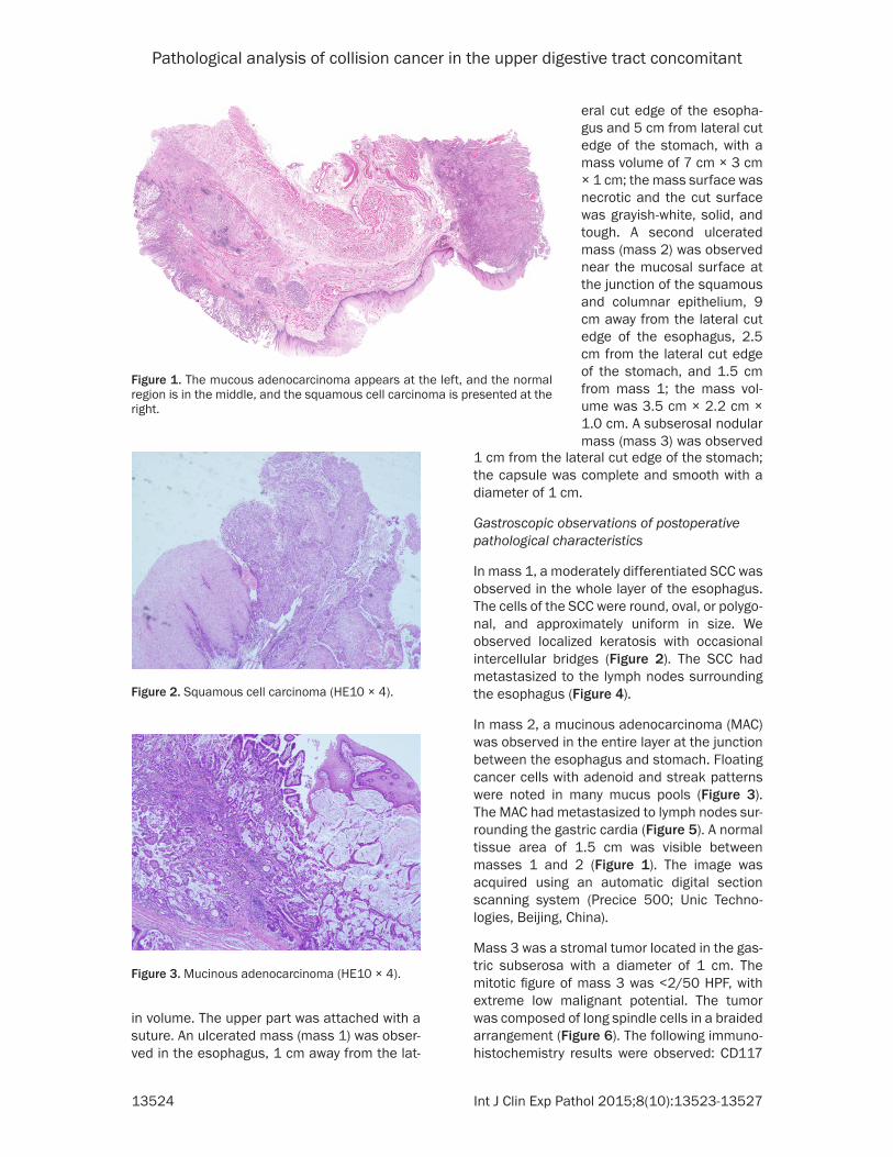
Figure 1. The mucous adenocarcinoma appears at the left, and the normal region is in the middle, and the squamous cell carcinoma is presented at the right.
Figure 2. Squamous cell carcinoma (HE10 × 4).
Figure 3. Mucinous adenocarcinoma (HE10 × 4).
1 cm from the lateral cut edge of the stomach; the capsule was complete and smooth with a diameter of 1 cm.
Gastroscopic observations of postoperative pathological characteristics
In mass 1, a moderately differentiated SCC was observed in the whole layer of the esophagus. The cells of the SCC were round, oval, or polygo-nal, and approximately uniform in size. We observed localized keratosis with occasional intercellular bridges (Figure 2). The SCC had metastasized to the lymph nodes surrounding the esophagus (Figure 4).
In mass 2, a mucinous adenocarcinoma (MAC) was observed in the entire layer at the junction between the esophagus and stomach. Floating cancer cells with adenoid and streak patterns were noted in many mucus pools (Figure 3). The MAC had metastasized to lymph nodes sur-rounding the gastric cardia (Figure 5). A normal tissue area of 1.5 cm was visible between masses 1 and 2 (Figure 1). The image was acquired using an automatic digital section scanning system (Precice 500; Unic Techno- logies, Beijing, China).
Mass 3 was a stromal tumor located in the gas-tric subserosa with a diameter of 1 cm. The mitotic figure of mass 3 was <2/50 HPF, with extreme low malignant potential. The tumor was composed of long spindle cells in a braided arrangement (Figure 6). The following immuno-histochemistry results were observed: CD117

Pathological analysis of collision cancer in the upper digestive tract concomitant
13525 Int J Clin Exp Pathol 2015;8(10):13523-13527
(+), Dog-1 (+), CD34 (+), Ki-67 (+, <1%), S-100 (-), Desmin (-), and SMA (-) (Figures 7-9). For histo-logical observation, raw materials and speci-mens were subject to hematoxylin and eosin
staining and Envision immunohistochemical staining, and labeled with CDll7, Dog-1, CD34, SMA, Desmin, S-100, and antibodies. Immuno- histochemistry reagents were obtained from
Figure 4. Lymph node metastases of squamous cell carcinoma (HE10 × 4).
Figure 5. Lymph node metastatic of mucinous ad-enocarcinoma (HE10 × 4).
Figure 6. Gastrointestinal stromal tumor (HE10 × 10).
Figure 7. Gastrointestinal stromal tumor shows CD117 (+) (10 × 20).
Figure 8. Gastrointestinal stromal tumor shows Deg-1 (+) (10 × 20).
Figure 9. Gastrointestinal stromal tumor shows CD34 (+) (10 × 20).

Pathological analysis of collision cancer in the upper digestive tract concomitant
13526 Int J Clin Exp Pathol 2015;8(10):13523-13527
the S-P kit (Maixin, Fuzhou, China). Grading and prognostic evaluation were performed follow-ing the postoperative risk grading standards for primary stromal tumors after resection, as described by Joensuu et al. [2] and the Chinese Consensus on Diagnosis and Treatment of Gastrointestinal Stromal Tumor (2013 edition) [3].
Discussion
Although esophageal and gastric cardiac can-cers are both common malignant tumors, dou-ble primary esophageal and gastric cardiac cancer is relatively rare and its incidence has been reported to be 0.08-0.87% in China [4, 5]. A double primary cancer concomitant with GST is even less common, with no reports available in China or other countries. The definition of multiple primary cancers was proposed by Warren and Gates in 1932 [6], which refers to the simultaneous or successive development of two or more primary malignant tumors in the same or different tissues or organs of an indi-vidual (excluding metastatic cancer). Simul- taneous multiple primary cancer is also known as synchronous cancer, which means that the first and second tumors are diagnosed simulta-neously or within a diagnostic interval of 6 months. If the diagnostic interval is >6 months, the tumor is defined as metachronous cancer [7]. The patient reported here met the diagnos-tic criteria of simultaneous multiple primary cancer, namely, a double primary cancer. Esophageal SCC occurred at the lower esopha-gus and metastasized to the lymph nodes sur-rounding the esophagus, while MAC occurred at the gastric cardia and metastasized to lymph nodes around the gastric cardia in this patient. Moreover, there was a normal tissue area of 1.5 cm between the esophageal SCC and MAC, excluding the possibility of metastasis and infil-tration and indicating two independent primary lesions. The incidence of esophageal cancer concomitant with gastric cancer is higher in men than in women, and the cardia is the most common region for gastric cancer [8, 9]. In this case, gastric cancer was observed in the gas-tric cardia, which was consistent with previous literature. Furthermore, a subserosal nodular mass with a diameter of 1.0 cm diameter and located 1 cm away from the lateral cut edge of the stomach was confirmed as a stromal tumor by immunohistochemistry. Stromal tumors are
among the most common mesenchymal tumors of the gastrointestinal tract. It is generally believed that stromal tumors originate from Cajal cells in the gastrointestinal tract or mes-enchymal stem cells, with mutations in the KIT or PDGFRA genes [10].
There are few reports of stromal tumors con-comitant with gastrointestinal cancer in the world. Currently, the largest cohort study report-ed in the literature was a retrospective study of 228 cases by Agaimy et al. [11]. With recent developments of diagnostic technology, there has been attention from domestic and interna-tional scholars focusing on the diagnosis and treatment of gastrointestinal stromal tumor, and therefore, reports of gastrointestinal can-cers complicated with GST are gradually inc- reasing. There are mixed views with regard to the prognostic impact of such cancers. Liu et al. [12] suggested that the prognosis of patients with gastric cancer concomitant with GST main-ly depends on the gastric cancer itself and the concomitant GST. A concomitant GST that is usually observed during intraoperative or post-operative pathological examination has a small diameter, low Fletcher grade, low recurrence rate after complete resection, and overall good prognosis, and therefore, does not have a large influence on the patients’ prognosis. The main cause of poor overall prognosis for these patients is mostly concomitant gastric cancer observed during the progressive stage, which has an overall low long-term survival rate and poor prognosis, which has a greater influence on the prognosis of patients with gastric cancer concomitant with GST. However, Lee et al. [13] believed that the prognosis for patients with GST is relatively poor if it is concomitant with gastric cancer, regardless of the Fletcher grade. In patients with GST concomitant with gastroin-testinal cancer, the stromal tumors have short diameters and often occur in the serosa. These tumors are difficult identified preoperatively and therefore, they are mostly discovered dur-ing intraoperative or postoperative pathological examination of the gastric cancer. Additionally, concomitant GST can be easily missed in post-operative diagnosis. Therefore, it is necessary to prompt the surgeon and pathologist to exam-ine the entire primary disease organ carefully and other abdominal organs intraoperatively or postoperatively to prevent misdiagnosis.

Pathological analysis of collision cancer in the upper digestive tract concomitant
13527 Int J Clin Exp Pathol 2015;8(10):13523-13527
Disclosure of conflict of interest
None.
Address correspondence to: Dr. Xiangwei Meng, De- partment of Gastroenterology, First Hospital of Jilin University, 71 Xinmin Street, Changchun 130021, China. E-mail: [email protected]
References
[1] Xie J, Wang LS, Meng F, et al. Missed Diagnosis and Prevention of Multicentric Tumors of Up-per Alimentary Tract-Report of 28 Patients. Chinese Journal of Clinical Oncology and Reha-bilitation 1998; 5: 43-45.
[2] Joensuu H. Risk stratification of patients diag-nosed with gastrointestinal stromal tumor. Hum Pathol 2008; 39: 1411-9.
[3] COCS gastrointestinal stromal tumor Expert Committee. The consensus of diagnose and treatment for gastrointestinal stromal tumors in China. Chinese Clinical Oncology 2013; 18: 1030-1037.
[4] Ruose S, Qishan W. Clinical analysis of 32 cas-es of esophageal and gastric multiple primary carcinoma. China Journal of Endoscopy 2004; 10: 67-68.
[5] Guan FS, Liu ZC, Zhao XJ, et al. Early diagnosis and treatment of multi-primary esophageal cancer and double primary esophageal and cardiac cancer. Chin J Thorac Cardiovasc Surg 2003; 19: 80-81.
[6] Warren S, Gates 0. Multiple primary malignant tumors: a survey of the literature and a statisti-cal study. Am J Cancer 1932; 16: 1358-1414.
[7] Natsugoe S, Matsumoto M, Okumura H, Ishi-gami S, Uenosono Y, Owaki T, Takao S, Aikou T. Multiple primary carcinomas with esophageal squamous cell cancer: clinicopathologic out-coule. Wodd J Surg 2005; 29: 4649.
[8] Kato H, Tachimori Y, Watanabe H, Mizobuchi S, Igaki H, Yamaguchi H, Ochiai A. Esophageal carc inoma simultaneously associated with gastric carcinoma: Analysis of clinicopatholog-ic feaures and treatments. J Surg Oncol 1994; 56: 122-127.
[9] Hamabe Y, Ikuta H, Nakamura Y, Kawasaki K, Yamamoto M. Clinicopathological features of esophageal cancer simultaneously associated with gastric cancer. J Surg Oncol 1998; 68: 179-182.
[10] Lasota J, Miettinen M. KIT and PDGFRA muta-tions in gastrointestinal stromal tumors (GIST). Semin Diagn Pathol 2006; 23: 91-102.
[11] Agaimy A, Wunsh PH, Sobin LH, Lasota J, Miet-tinen M. Occurrence of other malignancies in Patients with gastrointestinal stromal tumors. Semin Diagn Pathol 2006; 23: 120-9.
[12] Liu XL, Wang JB, Huang CM, Zheng CH, Li P, Xie JW, Lin JX. Clinicopathologic features and prog-nostic factors of gastric gastrointestinal stro-mal with synchronous gastric cancer. Zhong-hua Wei Chang Wai Ke Za Zhi 2012; 15: 247-250.
[13] Lee FY, Jan YJ, Wang J, Yu CC, Wu CC. Synchro-nous gastric gastrointestinal stromal tumor and signet-ring cell adenocarcinoma: a case report. Int J Surg Pathol 2007; 15: 397-400.