case report open access the fabella syndrome - a rare
TRANSCRIPT

Driessen et al. BMC Musculoskeletal Disorders 2014, 15:100http://www.biomedcentral.com/1471-2474/15/100
CASE REPORT Open Access
The fabella syndrome - a rare cause ofposterolateral knee pain: a review of theliterature and two case reportsArne Driessen1,2, Maurice Balke2, Christoph Offerhaus3, William James White4, Sven Shafizadeh2, Christoph Becher5,Bertil Bouillon2 and Jürgen Höher6*
Abstract
Background: The purpose of this article was to evaluate the risks and benefits of non-operative treatment versussurgical excision of a fabella causing posterolateral knee pain. We performed a systematic review of literature andalso present two case reports.Twelve publications were found in a PubMed literature review searching the word “fabella syndrome”. Non-operativetreatment and surgical excision of the fabella has been described.
Case presentation: Two patients presented to our outpatient clinic with persisting posterolateral knee pain. In bothcases the presence of a fabella was identified, located in close proximity to the posterolateral femoral condyle. All othercommon causes of intra- and extra articular pathologies possibly causing the posterolateral knee pain were excluded.Following failure to respond to physiotherapy both patients underwent arthroscopy which excluded other possiblecauses for posterolateral knee pain. The decision was made to undertake surgical excision of the fabella in both caseswithout complication.Both patients were examined 6 month and one year after surgery with the Tegner activity score, the Visual AnalogueScale (VAS), and International Knee Documentation Committee Score (IKDC).
Conclusion: Consistent posterolateral pain during exercise might indicate the presence of a fabella syndrome.Resecting the fabella can be indicated and is a minor surgical procedure with minimal risk. Despite good results in theliterature posterolateral knee pain can persist and prevent return to a high level of sports. Level of evidence: IV, casereports and analysis of literature.
Keywords: Fabella syndrome, Posterolateral knee pain, Fabellectomy, Sesamoid bone, Return to sports,Review of literature
BackgroundThe fabella is a sesamoid bone in the posterolateral capsuleof the human knee joint. The presence of the fabella inhumans varies widely and is reported in the literature torange from 20% to 87% [1-7].The fabella is located in the posterior aspect of the
knee where lines of tensile stress intersect.
* Correspondence: [email protected] of Traumatology, Clinic for Sports Traumatology, OrthopaedicSurgery and Sports Traumatology, Cologne-Merheim Medical Centre (CMMC),University of Witten/Herdecke (Campus Cologne-Merheim), Ostmerheimerstrasse200, 51109 Köln, GermanyFull list of author information is available at the end of the article
© 2014 Driessen et al.; licensee BioMed CentraCommons Attribution License (http://creativecreproduction in any medium, provided the orDedication waiver (http://creativecommons.orunless otherwise stated.
It articulates with the posterior part of the articularsurface of the lateral femoral condyle and is embeddedin the muscular fibres of the gastrocnemius muscle [5].Anteriorly the fabella is bordered by the posterior
capsule of the knee joint and posteriorly it is situated atthe endpoint of the oblique popliteal ligament and the lat-eral gastrocnemius tendon. In addition the fabellofibularligament (or lig. of Vallois) runs to its distal insertion atthe fibular head.Recent anatomic studies suggest that the presence of a
fabella is higher in the Asian population [3,4,6].Functionally, the fabella is believed to have a role similar
to the patella in redirecting extension forces of the knee
l Ltd. This is an Open Access article distributed under the terms of the Creativeommons.org/licenses/by/2.0), which permits unrestricted use, distribution, andiginal work is properly credited. The Creative Commons Public Domaing/publicdomain/zero/1.0/) applies to the data made available in this article,
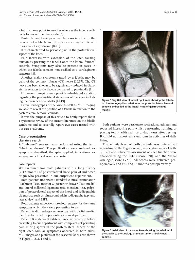
Figure 1 Sagittal view of lateral right knee showing the fabellain close topographical relation to the posterior lateral femoralcondyle embedded in the lateral head of gastrocnemiusmuscle.
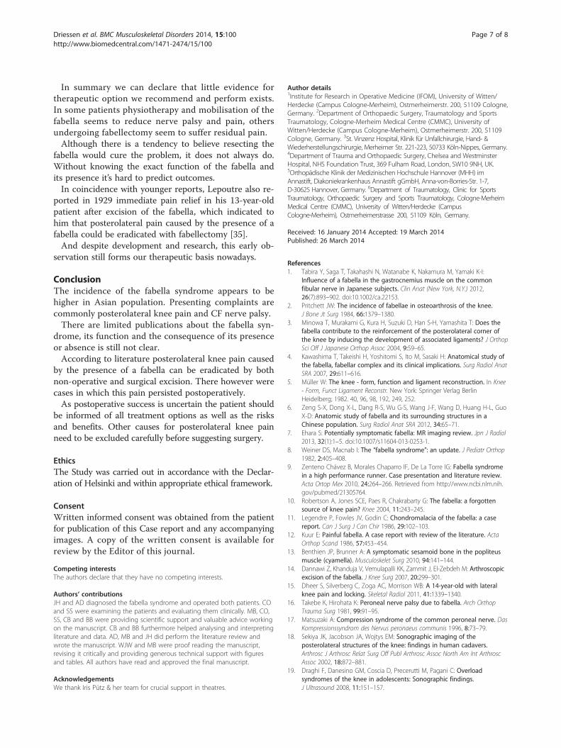
Figure 2 Axial view of the same knee showing the relation ofthe fabella to the cartilage of the posterior lateral femoralcondyle.
Driessen et al. BMC Musculoskeletal Disorders 2014, 15:100 Page 2 of 8http://www.biomedcentral.com/1471-2474/15/100
joint from one point to another whereas the fabella redi-rects forces on the flexor side [5].Posterolateral knee pain can be associated with the
presence of a fabella and this incidence may be referredto as a fabella syndrome [8-15].It is characterized by periodic pain in the posterolateral
aspect of the knee.Pain increases with extension of the knee causing
tension by pressing the fabella onto the lateral femoralcondyle. Symptoms may also be present in cases inwhich the fabella remains non ossified as a cartilaginousstructure [8].Another major symptom caused by a fabella may be
palsy of the common fibular (CF) nerve [16,17]. The CFnerve has been shown to be significantly reduced in diam-eter in relation to the fabella compared to proximally [1].Ultrasound imaging may provide valuable information
regarding the posterolateral structures of the knee includ-ing the presence of a fabella [18,19].Lateral radiographs of the knee as well as MRI Imaging
are able to reveal the position of a fabella in relation to theposterolateral femoral condyle.It was the purpose of this article to firstly report about
a systematic review of the current literature on the fabellasyndrome and to secondly report two cases treated withthis rare syndrome.
Case presentationLiterature searchA “pub med” research was performed using the term“fabella syndrome”. The publications were analysed forsymptoms described, therapies applied, indications forsurgery and clinical results reported.
Case reportsWe examined two male patients with a long history(> 12 month) of posterolateral knee pain of unknownorigin who presented in our outpatient department.Both patients underwent standard clinical examination
(Lachman Test, anterior & posterior drawer Test, medialand lateral collateral ligament test, mensicus test, palpa-tion of posterolateral aspect of the knee) and radiographicdiagnostics such as ultrasound, plain radiographs (a.p. andlateral view) and MRI.Both patients underwent previous surgery for the same
symptoms which they were presenting to us.Patient A did undergo arthroscopy with partial medial
meniscectomy before presenting at our department.Patient B underwent bilateral knee arthroscopy before
presenting to our department with complaints of persistingpain during sports in the posterolateral aspect of theright knee. Similar symptoms occurred in both sides.MRI images and pictures of the resected fabella are shownin Figure 1, 2, 3, 4 and 5.
Both patients were passionate recreational athletes andreported increasing pain whilst performing running orplaying tennis with pain resolving hours after resting.Both did not report any symptoms in activities of dailyliving.The activity level of both patients was determined
according to the Tegner score (preoperative value of both:3). Pain and subjective assessment of knee function wereanalysed using the IKDC score [20], and the VisualAnalogue score (VAS). All scores were delivered pre-operatively and at 6 and 12 months postoperatively.

Figure 3 Resected fabella with cartilaginous surface.Figure 5 Sagittal view after 3 month after resection of thefabella; intact posterolateral capsule & gastrocnemius musclewith little scar tissue.
Driessen et al. BMC Musculoskeletal Disorders 2014, 15:100 Page 3 of 8http://www.biomedcentral.com/1471-2474/15/100
ResultsLiterature reviewThe review of the literature searching the PubMed onlinedata revealed five case reports and three studies with morethan 10 patients in each paper [8-10,12,14,16,17,21].The findings and outcome results of reported patients
with the treatment of a fabella syndrome are summarizedin Table 1.Weiner et al. described the largest number of patients
(n = 16) treated for pain associated with the presence ofa fabella.According to their publication five patients responded
to conservative treatment. Eleven patients required surgery,
Figure 4 Resecting the fabella through a lateral incisionbetween M. biceps femoris, iliotibial band and posterolateralfemoral condyle.
all of which obtained immediate relief of symptoms withremoval of the ossified fabella, cartilaginous fabella, orthickened gastrocnemius fibres [8].Dannawi et al. presented two cases of symptomatic
pain caused by the presence of a fabella. Both underwentarthroscopic resection and are reported to suffer no pain12 month after surgery [14].Müller also described an interesting case that undermines
the theory of the fabella forming in response to appropriatestress. An 18-year-old female presented to their departmenthaving sustained a complex knee injury. Radiographs wereobtained which showed no evidence of a fabella. Thepatient was treated non-operatively. Several years laterfollowing further presentation to the department forongoing symptoms further imaging was obtained. Theseshowed evidence of a fabella which had not previouslybeen present at first review [5].
Common fibular nerve palsyTwo reports from Japan were found describing the nervepalsy in patients as the major symptom for the fabellasyndrome.Takebe reported in 1981 about seven patients suffering
from common fibular nerve palsy due to compressionfrom fabella. Three cases were treated by surgery and fourcases by conservative methods [16]. Surgery was performedmore than 1,5 month after onset of symptoms. All threepatients treated with surgical excision of the fabella hadpreoperative sensory neuropathies and one patient inaddition a foot drop, which was originally thought tobe the result of a lumbar disc herniation. All patientsrecovered after surgery with one patient regaining sensoryfunction the day after surgery. Furthermore Takebe reports

Table 1 Details derived from six case reports found through PubMed research
Weiner, D.S. 1982 [27] Kuur, E. 1986 [12] Zipple, J.2003 [31] Robertson, A. 2004 [22] Dannawi, Z. 2007 [2] Zenteno, B. 2010 [30]
(n) number of patients &(morphology of fabella)
16 (9 bony & 2 cartilaginous,5 thickened fibres ofgastrocnemius muscle
1 1 (bony) 1 (bony) 2 (bony) 1 (bony)
Symptoms described inthe text
Pain in extension of the knee,could be reproduced bydirect pressure
3-4 year history ofintermittent pain andslight swelling, painfulknee extension, painstarted after specialintensive training
Chief complaint: leftposterolateral pain,weakness and footdrop symptoms
2 month history of pain andswelling in posterolateralregion of the right kneeassociated with a clickingsensation symptoms worsewhile squatting, stair ascent& descent
Case 1: sharp intermittentpain posterolateral knee,exacerbated & catchingin extension case 2: pain& swelling posterolateralknee
Pain while running morethan 2 km; posterolateralpain
Level of activity Not described Active soccer player Lifetime habit ofroutine engagementin vigorous exercise5–6 times per week
65 years old, no descriptionabout sports
58 year old women &45 year old man, notdescribed
High performance runner
(n) non-surgical treatment Injections of steroid,immobilization with splinting& casting, restriction of activity,analgesics
Temporary restrictionof activities, injectionof steroid andanti-inflam- matorymedication
1; mobilisation ofthe pisiform bonein the wrist
1; ultrasound guidedinjection of cortisone &local anaesthetic
Case 1: physical therapyfor 1 year with remainingpain case 2: NSAID, noinjection
Multiple conservativetreatment whichfailed; ozone therapy,physiotherapy, ultrasound
(n) surgery 11 1 No No 2 1
Follow up 0,5 y – 7 y 2,5 years 16 month 12 month 12 & 18 month 11 month
Symptoms & problemsafter procedure
12 pain free (surgical treatment);1 minimal periodic pain, 2periodic pain, 1 significantperiodic pain (non surgicaltreatment); 5, injections ofsteroid, immobilization withsplinting & casting,restriction ofactivity, analgesics
Work & sportswithout pain
None No symptoms 12 monthafter intervention
Case 1: pain subsided,pain free 12 month afterprocedure case 2: no pain
None
Activity after surgery No report No report Full 0 (0–10) No report No report High, international levelcompetitions, participantin Olympic games 200811 month after surgery
Nerve palsy - - No report -
Driessen
etal.BM
CMusculoskeletalD
isorders2014,15:100
Page4of
8http://w
ww.biom
edcentral.com/1471-2474/15/100

Driessen et al. BMC Musculoskeletal Disorders 2014, 15:100 Page 5 of 8http://www.biomedcentral.com/1471-2474/15/100
that ankle dorsiflexion steadily became stronger in thepatient with the motor disfunction symptoms. Althoughsome patients from the conservative treatment grouphad pathological electrophysiological examinations theywere not operated. Takebe recommends conservativemethods first as he observed sufficient recovery withinthis group, despite the underlying neuropathies provenon electrophysiological studies [16].Matsuzaki et al. described in a book chapter nerve
compression in 112 patients from constricting fascia orfabella of whom 19 were operated and followed up to anaverage of 4 years. Pain and dysaesthesia disappeared inall cases [17].
Anatomical studies of the fabella complex and therelation to the common fibular nerveMüllers description of the anatomical structures adjacentto the fabella and its function are fortified by a group ofthe following Japanese anatomists [5].Kawashima et al. [4] studied the fabella and its surround-
ing structures in 75 knees (150 heads of the gastrocnemiusmuscle) from 39 Japanese cadavers. They observed 99fabellae (66.0%) including 44 complete bony fabellae(29.3%). Of these bony fabellae, 43 (97.7%) were located inthe lateral head of the gastrocnemius muscle with itssurrounding structures and were positioned only on the lat-eral condyle of the femur. Furthermore the bony fabellaeand the cartilage formed small articular cavity by cooper-ating with the femoral condyle. Their suggestion is thatthe fabella may play an important role in stabilizing thefabella complex and the femoral condyle [4].Another anatomical study was published in 2012 in-
vestigating the role of the fabella and the relation to thecommon fibular nerve.Their study describes the presence of a bony or cartilage
fabella in 86,9% in the lateral head of the gastrocnemiusmuscle in a Chinese population [6].They dissected 61 formalin fixed specimen to record
the relationship between the common peroneal nerve &the fabella.The anatomical relation of the fabella to the CF nerve
and the head of the gastrocnemius muscle are investigatedin 102 knees of 51 Japanese cadavers by Tabira et al. [1].The presence of a fabella was observed in 70 knees (68.6%).There was a significant difference in the thickness andthe width of the CF nerve adjacent to a fabella thanproximal to the region. The increase in width of the CFnerve was greater for knees with fabellae weather boneor cartilage than for knees with absent fabellae. The CFnerve in the bony area directly adjacent to the fabellawas noted to be thinner than in the absent fabellagroup. Furthermore Tabira et al. describe difference inthickness of the CF nerve between the bony and cartilagefabella.
Miscellaneous publicationsErichsen et al. [22] described a case of a 68-year-oldwoman who underwent total knee replacement surgeryon both knees and developed local tenderness and painfulclicking in the posterolateral aspect of the knee approxi-mately one year after arthroplasty. The lateral radiographof the knee revealed a large fabella impinging on theprosthesis. The symptoms and signs were alleviated after abilateral surgical excision of the fabella.According to their experience Larson et al. [23] rec-
ommended provident excision of the fabella through theanterior approach during total knee replacement due topossible impingement.Several other reports such as a dislocation of a fabella
causing catching problems is described by Frey et al. [24] aswell as posttraumatic osteoarthritic changes of the fabellacausing problems could be found searching PubMed [25].Ehara [7] describes in his recent published article in
which he reviewed routine MRI of the knee of 653 casesthe incidence of osteocartilaginous degeneration localizedin the fabellofemoral joint in patients with osteoarthriticchanges as rare (1,1%; 7 out of 623 patients mean age64 years in Japanese population).Literature reveals one case of a stress fracture in fabella
[26]. Rare as well but also described, as accompanyingmore complex traumatic fracture pattern of the knee jointare fractures of the fabella [27].Even though the fabella syndrome is seldom, one descrip-
tion of a case could be found, in which the posterolateralknee pain caused by an intra articular osteoid osteoma withthe presence of a fabella was mistaken for a painful fabellasyndrome [28].
Case reportsOf the two patients presented in outpatients with persist-ing posterolateral knee pain, both showed the presence ofa fabella adjacent to the posterolateral femoral condyle.Standard examination and radiographic imaging suchas X-Ray and MRI could exclude other intra- and extraarticular pathologies possibly causing pain.We found the clinical examination very much beneficial
to fortify the diagnosis as both patients presented with thetypical symptoms such as pain while extending the kneeand pain that could be reproduced by direct pressure onthe fabella.In addition and correlating with the literature both
reported having pain while doing exercise but havinglittle or no pain during all day activities [8,9,12,21].Both patients underwent surgical excision of the fabella
(patient A unilateral, patient B bilateral) through a dorsolat-eral incision between the biceps muscle and the iIiotibialband after having had unsatisfying reduction of pain underphysiotherapy. The fabellae were resected and closure oflayers was performed in standard procedure [29-31]. Other

Driessen et al. BMC Musculoskeletal Disorders 2014, 15:100 Page 6 of 8http://www.biomedcentral.com/1471-2474/15/100
possible causes for posterolateral pain were excludedarthroscopically prior to this.The results of the different scores are shown in Table 2
showing an improvement in activity and reduction ofpain.Patient A reported to have persistent pain six month
after surgery without being back to the same level ofsports. Investigations couldn’t reveal any obvious reasonfor these problems. But significantly important both re-ported to be finally pain free at 12 months.
DiscussionSince Pancoast stated in 1909 that the presence of thefabella “has apparently little or no interest beyond the merefact of its occasional occurrence” more than hundred yearspassed by [32].Within these years science developed new methods
trying to describe and understand the function of severalbones of the body – also of the fabella.In comparison to other anatomical structures, little
interest has been paid to this sesamoid bone that isrelatively common – and in most cases is a incidentalfinding on imaging.This would support the theory of Müller that the fabella
does play an important role as a structure where lines oftensile stress interact. Its presence might be explainedfunctioning as a sesamoid structure redirecting tensileforces [5].In his review of knee MRI of 653 patients of a Japanese
population 200 (31%; age range 4–89, median 61) had afabella. Ehara noted that the pure existence in anatomicalstudies is higher which might be explained with thehigher age of the cadavers (age range 64 – 83, no meanage given) [7].The increased incidence in Asian population might
refer to different habits according to kneeling and squattingand therefore increased tensile forces on the flexor side ofthe knee. But the reason - if so - remains unclear.As well the reports existing show an increased incidence
in young active athletes without evidence for a specificsport [9,12,14,21]. The occurrence of problems in thesepatients might be explained with increased tensile forceson the posterior knee as well. But it remains unclear whysome develop problems and others don’t.According to our clinical experience the examination
of patients with posterolateral knee pain should includepalpation of the posterolateral structures as well as
Table 2 Results of scores
Preop (A) Preop (B) 6 month (A
IKDC 66,7 59,8 75,9
VAS 10,7 (1,7/6,8/2,2) 17,5 (5,3/8,0/4,2) 22,3 (6,3/7,9/8
Tegner 3 3 4
ultrasound examination. If specific pain can be reproducedby applying pressure on the fabella, it would be a clearindication for the fabella being the underlying cause.Other extra- and intra-articular causes for posterolateral
knee pain such as Baker’s cyst, foreign bodies, meniscaltears, localized pigmented villonodular synovitis andosteochondral fragments need to be excluded. Withhistory of trauma ligamentous instability, tibiofibularjoint mobility and fracture of fabella should be consid-ered [10,12,31,33].Injecting local anaesthetic and steroid for diagnostic
and therapeutic purpose should be performed as firstintervention.In the majority of the few reported case particularly
physically active patients are most likely to develop pos-terolateral knee pain caused by the fabella [9,12,21].The problems occurring are described to be a quartet of
symptoms such as “intermittent posterolateral mechanicalknee pain, pain accentuation by full knee extension, local-ized tenderness with compression of the fabella againstits corresponding condylar surface, and immediate andpersistent relief with fabellectomy” [34].This quartet introduced by Weiner in 1977 confirms
the indication for surgery after already having performedit due to an eradication of pain following excision. Wewould therefore postulate that.Approaching the surgical pathway should include an
ultrasound-guided injection of local anaesthetic andsteroids for both diagnostics and therapeutics.Reviewing literature revealed CF nerve palsy in some
patients, which could be cured by resecting the fabella[16,21].Anatomical dissections of the posterolateral knee show
a close topographical relation [16,17] between the poster-ior femoral condyle but as well to the CF nerve, which infact could explain both the posterolateral pain and thenerve palsy.Furthermore this would indicate that the most favourable
therapy of the mechanical compression of other structureswould be a decompression fabellectomy.But there are reports from successful conservative
treatment despite patients suffering from nerve palsy[8,16].These patients suffering from “hyper compression” pain
need to be distinguished carefully from those with mech-anical locking such in arthroplasty and those with arthriticchanges of the fabella causing pain.
) 6 month (B) 12 month (A) 12 month (B)
82,8 85,1 88,5
,1) 27,5 (8,8/9,3/9,2) 28,6 (9,5/9,1/10) 30 (10/10/10)
4 4 5
Driessen et al. BMC Musculoskeletal Disorders 2014, 15:100 Page 7 of 8http://www.biomedcentral.com/1471-2474/15/100
In summary we can declare that little evidence fortherapeutic option we recommend and perform exists.In some patients physiotherapy and mobilisation of thefabella seems to reduce nerve palsy and pain, othersundergoing fabellectomy seem to suffer residual pain.Although there is a tendency to believe resecting the
fabella would cure the problem, it does not always do.Without knowing the exact function of the fabella andits presence it’s hard to predict outcomes.In coincidence with younger reports, Lepoutre also re-
ported in 1929 immediate pain relief in his 13-year-oldpatient after excision of the fabella, which indicated tohim that posterolateral pain caused by the presence of afabella could be eradicated with fabellectomy [35].And despite development and research, this early ob-
servation still forms our therapeutic basis nowadays.
ConclusionThe incidence of the fabella syndrome appears to behigher in Asian population. Presenting complaints arecommonly posterolateral knee pain and CF nerve palsy.There are limited publications about the fabella syn-
drome, its function and the consequence of its presenceor absence is still not clear.According to literature posterolateral knee pain caused
by the presence of a fabella can be eradicated by bothnon-operative and surgical excision. There however werecases in which this pain persisted postoperatively.As postoperative success is uncertain the patient should
be informed of all treatment options as well as the risksand benefits. Other causes for posterolateral knee painneed to be excluded carefully before suggesting surgery.
EthicsThe Study was carried out in accordance with the Declar-ation of Helsinki and within appropriate ethical framework.
ConsentWritten informed consent was obtained from the patientfor publication of this Case report and any accompanyingimages. A copy of the written consent is available forreview by the Editor of this journal.
Competing interestsThe authors declare that they have no competing interests.
Authors’ contributionsJH and AD diagnosed the fabella syndrome and operated both patients. COand SS were examining the patients and evaluating them clinically. MB, CO,SS, CB and BB were providing scientific support and valuable advice workingon the manuscript. CB and BB furthermore helped analysing and interpretingliterature and data. AD, MB and JH did perform the literature review andwrote the manuscript. WJW and MB were proof reading the manuscript,revising it critically and providing generous technical support with figuresand tables. All authors have read and approved the final manuscript.
AcknowledgementsWe thank Iris Pütz & her team for crucial support in theatres.
Author details1Institute for Research in Operative Medicine (IFOM), University of Witten/Herdecke (Campus Cologne-Merheim), Ostmerheimerstr. 200, 51109 Cologne,Germany. 2Department of Orthopaedic Surgery, Traumatology and SportsTraumatology, Cologne-Merheim Medical Centre (CMMC), University ofWitten/Herdecke (Campus Cologne-Merheim), Ostmerheimerstr. 200, 51109Cologne, Germany. 3St. Vinzenz Hospital, Klinik für Unfallchirurgie, Hand- &Wiederherstellungschirurgie, Merheimer Str. 221-223, 50733 Köln-Nippes, Germany.4Department of Trauma and Orthopaedic Surgery, Chelsea and WestminsterHospital, NHS Foundation Trust, 369 Fulham Road, London, SW10 9NH, UK.5Orthopädische Klinik der Medizinischen Hochschule Hannover (MHH) imAnnastift, Diakoniekrankenhaus Annastift gGmbH, Anna-von-Borries-Str. 1-7,D-30625 Hannover, Germany. 6Department of Traumatology, Clinic for SportsTraumatology, Orthopaedic Surgery and Sports Traumatology, Cologne-MerheimMedical Centre (CMMC), University of Witten/Herdecke (CampusCologne-Merheim), Ostmerheimerstrasse 200, 51109 Köln, Germany.
Received: 16 January 2014 Accepted: 19 March 2014Published: 26 March 2014
References1. Tabira Y, Saga T, Takahashi N, Watanabe K, Nakamura M, Yamaki K-I:
Influence of a fabella in the gastrocnemius muscle on the commonfibular nerve in Japanese subjects. Clin Anat (New York, N.Y.) 2012,26(7):893–902. doi:10.1002/ca.22153.
2. Pritchett JW: The incidence of fabellae in osteoarthrosis of the knee.J Bone Jt Surg 1984, 66:1379–1380.
3. Minowa T, Murakami G, Kura H, Suzuki D, Han S-H, Yamashita T: Does thefabella contribute to the reinforcement of the posterolateral corner ofthe knee by inducing the development of associated ligaments? J OrthopSci Off J Japanese Orthop Assoc 2004, 9:59–65.
4. Kawashima T, Takeishi H, Yoshitomi S, Ito M, Sasaki H: Anatomical study ofthe fabella, fabellar complex and its clinical implications. Surg Radiol AnatSRA 2007, 29:611–616.
5. Müller W: The knee - form, function and ligament reconstruction. In Knee- Form, Funct Ligament Reconstr. New York: Springer Verlag BerlinHeidelberg; 1982. 40, 96, 98, 192, 249, 252.
6. Zeng S-X, Dong X-L, Dang R-S, Wu G-S, Wang J-F, Wang D, Huang H-L, GuoX-D: Anatomic study of fabella and its surrounding structures in aChinese population. Surg Radiol Anat SRA 2012, 34:65–71.
7. Ehara S: Potentially symptomatic fabella: MR imaging review. Jpn J Radiol2013, 32(1):1–5. doi:10.1007/s11604-013-0253-1.
8. Weiner DS, Macnab I: The “fabella syndrome”: an update. J Pediatr Orthop1982, 2:405–408.
9. Zenteno Chávez B, Morales Chaparro IF, De La Torre IG: Fabella syndromein a high performance runner. Case presentation and literature review.Acta Ortop Mex 2010, 24:264–266. Retrieved from http://www.ncbi.nlm.nih.gov/pubmed/21305764.
10. Robertson A, Jones SCE, Paes R, Chakrabarty G: The fabella: a forgottensource of knee pain? Knee 2004, 11:243–245.
11. Legendre P, Fowles JV, Godin C: Chondromalacia of the fabella: a casereport. Can J Surg J Can Chir 1986, 29:102–103.
12. Kuur E: Painful fabella. A case report with review of the literature. ActaOrthop Scand 1986, 57:453–454.
13. Benthien JP, Brunner A: A symptomatic sesamoid bone in the popliteusmuscle (cyamella). Musculoskelet Surg 2010, 94:141–144.
14. Dannawi Z, Khanduja V, Vemulapalli KK, Zammit J, El-Zebdeh M: Arthroscopicexcision of the fabella. J Knee Surg 2007, 20:299–301.
15. Dheer S, Silverberg C, Zoga AC, Morrison WB: A 14-year-old with lateralknee pain and locking. Skeletal Radiol 2011, 41:1339–1340.
16. Takebe K, Hirohata K: Peroneal nerve palsy due to fabella. Arch OrthopTrauma Surg 1981, 99:91–95.
17. Matsuzaki A: Compression syndrome of the common peroneal nerve. DasKompressionssyndrom des Nervus peronaeus communis 1996, 8:73–79.
18. Sekiya JK, Jacobson JA, Wojtys EM: Sonographic imaging of theposterolateral structures of the knee: findings in human cadavers.Arthrosc J Arthrosc Relat Surg Off Publ Arthrosc Assoc North Am Int ArthroscAssoc 2002, 18:872–881.
19. Draghi F, Danesino GM, Coscia D, Precerutti M, Pagani C: Overloadsyndromes of the knee in adolescents: Sonographic findings.J Ultrasound 2008, 11:151–157.
Driessen et al. BMC Musculoskeletal Disorders 2014, 15:100 Page 8 of 8http://www.biomedcentral.com/1471-2474/15/100
20. Hefti F, Müller W, Jakob RP, Stäubli HU: Evaluation of knee ligamentinjuries with the IKDC form. Knee Surg Sports Traumatol Arthrosc 1993,1:226–234.
21. Zipple JT, Hammer RL, Loubert PV: Treatment of fabella syndrome withmanual therapy: a case report. J Orthop Sports Phys Ther 2003, 33:33–39.
22. Erichsen H: Bilateral fabellar impingement after total knee replacement-acase report. Acta Orthop Scand 1997, 68(4):403.
23. Larson JE, Becker DA: Fabellar impingement in total knee arthroplasty. Acase report. J Arthroplasty 1993, 8:95–97.
24. Frey C, Bjorkengen A, Sartoris D, Resnick D: Knee dysfunction secondary todislocation of the fabella. Clin Orthop Relat Res 1987, Sep:223–227.
25. Franceschi F, Longo UG, Ruzzini L, Leonardi F, Rojas M, Gualdi G, Denaro V:Dislocation of an enlarged fabella as uncommon cause of knee pain: acase report. Knee 2007, 14:330–332.
26. Woo CC: Fracture of the fabella. J Manipulative Physiol Ther 1988,11:422–425.
27. Heideman GM, Baynes KE, Mautz AP, DuBois MS, Roberts JW: Fabellafracture with CT imaging: a case report. Emerg Radiol 2011, 18:357–361.
28. García-Germán D, Sánchez-Gutiérrez S, Bueno A, Carballo F, López-GonzálezD, Canillas F, Martel J: Intra-articular osteoid osteoma simulating a painfulfabella syndrome. Knee 2010, 17:310–312.
29. Osti M, Tschann P, Künzel KH, Benedetto KP: Posterolateral corner of theknee: microsurgical analysis of anatomy and morphometry. Orthopedics2013, 36:e1114–e1120.
30. Charalambous CP, Kwaees TA: Anatomical considerations in hamstringtendon harvesting for anterior cruciate ligament reconstruction. MusclesLigaments Tendons J 2012, 2:253–257.
31. Kroh F: Indikationen zur Eröffnung der hinteren Kapseltaschen desKniegelenkes. Arch Orthop Unfallchir 1942, 42:95–115.
32. Pancoast HK: Radiographic statistics of the sesamoid in the tendon of thegastrocnemius. Univ Penn Med Bull 1909, 22:213.
33. Oliva F, Frizziero A: One step open synovectomy without adjuvanttherapy for diffuse pigmented villonodular synovitis of the knee in asoccer player. Muscles Ligaments Tendons J 2011, 1:36–39.
34. Weiner D, Macnab I, Turner M: The fabella syndrome. Clin Orthop Relat Res1977, Jul-Aug:213–215.
35. Lepoutre C: Sesamoide douloureux (sesamoide du jumeau externe). RevOrthop 1929, 16:234–236.
doi:10.1186/1471-2474-15-100Cite this article as: Driessen et al.: The fabella syndrome - a rarecause of posterolateral knee pain: a review of the literature andtwo case reports. BMC Musculoskeletal Disorders 2014 15:100.
Submit your next manuscript to BioMed Centraland take full advantage of:
• Convenient online submission
• Thorough peer review
• No space constraints or color figure charges
• Immediate publication on acceptance
• Inclusion in PubMed, CAS, Scopus and Google Scholar
• Research which is freely available for redistribution
Submit your manuscript at www.biomedcentral.com/submit