case report open access leser-trélat syndrome in patients
TRANSCRIPT

CASE REPORT Open Access
Leser-Trélat syndrome in patients affected by sixmultiple metachronous primitive cancersGiovanni Ponti1*, Gabriele Luppi1, Lorena Losi2, Alberto Giannetti3, Stefania Seidenari3
Abstract
Leser-Trélat syndrome is characterized by the eruptive appearance of multiple seborrheic keratoses in associationwith underlying malignant disease. Usually, the sign of Leser-Trélat is associated with adenocarcinoma, most fre-quently of the colon, breast, or stomach, but also of the lung, kidney, liver, and pancreas. Herein, we present a casethat we believe is the first report of the sign of Leser-Trélat in association with occult gastric adenocarcinoma andthe anamnestic oncologic history of five other multiple primitive cancers. Epidermal growth factor receptor (EGFR)immunohistochemical expression analysis of multiple seborrheic keratoses revealed an intense membranous stain-ing in the basal keratinocytes and in all the upper epidermal layers. Patients with the sign of Leser-Trélat shouldundergo a diagnostic screening programme for malignant disease along with patients with known Leser-Trélatsyndrome who present with a recent acute and florid eruption of their seborrheic keratoses. We propose theimportance of combining the molecular features of multiple seborrheic keratoses with EGFR immunohistochemistryanalyses to determine the likelihood of Leser-Trélat syndrome and the consequent high risk of underlying multiplevisceral malignancies.
BackgroundSkin manifestations are frequently observed in manyinternal malignancies. Cutaneous lesions, includingbenign neoplasms or pigmented lesions, can appear inthe context of specific genodermatosis [1,2], lentiginosis[3], or paraneoplastic syndrome [4] representing a cru-cial cutaneous marker of internal malignancy in boththe sporadic and genetic setting. Several familial cancersyndromes have additional malignant or non-malignantcutaneous features that form part of the clinical diag-nostic criteria constituting a critical tool for clinicaldiagnosis.Several cutaneous paraneoplastic syndromes may be
associated with underlying tumours. They include mus-culoskeletal disorders (clubbing, hypertrophic osteoar-thropathy, dermatomyositis, and multicentricreticulohistiocytosis), reactive erythemas (erythema gyra-tum repens and necrolytic migratory erythema), vasculardermatoses (Trousseau’s syndrome), papulosquamousdisorders (acanthosis nigricans, tripe palms, palmarhyperkeratosis, acquired ichthyosis, pityriasis rotunda,Bazex’s syndrome, florid cutaneous papillomatosis and
extramammary Paget’s disease), and disorders of hairgrowth (hypertrichosis lanuginosa acquisita) (Table 1)[4]. The clinical manifestations of these dermatoses mayprecede, coincide with, or follow the diagnosis of cancer.Most paraneoplastic dermatoses disappear when the pri-mary tumour is removed and reappear in the case ofrecurrence or metastases of the cancer.The sign of Leser-Trélat is characterized by the erup-
tive appearance of multiple seborrheic keratoses (SK) inassociation with underlying malignant disease. Two Eur-opean surgeons, Edmund Leser and Ulisse Trélat, werethe first to describe the sign, and although they gave nomention of the presence of seborrheic keratoses report-ing a specific association with angiomas - that probablydoes not exist - their names continue to be linked to adifferent paraneoplastic association [5]. In 1900, a subse-quent report clearly defined the association of sebor-rheic keratosis and cancer [6]. Usually, the sign ofLeser-Trélat is associated with adenocarcinoma, mostfrequently of the colon, breast, or stomach, but also ofthe kidney, liver, and pancreas, among others [7,8].Endogenous mediators of hyperproliferative skin dis-
ease such as epidermal growth factor (EGF), transform-ing growth factor-alpha (TGF-a), or amphiregulin thatmay function in a localized autocrine manner or be
* Correspondence: [email protected] of Oncology and Haematology, University of Modena andReggio Emilia, Modena, Italy
Ponti et al. Journal of Hematology Oncology 2010, 3:2http://www.jhoonline.org/content/3/1/2 JOURNAL OF HEMATOLOGY
& ONCOLOGY
© 2010 Ponti et al; licensee BioMed Central Ltd. This is an Open Access article distributed under the terms of the Creative CommonsAttribution License (http://creativecommons.org/licenses/by/2.0), which permits unrestricted use, distribution, and reproduction inany medium, provided the original work is properly cited.
found in the circulation affecting the epidermis morediffusely, may induce some seborrheic keratoses [9,10].Herein, we present a case that we believe is the first
report of Leser-Trélat syndrome in association withoccult gastric adenocarcinoma and the anamnesticoncologic history of five other multiple primitivecancers.
Case presentationA 83-year-old man was admitted to our hospital inAugust 2009 with a recent history of dyspnea, coughand dyspepsia. His medical history was characterized bysigmoidectomy and ileal-cecal resection for two syn-chronous adenocarcinomas (T1N0M0) in August 2008;localized adenocarcinoma of the prostate diagnosed in2007 treated with hormonotherapy and radiotherapywith radical intent; cutaneous basal cell carcinoma andsquamous cell carcinoma removed about 10 yearsbefore. For the past 25 years, he had noted multipleasymptomatic lesions, diagnosed as multiple seborrheickeratosis, of the face and trunk and these had recentlyincreased in size and number with generalized pruritis.On admission, dermatologic examination noted florideruption of innumerable keratotic lesions of the face,neck and thorax, ranging in size from 3 to 15 mm (Fig-ure 1). Histopathological examination of a surgical skinbiopsy confirmed the clinical diagnosis of seborrheickeratosis and was used for EGFR immunohistochemicalanalysis (Figure 2). Immunoperoxidase staining usingdiaminobenzidine as chromogen was run with theBenchmark XT automatic staining system (VentanaMedical Systems Inc, Strasbourg, France). Mouse
monoclonal antibody anti-EGFR (clone 31G7; Ventana,Strasbourg, France) was used at a dilution of 1:100.In July 2009, computed tomography (CT) of the chest
and abdomen revealed a 2.5 by 2.0-cm solid mass in theupper lobe of his right lung. Diagnosis of gastric adeno-carcinoma was obtained at histological analysis by lungbiopsy which revealed signet ring cells, and at clinicalevaluation of his gastric symptoms (gastric pain, dyspep-sia). Contemporaneously, due to haematologic pancyto-penia, the patient was studied by bone marrow biopsy(BOM) that revealed a diffuse infiltration of adenocarci-noma with signet ring cells at histological analysis. Thepatient died of disease progression in September 2009.The patient’s sister had died of neck cancer at the age
of 50 years. There was no history of malignancies in thepatient’s mother, father or other first-degree relatives.
DiscussionThe clinical association of florid eruption of seborrheickeratoses and diagnosis of malignancy clearly identifyLeser-Trélat syndrome. Heaphy et al. [11] suggest that itwould be useful to distinguish between a “sign of Leser-Trélat” and a “syndrome of Leser-Trélat.” They proposethat the “sign of Leser-Trélat” be defined as a suddenacute efflorescence of seborrheic keratoses sometimesaccompanied by pruritus or acanthosis nigricans (orboth). According to this definition, the sign may be pre-sent with or without occult malignancy and is detectableon history and physical examination alone. The term“syndrome of Leser-Trélat” would then be used todescribe a paraneoplastic syndrome in patients with the“sign of Leser-Trélat” in whom an occult malignancywas identified after the appearance of the sign.
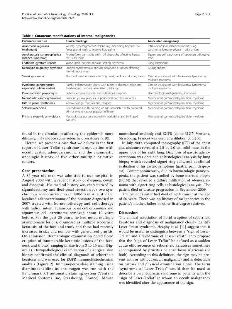
Table 1 Cutaneous manifestations of internal malignancies
Cutaneous feature Clinical findings Associated malignancy
Acanthosis nigricans(malignant)
Velvety hyperpigmented thickening extending beyond theflexures and neck to involve lips, palms
Intra-abdominal adenocarcinoma, lungcarcinoma, lymphoreticular malignancies
Acrokeratosis paraneoplastica(Bazex’s syndrome)
Psoriasiform dermatitis with nail dystrophy affecting hands,feet, ears, nose
Squamous cell carcinoma of upper aerodigestivetract
Erythema gyratum repens Wood grain pattern annular, scaling erythema Lung carcinoma
Necrolytic migratory erythema Eroded erythematous annular polycyclic eruption affectingintertriginous areas
Glucagonoma
Sweet syndrome Plum coloured nodules affecting head, neck and dorsae, hands Can be associated with leukaemia, lymphoma,multiple myeloma
Pyoderma gangrenosumespecially bullous variant
Painful inflammatory ulcers with raised violaceous edge andoverhanging borders; associated pathergy
Can be associated with leukaemia, lymphoma,multiple myeloma
Paraneoplastic pemphigus Bullous, erosive mucosal +/- cutaneous eruption Haematologic malignancies, thymoma
Necrobiotic xanthogranuloma Purpuric yellow plaques in periorbital and flexural areas Monoclonal gammopathy/multiple myeloma
Diffuse plane xanthomas Yellow-orange macules and plaques Monoclonal gammopathy/multiple myeloma
Scleromyxoedema Scleroderma-like thickening of skin associated with colouredskin or erythematous papular infiltrate
Monoclonal gammopathy/multiple myeloma
Primary systemic amyloidosis Macroglossia, purpura especially periorbital and infiltratedpapules
Monoclonal gammopathy/multiple myeloma
Ponti et al. Journal of Hematology Oncology 2010, 3:2http://www.jhoonline.org/content/3/1/2
Page 2 of 5
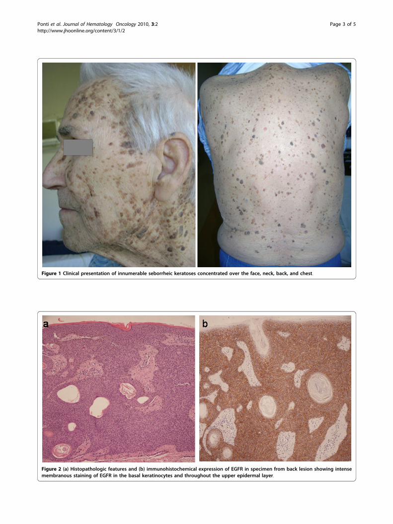
Figure 1 Clinical presentation of innumerable seborrheic keratoses concentrated over the face, neck, back, and chest.
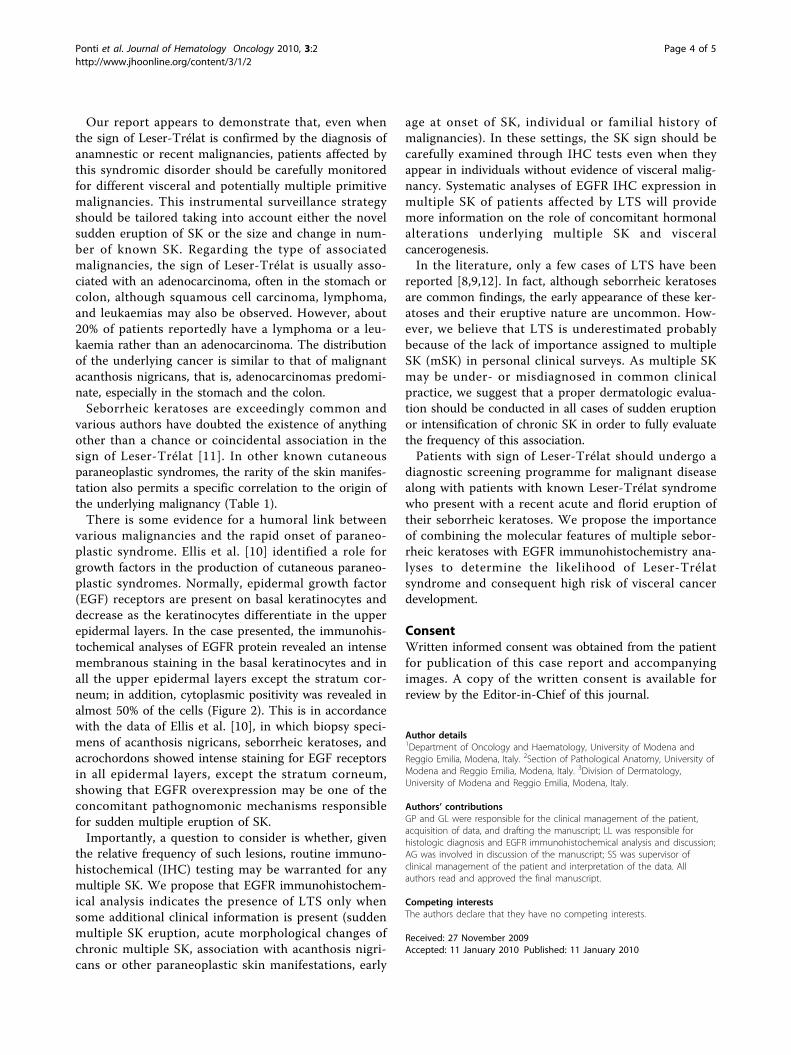
Figure 2 (a) Histopathologic features and (b) immunohistochemical expression of EGFR in specimen from back lesion showing intensemembranous staining of EGFR in the basal keratinocytes and throughout the upper epidermal layer.
Ponti et al. Journal of Hematology Oncology 2010, 3:2http://www.jhoonline.org/content/3/1/2
Page 3 of 5
Our report appears to demonstrate that, even whenthe sign of Leser-Trélat is confirmed by the diagnosis ofanamnestic or recent malignancies, patients affected bythis syndromic disorder should be carefully monitoredfor different visceral and potentially multiple primitivemalignancies. This instrumental surveillance strategyshould be tailored taking into account either the novelsudden eruption of SK or the size and change in num-ber of known SK. Regarding the type of associatedmalignancies, the sign of Leser-Trélat is usually asso-ciated with an adenocarcinoma, often in the stomach orcolon, although squamous cell carcinoma, lymphoma,and leukaemias may also be observed. However, about20% of patients reportedly have a lymphoma or a leu-kaemia rather than an adenocarcinoma. The distributionof the underlying cancer is similar to that of malignantacanthosis nigricans, that is, adenocarcinomas predomi-nate, especially in the stomach and the colon.Seborrheic keratoses are exceedingly common and
various authors have doubted the existence of anythingother than a chance or coincidental association in thesign of Leser-Trélat [11]. In other known cutaneousparaneoplastic syndromes, the rarity of the skin manifes-tation also permits a specific correlation to the origin ofthe underlying malignancy (Table 1).There is some evidence for a humoral link between
various malignancies and the rapid onset of paraneo-plastic syndrome. Ellis et al. [10] identified a role forgrowth factors in the production of cutaneous paraneo-plastic syndromes. Normally, epidermal growth factor(EGF) receptors are present on basal keratinocytes anddecrease as the keratinocytes differentiate in the upperepidermal layers. In the case presented, the immunohis-tochemical analyses of EGFR protein revealed an intensemembranous staining in the basal keratinocytes and inall the upper epidermal layers except the stratum cor-neum; in addition, cytoplasmic positivity was revealed inalmost 50% of the cells (Figure 2). This is in accordancewith the data of Ellis et al. [10], in which biopsy speci-mens of acanthosis nigricans, seborrheic keratoses, andacrochordons showed intense staining for EGF receptorsin all epidermal layers, except the stratum corneum,showing that EGFR overexpression may be one of theconcomitant pathognomonic mechanisms responsiblefor sudden multiple eruption of SK.Importantly, a question to consider is whether, given
the relative frequency of such lesions, routine immuno-histochemical (IHC) testing may be warranted for anymultiple SK. We propose that EGFR immunohistochem-ical analysis indicates the presence of LTS only whensome additional clinical information is present (suddenmultiple SK eruption, acute morphological changes ofchronic multiple SK, association with acanthosis nigri-cans or other paraneoplastic skin manifestations, early
age at onset of SK, individual or familial history ofmalignancies). In these settings, the SK sign should becarefully examined through IHC tests even when theyappear in individuals without evidence of visceral malig-nancy. Systematic analyses of EGFR IHC expression inmultiple SK of patients affected by LTS will providemore information on the role of concomitant hormonalalterations underlying multiple SK and visceralcancerogenesis.In the literature, only a few cases of LTS have been
reported [8,9,12]. In fact, although seborrheic keratosesare common findings, the early appearance of these ker-atoses and their eruptive nature are uncommon. How-ever, we believe that LTS is underestimated probablybecause of the lack of importance assigned to multipleSK (mSK) in personal clinical surveys. As multiple SKmay be under- or misdiagnosed in common clinicalpractice, we suggest that a proper dermatologic evalua-tion should be conducted in all cases of sudden eruptionor intensification of chronic SK in order to fully evaluatethe frequency of this association.Patients with sign of Leser-Trélat should undergo a
diagnostic screening programme for malignant diseasealong with patients with known Leser-Trélat syndromewho present with a recent acute and florid eruption oftheir seborrheic keratoses. We propose the importanceof combining the molecular features of multiple sebor-rheic keratoses with EGFR immunohistochemistry ana-lyses to determine the likelihood of Leser-Trélatsyndrome and consequent high risk of visceral cancerdevelopment.
ConsentWritten informed consent was obtained from the patientfor publication of this case report and accompanyingimages. A copy of the written consent is available forreview by the Editor-in-Chief of this journal.
Author details1Department of Oncology and Haematology, University of Modena andReggio Emilia, Modena, Italy. 2Section of Pathological Anatomy, University ofModena and Reggio Emilia, Modena, Italy. 3Division of Dermatology,University of Modena and Reggio Emilia, Modena, Italy.
Authors’ contributionsGP and GL were responsible for the clinical management of the patient,acquisition of data, and drafting the manuscript; LL was responsible forhistologic diagnosis and EGFR immunohistochemical analysis and discussion;AG was involved in discussion of the manuscript; SS was supervisor ofclinical management of the patient and interpretation of the data. Allauthors read and approved the final manuscript.
Competing interestsThe authors declare that they have no competing interests.
Received: 27 November 2009Accepted: 11 January 2010 Published: 11 January 2010
Ponti et al. Journal of Hematology Oncology 2010, 3:2http://www.jhoonline.org/content/3/1/2
Page 4 of 5

References1. Winship IM, Dubbing TE: Lesson from the skin-cutaneous features of
familial cancer. Lancet Oncol 2008, 9:462-472.2. Ponti G, Ponz de Leon M: Muir-Torre Syndrome. Lancet Oncol 2005, 6:980-
987.3. Bauer AJ, Stratakis CA: The lentiginoses: cutaneous markers of systemic
disease and a window to new aspects of tumourigenesis. J Med Genet2005, 42:801-810.
4. Lee A: Skin manifestation of systemic disease. Aust Fam Physician 2009,38(7):498-505.
5. Leser E: Ueber ein die Krebskrankheit beim Menschen haufigbegleitendes, noch wenig gekanntes Symptom. Munchener MedWochenschr 1901, 51:2035-2036.
6. Hollander EV: Beitrage zur Fruhdiagnose des Darmcarcinomas(Hereditatsverhaltnisse und Hautveranderungen). Dtsch Med Wochenschr1900, 26:483-485.
7. Lindelöf B, Sigurgeirsson B, Melander S: Seborrheic keratoses and cancer. JAm Acad Dermatol 1992, 26(6):947-950.
8. Schwengle LE, Rampen FH, Wobbes T: Seborrhoeic keratoses and internalmalignancies. A case control study. Clin Exp Dermatol 1988, 13(3):177-179.
9. Grob JJ, Rava MC, Gouvernet J, et al: The relation between seborrheickeratoses and malignant solid tumours. A case-control study. Acta DermVenereol 1991, 71(2):166-169.
10. Ellis DL, Kafka SP, Chow JC, et al: Melanoma, growth factors, acanthosisnigricans, the sign of Leser-Trelat, and multiple acrochordons. A possiblerole for alpha-transforming growth factor in cutaneous paraneoplasticsyndromes. N Engl J Med 1987, 317(25):1582-1587.
11. Heaphy MR Jr, Millns JL, Schroeter AL: The sign of Leser-Trelat in a case ofadenocarcinoma of the lung. J Am Acad Dermatol 2000, 43:386-390.
12. Lindelöf B, Sigurgeirsson B, Melander S: Seborrheic keratoses and cancer. JAm Acad Dermatol 1992, 26:947-950.
doi:10.1186/1756-8722-3-2Cite this article as: Ponti et al.: Leser-Trélat syndrome in patientsaffected by six multiple metachronous primitive cancers. Journal ofHematology & Oncology 2010 3:2.
Publish with BioMed Central and every scientist can read your work free of charge
"BioMed Central will be the most significant development for disseminating the results of biomedical research in our lifetime."
Sir Paul Nurse, Cancer Research UK
Your research papers will be:
available free of charge to the entire biomedical community
peer reviewed and published immediately upon acceptance
cited in PubMed and archived on PubMed Central
yours — you keep the copyright
Submit your manuscript here:http://www.biomedcentral.com/info/publishing_adv.asp
BioMedcentral
Ponti et al. Journal of Hematology Oncology 2010, 3:2http://www.jhoonline.org/content/3/1/2
Page 5 of 5