case report open access dress with delayed onset acute
TRANSCRIPT
CASE REPORT Open Access
DRESS with delayed onset acute interstitialnephritis and profound refractory eosinophiliasecondary to VancomycinPaloma O’Meara1, Rozita Borici-Mazi1, A Ross Morton1 and Anne K Ellis1,2*
Abstract
Background: Drug Reaction with Eosinophilia and Systemic Symptoms (DRESS) is a relatively rare clinical entity;even more so in response to vancomycin.
Methods: Case report.
Results: We present a severe case of vancomycin-induced DRESS syndrome, which on presentation included onlyskin, hematological and mild liver involvement. The patient further developed severe acute interstitial nephritis,eosinophilic pneumonitis, central nervous system (CNS) involvement and worsening hematological abnormalitiesdespite immediate discontinuation of vancomycin and parenteral corticosteroids. High-dose corticosteroids for aprolonged period were necessary and tapering of steroids a challenge due to rebound-eosinophilia and skininvolvement.
Conclusion: Patients with DRESS who are relatively resistant to corticosteroids with delayed onset of certain organinvolvement should be treated with a more prolonged corticosteroid tapering schedule. Vancomycin is increasinglybeing recognized as a culprit agent in this syndrome.
IntroductionWe present a case of severe Drug Reaction with Eosino-philia and Systemic Symptoms (DRESS) [1] syndromesecondary to vancomycin, with associated multiorgandysfunction. The relatively high mortality of this syn-drome warrants prompt recognition and elimination ofthe culprit drug and often treatment with high-dosecorticosteroids.
Case ReportA 66 year-old male presented to the emergency depart-ment (ED) with a one-week history of progressive pruri-tic erythematous rash, dry cough and two days ofepisodic high fevers. He had suffered a fall 12 weeksprior that had resulted in a pelvic fracture requiring anopen-reduction internal fixation, which subsequentlybecame infected with methicillin-resistant Staphylococ-cus aureus (MRSA) and treatment with intravenous
vancomycin was initiated. After four weeks of vancomy-cin therapy he developed a rash. This was initiallythought to be due to a red-man syndrome variant. Infu-sion rates were slowed, and premedication with diphen-hydramine was initiated, but the rash worsened, withthe subsequent development of episodic daily fevers,documented to be as high as 40°C.His past medical history was significant for heterozy-
gous hemochromatosis, a remote splenectomy secondaryto traumatic rupture, and non-anaphylactic adversereactions to penicillin and sulfa antibiotics. He had nohistory of reactive airway disease and had no travel his-tory, living in an Ontario city.In the ED he was hemodynamically stable with a nor-
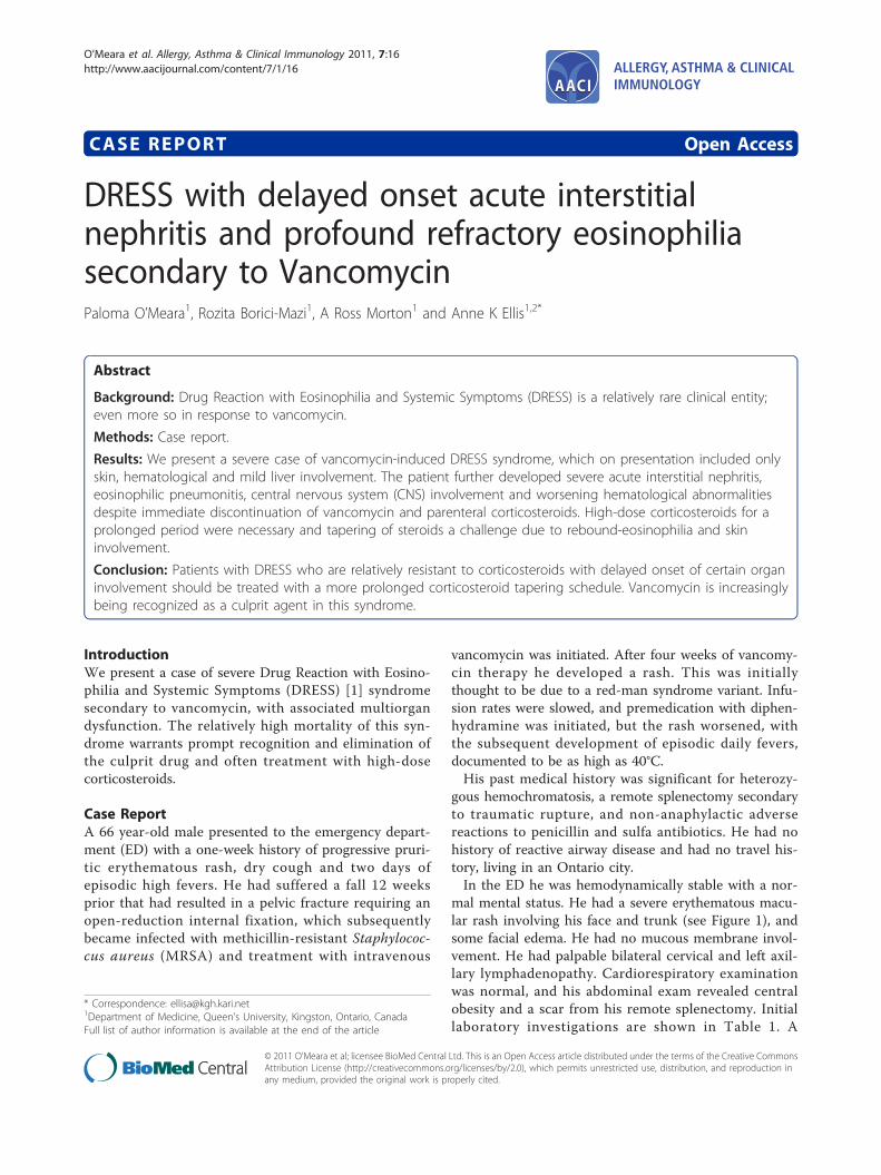
mal mental status. He had a severe erythematous macu-lar rash involving his face and trunk (see Figure 1), andsome facial edema. He had no mucous membrane invol-vement. He had palpable bilateral cervical and left axil-lary lymphadenopathy. Cardiorespiratory examinationwas normal, and his abdominal exam revealed centralobesity and a scar from his remote splenectomy. Initiallaboratory investigations are shown in Table 1. A
* Correspondence: [email protected] of Medicine, Queen’s University, Kingston, Ontario, CanadaFull list of author information is available at the end of the article
O’Meara et al. Allergy, Asthma & Clinical Immunology 2011, 7:16http://www.aacijournal.com/content/7/1/16 ALLERGY, ASTHMA & CLINICAL
IMMUNOLOGY
© 2011 O’Meara et al; licensee BioMed Central Ltd. This is an Open Access article distributed under the terms of the Creative CommonsAttribution License (http://creativecommons.org/licenses/by/2.0), which permits unrestricted use, distribution, and reproduction inany medium, provided the original work is properly cited.
computed tomography (CT) scan of his pelvis showedno abscess formation. A skin biopsy of the initial rashshowed a mild perivascular lymphocytic infiltrate, con-sistent with a drug reaction.Our initial working diagnosis was Drug Reaction with
Eosinophilia and Systemic Symptoms (DRESS) second-ary to vancomycin, which was immediately discontinued.
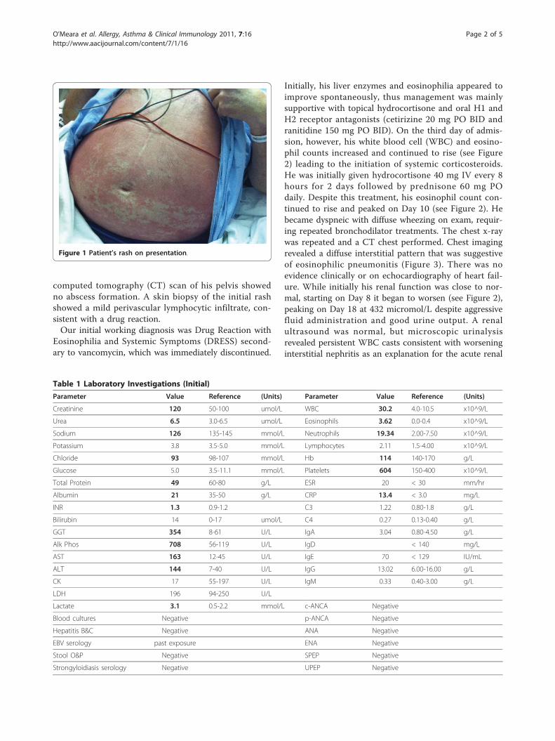
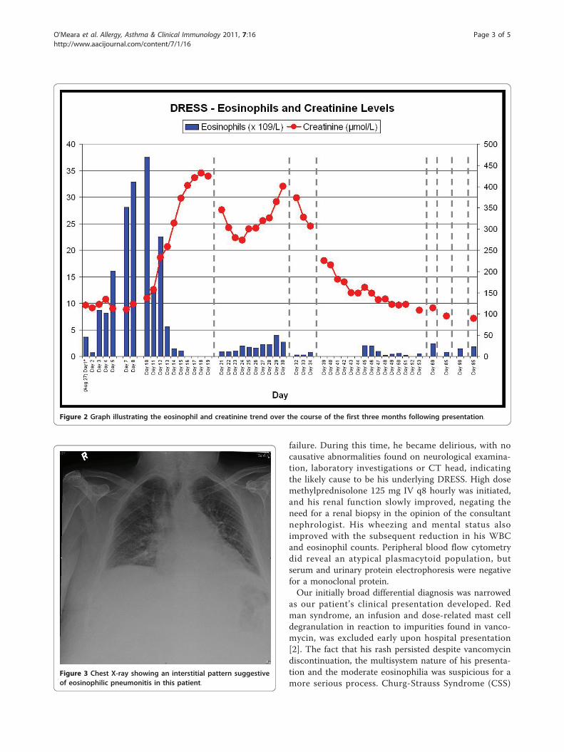
Initially, his liver enzymes and eosinophilia appeared toimprove spontaneously, thus management was mainlysupportive with topical hydrocortisone and oral H1 andH2 receptor antagonists (cetirizine 20 mg PO BID andranitidine 150 mg PO BID). On the third day of admis-sion, however, his white blood cell (WBC) and eosino-phil counts increased and continued to rise (see Figure2) leading to the initiation of systemic corticosteroids.He was initially given hydrocortisone 40 mg IV every 8hours for 2 days followed by prednisone 60 mg POdaily. Despite this treatment, his eosinophil count con-tinued to rise and peaked on Day 10 (see Figure 2). Hebecame dyspneic with diffuse wheezing on exam, requir-ing repeated bronchodilator treatments. The chest x-raywas repeated and a CT chest performed. Chest imagingrevealed a diffuse interstitial pattern that was suggestiveof eosinophilic pneumonitis (Figure 3). There was noevidence clinically or on echocardiography of heart fail-ure. While initially his renal function was close to nor-mal, starting on Day 8 it began to worsen (see Figure 2),peaking on Day 18 at 432 micromol/L despite aggressivefluid administration and good urine output. A renalultrasound was normal, but microscopic urinalysisrevealed persistent WBC casts consistent with worseninginterstitial nephritis as an explanation for the acute renal
Figure 1 Patient’s rash on presentation.
Table 1 Laboratory Investigations (Initial)
Parameter Value Reference (Units) Parameter Value Reference (Units)
Creatinine 120 50-100 umol/L WBC 30.2 4.0-10.5 x10^9/L
Urea 6.5 3.0-6.5 umol/L Eosinophils 3.62 0.0-0.4 x10^9/L
Sodium 126 135-145 mmol/L Neutrophils 19.34 2.00-7.50 x10^9/L
Potassium 3.8 3.5-5.0 mmol/L Lymphocytes 2.11 1.5-4.00 x10^9/L
Chloride 93 98-107 mmol/L Hb 114 140-170 g/L
Glucose 5.0 3.5-11.1 mmol/L Platelets 604 150-400 x10^9/L
Total Protein 49 60-80 g/L ESR 20 < 30 mm/hr
Albumin 21 35-50 g/L CRP 13.4 < 3.0 mg/L
INR 1.3 0.9-1.2 C3 1.22 0.80-1.8 g/L
Bilirubin 14 0-17 umol/L C4 0.27 0.13-0.40 g/L
GGT 354 8-61 U/L IgA 3.04 0.80-4.50 g/L
Alk Phos 708 56-119 U/L IgD < 140 mg/L
AST 163 12-45 U/L IgE 70 < 129 IU/mL
ALT 144 7-40 U/L IgG 13.02 6.00-16.00 g/L
CK 17 55-197 U/L IgM 0.33 0.40-3.00 g/L
LDH 196 94-250 U/L
Lactate 3.1 0.5-2.2 mmol/L c-ANCA Negative
Blood cultures Negative p-ANCA Negative
Hepatitis B&C Negative ANA Negative
EBV serology past exposure ENA Negative
Stool O&P Negative SPEP Negative
Strongyloidiasis serology Negative UPEP Negative
O’Meara et al. Allergy, Asthma & Clinical Immunology 2011, 7:16http://www.aacijournal.com/content/7/1/16
Page 2 of 5
failure. During this time, he became delirious, with nocausative abnormalities found on neurological examina-tion, laboratory investigations or CT head, indicatingthe likely cause to be his underlying DRESS. High dosemethylprednisolone 125 mg IV q8 hourly was initiated,and his renal function slowly improved, negating theneed for a renal biopsy in the opinion of the consultantnephrologist. His wheezing and mental status alsoimproved with the subsequent reduction in his WBCand eosinophil counts. Peripheral blood flow cytometrydid reveal an atypical plasmacytoid population, butserum and urinary protein electrophoresis were negativefor a monoclonal protein.Our initially broad differential diagnosis was narrowed
as our patient’s clinical presentation developed. Redman syndrome, an infusion and dose-related mast celldegranulation in reaction to impurities found in vanco-mycin, was excluded early upon hospital presentation[2]. The fact that his rash persisted despite vancomycindiscontinuation, the multisystem nature of his presenta-tion and the moderate eosinophilia was suspicious for amore serious process. Churg-Strauss Syndrome (CSS)
Figure 2 Graph illustrating the eosinophil and creatinine trend over the course of the first three months following presentation.
Figure 3 Chest X-ray showing an interstitial pattern suggestiveof eosinophilic pneumonitis in this patient.
O’Meara et al. Allergy, Asthma & Clinical Immunology 2011, 7:16http://www.aacijournal.com/content/7/1/16
Page 3 of 5
was also considered, but our patient had no history ofasthma or any bronchodilator or corticosteroid use inthe past, and therefore it was lower on our differential.Nonetheless, anti-neutrophil cytoplasmic antibodies(ANCAs), a test with only about 40% sensitivity for CSS,was sent and found to be negative [3]. This was sent 8days after initiation of corticosteroids, which may havedecreased the test’s sensitivity. However the predictivevalue of a negative ANCA in the face of our low clinicalprobability was sufficient to exclude this diagnosis. CSS,often described as having three phases, the asthmatic,the hypereosinophilic and the vasculitic phases, isalmost always preceded by a usually escalating asthmaticphase that can last up to years and is rarely subtle [3].Seldom, asthma can be a late feature. However, we havefollowed our patient over time, and complete taperingof corticosteroid left him with no residual respiratorysymptoms, making CSS highly unlikely.Microscopic stool examination for ova and parasites
was negative as was Stongyloides stercoralis serology. Nofurther parasitic work-up was completed as the patienthad no risk factors or gastrointestinal symptoms. Severesystemic bacterial infections, with the exception of scar-let fever, cause eosinopenia [4], and at the time ofadmission our patient had in fact been recovering froman MRSA bacteremia. Genetic testing for hypereosino-philic syndromes (HESs) with FIP1L1 and PDGFRAwere negative, and although these are positive in onlyabout 30% of patients with HESs [5], a secondary causeof hypereosinophilia was apparent and thus helpedexclude this diagnosis with more certainty.On day 39 from initial presentation, our patient
returned to hospital with a methicillin-sensitive Staphy-lococcus aureus (MSSA) bacteremia and severe leftshoulder pain. He was admitted under orthopedic sur-gery for exploration and debridement of both theshoulder and pelvis as well as removal of the pelvichardware. Evidence of osteomyelitis in the pelvic bonewas found during the surgery. He remains on cefazolinand is being followed by the infectious diseases service.Meanwhile, a taper of his prednisone was attemptedaround day 50 over a two week period. Unfortunately,his rash and eosinophilia returned shortly after its dis-continuation, requiring re-initiation and a slower taper-ing attempt. This was finally successful more than fourmonths after initial presentation. He is currently mana-ged with an oral H1 receptor antagonist alone, and hisperipheral eosinophil count has remained suppressed.
DiscussionDrug Reaction with Eosinophilia and Systemic Symp-toms (DRESS) is an idiosyncratic hypersensitivityresponse characterized by a maculopapular erythema-tous eruption that typically develops 2-6 weeks following
initiation of the culprit drug. The typical findingsinclude fever, lymphadenopathy, multisystem organ fail-ure and eosinophilia or atypical lymphocytosis. Theterm DRESS was coined in 1996 by Bocquet et al., in anattempt to unify the many names given to different drugreactions thought to have a common pathophysiologicalmechanism [6]. It has been postulated that concomitantinfection with herpes-simplex virus-6 (HSV-6) predis-poses to development of DRESS [7] and recently sug-gested as a diagnostic requirement [8]. Multi-organfailure often presents in a stepwise fashion despite dis-continuation of the culprit drug. The affected organsinclude, in order of frequency, the skin, liver, kidneys,lungs, heart, and more rarely CNS, thyroid, pancreas,colon, muscles and serosa. The most common drugsthat cause DRESS are anti-epileptics, the first describedbeing phenytoin in 1939 [9]. Nine cases of vancomycin-induced DRESS syndrome have been described so far inthe English literature [10-18]. As well, a tenth case,although not labeled as such, fulfills the criteria ofDRESS [19]. None of the described cases appear to havebeen as severe as what was observed in our case, andour patient was initially refractory to corticosteroidswith a late onset to his acute kidney injury from intersti-tial nephritis.Identifying patients with this syndrome is important,
as mortality approaches 10% [1]. Treatment includesstrict discontinuation of the culprit drug(s). Also, prob-ability tools exist to help identify the most likely agents[20]. Supportive care with symptomatic treatment usingH1 and H2 receptor antagonists and topical steroidtreatment may be sufficient for some. It is recom-mended to start systemic corticosteroids when internalorgan involvement is present [21]. Despite the lack ofrandomized controlled trials comparing supportive carealone to systemic steroids in the treatment of DRESS,experience has dictated their use and they are recom-mended by experts [15]. The use of systemic corticoster-oids is further supported by observations of clinicalworsening with early tapering of the same [22]. Occa-sionally, additional immunosuppressive therapy is neces-sary and has been observed to improve organ function[8].
ConclusionWe present a case of a patient with a relatively severeDRESS syndrome secondary to vancomycin with multi-ple organ systems affected, including skin, hematologi-cal, liver, lung, brain and kidneys in a stepwise fashion.Onset of renal injury from acute interstitial nephritiswas delayed and the response to standard doses of par-enteral corticosteroids insufficient despite initial sponta-neous improvements with the discontinuation of theoffending drug. Additionally, skin and hematological
O’Meara et al. Allergy, Asthma & Clinical Immunology 2011, 7:16http://www.aacijournal.com/content/7/1/16
Page 4 of 5
abnormalities recurred once corticosteroids weretapered. Patients with DRESS who are relatively resistantto corticosteroids with delayed onset of certain organinvolvement should be treated with a more prolongedcorticosteroid tapering schedule. Vancomycin is increas-ingly being recognized as a culprit agent in thissyndrome.
Consent StatementWritten informed consent was obtained from the patientfor publication of this case report and accompanyingimages. A copy of the written consent is available forreview by the Editor-in-Chief of this journal.
Author details1Department of Medicine, Queen’s University, Kingston, Ontario, Canada.2Department of Biomedical and Molecular Sciences, Queen’s University,Kingston, Ontario, Canada.
Authors’ contributionsPO: Involved in care of patient, Literature review, Created initial drafts ofmanuscript and table, completed first round of revisions following reviewerfeedback. RBM: Involved in care of patient, review/revisions to and approvalof manuscript final draft. ARM: Involved in care of patient, review/revisionsto and approval of manuscript final draft.AKE: Involved in care of patient, critical review and revisions to manuscriptprior to submission and post-reviewer feedback; created Figure 2; approvalof manuscript final draft.All authors read and approved the final manuscript.
Competing interestsThe authors declare that they have no competing interests.
Received: 8 March 2011 Accepted: 3 October 2011Published: 3 October 2011
References1. Walsh SA, Creamer D: Drug reaction with eosinophilia and systemic
symptoms (DRESS): a clinical update and review of current thinking.[Review]. Clin Exp Dermatol 2011, 36(1):6-11.
2. Sivagnanam S, Deleu D: Red man syndrome. Crit Care 2003, 7(2):119-120.3. Baldini C, Talarico R, Della Rossa A, Bombardieri S: Clinical Manifestations
and Treatment of Churg-Strauss Syndrome. Rheum Dis Clin N Am 2010,36:527-543.
4. Bass DA, Gonwa TA, Szejda P, Cousart MS, DeChatelet LR, McCall CE:Eosinopenia of acute infection: Production of eosinopenia bychemotactic factors of acute inflammation. J Clin Invest 1980, 65(6):1265.
5. Roufosse F, Weller PF: Practical approach to the patient withhypereosinophilia. J Allergy Clin Immunol 2010, 126(1):39-44.
6. Bocquet H, Bagot M, Roujeau JC: Drug-induced pseudolymphoma anddrug hypersensitivity syndrome (drug rash with eosinophilia andsystemic symptoms: DRESS). Semin Cutan Med Surg 1996, 15(4):250-257.
7. Descamps V, Valance A, Edlinger C, Fillet AM, Grossin M, Lebrun-Vignes B,Belaich S, Crickx B: Association of human herpesvirus 6 infection withdrug reaction with eosinophilia and systemic symptoms. Archives ofDermatology 2001, 137(3):301-304.
8. Kano Y, Shiohara T: The variable clinical picture of drug-inducedhypersensitivity syndrome/drug rash with eosinophilia and systemicsymptoms in relation to the eliciting drug [Review]. Immunol Allergy ClinNorth Am 2009, 29(3):481-501.
9. Cooper R, Burrows RGR: Treatment of epilepsy with sodiumdiphenylhydantoine. Lancet 1940, 1:490-492.
10. Boet S, Noblet C, Haas-Hubscher C, Picard D, Musette P, Dureuil B: Severevancomycin-induced drug rash with eosinophilia and systemicsymptoms syndrome imitating septic shock. European Journal ofAnaesthesiology 2009, 26(9):791-793.
11. Vauthey L, Uckay I, Abrassart S, Bernard L, Assal M, Ferry T, Djordjevic M,Roussos C, Vaudaux P: Vancomycin-induced DRESS syndrome in a femalepatient. Pharmacology 2008, 82(2):138-141.
12. Kwon HS, Chang YS, Jeong YY, Lee SM, Song WJ, Kim HB, Kim YK, Cho SH,Kim YY, Min KU: A case of hypersensitivity syndrome to bothvancomycin and teicoplanin. Journal of Korean Medical Science 2006,21(6):1108-1110.
13. Yazganoglu KD, Ozkaya E, Ergin-Ozcan P, Cakar N: Vancomycin-induceddrug hypersensitivity syndrome. Journal of the European Academy ofDermatology & Venereology 2005, 19(5):648-650.
14. Zuliani E, Zwahlen H, Gilliet F, Marone C: Vancomycin-inducedhypersensitivity reaction with acute renal failure: resolution followingcyclosporine treatment. Clinical Nephrology 2005, 64(2):155-158.
15. Hsu SIH: Biopsy-proved acute tubulointerstitial nephritis and toxicepidermal necrolysis associated with vancomycin. Pharmacotherapy 2001,21(10):1233-1239.
16. Marik PE, Ferris N: Delayed hypersensitivity reaction to vancomycin.Pharmacotherapy 1997, 17(6):1341-1344.
17. Vinson AE, Dufort EM, Willis MD, Eberson CP, Harwell JI: Drug rash,eosinophilia, and systemic symptoms syndrome: Two pediatric casesdemonstrating the range of severity in presentation-A case ofvancomycin-induced drug hypersensitivity mimicking toxic shocksyndrome and a milder case induced by minocycline. Pediatric CriticalCare Medicine 2010, 11(4):e38-e43.
18. Wai AO, Lo AMS, Abdo A, Marra F: Vancomycin-induced acute interstitialnephritis. Annals of Pharmacotherapy 1998, 32(11):1160-1164.
19. Alexander II: Vancomycin-induced Stevens-Johnson syndrome. Allergy andAsthma Proceedings 1996, 17(2):75-78.
20. Rehan HS, Chopra D, Kakkar AK: Physician’s guide to pharmacovigilance:Terminology and causality assessment. European Journal of InternalMedicine 2009, 20(1):3-8.
21. Tas S, Simonart T: Management of drug rash with eosinophilia andsystemic symptoms (DRESS syndrome) an update. Dermatology 2003,206(4):353-356.
22. Chopra S, Levell NJ, Cowley G, Gilkes JJ: Systemic corticosteroids in thephenytoin hypersensitivity syndrome. British Journal of Dermatology 1996,134(6):1109-1112.
doi:10.1186/1710-1492-7-16Cite this article as: O’Meara et al.: DRESS with delayed onset acuteinterstitial nephritis and profound refractory eosinophilia secondary toVancomycin. Allergy, Asthma & Clinical Immunology 2011 7:16.
Submit your next manuscript to BioMed Centraland take full advantage of:
• Convenient online submission
• Thorough peer review
• No space constraints or color figure charges
• Immediate publication on acceptance
• Inclusion in PubMed, CAS, Scopus and Google Scholar
• Research which is freely available for redistribution
Submit your manuscript at www.biomedcentral.com/submit
O’Meara et al. Allergy, Asthma & Clinical Immunology 2011, 7:16http://www.aacijournal.com/content/7/1/16
Page 5 of 5