case report lipoma arborescens: review of an uncommon...
TRANSCRIPT
Case ReportLipoma Arborescens: Review of an Uncommon Cause forSwelling of the Knee
M. De Vleeschhouwer,1 E. Van Den Steen,2 G. Vanderstraeten,1 W. Huysse,3
J. De Neve,2 and L. Vanden Bossche1
1Physical Medicine and Rehabilitation, Ghent University Hospital, 9000 Ghent, Belgium2Department of Physical Medicine and Rehabilitation, AZ Sint-Jan, 8000 Bruges, Belgium3Department of Radiology, Ghent University Hospital, 9000 Ghent, Belgium
Correspondence should be addressed to M. De Vleeschhouwer; [email protected]
Received 26 February 2016; Accepted 4 May 2016
Academic Editor: Werner Kolb
Copyright © 2016 M. De Vleeschhouwer et al. This is an open access article distributed under the Creative Commons AttributionLicense, which permits unrestricted use, distribution, and reproduction in any medium, provided the original work is properlycited.
Lipoma arborescens is a rare cause of chronic monoarticular arthritis, with only a few cases reported in the literature. It is mostcommonly seen in the knee, but cases in other joints such as the wrist, shoulder, and elbow have also been described. It is a benigncondition, in which the subsynovial tissue is replaced diffusely bymature fat cells.We describe a case involving the knee and discussthe symptoms, diagnosis, and treatment.
1. Introduction
Lipoma arborescens, also referred to as villous lipomatousproliferation of synovial membrane, diffuse lipoma of thejoint, or diffuse synovial lipoma, is a rare benign intra-articular lesion characterized by villous proliferation of thesynovial membrane [1]. The etiology of this condition stillremains unclear.
Lipoma arborescens typically affects adults. It most com-monly involves the knee, but other locations have also beendescribed. People present with joint pain, swelling, andeffusion. The diagnosis is based on the typical appearanceon MRI, and the recommended treatment is open or arthro-scopic synovectomy. Recurrence is uncommon.
2. Case Report
A 31-year-old Dutch man of Turkish origin presented to thephysical and rehabilitation medicine department with lowback pain. On physical examination, a marked effusion inthe right knee joint was noted. According to the patient, theswelling had been present since two years in a varying degree,with intermittent episodes of pain. There was no history oftrauma. The patient’s father had type-2 diabetes mellitus, but
our patient was not diagnosed with it.The patient was knownto have psoriasis.
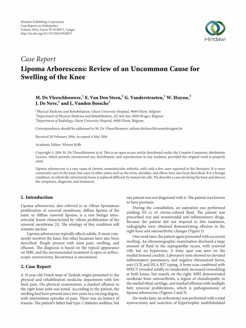
During the consultation, an aspiration was performedyielding 113 cc of citrine-colored fluid. The patient wasprescribed rest and nonsteroidal anti-inflammatory drugs.Because the patient did not respond to this treatment,radiographs were obtained demonstrating effusion in theright knee and osteoarthritic changes (Figure 1).
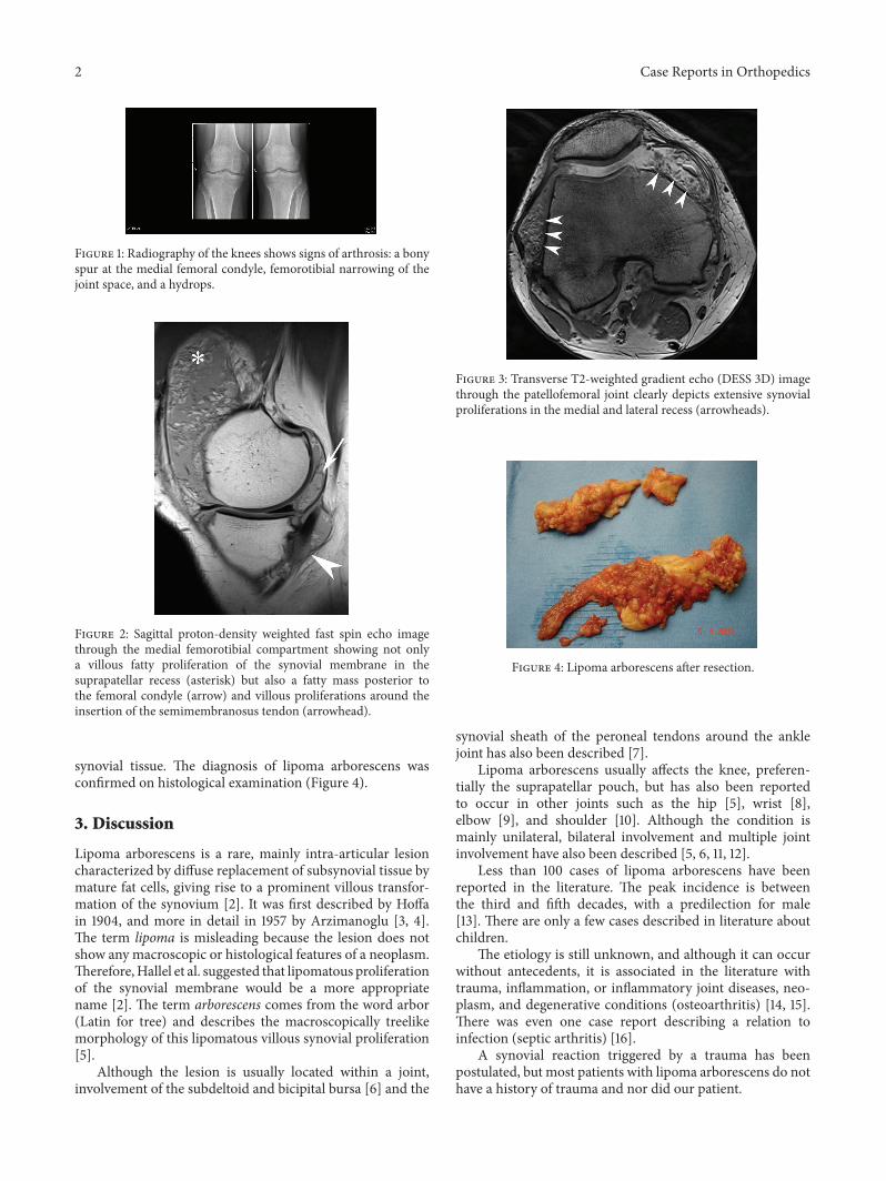
Oneweek later, the patient again presentedwith recurrentswelling. An ultrasonographic examination disclosed a largeamount of fluid in the suprapatellar recess, with synovialvilli, but no hyperemia. A bony spur was seen on themedial femoral condyle. Laboratory tests showed no elevatedinflammatory parameters, and negative rheumatoid factor,anti-CCP, and HLA-B27 typing. A bone scan combined withSPECT revealed mildly-to-moderately increased remodelingat both knees, but mainly on the right. MRI demonstratedmoderate knee osteoarthritis, a region of chondropathy inthe medial tibial cartilage, andmarked effusion with multiplefatty synovial proliferations, which is pathognomonic oflipoma arborescens (Figures 2 and 3).
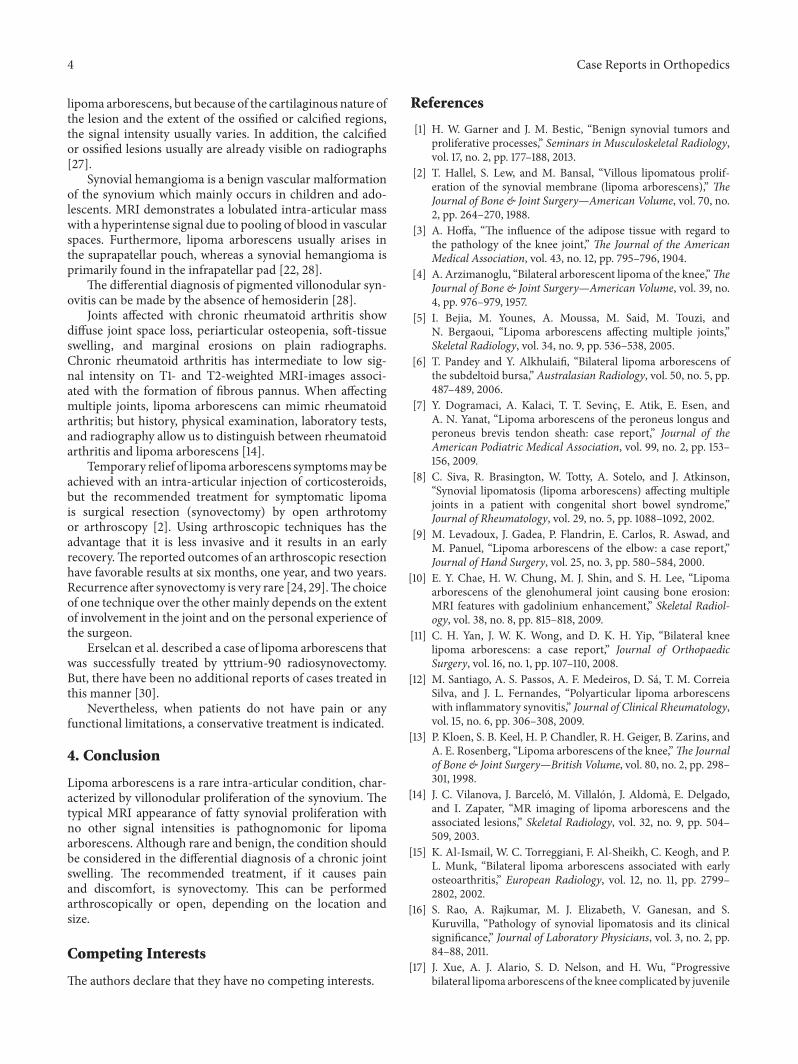
Six weeks later, an arthrotomy was performed with a totalsynovectomy and resection of hypertrophic multilobulated
Hindawi Publishing CorporationCase Reports in OrthopedicsVolume 2016, Article ID 9538075, 5 pageshttp://dx.doi.org/10.1155/2016/9538075
2 Case Reports in Orthopedics
Figure 1: Radiography of the knees shows signs of arthrosis: a bonyspur at the medial femoral condyle, femorotibial narrowing of thejoint space, and a hydrops.
Figure 2: Sagittal proton-density weighted fast spin echo imagethrough the medial femorotibial compartment showing not onlya villous fatty proliferation of the synovial membrane in thesuprapatellar recess (asterisk) but also a fatty mass posterior tothe femoral condyle (arrow) and villous proliferations around theinsertion of the semimembranosus tendon (arrowhead).
synovial tissue. The diagnosis of lipoma arborescens wasconfirmed on histological examination (Figure 4).
3. Discussion
Lipoma arborescens is a rare, mainly intra-articular lesioncharacterized by diffuse replacement of subsynovial tissue bymature fat cells, giving rise to a prominent villous transfor-mation of the synovium [2]. It was first described by Hoffain 1904, and more in detail in 1957 by Arzimanoglu [3, 4].The term lipoma is misleading because the lesion does notshow any macroscopic or histological features of a neoplasm.Therefore,Hallel et al. suggested that lipomatous proliferationof the synovial membrane would be a more appropriatename [2]. The term arborescens comes from the word arbor(Latin for tree) and describes the macroscopically treelikemorphology of this lipomatous villous synovial proliferation[5].
Although the lesion is usually located within a joint,involvement of the subdeltoid and bicipital bursa [6] and the
Figure 3: Transverse T2-weighted gradient echo (DESS 3D) imagethrough the patellofemoral joint clearly depicts extensive synovialproliferations in the medial and lateral recess (arrowheads).
Figure 4: Lipoma arborescens after resection.
synovial sheath of the peroneal tendons around the anklejoint has also been described [7].
Lipoma arborescens usually affects the knee, preferen-tially the suprapatellar pouch, but has also been reportedto occur in other joints such as the hip [5], wrist [8],elbow [9], and shoulder [10]. Although the condition ismainly unilateral, bilateral involvement and multiple jointinvolvement have also been described [5, 6, 11, 12].
Less than 100 cases of lipoma arborescens have beenreported in the literature. The peak incidence is betweenthe third and fifth decades, with a predilection for male[13]. There are only a few cases described in literature aboutchildren.
The etiology is still unknown, and although it can occurwithout antecedents, it is associated in the literature withtrauma, inflammation, or inflammatory joint diseases, neo-plasm, and degenerative conditions (osteoarthritis) [14, 15].There was even one case report describing a relation toinfection (septic arthritis) [16].
A synovial reaction triggered by a trauma has beenpostulated, but most patients with lipoma arborescens do nothave a history of trauma and nor did our patient.

Case Reports in Orthopedics 3
The hypothesis of lipoma arborescens being a reactionto chronic inflammation is supported by the histologicalfinding of a mononuclear cell infiltrate in the underlyingsynovial membrane [13]. Furthermore, inflammatory con-ditions, such as rheumatoid arthritis, psoriatic arthritis, orpsoriasis, uveitis, and juvenile spondyloarthropathy, are inseveral reports associated with lipoma arborescens [17, 18].
A causal relationship between lipoma arborescens anddegenerative joint disease is suspected but has yet to be con-firmed. It has been suggested that the lesion can be dividedinto a primary and a secondary type.The primary type is rareand has been described as a form of synovial lipomatosis withhypertrophy as the cardinal feature; it is rarely considereda cause of degenerative knee joint changes. The secondarytype has been defined as lipomatosis resulting from chronicirritation of the synovium (as seen with degenerative jointdisease or arthritis) rather than a true neoplasm and is byfar the most common form of lipoma arborescens. Mostauthors have accepted the last hypothesis [14, 15, 19, 20];this suggests that osteoarthritic changes are secondary to thepresence of lipoma arborescens. Natera even concludes inhis retrospective review that progressive joint degenerationcould be prevented or at least delayed, if prompt synovectomyis performed [20, 21].
Patients with lipoma arborescens typically present withone or more of the following symptoms: slowly progressiveswelling, pain, intermittent episodes of joint effusion, limitedrange of motion, and locking. Only rarely can a soft-tissuemass be identified on palpation [22]. Most patients haveno history of recent trauma. The symptoms are cyclic, withintermittent exacerbations caused by mechanical trapping ofthe lipomatous villi inside the joint space.
The diagnosis of lipoma arborescens is based on thetypical findings on MRI, but because of the clinical appear-ance in this case other more common pathologies had to beexcluded by using laboratory tests, joint fluid aspiration, andother imaging studies. In general the laboratory findings arenonspecific.The joint fluid aspirate is negative for crystals andmicroorganisms and ismainly used to exclude other causes ofjoint swelling [2, 15].
Plain radiographs sometimes show a soft-tissue density,nonspecific bone erosion, or other osteoarthritic changes(joint space narrowing, osteophytes, subchondral sclerosis,and bone cyst formation) [6, 10].
On ultrasonography, a frondlike hyperechoicmass is seenthat waves during manipulation of the joint [23].
CT scanning shows a villous synovial mass of a densitysimilar to fat. The signal is not enhanced with intravenouscontrast administration [22].
On MRI it has a pathognomonic aspect, which makesMRI the diagnostic imagingmodality of choice [13].The find-ings have a high degree of accuracy in the identification andanatomic characterization, and they give a correct evaluationof size and grade, which can be useful for choosing the mostappropriate therapy.
These findings firstly include a large frondlike massarising from the synovium. Secondly, the mass has a sub-cutaneous fat-equivalent signal on all pulse sequences. Thesubsynovial component has a high signal intensity on T1-
and T2-weighted images and signal suppression on short T1inversion recovery sequences (STIR) or after presaturation ofthe fat [10, 14, 24, 25]. There is a third characteristic: as onCT, there is no enhancement of the (sub)synovial tissue withintravenous administration of contrast medium. However,the joint fluid and synovial layer may show enhancementrelated to the presence of inflammatory cells [24]. A fourthfeature is joint effusion, which can be present in differentdegrees. And finally, there is an absence of magnetic suscep-tibility effects from hemosiderin [26].
Other features are bone erosions, synovial cyst, degen-erative changes, and chondromatosis. On MRI, our patientpresented both the typical signal characteristics and amarkedeffusion. Furthermore, some degenerative changes such aschondropathy, osteophyte formation, and other signs of kneeosteoarthritis were also seen.
The most common presentation of lipoma arborescens isa diffuse villous proliferation, but in aminority of cases it mayalso present as a focal pseudomass. According to Soler et al.,the pseudomass type presumably is associated with primarylipoma arborescens, that is, without degenerative changes,but this was later refuted byVilanova et al. In his retrospectiveseries of thirty-two cases he found that 87% of the patientsshowed associated degenerative changes of the joint and 72%had a meniscal tear. We have to note that his study groupmainly consisted of elderly patients [14, 25].
In the past, a biopsywas considered essential formaking afinal diagnosis, but now the typicalMRI findings are sufficientto permit a reliable diagnosis [14].
Macroscopically, the lesion has a white-yellowish aspectand shows villous proliferation. Histologically, the villi arefilled with mature fat cells and enlarged hyperaemic capil-laries may be present. The underlying synovial membranemay containmononuclear chronic inflammatory cells and thesynovial cells may appear to be enlarged and reactive, withabundant eosinophilic cytoplasm [6, 10, 11, 25].
Lipoma arborescens forms part of the differential diag-nosis of a chronic joint swelling; including pigmented vil-lonodular synovitis, synovial osteochondromatosis, rheuma-toid arthritis, intra-articular or synovial lipoma, synovialhemangioma, amyloid arthropathy, and xanthoma. Intra-articular lipoma and synovial osteochondromatosis are theonly among these entities that can demonstrate similar MRIsignal characteristics [25].
Intra-articular lipoma can be differentiated from lipomaarborescens based on its macroscopic and microscopic fea-tures. It appears as a solitary round or oval mass, as opposedto the multiple villous lipomatous proliferations and thefrondlike morphology of lipoma arborescens. Intra-articularsynovial lipoma (IASL) is composed of mature fat cellscovered by a thin fibrous layer, usuallywith no synovial lining.Histologically, lipoma arborescens is characterized by diffusereplacement of the subsynovial layer by mature fat cells witha moderate infiltration of mononuclear cells. Intra-articularlipoma does not arise from the synovial layer, nor does itreplace it.
Synovial osteochondromatosis is characterized by anodular proliferation and metaplasia of the synovial mem-brane. On MRI, the signal intensity of it is similar to that of
4 Case Reports in Orthopedics
lipoma arborescens, but because of the cartilaginous nature ofthe lesion and the extent of the ossified or calcified regions,the signal intensity usually varies. In addition, the calcifiedor ossified lesions usually are already visible on radiographs[27].
Synovial hemangioma is a benign vascular malformationof the synovium which mainly occurs in children and ado-lescents. MRI demonstrates a lobulated intra-articular masswith a hyperintense signal due to pooling of blood in vascularspaces. Furthermore, lipoma arborescens usually arises inthe suprapatellar pouch, whereas a synovial hemangioma isprimarily found in the infrapatellar pad [22, 28].
The differential diagnosis of pigmented villonodular syn-ovitis can be made by the absence of hemosiderin [28].
Joints affected with chronic rheumatoid arthritis showdiffuse joint space loss, periarticular osteopenia, soft-tissueswelling, and marginal erosions on plain radiographs.Chronic rheumatoid arthritis has intermediate to low sig-nal intensity on T1- and T2-weighted MRI-images associ-ated with the formation of fibrous pannus. When affectingmultiple joints, lipoma arborescens can mimic rheumatoidarthritis; but history, physical examination, laboratory tests,and radiography allow us to distinguish between rheumatoidarthritis and lipoma arborescens [14].
Temporary relief of lipoma arborescens symptomsmay beachieved with an intra-articular injection of corticosteroids,but the recommended treatment for symptomatic lipomais surgical resection (synovectomy) by open arthrotomyor arthroscopy [2]. Using arthroscopic techniques has theadvantage that it is less invasive and it results in an earlyrecovery.The reported outcomes of an arthroscopic resectionhave favorable results at six months, one year, and two years.Recurrence after synovectomy is very rare [24, 29].The choiceof one technique over the other mainly depends on the extentof involvement in the joint and on the personal experience ofthe surgeon.
Erselcan et al. described a case of lipoma arborescens thatwas successfully treated by yttrium-90 radiosynovectomy.But, there have been no additional reports of cases treated inthis manner [30].
Nevertheless, when patients do not have pain or anyfunctional limitations, a conservative treatment is indicated.
4. Conclusion
Lipoma arborescens is a rare intra-articular condition, char-acterized by villonodular proliferation of the synovium. Thetypical MRI appearance of fatty synovial proliferation withno other signal intensities is pathognomonic for lipomaarborescens. Although rare and benign, the condition shouldbe considered in the differential diagnosis of a chronic jointswelling. The recommended treatment, if it causes painand discomfort, is synovectomy. This can be performedarthroscopically or open, depending on the location andsize.
Competing Interests
The authors declare that they have no competing interests.
References
[1] H. W. Garner and J. M. Bestic, “Benign synovial tumors andproliferative processes,” Seminars in Musculoskeletal Radiology,vol. 17, no. 2, pp. 177–188, 2013.
[2] T. Hallel, S. Lew, and M. Bansal, “Villous lipomatous prolif-eration of the synovial membrane (lipoma arborescens),” TheJournal of Bone & Joint Surgery—American Volume, vol. 70, no.2, pp. 264–270, 1988.
[3] A. Hoffa, “The influence of the adipose tissue with regard tothe pathology of the knee joint,” The Journal of the AmericanMedical Association, vol. 43, no. 12, pp. 795–796, 1904.
[4] A. Arzimanoglu, “Bilateral arborescent lipoma of the knee,”TheJournal of Bone & Joint Surgery—American Volume, vol. 39, no.4, pp. 976–979, 1957.
[5] I. Bejia, M. Younes, A. Moussa, M. Said, M. Touzi, andN. Bergaoui, “Lipoma arborescens affecting multiple joints,”Skeletal Radiology, vol. 34, no. 9, pp. 536–538, 2005.
[6] T. Pandey and Y. Alkhulaifi, “Bilateral lipoma arborescens ofthe subdeltoid bursa,” Australasian Radiology, vol. 50, no. 5, pp.487–489, 2006.
[7] Y. Dogramaci, A. Kalaci, T. T. Sevinc, E. Atik, E. Esen, andA. N. Yanat, “Lipoma arborescens of the peroneus longus andperoneus brevis tendon sheath: case report,” Journal of theAmerican Podiatric Medical Association, vol. 99, no. 2, pp. 153–156, 2009.
[8] C. Siva, R. Brasington, W. Totty, A. Sotelo, and J. Atkinson,“Synovial lipomatosis (lipoma arborescens) affecting multiplejoints in a patient with congenital short bowel syndrome,”Journal of Rheumatology, vol. 29, no. 5, pp. 1088–1092, 2002.
[9] M. Levadoux, J. Gadea, P. Flandrin, E. Carlos, R. Aswad, andM. Panuel, “Lipoma arborescens of the elbow: a case report,”Journal of Hand Surgery, vol. 25, no. 3, pp. 580–584, 2000.
[10] E. Y. Chae, H. W. Chung, M. J. Shin, and S. H. Lee, “Lipomaarborescens of the glenohumeral joint causing bone erosion:MRI features with gadolinium enhancement,” Skeletal Radiol-ogy, vol. 38, no. 8, pp. 815–818, 2009.
[11] C. H. Yan, J. W. K. Wong, and D. K. H. Yip, “Bilateral kneelipoma arborescens: a case report,” Journal of OrthopaedicSurgery, vol. 16, no. 1, pp. 107–110, 2008.
[12] M. Santiago, A. S. Passos, A. F. Medeiros, D. Sa, T. M. CorreiaSilva, and J. L. Fernandes, “Polyarticular lipoma arborescenswith inflammatory synovitis,” Journal of Clinical Rheumatology,vol. 15, no. 6, pp. 306–308, 2009.
[13] P. Kloen, S. B. Keel, H. P. Chandler, R. H. Geiger, B. Zarins, andA. E. Rosenberg, “Lipoma arborescens of the knee,”The Journalof Bone & Joint Surgery—British Volume, vol. 80, no. 2, pp. 298–301, 1998.
[14] J. C. Vilanova, J. Barcelo, M. Villalon, J. Aldoma, E. Delgado,and I. Zapater, “MR imaging of lipoma arborescens and theassociated lesions,” Skeletal Radiology, vol. 32, no. 9, pp. 504–509, 2003.
[15] K. Al-Ismail, W. C. Torreggiani, F. Al-Sheikh, C. Keogh, and P.L. Munk, “Bilateral lipoma arborescens associated with earlyosteoarthritis,” European Radiology, vol. 12, no. 11, pp. 2799–2802, 2002.
[16] S. Rao, A. Rajkumar, M. J. Elizabeth, V. Ganesan, and S.Kuruvilla, “Pathology of synovial lipomatosis and its clinicalsignificance,” Journal of Laboratory Physicians, vol. 3, no. 2, pp.84–88, 2011.
[17] J. Xue, A. J. Alario, S. D. Nelson, and H. Wu, “Progressivebilateral lipoma arborescens of the knee complicated by juvenile

Case Reports in Orthopedics 5
spondyloarthropathy: a case report and review of the literature,”Seminars in Arthritis and Rheumatism, vol. 43, no. 2, pp. 259–263, 2013.
[18] C. Nguyen, B. B. Jean-Luc, A. Papelard, S. Poiraudeau,M. Revel,and F. Rannou, “The role of magnetic resonance imaging for thediagnosis of lipoma arborescens in polyarthritic patients withpersistent single-joint effusion,” Journal of Clinical Rheumatol-ogy, vol. 15, no. 8, p. 431, 2009.
[19] J. Xiao, Y. Xu, J.Wang, J. Feng, and Z. Shi, “Bilateral knee lipomaarborescens combined with osteoarthritis in elderly patients,”Journal of International Medical Research, vol. 39, no. 4, pp.1563–1569, 2011.
[20] K. Ikushima, T. Ueda, I. Kudawara, and H. Yoshikawa, “Lipomaarborescens of the knee as a possible cause of osteoarthrosis,”Orthopedics, vol. 24, no. 6, pp. 603–605, 2001.
[21] L. Natera, P. E. Gelber, J. I. Erquicia, and J. C.Monllau, “Primarylipoma arborescens of the knee may involve the development ofearly osteoarthritis if prompt synovectomy is not performed,”Journal of Orthopaedics and Traumatology, vol. 16, no. 1, pp. 47–53, 2015.
[22] M. A. Adelani, R. M. Wupperman, and G. E. Holt, “Benignsynovial disorders,” Journal of the American Academy ofOrthopaedic Surgeons, vol. 16, no. 5, pp. 268–275, 2008.
[23] T. J. Learch and M. Braaton, “Lipoma arborescens: high-resolution ultrasonographic findings,” Journal of Ultrasound inMedicine, vol. 19, no. 6, pp. 385–389, 2000.
[24] G. Chaljub and P. R. Johnson, “In vivo MRI characteristicsof lipoma arborescens utilizing fat suppression and contrastadministration,” Journal of Computer Assisted Tomography, vol.20, no. 1, pp. 85–87, 1996.
[25] R. Soler, E. Rodrıguez, A. Bargiela, and M. Da Riba, “Lipomaarborescens of the knee:MR characteristics in 13 joints,” Journalof Computer Assisted Tomography, vol. 22, no. 4, pp. 605–609,1998.
[26] J. F. Feller, M. Rishi, and E. C. Hughes, “Lipoma arborescens ofthe knee: MR demonstration,” American Journal of Roentgenol-ogy, vol. 163, no. 1, pp. 162–164, 1994.
[27] J. U. V. Monu and M. Oka, Synovial Osteochondromatosis,eMedicine, 2007, http://www.emedicine.com/radio/topic667.htm.
[28] J.-H. Ji, Y.-S. Lee, and M. Shafi, “Spontaneous recurrenthemarthrosis of the knee joint in elderly patients withosteoarthritis: an infrequent presentation of synovial lipomaarborescens,” Knee Surgery, Sports Traumatology, Arthroscopy,vol. 18, no. 10, pp. 1352–1355, 2010.
[29] M. Franco, J. M. Puch, M. J. Carayon, D. Bortolotti, L. Albano,and A. Lallemand, “Lipoma arborescens of the knee: report of acase managed by arthroscopic synovectomy,” Joint Bone Spine,vol. 71, no. 1, pp. 73–75, 2004.
[30] T. Erselcan, O. Bulut, S. Bulut et al., “Lipoma arborescens;successfully treated by yttrium-90 radiosynovectomy,” Annalsof Nuclear Medicine, vol. 17, no. 7, pp. 593–596, 2003.

Submit your manuscripts athttp://www.hindawi.com
Stem CellsInternational
Hindawi Publishing Corporationhttp://www.hindawi.com Volume 2014
Hindawi Publishing Corporationhttp://www.hindawi.com Volume 2014
MEDIATORSINFLAMMATION
of
Hindawi Publishing Corporationhttp://www.hindawi.com Volume 2014
Behavioural Neurology
EndocrinologyInternational Journal of
Hindawi Publishing Corporationhttp://www.hindawi.com Volume 2014
Hindawi Publishing Corporationhttp://www.hindawi.com Volume 2014
Disease Markers
Hindawi Publishing Corporationhttp://www.hindawi.com Volume 2014
BioMed Research International
OncologyJournal of
Hindawi Publishing Corporationhttp://www.hindawi.com Volume 2014
Hindawi Publishing Corporationhttp://www.hindawi.com Volume 2014
Oxidative Medicine and Cellular Longevity
Hindawi Publishing Corporationhttp://www.hindawi.com Volume 2014
PPAR Research
The Scientific World JournalHindawi Publishing Corporation http://www.hindawi.com Volume 2014
Immunology ResearchHindawi Publishing Corporationhttp://www.hindawi.com Volume 2014
Journal of
ObesityJournal of
Hindawi Publishing Corporationhttp://www.hindawi.com Volume 2014
Hindawi Publishing Corporationhttp://www.hindawi.com Volume 2014
Computational and Mathematical Methods in Medicine
OphthalmologyJournal of
Hindawi Publishing Corporationhttp://www.hindawi.com Volume 2014
Diabetes ResearchJournal of
Hindawi Publishing Corporationhttp://www.hindawi.com Volume 2014
Hindawi Publishing Corporationhttp://www.hindawi.com Volume 2014
Research and TreatmentAIDS
Hindawi Publishing Corporationhttp://www.hindawi.com Volume 2014
Gastroenterology Research and Practice
Hindawi Publishing Corporationhttp://www.hindawi.com Volume 2014
Parkinson’s Disease
Evidence-Based Complementary and Alternative Medicine
Volume 2014Hindawi Publishing Corporationhttp://www.hindawi.com