case report granulomatosis with polyangiitis with...
TRANSCRIPT

Case ReportGranulomatosis with Polyangiitis with Bilateral Facial Palsy andSevere Mixed Hearing Loss
Agnieszka Wawrzecka,1 Anna SzymaNska,1,2 RadosBaw Jeleniewicz,1,3
and Marcin SzymaNski1
1Department of Otolaryngology Head and Neck Surgery, Medical University of Lublin, Jaczewskiego 8, 20-954 Lublin, Poland2Department of Neuroradiology and Interventional Radiology, Medical University of Lublin, Jaczewskiego 8, 20-954 Lublin, Poland3Department of Rheumatology, Medical University of Lublin, Jaczewskiego 8, 20-954 Lublin, Poland
Correspondence should be addressed to Marcin Szymanski; [email protected]
Received 25 March 2016; Accepted 9 June 2016
Academic Editor: Harukazu Hiraumi
Copyright © 2016 Agnieszka Wawrzecka et al. This is an open access article distributed under the Creative Commons AttributionLicense, which permits unrestricted use, distribution, and reproduction in any medium, provided the original work is properlycited.
Granulomatosis with polyangiitis is autoimmune and rare disease. It affects many organs, but the most often affected organs arethe nose, lungs, and kidneys. It is part of vasculitis and causes an autoimmune attack by an abnormal type of circulating antibodytermed ANCAs against small blood vessels. Disease concerns both men and women with a peak age of presentation in the sixthand seven decades. Typically upper and lower respiratory tract and kidneys are involved. Otitis externa, otitis media, or mastoiditisrarely occurs in granulomatosis with polyangiitis. Deafness is the most dangerous aural complication. Histological examination ofbiopsy is often not specific. A case of GPA with bilateral otitis media, bilateral deafness, and bilateral facial palsy with fatal courseis presented.
1. Introduction
Granulomatosis with polyangiitis (GPA) is a rare, autoim-mune, multisystemic disease first described by FriedrichWegener in 1936 [1, 2] and has been named Wegener’sgranulomatosis for many years. In 2012, the name Wegener’sgranulomatosis (WG) was adopted by the 2012 InternationalChapelHill Consensus Conference and it is nownamed gran-ulomatosis with polyangiitis. GPA is necrotizing granuloma-tous inflammation involving the upper and lower respira-tory tract and necrotizing vasculitis affecting predominantlysmall-to-medium vessels. Necrotizing glomerulonephritis iscommon.GPAbelongs toAAV (ANCA-associated vasculitis)[3].
GPA is a relatively rare condition, with a peak age ofpresentation in the sixth and seven decades of life. However,it can appear at any age, with no gender predilection [4].The prevalence of the disease ranges between 12 cases permillion inhabitants per year in Norway and 3 cases permillion inhabitants in Spain [5, 6]. Typically upper and lowerrespiratory tract and kidneys are involved. The diagnostic
criteria established by the American College of Rheumatol-ogy in 1990 include the following: hematuria, abnormal chestradiograph, ulceration in themouth and/or nose, and positivehistopathological evaluation [7]. To confirm the diagnosisof GPA, two of these have to be stated. Approximately one-third of patients may present with a limited, locoregionalform of the disease, without renal involvement [8]. Otologicsymptoms may be present in the course of the disease butrarely are the first to appear [9–11]. Facial paralysis anddeafness as primary manifestation of GPA are uncommon.We present a rare case of progressive GPA with unusualpresentation and fatal course.
2. Case Report
A 56-year-old female presented with bilateral otalgia andhypoacusis gradually progressing for the past two weeks andleft sided facial palsy significantly increasing within two days.She has been previously twice unsuccessfully treated withantibiotics in another hospital due to chronic otitismedia. Shealso had a history of psoriasis and hyperthyreosis. AAV may
Hindawi Publishing CorporationCase Reports in OtolaryngologyVolume 2016, Article ID 5206170, 4 pageshttp://dx.doi.org/10.1155/2016/5206170
2 Case Reports in Otolaryngology
occur with antithyroid drug therapy. However, the patientwas treated only one month before the blood test; thus thepossibility of drug-induced AAV is low.
Otoscopic examination revealed bilaterally thickened andreddish eardrums. There was a subtotal perforation of theright tympanic membrane and an anterior perforation of theleft tympanic membrane with effusion. Left facial nerve palsywas categorized as grade IV according to House-Brackmann.Nose examination was normal. The pure tone audiometryshowed severe bilateral mixed hearing loss on the level of80–100 dB with air bone. On the right the threshold is onthe level of 95–100 dB with air bone gap of 50 dB. On theleft the threshold gap is 70 dB. Chest X-ray revealed signs ofbronchitis. Urine analysis was normal.
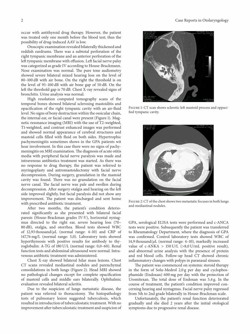
High resolution computed tomography scans of thetemporal bones showed bilateral sclerosing mastoiditis andopacification of the right tympanic cavity with an air-fluidlevel. No signs of bony destruction within the ossicular chain,the internal ear, or facial canal were present (Figure 1). Mag-netic resonance imaging (MRI) with the use of T2-weighted,T1-weighted, and contrast enhanced images was performedand showed normal appearance of cerebral structures andmastoid cells filled with fluid on both sides. Hypertrophicpachymeningitis sometimes shows in the GPA patients withear involvement. In this case there were no signs of pachy-meningitis onMRI examination.The diagnosis of acute otitismedia with peripheral facial nerve paralysis was made andintravenous antibiotics treatment was started. As there wasno response to drug therapy, the patient was referred tomyringoplasty and antromastoidectomy with facial nervedecompression. During surgery, granulation in the mastoidcavity was found. There was no granulation on the facialnerve canal. The facial nerve was pale and swollen duringdecompression. After surgery otalgia and hearing on the leftside improved slightly, but facial paralysis did not show anyimprovement. The patient was discharged and sent homewith prescribed antibiotic treatment.
After two months, the patient’s condition deterio-rated significantly as she presented with bilateral facialparesis (House-Brackman grades IV-V), horizontal nystag-mus directed to the right ear, severe hearing loss (70–80 dB), otalgia, and otorrhea. Blood tests showed WBCof 12,93 thousand/𝜇L (normal range: 4–10) and CRP of147,76mg/L (normal range: 5,0). Laboratory tests showedhyperthyreosis with positive results for antibody to thy-roglobulin: A-TG of 180U/L (normal range: 0,0–60). Renalfunction tests and abdominal ultrasound were normal. Intra-venous antibiotic treatment was administered.
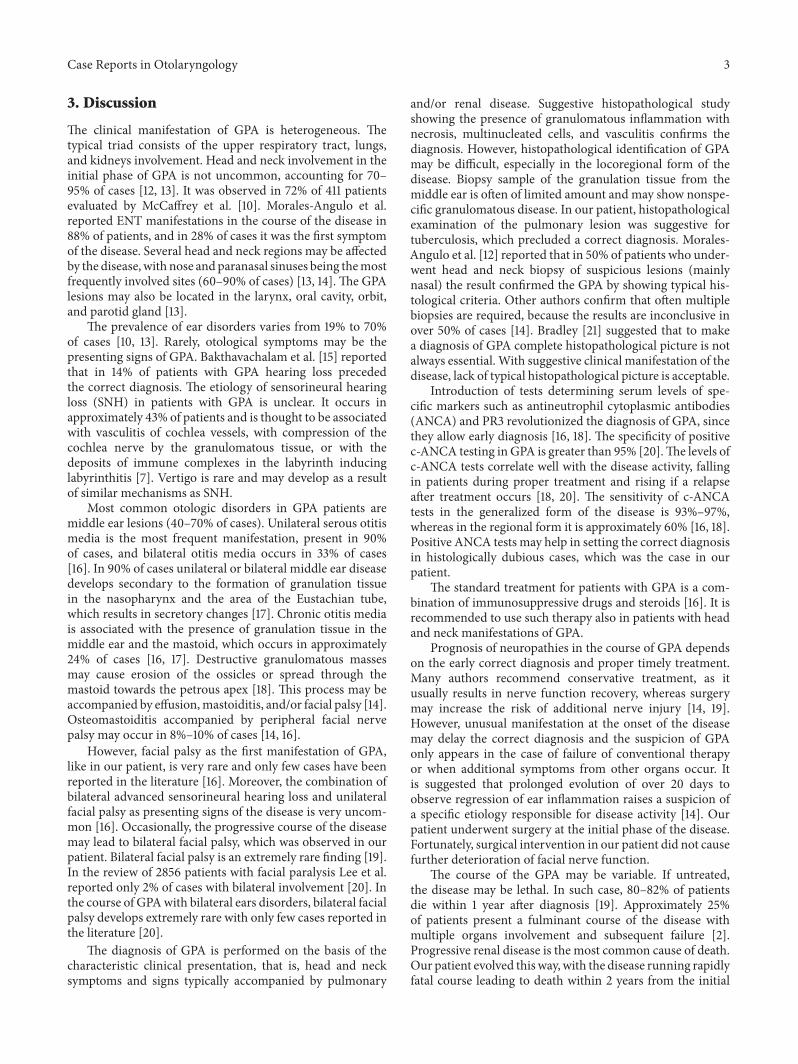
Chest X-ray showed bilateral hilar mass lesions. ChestCT scans revealed mediastinal nodules and parenchymalconsolidations in both lungs (Figure 2). Head MRI showedno pathological changes except for complete opacificationof mastoid cells and middle ears bilaterally. Ophthalmicevaluation revealed bilateral scleritis.
Due to the suspicion of lungs metastatic disease, thepatient was referred to thoracotomy. The histopathologytests of pulmonary lesion suggested tuberculosis, whichresulted in introduction of tuberculostatic treatment.With noimprovement after tuberculostatic treatment and suspicion of
Figure 1: CT scan shows sclerotic left mastoid process and oppaci-fied tympanic cavity.
Figure 2: CT of the chest shows twometastatic focuses in both lungsand mediastinal nodules.
GPA, serological ELISA tests were performed and c-ANCAtests were positive. Subsequently the patient was transferredto Rheumatology Department, where the diagnosis of GPAwas confirmed. Control laboratory tests showed WBC of14,9 thousand/𝜇L (normal range: 4–10), markedly increasedvalue of c-ANKA > 150U/L (>8,0U/mL positive result),and abnormal urine analysis with the presence of proteinand red blood cells. Follow-up head CT showed chronicinflammatory changes with polyps in paranasal sinuses.
The patient was commenced on systemic steroid therapyin the form of Solu-Medrol 2,0 g per day and cyclophos-phamide (Endoxan) 600mg per day with the protection ofUromitexan. The total dose of Endoxan was 5,4 g. In thecourse of treatment, the patient’s condition improved con-cerning hearing and nystagmus. Facial nerve palsy regressedfrom 5th to 2nd grade bilaterally in House-Brackman scale.
Unfortunately, the patient’s renal function deterioratedgradually and she died 2 years after the initial otologicalsymptoms due to progressive renal disease.
Case Reports in Otolaryngology 3
3. Discussion
The clinical manifestation of GPA is heterogeneous. Thetypical triad consists of the upper respiratory tract, lungs,and kidneys involvement. Head and neck involvement in theinitial phase of GPA is not uncommon, accounting for 70–95% of cases [12, 13]. It was observed in 72% of 411 patientsevaluated by McCaffrey et al. [10]. Morales-Angulo et al.reported ENT manifestations in the course of the disease in88% of patients, and in 28% of cases it was the first symptomof the disease. Several head and neck regions may be affectedby the disease, with nose andparanasal sinuses being themostfrequently involved sites (60–90% of cases) [13, 14]. The GPAlesions may also be located in the larynx, oral cavity, orbit,and parotid gland [13].
The prevalence of ear disorders varies from 19% to 70%of cases [10, 13]. Rarely, otological symptoms may be thepresenting signs of GPA. Bakthavachalam et al. [15] reportedthat in 14% of patients with GPA hearing loss precededthe correct diagnosis. The etiology of sensorineural hearingloss (SNH) in patients with GPA is unclear. It occurs inapproximately 43% of patients and is thought to be associatedwith vasculitis of cochlea vessels, with compression of thecochlea nerve by the granulomatous tissue, or with thedeposits of immune complexes in the labyrinth inducinglabyrinthitis [7]. Vertigo is rare and may develop as a resultof similar mechanisms as SNH.
Most common otologic disorders in GPA patients aremiddle ear lesions (40–70% of cases). Unilateral serous otitismedia is the most frequent manifestation, present in 90%of cases, and bilateral otitis media occurs in 33% of cases[16]. In 90% of cases unilateral or bilateral middle ear diseasedevelops secondary to the formation of granulation tissuein the nasopharynx and the area of the Eustachian tube,which results in secretory changes [17]. Chronic otitis mediais associated with the presence of granulation tissue in themiddle ear and the mastoid, which occurs in approximately24% of cases [16, 17]. Destructive granulomatous massesmay cause erosion of the ossicles or spread through themastoid towards the petrous apex [18]. This process may beaccompanied by effusion,mastoiditis, and/or facial palsy [14].Osteomastoiditis accompanied by peripheral facial nervepalsy may occur in 8%–10% of cases [14, 16].
However, facial palsy as the first manifestation of GPA,like in our patient, is very rare and only few cases have beenreported in the literature [16]. Moreover, the combination ofbilateral advanced sensorineural hearing loss and unilateralfacial palsy as presenting signs of the disease is very uncom-mon [16]. Occasionally, the progressive course of the diseasemay lead to bilateral facial palsy, which was observed in ourpatient. Bilateral facial palsy is an extremely rare finding [19].In the review of 2856 patients with facial paralysis Lee et al.reported only 2% of cases with bilateral involvement [20]. Inthe course of GPAwith bilateral ears disorders, bilateral facialpalsy develops extremely rare with only few cases reported inthe literature [20].
The diagnosis of GPA is performed on the basis of thecharacteristic clinical presentation, that is, head and necksymptoms and signs typically accompanied by pulmonary
and/or renal disease. Suggestive histopathological studyshowing the presence of granulomatous inflammation withnecrosis, multinucleated cells, and vasculitis confirms thediagnosis. However, histopathological identification of GPAmay be difficult, especially in the locoregional form of thedisease. Biopsy sample of the granulation tissue from themiddle ear is often of limited amount and may show nonspe-cific granulomatous disease. In our patient, histopathologicalexamination of the pulmonary lesion was suggestive fortuberculosis, which precluded a correct diagnosis. Morales-Angulo et al. [12] reported that in 50% of patients who under-went head and neck biopsy of suspicious lesions (mainlynasal) the result confirmed the GPA by showing typical his-tological criteria. Other authors confirm that often multiplebiopsies are required, because the results are inconclusive inover 50% of cases [14]. Bradley [21] suggested that to makea diagnosis of GPA complete histopathological picture is notalways essential. With suggestive clinical manifestation of thedisease, lack of typical histopathological picture is acceptable.
Introduction of tests determining serum levels of spe-cific markers such as antineutrophil cytoplasmic antibodies(ANCA) and PR3 revolutionized the diagnosis of GPA, sincethey allow early diagnosis [16, 18]. The specificity of positivec-ANCA testing in GPA is greater than 95% [20].The levels ofc-ANCA tests correlate well with the disease activity, fallingin patients during proper treatment and rising if a relapseafter treatment occurs [18, 20]. The sensitivity of c-ANCAtests in the generalized form of the disease is 93%–97%,whereas in the regional form it is approximately 60% [16, 18].Positive ANCA tests may help in setting the correct diagnosisin histologically dubious cases, which was the case in ourpatient.
The standard treatment for patients with GPA is a com-bination of immunosuppressive drugs and steroids [16]. It isrecommended to use such therapy also in patients with headand neck manifestations of GPA.
Prognosis of neuropathies in the course of GPA dependson the early correct diagnosis and proper timely treatment.Many authors recommend conservative treatment, as itusually results in nerve function recovery, whereas surgerymay increase the risk of additional nerve injury [14, 19].However, unusual manifestation at the onset of the diseasemay delay the correct diagnosis and the suspicion of GPAonly appears in the case of failure of conventional therapyor when additional symptoms from other organs occur. Itis suggested that prolonged evolution of over 20 days toobserve regression of ear inflammation raises a suspicion ofa specific etiology responsible for disease activity [14]. Ourpatient underwent surgery at the initial phase of the disease.Fortunately, surgical intervention in our patient did not causefurther deterioration of facial nerve function.
The course of the GPA may be variable. If untreated,the disease may be lethal. In such case, 80–82% of patientsdie within 1 year after diagnosis [19]. Approximately 25%of patients present a fulminant course of the disease withmultiple organs involvement and subsequent failure [2].Progressive renal disease is the most common cause of death.Our patient evolved this way, with the disease running rapidlyfatal course leading to death within 2 years from the initial

4 Case Reports in Otolaryngology
otological symptoms. Preuss et al. [9] presented similar caseof patient withGP-induced bilateral facial palsy together withrenal and pulmonary involvement, which despite intensivetreatment had a fatal course due to multiorgan failure [19].The average survival of patients with GPA is 5 months [19].However, prompt initiation of suitable treatment may resultin long-term remissions in up to 90% of patients, especiallybefore renal involvement [14].
4. Conclusions
In patients with GPA, facial nerve palsy may develop notonly in a classical form of a disease subsequent to middleear involvement but also occasionally in a locoregionalform as primary otological manifestation accompanied bysensorineural hearing loss. In such cases, otolaryngologistshave a crucial role in recognizing the early symptoms of thedisease and starting the appropriate therapy. Otitis mediafailing in responding to conventional treatment, facial nervepalsy accompanying the middle ear disease, or symptomsfrom other organs suggesting the multisystemic involvementshould always raise a suspicion of GPA. Positive ANCA testscontribute to correct diagnosis in dubious cases. Early diag-nosis may prevent the unnecessary and potentially hazardoussurgical treatment and reduce the mortality and morbidityassociated with the disease.
Competing Interests
The authors declare that they have no competing interests.
References
[1] R. J. Falk, W. L. Gross, L. Guillevin et al., “Granulomatosiswith polyangiitis (Wegener’s): an alternative name forWegener’sgranulomatosis,” Arthritis and Rheumatism, vol. 63, no. 4, pp.863–864, 2011.
[2] M. Wierzbicka, M. Puszczewicz, A. Bartochowska, and W.Szyfter, “The otologic manifestation ofWegener’s granulomato-sis—review of contemporary achievements in diagnostics andtreatment,” Otolaryngologia Polska, vol. 66, no. 4, pp. 254–258,2012.
[3] J. C. Jennette, R. J. Falk, P. A. Bacon et al., “2012 RevisedInternational Chapel Hill consensus conference nomenclatureof vasculitides,” Arthritis & Rheumatism, vol. 65, no. 1, pp. 1–11,2013.
[4] M. A. Gonzalez-Gay and C. Garcıa-Porrua, “Epidemiology ofthe vasculitides,” Rheumatic Disease Clinics of North America,vol. 27, no. 4, pp. 729–749, 2001.
[5] M. A. Gonzalez-Gay, C. Garcia-Porrua, J. Guerrero, P.Rodriguez-Ledo, and J. Llorca, “The epidemiology of theprimary systemic vasculitides in northwest Spain: implicationsof the Chapel Hill Consensus Conference definitions,” ArthritisCare and Research, vol. 49, no. 3, pp. 388–393, 2003.
[6] W. Koldingsnes and H. Nossent, “Epidemiology of Wegener’sgranulomatosis in northern Norway,” Arthritis and Rheuma-tism, vol. 43, no. 11, pp. 2481–2487, 2000.
[7] D. Takagi, Y. Nakamaru, S. Maguchi, Y. Furuta, and S. Fukuda,“Otologic manifestations of Wegener’s granulomatosis,” Laryn-goscope, vol. 112, no. 9, pp. 1684–1690, 2002.
[8] S. M. Cassan, D. T. Coles, and E. G. Harrison Jr., “The conceptof limited forms of Wegener’s granulomatosis,” The AmericanJournal of Medicine, vol. 49, no. 3, pp. 366–379, 1970.
[9] S. F. Preuss, M. Stenner, D. Beutner, M. Laudes, and J.P. Klussmann, “Fatal course of Wegener’s granulomatosiswith bilateral otomastoiditis and bilateral facial nerve palsy,”Otolaryngology—Head and Neck Surgery, vol. 138, no. 6, pp.799–800, 2008.
[10] T. V. McCaffrey, T. J. McDonald, G. W. Facer, and R. A. DeRe-mee, “Otologic manifestations of Wegener’s granulomatosis,”Otolaryngology-Head and Neck Surgery, vol. 88, no. 5, pp. 586–593, 1980.
[11] A. D. Kornblut, S. M. Wolff, and A. S. Fauci, “Ear disease inpatients with wegener’s granulomatosis,” Laryngoscope, vol. 92,no. 7, pp. 713–717, 1982.
[12] C. Morales-Angulo, R. Garcıa-Zornoza, S. Obeso-Aguera, J.Calvo-Alen, and M. A. Gonzalez-Gay, “Ear, nose and throatmanifestations of Wegener’s granulomatosis (granulomatosiswith polyangiitis),”ActaOtorrinolaringologica Espanola, vol. 63,no. 3, pp. 206–211, 2012.
[13] S. Gottschlich, P. Ambrosch, D. Kramkowski et al., “Head andneck manifestations of Wegener’s granulomatosis,” Rhinology,vol. 44, no. 4, pp. 227–233, 2006.
[14] A. S. D. A.Maranhao, V. G. Chen, B. A. A. Rossini, J. R. G. Testa,and N. D. O. Penido, “Mastoiditis and facial paralysis as initialmanifestations ofWegener’s Granulomatosis,” Brazilian Journalof Otorhinolaryngology, vol. 78, no. 2, pp. 80–86, 2012.
[15] S. Bakthavachalam, M. S. Driver, C. Cox, J. H. Spiegel, K.M. Grundfast, and P. A. Merkel, “Hearing loss in Wegener’sgranulomatosis,” Otology and Neurotology, vol. 25, no. 5, pp.833–837, 2004.
[16] N. Verma andA.Gupta, “Wegener’s granulomatosis: an unusualpresentation case report and review of the literature,” TheInternet Journal of Otorhinolaryngology, vol. 14, no. 1, 2012.
[17] E. Ferri, E. Armato, P. Capuzzo, S. Cavaleri, and F. Ianniello,“Early diagnosis of Wegener’s granulomatosis presenting withbilateral facial paralysis and bilateral serous otitis media,” AurisNasus Larynx, vol. 34, no. 3, pp. 379–382, 2007.
[18] A. Sharma, S. Deshmukh, and J. Dabholkar, “ENT manifesta-tions ofWegeners granulomatosis,”Otolaryngologia Polska, vol.67, no. 5, pp. 257–260, 2013.
[19] A. Roszkowska, M. Morawska-Kochman, H. Temporale, M.Sikorska-Zuk, and T. Kręcicki, “Bilateral facial palsy in rapidlyprogressive course of wegener’s granulomatosis: a case report,”Case Reports in Otolaryngology, vol. 2013, Article ID 875108, 5pages, 2013.
[20] J. H. Lee, K. W. Kim, N. H. Myong, and J. Y. Jung, “Wegener’sgranulomatosis presenting as bilateral otalgia with facial palsy:a case report,” Korean Journal of Audiology, vol. 17, no. 1, pp. 35–37, 2013.
[21] P. J. Bradley, “Wegener’s granulomatosis of the ear,”The Journalof Laryngology & Otology, vol. 97, no. 7, pp. 623–626, 1983.

Submit your manuscripts athttp://www.hindawi.com
Stem CellsInternational
Hindawi Publishing Corporationhttp://www.hindawi.com Volume 2014
Hindawi Publishing Corporationhttp://www.hindawi.com Volume 2014
MEDIATORSINFLAMMATION
of
Hindawi Publishing Corporationhttp://www.hindawi.com Volume 2014
Behavioural Neurology
EndocrinologyInternational Journal of
Hindawi Publishing Corporationhttp://www.hindawi.com Volume 2014
Hindawi Publishing Corporationhttp://www.hindawi.com Volume 2014
Disease Markers
Hindawi Publishing Corporationhttp://www.hindawi.com Volume 2014
BioMed Research International
OncologyJournal of
Hindawi Publishing Corporationhttp://www.hindawi.com Volume 2014
Hindawi Publishing Corporationhttp://www.hindawi.com Volume 2014
Oxidative Medicine and Cellular Longevity
Hindawi Publishing Corporationhttp://www.hindawi.com Volume 2014
PPAR Research
The Scientific World JournalHindawi Publishing Corporation http://www.hindawi.com Volume 2014
Immunology ResearchHindawi Publishing Corporationhttp://www.hindawi.com Volume 2014
Journal of
ObesityJournal of
Hindawi Publishing Corporationhttp://www.hindawi.com Volume 2014
Hindawi Publishing Corporationhttp://www.hindawi.com Volume 2014
Computational and Mathematical Methods in Medicine
OphthalmologyJournal of
Hindawi Publishing Corporationhttp://www.hindawi.com Volume 2014
Diabetes ResearchJournal of
Hindawi Publishing Corporationhttp://www.hindawi.com Volume 2014
Hindawi Publishing Corporationhttp://www.hindawi.com Volume 2014
Research and TreatmentAIDS
Hindawi Publishing Corporationhttp://www.hindawi.com Volume 2014
Gastroenterology Research and Practice
Hindawi Publishing Corporationhttp://www.hindawi.com Volume 2014
Parkinson’s Disease
Evidence-Based Complementary and Alternative Medicine
Volume 2014Hindawi Publishing Corporationhttp://www.hindawi.com