case report good functional outcome after prolonged...
TRANSCRIPT

Hindawi Publishing CorporationCase Reports in Neurological MedicineVolume 2013, Article ID 872127, 5 pageshttp://dx.doi.org/10.1155/2013/872127
Case ReportGood Functional Outcome after Prolonged PostanoxicComatose Myoclonic Status Epilepticus in a PatientWho Had Undergone Bone Marrow Transplantation
Jennifer Accardo,1 Domenico De Lisi,2 Paola Lazzerini,3 and Alberto Primavera1
1 Centro di Fisiopatologia del Sonno, DINOGMI, Universita di Genova, Largo Rosanna Benzi, 10 16132 Genova, Italy2 U.O. Anestesia e Rianimazione, DIPEA, IRCCS Azienda Ospedaliera Universitaria San Martino-IST, Largo Rosanna Benzi,10 16132 Genova, Italy
3 U.O. Neurofisiopatologia, IRCCS Azienda Ospedaliera Universitaria San Martino-IST, Largo Rosanna Benzi,10 16132 Genova, Italy
Correspondence should be addressed to Jennifer Accardo; [email protected]
Received 16 September 2013; Accepted 22 October 2013
Academic Editors: I. L. Simone and M. Swash
Copyright © 2013 Jennifer Accardo et al. This is an open access article distributed under the Creative Commons AttributionLicense, which permits unrestricted use, distribution, and reproduction in any medium, provided the original work is properlycited.
In anoxic coma, myoclonic status epilepticus and other nonreactive epileptiform patterns are considered as signs of poor prognosis.We report the case of a good recovery in a prolonged comatosemyoclonic status epilepticus (MSE) after a cardiac arrest (CA) treatedwithmild therapeutic hypothermia (TH) in a patient who had undergone a bonemarrow transplantation for Hodgkin’s lymphoma.This case emphasizes the opportunity of performing an electroencephalogram (EEG) in the acute period after an hypoxic-ischemicinsult and underlines the diagnostic difficulties betweenMSE and Lance-Adams syndrome, which classically occurs after the patienthas regained consciousness, but can also begin while the patient is still comatose or sedated. Major problems in prognostication forpostarrest comatose patients will also be pointed out.
1. Introduction
Postanoxic status epilepticus, particularly myoclonic status,is traditionally considered a marker of unfavorable outcome.Overall, the prognosis is extremely poor, with only a fractionof patients surviving hospital discharge and often even thenreporting severe neurological or cognitive deficits [1–4].
With the advent of therapeutic hypothermia, an improve-ment in outcome was described in comatose survivors. Inparticular, some authors reported cases of patients with earlypost-anoxic MSE who presented a good recovery [5–7].There are also a handful of case reports that mention earlymyoclonus in patients who regained consciousness and hada good neurological outcome after cardiorespiratory arrest[8–12]. These findings stress the importance of consideringa combination of prognostic features before making anyoutcome prediction, with also bearing inmind the confound-ing effects of several factors, including but not limited tohypothermia and sedatives.
We describe the case of a patient who had undergone abonemarrow transplantation and had a good recovery after aprolonged post-anoxic MSE. Our case aims to demonstratethe possibility of reasonable neurological recovery despiteearly onset of myoclonic status even when very seriouscomorbidity is present. The diagnostic difficulties betweenMSE and Lance-Adams syndrome are also underlined.
2. Case Report
A 52-year-old man was diagnosed with Hodgkin’s lymphomain March 2009. He was treated with radiotherapy, five cyclesof chemotherapy, and then underwent autologous bonemarrow transplantation, all of which did not prove beneficialin terms of remission.
Finally, in March 2010 an allogenic transplantation wasperformed, obtaining a good hematologic response.
Approximately 60 days later, the patient complained ofchest pain and subsequently suffered a ventricular fibrillation

2 Case Reports in Neurological Medicine
CA. The paramedics resuscitated him, but he remainedcomatose, showing no withdrawal to painful stimuli, slug-gishly reactive pupils, and very weak corneal reflexes. Thepatient was then transported to an intensive care unit (ICU),where he was intubated and ventilated and amild therapeutichypothermia (target temperature 34∘C)was performed over aperiod of 24 hours. An urgent cranial computed tomography(CT) scanwas negative for hemorrhage, cerebral oedema, andstructural lesions.
After the sedative reduction, the patient was stillcomatose with a Glasgow coma scale motor (GCS-M) score<2, had no spontaneous respiration, and presented massivediffusemyoclonic jerks, related to the seizures and resistant tophenytoin, diazepammidazolam, and propofol. An EEG wasperformed 36 hours after insult, showing a pattern suggestiveof unreactive MSE (Figure 1). Levetiracetam was added atthe dose of 2000mg/die and later increased to 3000mg/die,without apparent improvement.
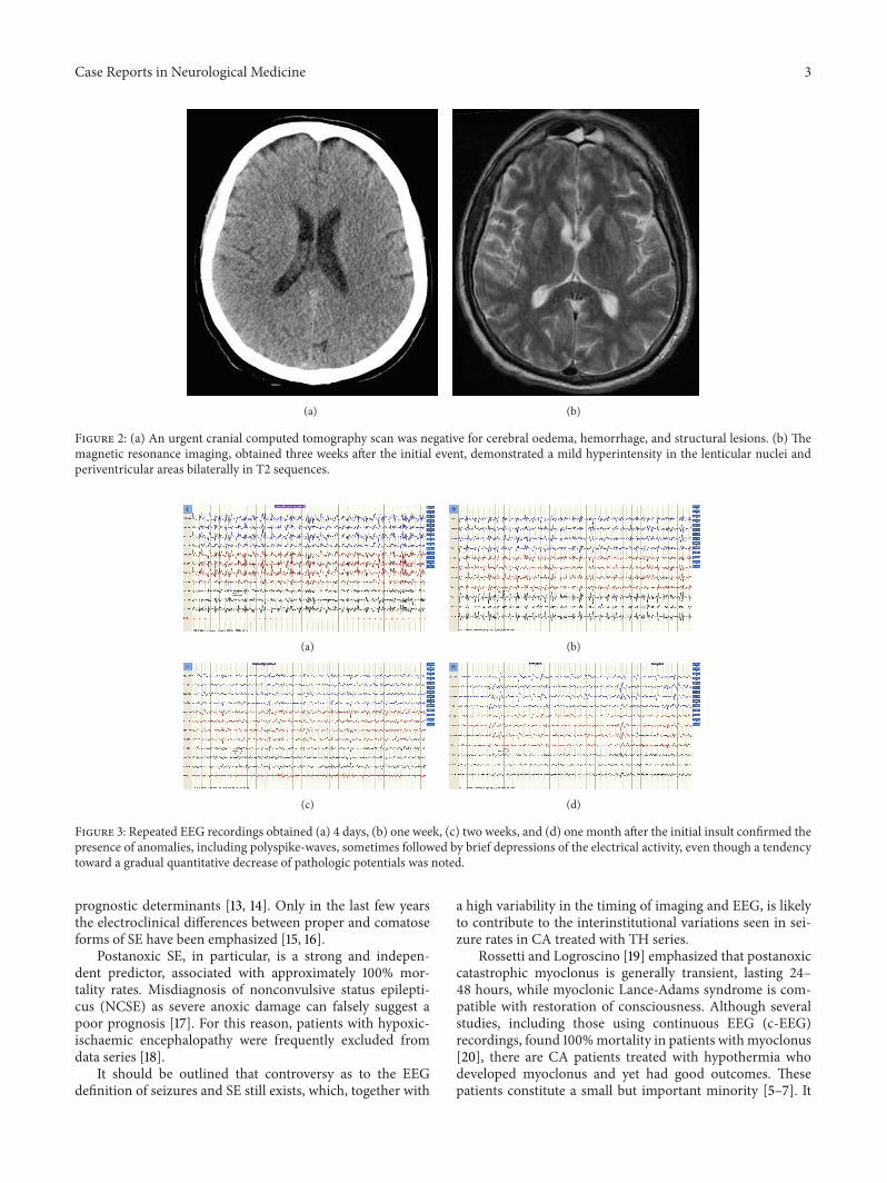
Magnetic resonance imaging (MRI), obtained threeweeks later, demonstrated a mild hyperintensity in the lentic-ular nuclei and periventricular areas bilaterally in FLAIR andT2 sequences (Figure 2).
Repeated EEG recordings confirmed the presence ofanomalies, including recurrent polyspike waves, sometimesfollowed by brief depressions of the electrical activity(Figure 3), even though a tendency toward a gradual quan-titative decrease of irritative potentials was noted. Diffuse,generalized, unrelenting myoclonic jerks involving the face,trunk, and limbs persisted and sometimes appeared to bestimulus related.
Due to the patient’s severe general conditions, immuno-suppressive therapy was suspended. On day 10 after theinitial event, a tracheostomy was performed. Eye openingoccurred on day 14, while at day 36 the patient occasionallypresented with brief deviation of eyes towards the source ofstimulation. In the next days, the patient slowly started toregain consciousness and was able to interact with the sur-rounding environment and feed autonomously, while untilthat moment a parenteral nutrition was needed. Myoclonicjerks persisted, but they became less frequent.The patient wasdischarged on day 80, after a cycle of rehabilitation to helphim reacquire the ability to ambulate.
During the following months, a mild cognitive recoverywas achieved (MMSE: 20/30), even though he presentedwith acalculia, agraphia, dysarthria, and action myoclonus,especially in relation with movement and emotional stim-uli. A follow-up EEG performed seven months after theinitial insult demonstrated a normal background activity,although isolated brief polyspike discharges appeared, inassociation with the myoclonic jerks (Figure 4). The patientalso underwent a follow-up cerebral MRI with Turbo-FLAIRand DWI sequences, which did not reveal the previouslyreported hyperintensity involving the lenticular nuclei, whileit showed a reduction of the hyperintensity involving thelateral periventricular areas bilaterally.
At present (three years after the initial event), the patienthas an improved cognitive performance with anMMSE scoreof 23/30 and is sufficiently autonomous with a Glasgow-Pittsburgh cerebral performance category scale score of 2,
Figure 1: EEG pattern 36 hours after the patient suffered a cardiacarrest and treated with mild therapeutic hypothermia. After thesedative discontinuation, the EEG appeared unreactive with diffuseepileptiformdischarges (polyspike-wave complexes) associatedwithcontinuous spontaneous generalized multifocal jerks, involving theface, limbs, and axial muscles.
while the intensity of myoclonus has reduced, althoughLance-Adams syndrome persists. His therapy consists of lowdoses of Diazepam, Levetiracetam 1000mg two times a day,and speech therapy. He still undergoes hematologic andneurologic follow-up visits on a three months basis.
3. Discussion
Theprognosis of a patient after a CA is influenced bymultiplefactors, including age, presence of comorbidities, circum-stances and duration of arrest, characteristics of resuscitationand cardiac rhythm, duration of impaired consciousness afterthe event, and presence of seizures.
In the case we presented, the patient had multiple factorssuggesting an unfavorable outcome.
The presence of serious comorbidities, which consisted inHodgkin’s lymphoma requiring two bone marrow transplan-tations, could be considered an important element indicatinga poor prognosis.
A prolonged coma following nontraumatic cerebralinjuries also carries a very poor prognosis. In postcardiacarrest patients treated with therapeutic hypothermia, time toawakening after resuscitation is highly variable and often nolonger than three days. Earlier awakening is associated withbetter neurologic status at hospital discharge. In our patient’scase, the impairment of mental status lasted for more than amonth.
Another indicator of severe prognosis in this patient wasthe presence of myoclonus after sedative medications werereduced, in association with an unreactive EEG pattern con-sistent with posthypoxic status epilepticus (SE) lasting at least20 days. The EEG classification and its variations over timehave been shown to hold a prognostic value for favorable andunfavorable outcomes. “Malignant” EEG patterns includemyoclonus with EEG correlate, a nonreactive background,burst suppression, and SE; nevertheless, sedation duringhypothermia might create an iatrogenic, reversible burst-suppression pattern and preclude reactivity in some patients.
In adult patients with SE, age, history of prior seizuresor epilepsy, SE aetiology, level of consciousness, seizure typeat SE onset, seizure duration, need for mechanical ventila-tion, and development of acute complications are the major
Case Reports in Neurological Medicine 3
(a) (b)
Figure 2: (a) An urgent cranial computed tomography scan was negative for cerebral oedema, hemorrhage, and structural lesions. (b) Themagnetic resonance imaging, obtained three weeks after the initial event, demonstrated a mild hyperintensity in the lenticular nuclei andperiventricular areas bilaterally in T2 sequences.
(a) (b)
(c) (d)
Figure 3: Repeated EEG recordings obtained (a) 4 days, (b) one week, (c) two weeks, and (d) one month after the initial insult confirmed thepresence of anomalies, including polyspike-waves, sometimes followed by brief depressions of the electrical activity, even though a tendencytoward a gradual quantitative decrease of pathologic potentials was noted.
prognostic determinants [13, 14]. Only in the last few yearsthe electroclinical differences between proper and comatoseforms of SE have been emphasized [15, 16].
Postanoxic SE, in particular, is a strong and indepen-dent predictor, associated with approximately 100% mor-tality rates. Misdiagnosis of nonconvulsive status epilepti-cus (NCSE) as severe anoxic damage can falsely suggest apoor prognosis [17]. For this reason, patients with hypoxic-ischaemic encephalopathy were frequently excluded fromdata series [18].
It should be outlined that controversy as to the EEGdefinition of seizures and SE still exists, which, together with
a high variability in the timing of imaging and EEG, is likelyto contribute to the interinstitutional variations seen in sei-zure rates in CA treated with TH series.
Rossetti and Logroscino [19] emphasized that postanoxiccatastrophic myoclonus is generally transient, lasting 24–48 hours, while myoclonic Lance-Adams syndrome is com-patible with restoration of consciousness. Although severalstudies, including those using continuous EEG (c-EEG)recordings, found 100%mortality in patients withmyoclonus[20], there are CA patients treated with hypothermia whodeveloped myoclonus and yet had good outcomes. Thesepatients constitute a small but important minority [5–7]. It

4 Case Reports in Neurological Medicine
Figure 4: A follow-up EEG obtained seven months later revealed anormal background activity, although isolated diffuse polyspike dis-charges time-locked with myoclonic jerks persisted, more evidentlyin the anterior regions.
is possible that hypothermia has an ameliorating effect, notonly on the hypoxic-ischemic insult but also on the damagingeffect of seizures.
The good recovery described in some cases and theoverlapping with Lance-Adams syndrome emphasize theopportunity of a subcategorization based, at least, on etiology,duration of the altered state of consciousness, neuroimagingfeatures, and clinical and neurophysiological findings.
Notwithstanding the presence of factors indicating asevere prognosis, some other elements were present in ourpatient that could indicate a more favorable outcome, such asthe arrest being due to a ventricular fibrillation as opposed topulseless electrical activity or asystole.
Data derived from neuroimaging also showed no oedemaor structural lesions in the acute phase, which could arguablyindicate that the cerebral suffering and damage were limitedand the patient’s compromised consciousness could havepartially been a result of SE rather than anatomic injury.Preliminary studies indicate that quantitativeMRI has strongpredictive value after CA and TH, since it can predictthe long-term functional and cognitive impact of hypoxic-ischemic encephalopathy, especially when performed withinfive days after the event [21, 22].
Other factors indicated the possibility of a favorableoutcome: more specifically, the sluggish pupil reactivity andweak corneal reflex suggested some preservation of brainstem functions.
In this case, it could be useful to consider that posthypoxicmyoclonus is generally divided into two entities, acute andchronic posthypoxic myoclonus.
Acute posthypoxic myoclonus, also known as myoclonicstatus epilepticus (MSE), typically begins within 24 hoursfrom the hypoxic insult and occurs in patients who are deeplycomatose, even after the discontinuation of sedative drugs.It consists of continuous (usually massive) myoclonus, withrhythmic or irregular bilateral synchronous jerking of face,trunk, and limbs, often with repetitive blinking, eye opening,upward eye rolling, and mouth twitching. The EEG showsgeneralized, bisynchronous polyspikes, spikes or sharp wavespreceding and time-locked with the clinical myoclonus,superimposed on a diffusely slow and suppressed backgroundor burst-suppression pattern.The treatment of this conditionis difficult and of questionable usefulness, since it is associatedwith poor prognosis. Nevertheless, prolonged seizures may
cause cerebral injury and should be treated promptly andeffectively with benzodiazepines, sodium valproate, propofol,levetiracetam, or a barbiturate.
Chronic posthypoxic myoclonus typically occurs withina few days to a few weeks after the hypoxic injury. Firstdescribed by Lance and Adams in 1963, it is a multifocalactionmyoclonus in combinationwith startle-sensitive, bilat-eral, and generalized jerks and is usually accompanied bydysmetria, dysarthria, and ataxia, with relative preservationof higher cognitive functions. It classically occurs after thepatient has regained mental status but can begin while thepatient is still comatose or sedated [8, 9, 12]. The prognosisis generally favorable and these patients continue to improveover time although cerebellar signs may persist. The EEGshows a cortical origin with responsive cortical rhythmswhich progressively regain normal patterns. Chronic posthy-poxic myoclonus has been shown to respond especially tovalproate, levetiracetam, and clonazepam.
It is vital to accurately distinguish between MSE andLance-Adams syndrome, due to their very different prog-noses. Although the distinction between these two conditionsmay be favored by the aforementioned aspects, a correct diag-nosis is sometimes complicated, since there is a considerableclinical overlap and several confounders are usually present.
These considerations lead us to think that it was appro-priate to describe our patient as initially suffering fromMSE,which later evolved to fulfill the criteria for Lance-Adamssyndrome that Yadavmali and Lane [11] suggested. However,the majority of authors prefer to consider this evolution as aninitial misdiagnosis of Lance-Adams syndrome [9, 23].
In summary, when evaluating comatose postarrestpatients with myoclonus, it should be borne in mindthat clinical assessment is often unreliable. Ancillaryinvestigations can add valuable information, although theyhave traditionally been underused and there is no establishedalgorithm of clinical signs or investigations which allow adefinitive prognosis. Blondin and Greer [24] suggested thatthe American Academy of Neurology practice parameters forassessing prognosis after CAmay not be accurate for patientstreated with TH and may lead to an overly pessimisticprognostication and premature withdrawal of care [9, 23].
4. Conclusion
The patient described here developed a post-anoxic MSE andwas unconscious for a prolonged time span, at least longerthan 35 days, however, in absence of remarkable neuroimag-ing signs of cerebral anoxia. Recovery of consciousness wasgradual, but a Lance-Adams syndrome persisted at followup,after 3 years.We suggest thatMSEwas the earlymanifestationof an hypoxic-ischemic encephalopathy and later evolved tofulfill the criteria for Lance-Adams syndrome.
It is mandatory to perform a global clinical, neurophys-iological, and radiological evaluation of the patient beforeestablishing a prognosis, since an inaccurate predictionof poor outcome may result in the patient being deniedpotentially life-saving treatments. As Benson and Young [25]suggested, myoclonus may not be always the “kiss of death.”

Case Reports in Neurological Medicine 5
Conflict of Interests
The authors declare that there is no conflict of interestsregarding the publication of this paper.
Acknowledgment
The authors would like to thank Dr. Van Lindt for herprecious help in handling and taking care of this patient.
References
[1] A. Krumholz, B. J. Stern, andH.D.Weiss, “Outcome from comaafter cardiopulmonary resuscitation: relation to seizures andmyoclonus,” Neurology, vol. 38, no. 3, pp. 401–405, 1988.
[2] G. G. Celesia, M. M. Grigg, and E. Ross, “Generalizedstatus myoclonicus in acute anoxic and toxic-metabolicencephalopathies,” Archives of Neurology, vol. 45, no. 7, pp.781–784, 1988.
[3] G. B. Young, J. J. Gilbert, andD.W. Zochodne, “The significanceof myoclonic status epilepticus in postanoxic coma,”Neurology,vol. 40, no. 12, pp. 1843–1848, 1990.
[4] E. F. M.Wijdicks, J. E. Parisi, and F.W. Sharbrough, “Prognosticvalue of myoclonus status in comatose survivors of cardiacarrest,” Annals of Neurology, vol. 35, no. 2, pp. 239–243, 1994.
[5] A. O. Rossetti, M. Oddo, L. Liaudet, and P. W. Kaplan, “Pre-dictors of awakening from postanoxic status epilepticus aftertherapeutic hypothermia,”Neurology, vol. 72, no. 8, pp. 744–749,2009.
[6] A. O. Rossetti, M. Oddo, G. Logroscino, and P. W. Kaplan,“Prognostication after cardiac arrest and hypothermia: aprospective study,” Annals of Neurology, vol. 67, no. 3, pp. 301–307, 2010.
[7] J. M. Lucas, M. N. Cocchi, J. Salciccioli et al., “Neurologicrecovery after therapeutic hypothermia in patients with post-cardiac arrest myoclonus,” Resuscitation, vol. 83, no. 2, pp. 265–269, 2012.
[8] H. R. Morris, R. S. Howard, and P. Brown, “Early myoclonicstatus and outcome after cardiorespiratory arrest,” Journal ofNeurology Neurosurgery and Psychiatry, vol. 64, no. 2, pp. 267–268, 1998.
[9] W. A. English, N. J. Giffin, and J. P. Nolan, “Myoclonus aftercardiac arrest: pitfalls in diagnosis and prognosis,” Anaesthesia,vol. 64, no. 8, pp. 908–911, 2009.
[10] S. Datta, G. K. Hart, H. Opdam, G. Gutteridge, and J. Archer,“Post-hypoxic myoclonic status: the prognosis is not alwayshopeless,” Critical Care and Resuscitation, vol. 11, no. 1, pp. 39–41, 2009.
[11] T. Yadavmali and A. S. Lane, “The Lance-Adams syndrome:helpful or just hopeful, after cardiopulmonary arrest,” Journalof the Intensive Care Society, vol. 12, no. 4, pp. 324–328, 2011.
[12] J. H. Shin, J.M. Park, A. R. Kim et al., “Lance-Adams syndrome,”Annals of Rehabilitation Medicine, vol. 36, no. 4, pp. 561–564,2012.
[13] B. F. Shneker and N. B. Fountain, “Assessment of acutemorbidity and mortality in nonconvulsive status epilepticus,”Neurology, vol. 61, no. 8, pp. 1066–1073, 2003.
[14] R. Sutter, P. Kaplan, and S. Ruegg, “Outcome predictors for sta-tus epilepticus—what really counts,”Nature Reviews Neurology,vol. 9, no. 9, pp. 525–534, 2013.
[15] G. Bauer and E. Trinka, “Nonconvulsive status epilepticus andcoma,” Epilepsia, vol. 51, no. 2, pp. 177–190, 2010.
[16] J. L. Fernandez-Torre, M. Rebollo, A. Gutierrez, F. Lopez-Espadas, and M. A. Hernandez-Hernandez, “Nonconvulsivestatus epilepticus in adults: electroclinical differences betweenproper and comatose forms,” Clinical Neurophysiology, vol. 123,no. 2, pp. 244–251, 2012.
[17] P. W. Kaplan, “EEG criteria for nonconvulsive status epilepti-cus,” Epilepsia, vol. 48, no. 8, pp. 39–41, 2007.
[18] S. E. Hocker, J. W. Britton, J. N. Mandrekar, E. F. Wijdicks, andA. A. Rabinstein, “Predictors of outcome in refractory statusepilepticus,” JAMA Neurology, vol. 70, no. 1, pp. 72–77, 2013.
[19] A. O. Rossetti and G. Logroscino, “In-hospital mortality ofgeneralized convulsive status epilepticus: a large us sample,”Neurology, vol. 70, no. 20, pp. 1939–1940, 2008.
[20] A. Z. Crepeau, A. A. Rabinstein, J. E. Fugate et al., “ContinuousEEG in therapeutic hypothermia after cardiac arrest: prognosticand clinical value,” Neurology, vol. 80, no. 4, pp. 339–344, 2013.
[21] M. Mlynash, D. M. Campbell, E. M. Leproust et al., “Temporaland spatial profile of brain diffusion-weightedMRI after cardiacarrest,” Stroke, vol. 41, no. 8, pp. 1665–1672, 2010.
[22] J. Kim, B. S. Choi, K. Kim et al., “Prognostic performanceof diffusion-weighted MRI combined with NSE in comatosecardiac arrest survivors treated with mild hypothermia,” Neu-rocritical Care, vol. 17, no. 3, pp. 412–420, 2012.
[23] J. Sreedharan, E. Gourlay,M. R. Evans, andM. Koutroumanidis,“Falsely pessimistic prognosis by EEG in post-anoxic comaafter cardiac arrest: the borderland of nonconvulsive statusepilepticus,” Epileptic Disorders, vol. 14, no. 3, pp. 340–344, 2012.
[24] N. A. Blondin and D. M. Greer, “Neurologic prognosis incardiac arrest patients treated with therapeutic hypothermia,”Neurologist, vol. 17, no. 5, pp. 241–248, 2011.
[25] C. Benson and G. B. Young, “EEG monitoring after cardiacarrest—the cold facts,”Neurology, vol. 80, no. 4, article 343, 2013.

Submit your manuscripts athttp://www.hindawi.com
Stem CellsInternational
Hindawi Publishing Corporationhttp://www.hindawi.com Volume 2014
Hindawi Publishing Corporationhttp://www.hindawi.com Volume 2014
MEDIATORSINFLAMMATION
of
Hindawi Publishing Corporationhttp://www.hindawi.com Volume 2014
Behavioural Neurology
EndocrinologyInternational Journal of
Hindawi Publishing Corporationhttp://www.hindawi.com Volume 2014
Hindawi Publishing Corporationhttp://www.hindawi.com Volume 2014
Disease Markers
Hindawi Publishing Corporationhttp://www.hindawi.com Volume 2014
BioMed Research International
OncologyJournal of
Hindawi Publishing Corporationhttp://www.hindawi.com Volume 2014
Hindawi Publishing Corporationhttp://www.hindawi.com Volume 2014
Oxidative Medicine and Cellular Longevity
Hindawi Publishing Corporationhttp://www.hindawi.com Volume 2014
PPAR Research
The Scientific World JournalHindawi Publishing Corporation http://www.hindawi.com Volume 2014
Immunology ResearchHindawi Publishing Corporationhttp://www.hindawi.com Volume 2014
Journal of
ObesityJournal of
Hindawi Publishing Corporationhttp://www.hindawi.com Volume 2014
Hindawi Publishing Corporationhttp://www.hindawi.com Volume 2014
Computational and Mathematical Methods in Medicine
OphthalmologyJournal of
Hindawi Publishing Corporationhttp://www.hindawi.com Volume 2014
Diabetes ResearchJournal of
Hindawi Publishing Corporationhttp://www.hindawi.com Volume 2014
Hindawi Publishing Corporationhttp://www.hindawi.com Volume 2014
Research and TreatmentAIDS
Hindawi Publishing Corporationhttp://www.hindawi.com Volume 2014
Gastroenterology Research and Practice
Hindawi Publishing Corporationhttp://www.hindawi.com Volume 2014
Parkinson’s Disease
Evidence-Based Complementary and Alternative Medicine
Volume 2014Hindawi Publishing Corporationhttp://www.hindawi.com