case report extramammary paget’s disease of the … · case report extramammary paget’s disease...
TRANSCRIPT
Int J Clin Exp Med 2017;10(12):16750-16754www.ijcem.com /ISSN:1940-5901/IJCEM0061848
Case Report Extramammary Paget’s disease of the scrotum: a case report and review of the literature
Riqiang Liu1, Xiuwang Wei2, Haiming Wei3, Bin Hu2
Departments of 1Hepatobiliary and Endocrine Surgery, 2Urology, 3Pathology, The People’s Hospital of Guangxi Zhuang Autonomous Region, Nanning, Guangxi, People’s Republic of China
Received July 19, 2017; Accepted November 1, 2017; Epub December 15, 2017; Published December 30, 2017
Abstract: Extramammary Paget’s disease (EMPD) is an uncommon cutaneous, intraepithelial adenocarcinoma which is slow-growing and developing in the apocrine gland-bearing areas. We describe the case of EMPD on the scrotum which was discovered in a 77-year-old male previously misdiagnosed and treated as other typical derma-tosis. EMPD can often mimic various types of other dermatosis such as eczema or dermatitis. Apart from clinical histology, the laboratory examination is of vital importance for accurate diagnosis. The diagnosis is combined with biopsy and immunohistochemical staining. Wide surgical excision for the treatment of EMPD is considered as the first choice. Long-term follow-up is mandatory in these patients because of the tendency of subsequent recurrence or concurrent malignancy of the disease.
Keywords: Paget’s disease, extramammary, scrotum neoplasm
Introduction
The Paget’s disease was first described by Paget in 1874, involving the nipple underlying ductal carcinoma of the breast [1]. Then Crocker described the first extramammary Paget’s dis-ease (EMPD) of the scrotum and penis in 1889 [2]. EMPD is an extremely rare cutaneous, intraepithelial adenocarcinoma (Since the first case was reported, there are only a few hun-dreds of reports that have been documented in peer-reviewed literature). It occurs mainly among postmenopausal women beyond 60 years of age. Also it has been reported in male patients, albeit more rarely [3-5]. The disease is usually observed in cutaneous apocrine gland-bearing regions, especially the vulva, perineum, perianal region, scrotum, penis, while it would also affect some less common areas like the axilla, groin, thigh, eyelid, exter-nal ear, and umbilicus [3, 6, 7]. Due to the rarity of EMPD and its early atypical and nonspecific symptoms, this tumor represents a diagnostic dilemma, and it is often misdiagnosed as other benign dermatologic diagnosis as eczema or dermatitis, leading to faulty or delay treatment [8]. The diagnosis of EMPD is clinical and must be confirmed by the histopathological examina-
tion of a tissue specimen. Some adjuvant treat-ment has been applied to EPMD such as che-motherapy, radiotherapy, and topical applica-tion of imiquimod, 5% cream or cytotoxic agent with 1% 5-fluorouracil cream [9-11]. Nonethel- ess, radical surgical excision is still the best treatment options.
We describe a case of EMPD affecting the scro-tum and initially misdiagnosed as eczema. We also present a review of the relevant literature to gain new insight in the diagnosis of this rare neoplasm.
Case report
A 77-year-old male was referred to our depart-ment of urology by a dermatologist, having per-formed biopsy of local excision of a scrotal lesion with the initial histology reported as EMPD. The patient presented to a 2-year history of recurrent, itchy, eczematous erosion, and indurated patchy lesion on his left side of scro-tum prior to presentation to the dermatologist, which had been treated with topical corticoste-roids and antibiotics. However, the skin lesion had failed treatment and progressed to an enlarging erosive lesion. Then the dermatolo-
Misdiagnosed extramammary Paget’s disease of the scrotum
16751 Int J Clin Exp Med 2017;10(12):16750-16754
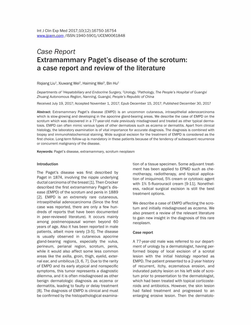
gist prompted referral for wide excision. The patient’s medical history was unremarkable except for hypertension, cataract, and thoraco-lumbar fracture, and he denied family history of similar disease. Physical examination revealed a 5 cm erythematous, fleshy, exophytic plaque at the left side of scrotum (Figure 1) without
palpable inguinal lymphadenopathy or other systemic symptoms. There was a less satellite lesion proximal to this.
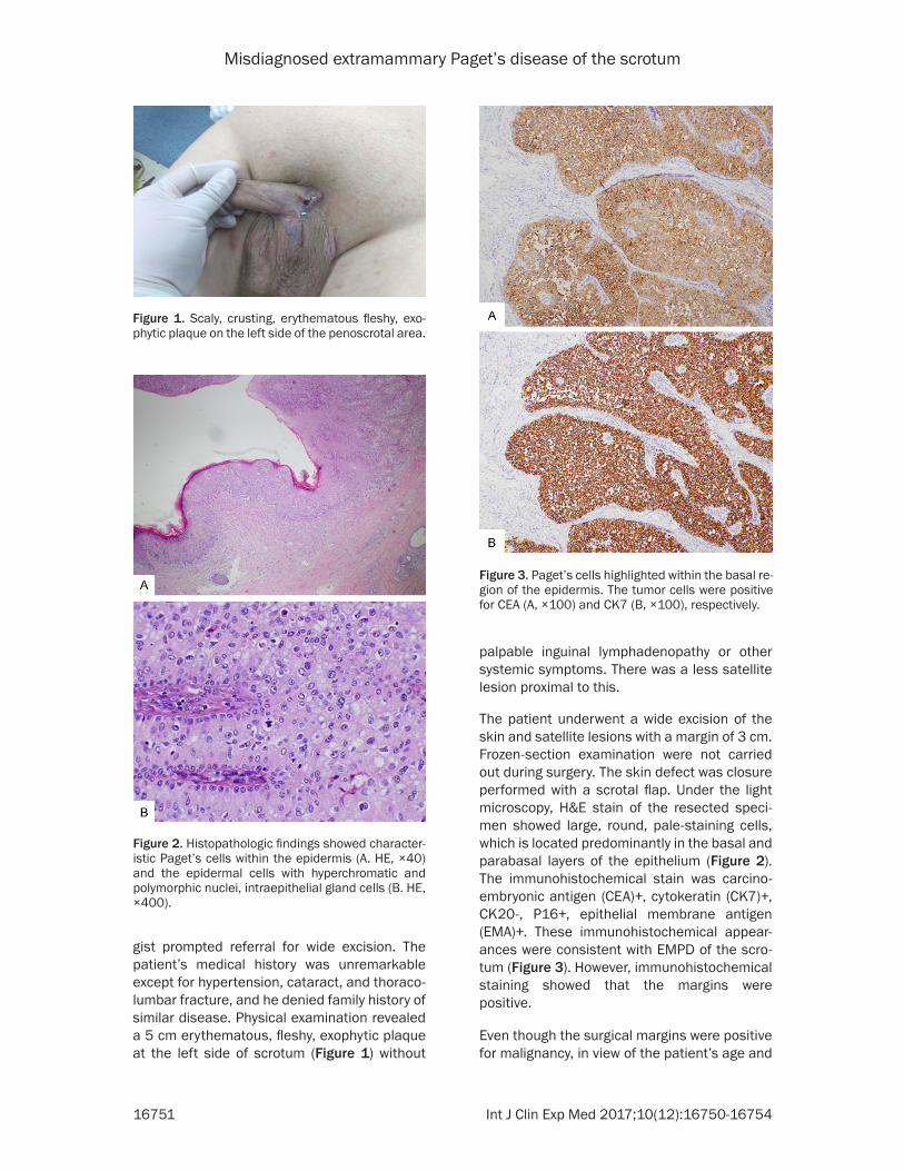
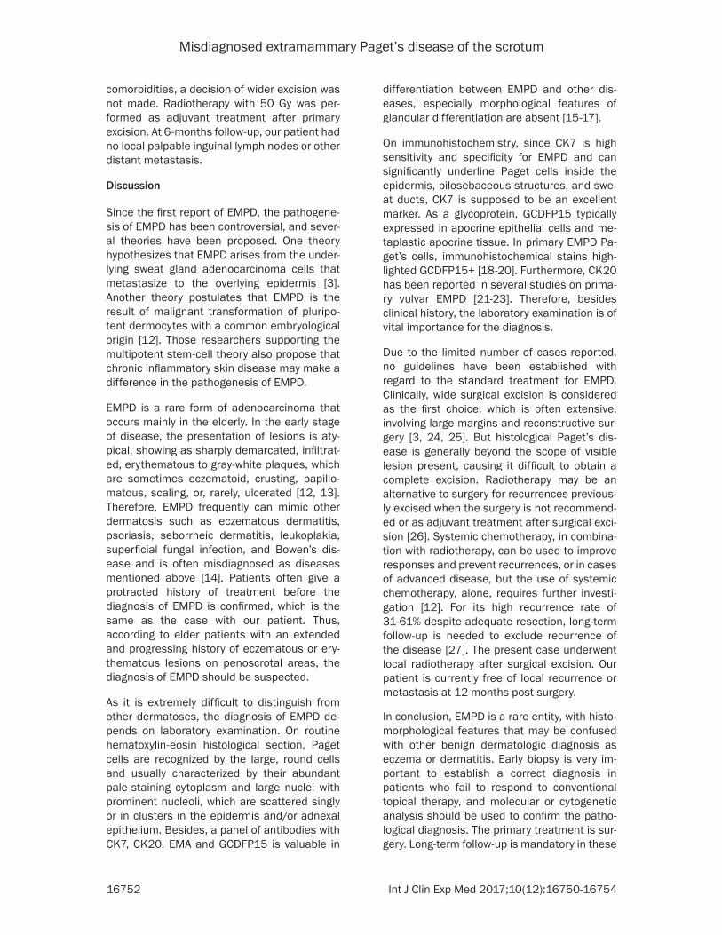
The patient underwent a wide excision of the skin and satellite lesions with a margin of 3 cm. Frozen-section examination were not carried out during surgery. The skin defect was closure performed with a scrotal flap. Under the light microscopy, H&E stain of the resected speci-men showed large, round, pale-staining cells, which is located predominantly in the basal and parabasal layers of the epithelium (Figure 2). The immunohistochemical stain was carcino-embryonic antigen (CEA)+, cytokeratin (CK7)+, CK20-, P16+, epithelial membrane antigen (EMA)+. These immunohistochemical appear-ances were consistent with EMPD of the scro-tum (Figure 3). However, immunohistochemical staining showed that the margins were positive.
Even though the surgical margins were positive for malignancy, in view of the patient’s age and
Figure 1. Scaly, crusting, erythematous fleshy, exo-phytic plaque on the left side of the penoscrotal area.
Figure 2. Histopathologic findings showed character-istic Paget’s cells within the epidermis (A. HE, ×40) and the epidermal cells with hyperchromatic and polymorphic nuclei, intraepithelial gland cells (B. HE, ×400).
Figure 3. Paget’s cells highlighted within the basal re-gion of the epidermis. The tumor cells were positive for CEA (A, ×100) and CK7 (B, ×100), respectively.
Misdiagnosed extramammary Paget’s disease of the scrotum
16752 Int J Clin Exp Med 2017;10(12):16750-16754
comorbidities, a decision of wider excision was not made. Radiotherapy with 50 Gy was per-formed as adjuvant treatment after primary excision. At 6-months follow-up, our patient had no local palpable inguinal lymph nodes or other distant metastasis.
Discussion
Since the first report of EMPD, the pathogene-sis of EMPD has been controversial, and sever-al theories have been proposed. One theory hypothesizes that EMPD arises from the under-lying sweat gland adenocarcinoma cells that metastasize to the overlying epidermis [3]. Another theory postulates that EMPD is the result of malignant transformation of pluripo-tent dermocytes with a common embryological origin [12]. Those researchers supporting the multipotent stem-cell theory also propose that chronic inflammatory skin disease may make a difference in the pathogenesis of EMPD.
EMPD is a rare form of adenocarcinoma that occurs mainly in the elderly. In the early stage of disease, the presentation of lesions is aty- pical, showing as sharply demarcated, infiltrat-ed, erythematous to gray-white plaques, which are sometimes eczematoid, crusting, papillo-matous, scaling, or, rarely, ulcerated [12, 13]. Therefore, EMPD frequently can mimic other dermatosis such as eczematous dermatitis, psoriasis, seborrheic dermatitis, leukoplakia, superficial fungal infection, and Bowen’s dis-ease and is often misdiagnosed as diseases mentioned above [14]. Patients often give a protracted history of treatment before the diagnosis of EMPD is confirmed, which is the same as the case with our patient. Thus, according to elder patients with an extended and progressing history of eczematous or ery-thematous lesions on penoscrotal areas, the diagnosis of EMPD should be suspected.
As it is extremely difficult to distinguish from other dermatoses, the diagnosis of EMPD de- pends on laboratory examination. On routine hematoxylin-eosin histological section, Paget cells are recognized by the large, round cells and usually characterized by their abundant pale-staining cytoplasm and large nuclei with prominent nucleoli, which are scattered singly or in clusters in the epidermis and/or adnexal epithelium. Besides, a panel of antibodies with CK7, CK20, EMA and GCDFP15 is valuable in
differentiation between EMPD and other dis-eases, especially morphological features of glandular differentiation are absent [15-17].
On immunohistochemistry, since CK7 is high sensitivity and specificity for EMPD and can significantly underline Paget cells inside the epidermis, pilosebaceous structures, and swe- at ducts, CK7 is supposed to be an excellent marker. As a glycoprotein, GCDFP15 typically expressed in apocrine epithelial cells and me- taplastic apocrine tissue. In primary EMPD Pa- get’s cells, immunohistochemical stains high-lighted GCDFP15+ [18-20]. Furthermore, CK20 has been reported in several studies on prima-ry vulvar EMPD [21-23]. Therefore, besides clinical history, the laboratory examination is of vital importance for the diagnosis.
Due to the limited number of cases reported, no guidelines have been established with regard to the standard treatment for EMPD. Clinically, wide surgical excision is considered as the first choice, which is often extensive, involving large margins and reconstructive sur-gery [3, 24, 25]. But histological Paget’s dis-ease is generally beyond the scope of visible lesion present, causing it difficult to obtain a complete excision. Radiotherapy may be an alternative to surgery for recurrences previous-ly excised when the surgery is not recommend-ed or as adjuvant treatment after surgical exci-sion [26]. Systemic chemotherapy, in combina-tion with radiotherapy, can be used to improve responses and prevent recurrences, or in cases of advanced disease, but the use of systemic chemotherapy, alone, requires further investi-gation [12]. For its high recurrence rate of 31-61% despite adequate resection, long-term follow-up is needed to exclude recurrence of the disease [27]. The present case underwent local radiotherapy after surgical excision. Our patient is currently free of local recurrence or metastasis at 12 months post-surgery.
In conclusion, EMPD is a rare entity, with histo-morphological features that may be confused with other benign dermatologic diagnosis as eczema or dermatitis. Early biopsy is very im- portant to establish a correct diagnosis in patients who fail to respond to conventional topical therapy, and molecular or cytogenetic analysis should be used to confirm the patho-logical diagnosis. The primary treatment is sur-gery. Long-term follow-up is mandatory in these
Misdiagnosed extramammary Paget’s disease of the scrotum
16753 Int J Clin Exp Med 2017;10(12):16750-16754
patients because of the tendency of subse-quent recurrence or concurrent malignancy of the disease. Due to limited experiences, more studies are required to provide the standard guideline for treatment as well as to help reduce the rate of reoccurrence.
Disclosure of conflict of interest
None.
Address correspondence to: Bin Hu, Department of Urology, The People’s Hospital of Guangxi Zhuang Autonomous Region, Nanning 530021, Guangxi, People’s Republic of China. Tel: +86-13978866989; E-mail: [email protected]
References
[1] Paget J. On disease of mammary areola pre-ceding cancer of mammary gland. Bartholo- mew Hosp Rep 1874; 10: 87-89.
[2] Crocker HR. Paget’s disease affecting the scro-tum and penis. Trans Pathol Soc Lond 1889; 40: 187-191.
[3] Kanitakis J. Mammary and extramammary Paget’s disease. J Eur Acad Dermatol Venereol 2007; 21: 581-590.
[4] Martin VG, Pellettiere EV, Gress D and Miller AW. Paget’s disease in an adolescent arising in a supernumerary nipple. J Cutan Pathol 1994; 21: 283-286.
[5] Ascenso AC, Marques MS and Capitao-Mor M. Paget’s disease of the nipple. Clinical and pathological review of 109 female patients. Dermatologica 1985; 170: 170-179.
[6] Shepherd V, Davidson EJ and Davies-Humph- reys J. Extramammary Paget’s disease. BJOG 2005; 112: 273-279.
[7] Sawada Y, Bito T, Kabashima R, Yoshiki R, Hino R, Nakamura M, Shiraishi M and Tokura Y. Ectopic extramammary Paget’s disease: case report and literature review. Acta Derm Venereol 2010; 90: 502-505.
[8] Liu HL, Liu DQ, Zhao YQ, Jie YH and Xu LZ. Photodynamic therapy for extramammary Paget’s disease: 5 cases report. Chin J Cancer Res 2007; 19: 230-232.
[9] Brown RS, Lankester KJ, McCormack M, Power DA and Spittle MF. Radiotherapy for perianal Paget’s disease. Clin Oncol (R Coll Radiol) 2002; 14: 272-284.
[10] Luk NM, Yu KH, Yeung WK, Choi CL and Teo ML. Extramammary Paget’s disease: outcome of radiotherapy with curative intent. Clin Exp Dermatol 2003; 28: 360-363.
[11] Berman B, Spencer J, Villa A, Poochareon V and Elgart G. Successful treatment of extrama-
mmary Paget’s disease of the scrotum with imiquimod 5% cream. Clin Exp Dermatol 2003; 28 Suppl 1: 36-38.
[12] Zollo JD and Zeitouni NC. The roswell park can-cer institute experience with extramammary Paget’s disease. Br J Dermatol 2000; 142: 59-65.
[13] Yang WJ, Kim DS, Im YJ, Cho KS, Rha KH, Cho NH and Choi YD. Extramammary Paget’s dis-ease of penis and scrotum. Urology 2005; 65: 972-975.
[14] Zhang N, Gong K, Zhang X, Yang Y and Na Y. Extramammary Paget’s disease of scrotum-re-port of 25 cases and literature review. Urol Oncol 2010; 28: 28-33.
[15] Hernandez JM and Copeland EM 3rd. Infiltra- ting apocrine adenocarcinoma with extramam-mary pagetoid spread. Am Surg 2007; 73: 307-309.
[16] Tsutsumida A, Yamamoto Y, Minakawa H, Yoshida T, Kokubu I and Sugihara T. Indications for lymph node dissection in the treatment of extramammary Paget’ s disease. Dermatol Surg 2003; 29: 21-24.
[17] Crowson AN, Magro CM and Mihm MC. Malig- nant adnexal neoplasms. Mod Pathol 2006; 19 Suppl 2: S93-S126.
[18] Goldblum JR and Hart WR. Perianal Paget’s disease: a histologic and immunohistochemi-cal study of 11 cases with and without associ-ated rectal adenocarcinoma. Am J Surg Pathol 1998; 22: 170-179.
[19] Grelck KW, Nowak MA and Doval M. Signet ring cell perianal Paget disease: loss of MUC2 ex-pression and loss of signet ring cell morpholo-gy associated with invasive disease. Am J Dermatopathol 2011; 33: 616-620.
[20] Yan D, Dai H, Jin M and Zhao Y. Clinicopathologic characteristics of extramammary Paget’s dis-ease of the scrotum associated with sweat gland adenocarcinoma-a clinical retrospective study. J Chin Med Assoc 2011; 74: 179-182.
[21] Shaco-Levy R, Bean SM, Vollmer RT, Papalas JA, Bentley RC, Selim MA and Robboy SJ. Paget disease of the vulva: a histologic study of 56 cases correlating pathologic features and dis-ease course. Int J Gynecol Pathol 2010; 29: 69-78.
[22] Liegl B, Leibl S, Gogg-Kamerer M, Tessaro B, Horn LC and Moinfar F. Mammary and extra-mammary Paget’s disease: an immunohisto-chemical study of 83 cases. Histopathology 2007; 50: 439-447.
[23] McCluggage WG. Recent developments in vul-vovaginal pathology. Histopathology 2009; 54: 156-173.
[24] Raspagliesi F, Fontanelli R, Rossi G, Ditto A, Solima E, Hanozet F and Kusamura S. Photody- namic therapy using a methyl ester of 5-ami-
Misdiagnosed extramammary Paget’s disease of the scrotum
16754 Int J Clin Exp Med 2017;10(12):16750-16754
nolevulinic acid in recurrent Paget’s disease of the vulva: a pilot study. Gynecol Oncol 2006; 103: 581-586.
[25] Terlou A, Blok LJ, Helmerhorst TJ and van Beurden M. Premalignant epithelial disorders of the vulva: squamous vulvar intraepithelial neoplasia, vulvar Paget’s disease and mela-noma in situ. Acta Obstet Gynecol Scand 2010; 89: 741-748.
[26] Minicozzi A, Borzellino G, Momo R, Steccanella F, Pitoni F and de Manzoni G. Perianal Paget’s disease: presentation of six cases and litera-ture review. Int J Colorectal Dis 2010; 25: 1-7.
[27] van Randenborgh H, Paul R, Nahrig J, Egelhof P and Hartung R. Extramammary Paget’s dis-ease of penis and scrotum. J Urol 2002; 168: 2540-2541.