case report a case of cryptococcal … vol. 3, no. 2, july...3. das bp, panda pl, malik rn, das b....
TRANSCRIPT
Journal of Krishna Institute of Medical Sciences University
JKIMSU, Vol. 3, No. 2, July-Dec 2014
CASE REPORT
ISSN 2231-4261
ÓÓ
AbstractA 15 years old boy was presented with inguinal lymph-adenopathy. The fine needle aspiration of lymph node revealed budding yeast cells and Cryptococcus neoformans was isolated on culture of the aspirate. On further investigations, patient was found to be Human Immunodeficiency Virus (HIV) positive with low CD4 count of 207cells/µl. Patient showed good response with antifungal and antiretroviral treatment. Cryptococcosis is the most common, life threatening, opportunistic, fungal disease in HIV infected individu-als. Lung, meninges and skin involvement have been described. Lymph node involvement in cryptococcosis is considered to be rare. Therefore a prompt diagnosis is mandatory for early initiation of specific treatment. We report here a case of cryptococcosis presenting as inguinal lymphadenopathy and diagnosed on fine needle aspiration cytology.
Keywords: Cryptococcosis, FNAC, Human Immuno-deficiency Virus, Lymph node
Introduction:Human Immunodeficiency Virus (HIV) infection has emerged as a global epidemic and India has a
significant share of this global burden [1]. Cryptococcosis caused by encapsulated yeast Cryptococcus neoformans is a life-threatening opportunistic fungal disease affecting 7-10 % of Acquired Immunodeficiency Syndrome (AIDS) patients [2]. Primary infection starts in the respira-tory tract but secondarily involves the central nervous system, skin, bone marrow, gastrointesti-
nal tract, retina and reticuloendothelial system[3].
122
A Case of Cryptococcal Lymphadenitis in HIV: A Chance Diagnosis by Fine Needle Aspiration Cytology
1* 1 1 2Rashmi K. Patil , S. Aruna , Kittur S. K. , Mantur B. G.1 2Department of Pathology, Department of Microbiology Belgaum Institute of Medical Sciences,
Belgaum - 590001(Karnataka) India.
There are very few reports, however, of crypto- coccal lymphadenitis as a presenting feature[2, 3].
Fine Needle Aspiration Cytology (FNAC) of involved lymphnode is an ideal first line diagnos-tic technique and provides an economical and rather quickly accomplished cytodiagnosis of cryptococcal lymphadenitis.
Case Report:A fifteen years old hospitalised boy was referred for FNAC with chief complaints of fever, general-ised weakness, vomiting, diarrhoea and swelling in the left inguinal region of three months dura-tion. On presentation, patient's immune status was unknown and he had been on empirical treatment for tuberculosis for 2 months. Both his parents were positive for HIV infection. Moreover, the mother died of HIV infection 6 years back. There was no history of contact of patient with known HIV individual. There was no history of blood transfusion given to the patient.On examination, he was of thin built and poorly nourished with two swellings evident over left inguinal region with largest measuring 5 x 3 cms, firm mobile and non tender. His blood investiga-tions revealed haemoglobin level of 6.8 gm %, total leukocyte count of 6500 cells/cumm. Eryth-rocyte sedimentation rate (ESR) by Westergrens method was 120 mm at the end of one hour. Biochemical investigations were within normal limits. His chest x-ray showed features of basal pneumonitis.
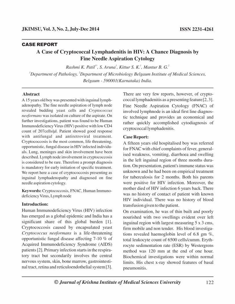
Fine needle aspiration of the inguinal lymph nodes was performed and yielded pus like mate-rial. Smears stained by Giemsa stain revealed extensive necrosis with few encapsulated budding yeast cells of varying sizes surrounded by halos (Fig. 1a). There were a few lymphocytes, macro-phages and neutrophils seen in the background without any granulomas. The capsule was better demonstrated by special stains like periodic acid Schiff (PAS) stain (Fig. 1b) and mucicarmine.
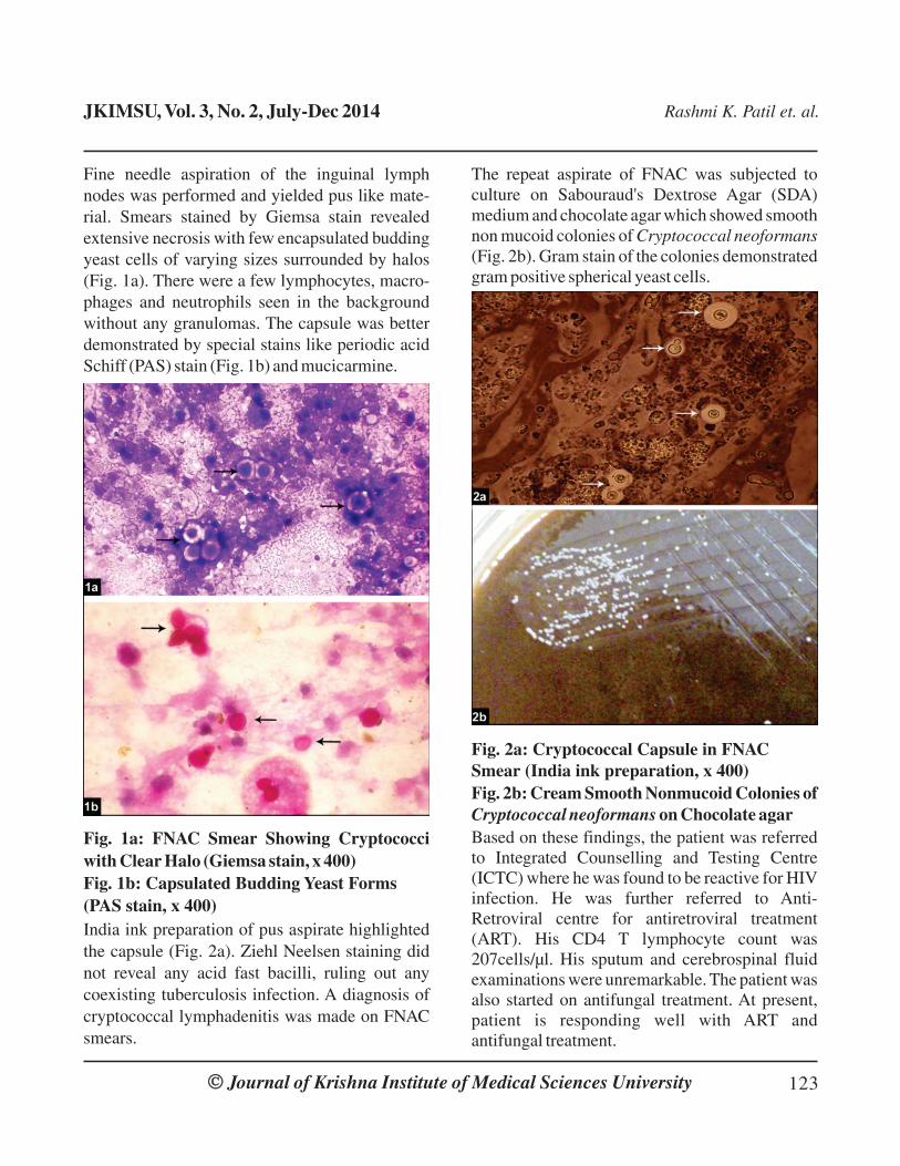
Fig. 1a: FNAC Smear Showing Cryptococci with Clear Halo (Giemsa stain, x 400)Fig. 1b: Capsulated Budding Yeast Forms (PAS stain, x 400) India ink preparation of pus aspirate highlighted the capsule (Fig. 2a). Ziehl Neelsen staining did not reveal any acid fast bacilli, ruling out any coexisting tuberculosis infection. A diagnosis of cryptococcal lymphadenitis was made on FNAC smears.
Journal of Krishna Institute of Medical Sciences UniversityÓÓ 123
JKIMSU, Vol. 3, No. 2, July-Dec 2014 Rashmi K. Patil et. al.
The repeat aspirate of FNAC was subjected to culture on Sabouraud's Dextrose Agar (SDA) medium and chocolate agar which showed smooth non mucoid colonies of Cryptococcal neoformans (Fig. 2b). Gram stain of the colonies demonstrated gram positive spherical yeast cells.
Fig. 2a: Cryptococcal Capsule in FNAC Smear (India ink preparation, x 400)Fig. 2b: Cream Smooth Nonmucoid Colonies of Cryptococcal neoformans on Chocolate agarBased on these findings, the patient was referred to Integrated Counselling and Testing Centre (ICTC) where he was found to be reactive for HIV infection. He was further referred to Anti-Retroviral centre for antiretroviral treatment (ART). His CD4 T lymphocyte count was 207cells/µl. His sputum and cerebrospinal fluid examinations were unremarkable. The patient was also started on antifungal treatment. At present, patient is responding well with ART and antifungal treatment.
DiscussionCryptococcosis is one of the opportunistic infec-tion in HIV/AIDS and therefore an expeditious diagnosis is of the utmost importance since once cryptococcal infection disseminates, it becomes life threatening [4]. The primary site of infection in humans is almost always pulmonary, following inhalation of the yeast of the fungus Cryptococcus neoformans which is found world wide in soil contaminated with pigeon or other bird droppings [5]. In humans, the spectrum of the disease varies from asymptomatic colonization of the airways to meningitis and other serious diseases, fever to pneumonia and less commonly lymph node enlargement [2]. In our case, patient presented with fever, features of pnuemonitis without central nervous system involvement.Cryptococcal meningitis and disseminated cryptococcosis have gained importance recently because of the rapid rise in the world wide inci-dence of HIV infection [3]. Cryptococcal lymphadenitis is an uncommon form of extra pulmonary cryptococcosis, which is one of the ‘AIDS defining criteria’ according to the Centre for Disease Control and prevention guidelines [1, 6].The cytological specimens in which cryptococcus is found include cerebrospinal fluid, sputum, bronchial washings and FNAC smears from lymphnodes, thyroid, spleen, adrenals, bone and lung [7]. Cryptococcal organisms are 5-15 µm, ovoid to spherical with narrow based budding, surrounded by a mucopolysaccharide capsule. These capsules are identified by special stains like PAS, mucicarmine, Gomori's Methanamine Silver (GMS) and India ink preparation [2, 5]. Unlike those in other fungal infections, the granulomatous and other inflammatory reactions are very minimal or absent [2]. Although, culture
Journal of Krishna Institute of Medical Sciences UniversityÓÓ 124
JKIMSU, Vol. 3, No. 2, July-Dec 2014 Rashmi K. Patil et. al.
is important for identification of the pathogenic species, diagnosis of cryptococcosis can be definitely made on cytologically obtained smears when the mucopolysaccharide capsule is visual-ised with special stains [8]. In present case, diagnosis of cryptococcal lymphadenitis was made on FNAC smears when the special stains facilitated the identification of organism. Further Cryptococcus neoformans species was isolated by culture of aspirate. In the present case, there was no history of blood transfusion given to the patient and / or contact of patient with known HIV individual. Hence there is a strong suspicion of transplacental transmission of HIV in our case. There are few reports of correlations of cryptococcal lymphadenitis in HIV patients with their CD4 T lymphocyte count, where authors have found that correlations of lesions with CD4 T lymphocyte count provides information about immune status and stage of the HIV disease. [9] They have concluded that cryptococcosis shows least CD4 count values as compared to other lesions in HIV [9, 10]. In our case also, CD4 count was as low as 207cells/µl reflecting advanced disease state.Lymph node FNAC can thus be a simple and useful technique in the diagnosis of fungal infec-tions. Its utility is enhanced by the ability to immediately prepare smears and simultaneously obtain samples for cultures. This method can expedite the potentially vast differential diagnoses in immunocompromised patients. Identification of these organisms, with or without cellular reaction can lead to a rapid diagnosis and impor-tantly an early initiation of specific and life – saving treatment.
1. Tahir M, Sharma SK, Sinha S, Das CJ. Immune reconstitution inflammatory syndrome in patient with cryptococcal lymphadenitis as the first manifestation of acquired immunodeficiency syndrome. J Postgrad Med 2007; 53:250-252.
2. Srinivasan R, Guptha N, Shifa R, Malhotra P, Rajwanshi A, Chakrabarti A. Cryptococcal Lymphadenitis Diagnosed by Fine Needle Aspiration Cytology:A review of 15 cases.Acta cytol 2010;54(1):1-4.
3. Das BP, Panda PL, Malik RN, Das B. Cryptococcal lymphadenitis and meningitis in human immunodefi-ciency virus infection: A case report. Indian J Pathol Microbiol 2002; 45:349-351.
4. Garbyal RS, Basu D, Roy S, Kumar P. Cryptococcal lymphadenitis:Report of a case with fine needle aspiration cytology. Acta cytol 2005; 49: 58-60.
5. Andola SK, Ahuja M, Shaik I. Cryptococcal lymphadenitis on Fine Needle Aspiration Cytology: A report of 2 cases. People's J Scientific Research 2012; 5(2):33-35.
References:
*Author for Correspondence: Dr Rashmi K. Patil, Associate Professor, Department of Pathology, Belgaum Institute of Medical Sciences, Belgaum - 590001, Karnataka, India.
Cell: 09986949387, Email: [email protected]
Journal of Krishna Institute of Medical Sciences UniversityÓÓ 125
JKIMSU, Vol. 3, No. 2, July-Dec 2014 Rashmi K. Patil et. al.
6. Schneider E, Whitmore S, Glynn KM, Dominguez K, Mitsch A,McKenna MT et al. Revised surveillance case definitions for HIV infection among adults, adolescents and children aged <18 months and for HIV infection and AIDS among children aged 18 months to <13years United states,2008.MMWR Recomm Rep 2008;57:1-12.
7. Lee MY, Chung JH, Shin Jh, Hwang TJ, Kim KS, Lee JH et al. Lymphonodular cryptococcosis diagnosed by fine needle aspiration cytology in hyper-IgM syn-drome: A case report. Acta cytol 2001; 45:241-244.
8. Suchitra S, Sheeladevi CS, Sunil R, Manjunath GV.Fine needle aspiration diagnosis of cryptococcal lymphadenitis: A window of opportunity. J cytol 2008; 25(4):147-149.
9. Jayaram G, Chew MT. Fine needle aspiration cytology of lymphnodes in HIV infected individuals. Acta cytol 2000; 44:960-966.
10. Kumarguru BN,Kulkarni MH, Kamakeri NS. FNAC of peripheral lymphnodes in HIV positive patients. Sci Med 2009; 19(2):4-12.