case presentation central giant cell...
TRANSCRIPT

1
Central Giant Cell Granuloma
Jane DahlstromAnatomical PathologistThe Canberra Hospital
Case presentationJames, 15 year old boyPresented with a < 6 month history of a rapidly growing right sided palatal mass and loose teethNo painCT scan of Maxilla
Expansile, lucent lesion associated with an unerupted upper right second molar tooth, not perforating the bone
Lesion involved the right maxillary alveolus, pterygoid plates and maxillary air sinus
Case presentation
Differential diagnosis on CT:
dentigerous cyst or ameloblastoma
Osteoclast like multinucleated giant cells, single spindled shaped cells

2
Case presentationFNAC – central giant cell granuloma –confirmed on incisional biopsyReferred to Sydney oral surgeon for second option in relation to management, including non surgical optionsRight partial maxillectomy rather than curettage was recommended due to the location and size of the tumour
Tumour measured approximately 45 x 30 x 25 mm
Anterior
Posterior
lateral Medial view
Multinucleated giant cells in a background of mononuclear fibrohistiocytic cells and red blood cells
3
Case presentation
Follow – up – Well – 5 years– No recurrence– Reconstructive surgery
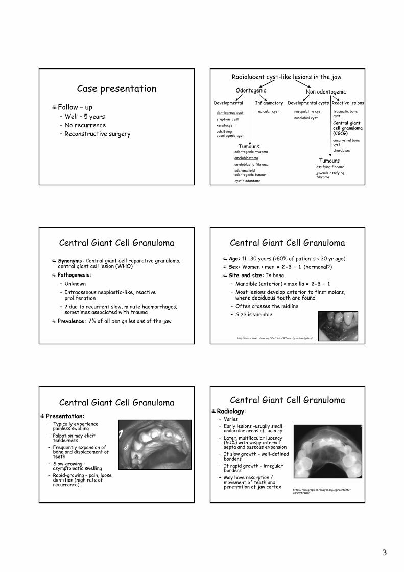
Radiolucent cyst-like lesions in the jaw
Odontogenic Non odontogenic
Developmental Inflammatory
dentigerous cyst
eruption cyst
keratocyst
calcifying odontogenic cyst
radicular cyst
Tumoursodontogenic myxoma
ameloblastoma
ameloblastic fibroma
adenomatoid odontogenic tumour
cystic odontoma
Developmental cysts
nasopalatine cyst
nasolabial cyst
Reactive lesions
Tumoursossifying fibroma
juvenile ossifying fibroma
traumatic bone cyst
Central giant cell granuloma (CGCG)aneurysmal bone cyst
cherubism
Central Giant Cell Granuloma
Synonyms: Central giant cell reparative granuloma; central giant cell lesion (WHO)Pathogenesis:– Unknown– Intraosseous neoplastic-like, reactive
proliferation– ? due to recurrent slow, minute haemorrhages;
sometimes associated with traumaPrevalence: 7% of all benign lesions of the jaw
Central Giant Cell GranulomaAge: 11- 30 years (>60% of patients < 30 yr age) Sex: Women > men = 2-3 : 1 (hormonal?) Site and size: In bone– Mandible (anterior) > maxilla = 2-3 : 1 – Most lesions develop anterior to first molars,
where deciduous teeth are found – Often crosses the midline– Size is variable
http://instruct.uwo.ca/anatomy/636/clinical%20cases/granuloma/gallery/
Central Giant Cell GranulomaPresentation:– Typically experience
painless swelling – Palpation may elicit
tenderness– Frequently expansion of
bone and displacement of teeth
– Slow-growing –asymptomatic swelling
– Rapid-growing – pain, loose dentition (high rate of recurrence)
Central Giant Cell GranulomaRadiology:– Varies – Early lesions -usually small,
unilocular areas of lucency– Later, multilocular lucency
(60%) with wispy internal septa and osseous expansion
– If slow growth - well-defined borders
– If rapid growth - irregular borders
– May have resorption / movement of teeth and penetration of jaw cortex
http://radiographics.rsnajnls.org/cgi/content/full/19/5/1107
4
Central Giant Cell GranulomaPathology:– Numerous osteoclast-like
giant cells, unevenly dispersed throughout a fibrovascular stroma
– Frequent mitotic figures; rare necrosis
– Hemorrhagic areas– Small foci of reactive
woven bone
Central Giant Cell GranulomaImmunohistochemistry:– CD68, vimentin (giant cells); ER negative– Vimentin, actin (stroma)
Electron microscopy:– Fibroblasts – Myofibroblasts – Histiocytes
Genetics:– Carinci F et al (Italy) 2005: Genetic profiling of central giant
cell granuloma of the jaws– ??associations Noonan syndrome and neurofibromatosis
Central Giant Cell Granuloma
Differential Diagnosis:Child:– Cherubism
Adult:– Hyperparathyroidism– ? Giant cell tumor (osteoclastoma)
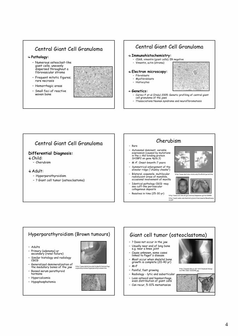
Cherubism– Rare– Autosomal dominant, variable
expression (caused by mutations in the c-Abl-binding protein SH3BP2 on gene 4p16.3)
– M >F. Onset 6month-7 years– Symmetrical enlargement of the
alveolar ridge (“chubby cheeks”)– Bilateral, expansile, multilocular
radiolucent areas of mandible, occasional involvement of maxilla
– Identical pathology CGCG –may see cuff-like perivascular collagenous deposits
– Resolves in time (25-30 yr)http://www.ncbi.nlm.nih.gov/entrez/dispomim.cgi?id=118400
http://web1.umkc.edu/dentistry/practition/assets/BoneDiseases.pdf
http://www.dent.ohio-state.edu/OralPath/parish.htm
Hyperparathyroidism (Brown tumours)
– Adults– Primary (adenoma) or
secondary (renal failure)– Similar histology and radiology
CGCG– Generalized demineralization of
the medullary bones of the jaw– Raised serum parathyroid
hormone– Hypercalcemia– Hypophosphatemia
http://www.dental.mu.edu/oralpath/lesions/hyperparathyroidism/hyperparathyroidism.htm
Giant cell tumor (osteoclastoma)– ? Does not occur in the jaw– Usually near end of long bone
e.g. near a knee joint– Cause unknown, some cases
linked to Paget's disease– Most occur when skeletal bone
growth is complete (20-40 yr)– M=F– Painful, fast growing– Radiology - lytic and subarticular – Less osteoid and haemorrhage,
even distribution of giant cells– Can recur, 5-10% metastasize
http://myweb.lsbu.ac.uk/~dirt/museum/margaret/451-3182-3320340.jpg

5
Central Giant Cell Granuloma
Treatment– Individualized treatment depending on
characteristics and location of tumorSurgical:– Curettage - recurrence 10-20% > maxilla– Extraction if unerupted tooth involved– Block resection (if aggressive lesion)Non-surgical:– Radiation – out of favor (risk of sarcoma)– Systemic Calcitonin therapy – Intralesional Glucocorticosteroids– Subcutanous interferon alpha-2a
Central Giant Cell GranulomaSystemic Calcitonin – 1993 (Harris, London)– Giant cell granulomas are rich in calcitonin receptors – Calcitonin inhibits osteoclast activity– Subcutaneous injection daily or nasal spray for about 1
year– Arrest the growth of lesion, until spontaneous healing (19
to 21 months)- Side effects: nausea, dizziness, vomiting, headaches,
diarrhea- Pathology: 6 months after treatment – absence of giant
cells and uniform cellular stroma
Central Giant Cell Granuloma
Intralesional glucocorticosteroids – 1998 (Jacoway, North Carolina)– Steroids cause decrease in secreted level of lysosomal
proteases from osteoclasts (eg:TRAP, cathepsin B) which are responsible for bone resorption
– Administer weekly or biweekly for least 6 weeks – 3 months
– Growth arrest of tumour, sometimes resolution– Problem: difficult to inject as lesion resolves
Central Giant Cell GranulomaSubcutaneous interferon alpha-2a– 1999 (Kaban, Boston)– Inhibits angiogenesis by suppressing over expression
basic fibroblast growth factor (bFGF)– Raised bFGF in urine– Dose of 1.1 – 6.16 million units/m2 daily, 1 year– Growth arrest of tumour, urinary bFGF levels return to
normal– Side effects: fever, flu-like symptoms, lethargy,
postnasal drip, skin rash, hair loss, mild neutropenia
Central Giant Cell Granuloma
Non-surgical treatmentsAdvantages: – Less invasive – Low cost – Low risk - Still able to treat lesion surgically if required
Disadvantages: – Long treatment duration – Side effects– Lack of long term studies
Central Giant Cell Granuloma• Troublesome lesion• Radiographic and pathological mimics
misdiagnosis with delayed treatment
• Treatment should be customisedpathologist
DIAGNOSIS
surgeon/ physician radiologist
Modified from IAP 2004 F Bonar

6
Acknowledgements
Patient, James McElelhinney and his familyDr Peter VickersDr Sanjiv JainA/Prof Ross O’NeilMrs Fiona Guymer
ReferencesCawson R, Binnie WH, Barrett AW et al. Oral disease. Clinical and pathological correlations. third edition , Mosby2001Regezi JA Odontogenic cysts, odontogenic tumors, fibroosseous, and giant cell lesions of the jaws. Mod Pathol 2002 Mar;15(3):331-41 Sousa FB, Etges A, Correa L, et al. Pediatric oral lesions: a 15-year review from Sao Paulo, Brazil. J Clin Pediatr Dent. 2002 Summer;26(4):413-8Scholl RJ, Kellett HM, Neumann PD et al. Cysts and Cystic Lesions of the Mandible: Clinical and Radiologic-Histopathologic Review. Radiographics. 1999;19:1107-24 Mark D. Murphey, MD, George C. et al. Imaging of Giant Cell Tumor and Giant Cell Reparative Granuloma of Bone: Radiologic-Pathologic Correlation Radiographics. 2001;21:1283-1309Dahlkemper P. Wolcott JF. Pringle GA. Hicks ML. Periapical central giant cell granuloma: a potential endodonticmisdiagnosis. Oral Surg Oral Med Oral Pathol Oral Radiol Endod. 90(6):739-45, 2000. Kurtz M, Mesa M, Alberto P. Treatment of a central giant cell lesion of the mandible with intralesionalglucocorticosteroids. Oral Surg Oral Med Oral Pathol Oral Radiol Endod. 2001;91(6):636-7 Pogrel MA, Regezi JA, Harris ST, Goldring SR. Calcitonin treatment for central giant cell granulomas of the mandible: report of two cases. J Oral Maxillofac Surg. 1999 Jul;57(7):848-53 Kaban LB, Mulliken JB, Ezekowitz Ra,et al. Antiangiogenic therapy of a recurrent giant cell tumor of the mandible with interferon alfa-2a. Pediatrics 1999; 103:1145-1149Kaban LB, Troulis MJ, Ebb D, et al. Antiangiogenic therapy with interferon alpha for giant cell lesions of the jaws. J Oral Maxillofac Surg. 2002 Oct;60(10):1103-11Oda D. Alternative treatment for central giant cell "reparative" granuloma. Adv Anat Pathol. 10(2):110, March 2003Selden HS. Central giant cell granuloma: a troublesome lesion. Journal of Endodontics. 26(6):371-3, 2000Waldron CA, Shafer WG. The central giant cell granuloma of the jaws: an analysis of 38 cases. Am J Clin Pathol1966; 45:437-447Horner K. Central giant cell granuloma of the jaw: a clinico-radiological study. Clin Radiol 1989; 40:622-626 Cohen MA, Hertzanu Y. Radiologic features, including those seen with computed tomography, of central giant cell granuloma of the jaws. Oral Surg Oral Med Oral Pathol 1988; 65:255-261 http://www.dent.ohio-state.edu/OralPath/; http://www.dental.mu.edu/oralpath/diagnosislist.htmKruse-Losler B, Diallo R, Gaertner C, et al. Central giant cell granuloma of the jaws: a clinical, radiologic, and histopathologic study of 26 cases. Oral Surg Oral Med Oral Pathol Oral Radiol Endod. 2006;101(3):346-54