case patient's b12 deficienq^ causes chiasmal lesion patient's b12 deficienq^ ... mate and...
TRANSCRIPT

Case
Patient's B12 Deficienq^Causes Chiasmal LesionThis patient's vitamin Bj, deficiency manifested through loss of visual fields, unevencup-to-disc ratios, elevated blood pressure, and dizzy spells.By Dianne Kowing, O.D., M.S.; and Elizabeth Kester, O.D.
69-year-old white male pre-sented for his annual eye
,exam. He had no new visualIcomplaints, but he men-
tioned a longstanding floater in hisright eye that had been diagnosedtwo years earlier as a posterior vit-reous detachment.
His medical history was signifi-cant for hypertension, hyperlipi-demia and depression. Medicationsincluded lisinopril,, Lipitor (ator-vastin calcium, Pfizer), Serzone(nefazodone, Bristol-Myers Squibb),and 81mg aspirin.
Diagnostic DataBest-corrected acuity was 20/25
O.U. Pupils were equal, round, reac-tive to light, and there was no affer-ent pupillary defect. Extraocularmuscles and adnexa were normal inboth eyes. Slit lamp examination ofthe lids, coniunctivas, corneas, ante-rior chambers and irides were unre-markable. Grade 1 nuclear sclerosiswas found O.U. IOP measured17mm Hg in each eye.
The dilated fundus exam revealeda cup-to-disc ratio of 0.4 O.D. and0.6 O.S. with temporal pallor O.U.Also, fine drusen was found in themaculae. A posterior vitreousdetachment was observed in the
r1. The initial fundus and optic nerve presentation O.D. (top) and O.S.
right eye. The retinal periphery wasflat and intact in both eyes.
We reviewed the patient's previ-ous exam records. He had beenexamined two years earlier, atwhich time he had a cup-to-discratio of 0.4 O.U. Last year, thesame observer again noted a 0.4cup-to-disc ratio O.U. The patientwas identified as a glaucoma sus-pect, and disc photos were taken(figure 1).
We ordered optical coherence
tomography (OCT) due to theasymmetry and changes in the cup-to-disc ratio (figures 2 and ^). Thin-ning of the peripapiilary retinalnerve fiber layer was found at the1% (red) level in both eyes (53|imO.D.,44pmO.S.).
The patient returned for Hum-phrey 24-2 visual fields two monthslater. These showed superotempo-ral depressions O.D. and temporaldepressions O.S. that were con-centrated near the vertical tnidline
122 REVIEW OF OPTOMETRY FEBRUARY 15, 2007

in each eye (figures 4 and 5). Thefalse negatives were 11 % O.D. and23% O.S. Repeat visual field testingyielded false negatives at 0% O.U.,suggesting repeatable bitemporalhemianopsias. A 120-point visualfield was then taicen, which verifieda bitemporal hemianopsia. The 24-2 visual fields were repeated twoadditional times over the next fivemonths.
The pachymetry readings were540pm O.D. and 551pm O.S. TheOCT was repeated and found to beunchanged with a high signalstrength and reliability. IOP was16mm Hg O.D. and 15mm Hg O.S.
The patient's recent lab work-up—a complete blood count, glyco-sylated hemoglobin (An), lipids,and comprehensive metabolicpanel—were unremarkable. A pro-lactin lab was ordered and was alsonormal. The patient reported expe-riencing elevated blood pressureand dizziness, and that he had beenworking with his primary carephysician to gain better control.
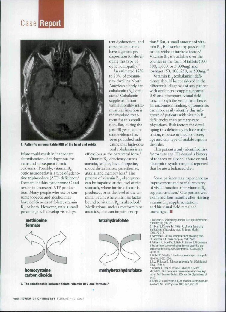
The patient was sent that day forcomputed tomography (CT) scan ofthe head with and without contrastto rule out a pituitary adenoma.The radiologist diagnosed mild gen-eralized cerebral atrophy consistentwith age. There were no masses oraneurysmal formation. He recom-mended a magnetic resonanceimaging/magnetic resonance angio-graph (MRI/MRA) of the brain,which found no sign of compressivelesions or other explanation of thepatient's symptoms (figure 6). Inparticular, there were no signs ofpituitary adenoma.
The patient was referred to oph-thalmology for further evaluation.He demonstrated normal colorvision and no peripheral neu-ropathies, except for an area ofbilateral numbness on the lateralthigh attributed to L3-5 disc dis-
0 20 40 60 80 1G0 120 14(j 160 180 200 :!20 240
T B * SUP Mi^ NF T B *
2. OCT right aye peripapiilary retinal nerve fiber layer scan.
300
200i
0 20 40 60 80 IOC 120 140 160 180 20C 220 240
lB<f SUP NAS l f ?ffl
T 28
3. OCT left eye peripapiilary retinal nerve fiber layer scan.
ease. The patient was sent back tooptometry for continued observa-tion without any identified cause ofthe field loss. After a review of allthe previous testing. Vitamin B,,(Cobalamin) and folate labs wereordered in hopes of explaining thebitemporal visual field loss.
DiagnosisThe patient's vitamin B,
(cobalamin) level was found to be
low, at 175 pg/niL. Normal range is240 pg/mL to 900 pg/mL.
Treatment and Follow-UpWe referred the patient to his pri-
mary-care provider for treatmentand management. He began a one-month course of oral B12 vitamins,to be modified to injections if hislevels remained low. But, at onemonth, his B12 level was 582Pg/mL, so he continues to be
REVIEW OF OPTOMETRY FEBRUARY 15, 2007 123

Case
ri J H •> • 1!• 1 * •"
a ••' •
• • • >>
B B ^B
1' • 1' a
* B
M BB • BB
•
H • • • •
• 1 • •
II a M
• • B B 0
D K tf «
a aa • •a B a a• B •a «• a
a •
• » • • • ' Ul
a nI H H a
n n • • n B4 >• • • •
" "
a aBB
a
a•
a
4. Right eye 24-2 Humptirey serial visual fields. 5. Left eye 24-2 Humplirey serial visual fleitfs.
mtmitored. His visual fields andperipapiilary nerve fiber layer haveremained stable.
DiscussionVisual field loss is often the initial
manifestation of disorders involvingthe optic chiasm.' Hemianopicdefects and preferential involvementof the temporal visual field are theearliest—^and most common—visualsymptoms.' There are multiple eti-ologies of chiasmal lesions, includ-ing congenital, traumatic, iatrogenic(secondary to radiation treatment),intrinsic (gliomas) and extrinsiclesions. Most chiasmal syndromescan be categorized as intrinsic(thickening of the chiasm itself) orextrinsic (compression of the chiasmfrom an adjacent structure).' Extrin-sic lesions are typically caused bypituitary tumors, craniopharyn-giomas, and meningiomas, but theycan also result from aneurysms andlesions of the sinuses.'
Vitamin B^, has a role in folic
acid metabolism, so it is necessaryfor DNA synthesis, hematopoiesis,nerve growth, myelin synthesis andnervous system integrity. VitaminBp forms a complex with intrinsicfactor before being absorbed fromthe ileum and stored in the liver.Inadequate dietary intake, malab-sorption syndromes, hypermeta-bolic state, pregnancy, andmegaloblastic anemia can alsocause vitamin B ^ deficiency.^ Nor-mal absorption of the vitaminalways exceeds body needs, soexcess B, is excreted in the urine.'
Vitamin B,, occurs naturally asthe product of specialized microor-ganisms that grow in, or serve asfood supply to, those animals beiowhumans in the food chain.' Neariyail dietary vitamin Bp comes fromanimal products.'
Documented cases of rare chias-mal lesions causing typical chiasmalvisual field defects, such as bitem-poral hemianopsia or junction sco-toma, include demyelinating
disease, vascuiitis, and cobalamindeficiency/ For example, one caseinvolved a 29-year-o!d female withbitemporal hemianopsia, normalMRI scan, normal computedtomography (CT) scan, and normalvisual evoked potential (VEP). Shehad only one-third of the normalamount of B,^, but her bloodcounts were almost normal; onlyher hemoglobin value wasdecreased, and there was no sign ofpernicious anemia. Vitamin B,deficiency may exist without hema-tological manifestations. The causeofthe vitamin B^ deficiency wasnot identified in this patient, andher intestinal update was normal.''
What is the mechanism in B ,that causes optic neuropathy? Vita-min B|,, in the form of methyl-cobalamin, is required as a cofactorfor the enzyme methionine syn-thetase, which then catalyzes therecycling of homocysteine tomethionine (figure 7)}
So, a deficiency of vitamin or
1Z4 REVIEW OF OPTOMETRY FEBRUARY 15, 2007
Case
6. Patient's unremarkable MRI of the head and orhlts.
folate could result in inadequatedetoxification of endogenous for-mate and subsequent formicacidemia.^ Possibly, vitamin B^optic neuropathy is a type of adeno-sine triphosphate (ATP) deficiency.*"Formate inhibits cytochrome C andresults in decreased ATP produc-tion. Many people who use or con-sume tobacco and alcohol mayhave deficiencies of folate, vitaminB,2 or both. However, only a smallpercentage will develop visual sys-
methioninefonnate
tem dysfunction, andthese patients mayhave a genetic pre-disposition for devel-oping this type ofoptic neuropathy.^
An estimated 12%to 20% of commu-nity dwelling NorthAmerican elderly arecobalamin (B, ) defi-cient/ Cobalaminsupplementationwith a monthly intra-muscular injection isthe standard treat-ment for this condi-tion. But, during thepast 40 years, abun-dant evidence hasbeen published indi-cating that high-doseoral cobalamin is as
efficacious as the parenterai form/
Vitamin Bp deficiency causesanemia, fatigue, loss of appetite,mood disturbances, paresthesias,ataxia, and memory loss.** Theprocess of vitamin Bp absorptioncan be impaired at the level of thestomach, where intrinsic factor isproduced, or at the Ievei of the ter-minal ileum, where intrinsic factorbound to vitamin Bp is absorbed.'*Medications, such as metformin orantacids, aiso can impair absorp-
tetrahydrofblate
homocysteinecart>on dioxide
methyltetrahydrofblate
7. The relationship between felate, vitamin B12 and formate.'
tion." But, a smail amount of vita-min Bj is absorbed by passive dif-fusion without intrinsic factor.^Vitamin B,, is avaiiabie over thecounter in the form of tablets (100,500, 1,000, or 5,000mg) andiozenges (50, 100, 250, or 500mg).''
Vitamin B, (cohaiamin) defi-ciency shouid be considered in thedifferentiai diagnosis of any patientwith optic nerve cupping, normalIOP and bitemporal visuai fieldloss. Though the visual field loss isan uncommon finding, optometristscan more easily identify this sub-group of patients with vitamin B,deficiencies than primary-carephysicians. Risk factors for devel-oping this deficiency include malnu-trition, tobacco or alcohol abuse,age and any type of malabsorptiondisorder.
This patient's only identified riskfactor was age. He denied a historyof tobacco or alcohol abuse or mal-absorption syndrome, and reportedthat he ate a balanced diet.
Some patients may experience animprovement and partial recoveryof visual function after vitamin B,supplementation.'' Our patient wasexamined four months after startingvitamin Bp supplementation,and his visual field remainedunchanged. •
1. Foraozan R. Chiasmal syndromes. Curr Opin Optittialnol2003 Dec:14(6):325-312. Tllkian S. Conover M. Tllkian A. Clinical & nursingimpiicalions ol iaboialory leslS- Si. Louis: Mosby:1995:3/7-378.3. Widmann F Clinical inlefprelalion ol laDoratofy lests.Philadelphia. FA Davis Company: 1963:79-61.4. Wilhelm H. Grodd W, Sctilefer U. Zrenner E, Uncommonchiasmal lesions; demyelinaling disease, vascjiitis andcobalamin deliciency. Ger J OplHtialmol. 1993 Aug;2(')-5):234-40.5. Golnik K. Schaible E. Folale-responsive opiic neuropathy,1994Sep;I4(3):163-96- Rizz JF, Lessel S- Tobacco amblyopia. Am J Ophttiainnol1993;! 16:84-8.7 Graham ID, Jelte N. Telroe J. Robinson fj, Milne S,Mitchell SL. Oral Cobalamin femains medicine's best Kepisecret. Arch G^onlol Gerialr. 2006 Apr 29; lEpub ahead otprint].8. Kripke C. Is oral Vitamin B, as etfeclive as inlramuscularinjeflion? Am Fam Physician. 2006 Jan1 ;73(1 ):55.
126 REVIEW OF OPTOMETRY FEBRUARY 15, 2007
