case 6yr boy is brought to the opd with complaints of recurrent painful swelling of the lt knee...
TRANSCRIPT

Case
6yr boy is brought to the OPD with complaints of recurrent painful swelling of the Lt Knee joint since 2yr of age. He also has a history of prolonged bleeding from cut sites.
One maternal uncle of the child died due to prolonged bleeding following a minor surgery.
O/E, No petechiae/purpura.Lt Knee joint swollen, tender
Hemophilia and Coagulation Disorders
Dr Nishant Verma
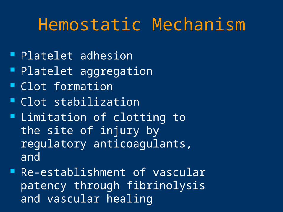
Hemostatic Mechanism
Platelet adhesion Platelet aggregation Clot formation Clot stabilization Limitation of clotting to the site of
injury by regulatory anticoagulants, and
Re-establishment of vascular patency through fibrinolysis and vascular healing
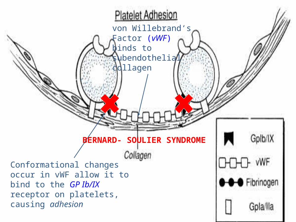
von Willebrand‘sFactor (vWF) binds to subendothelialcollagen.
Conformational changes occur in vWF allow it to bind to the GP Ib/IX receptor on platelets,causing adhesion
BERNARD- SOULIER SYNDROME

The GP IIb/IIIa receptor complex changes conformation allowing binding of fibrinogen.
Fibrinogen acts as a glue
binding platelets together.
GLANZMANN’s Thrombasthenia

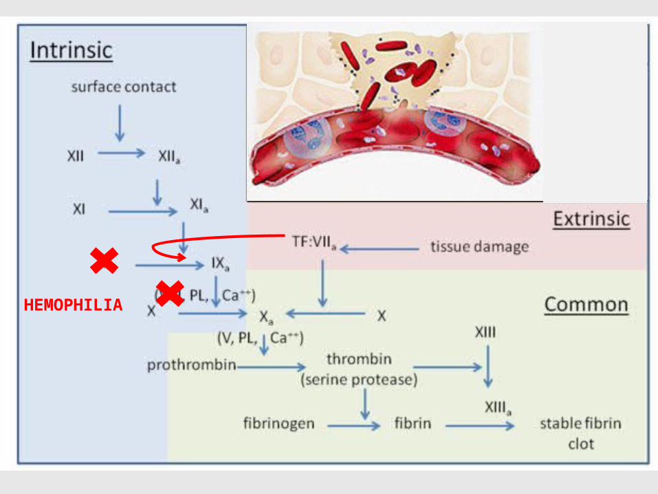
Clotting FactorsI FibrinogenII ProthrombinV Labile factor, proaccelerin
VII Stable factor or proconvertinVIII Antihemophilic factor (AHF)IX Christmas factorX Stuart-Power factorXI Plasma thromboplastin antecedentXII Hageman factorXIII Fibrin Stabilizing Factor

Waterfall cascade:
Prothrombin
Fibrinogen
Factor X
Intrinsic
pathway
Factor XII,
prekallikrein and
HMWK , factor X
I
, factor IX
,
factor VIII
Extrinsic
pathway
Tissue factor,
factor VII
Common
Pathway Fibrin
Thrombin
Factor Xa

HEMOPHILIA

Overview of fibrinolytic mechanism

von Willebrand Factor

Classification of bleeding disorders
• Primary Hemostatic defect– Platelet disorder
• Congenital• Acquired
– Von Willebrand Disease• Coagulation Disorders (Clotting factor deficiency)
– Acquired– Inherited
• Vascular

Clinical characteristic Primary Hemostatic Defect Coagulation Disorder
Site of bleeding Skin, mucous membrane Soft tissues, muscles, joints
Bleeding after minor cuts Yes No
Petechiae Yes No
Ecchymosis Small, superficial Larger, deeper
Hemarthrosis Rare Common
Bleeding after trauma/surgery Immediate Delayed
Example Platelet defect, vWD Hemophilia
Classification of bleeding disorders
Source: Nathan and Oski’s Hematology of Infancy and Childhood. 7th Edition, Pg 1450
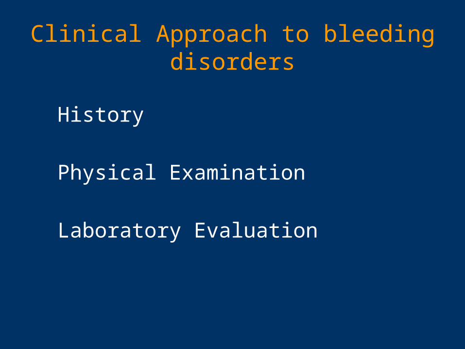
Clinical Approach to bleeding disorders
History
Physical Examination
Laboratory Evaluation

Clinical Approach to bleeding disorders
History Nature of bleeding-
- Immediate vs delayed
- Superficial vs deep
- Surgical / dental history
Family H/O bleeding-
- Others involved ?
- Males only? (x-linked)
- Consecutive generations
Medication history-
-NSAID, Heparin (patients with central lines)
Others- Liver / renal disease

Clinical Approach to bleeding disorders
Physical Examination Bruises-
- Number
- Location
- Site
Petechiae
Joint bleeding
Other Physical findings-
- Jaundice
- Skeletal deformity
- Hepatosplenomegaly
Clinical Approach to bleeding disorders
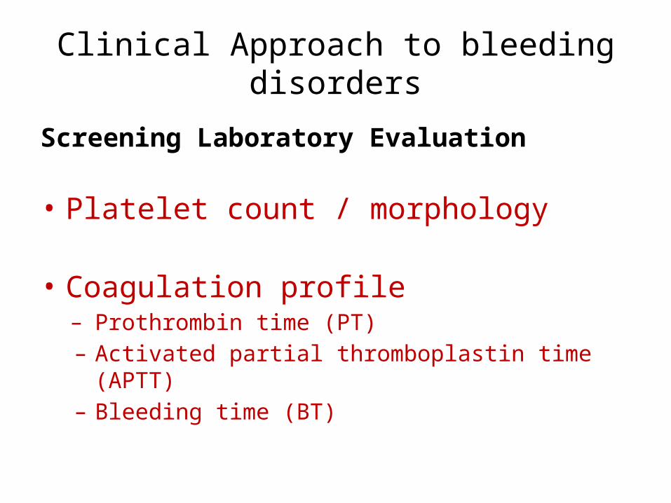
Screening Laboratory Evaluation
• Platelet count / morphology
• Coagulation profile– Prothrombin time (PT)– Activated partial thromboplastin time (APTT)– Bleeding time (BT)
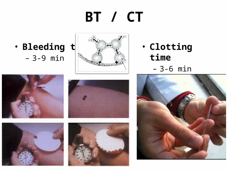
BT / CT
• Bleeding time– 3-9 min
• Clotting time– 3-6 min

Coagulation profile
• Sample collection
– Citrated tube
– Gently mixed
– Immediate transport

Coagulation profile• PT
– Method– Normal – 10-11s– INR = (Patient PT/Control PT)ISI – Isolated PT
• APTT– Method– Normal – 26-35s– Isolated APTT– PT + APTT
• TT
TT

Advanced tests
• Factor assays
• Testing for vWD
• Platelet function analyzer (PFA 100)
Classification of bleeding disorders
• Primary Hemostatic defect– Platelet disorder
• Congenital• Acquired
– Von Willebrand Disease• Coagulation Disorders (Clotting factor deficiency)
– Acquired– Inherited
• Vascular
Coagulation Disorders: Acquired
• Vitamin K deficiency
• Liver disease
• Accelerated destruction of coagulation factors
• Inhibitors of coagulation
• Miscellaneous

Vitamin K Dependent Proteins
Dietary Vitamin K
Vitamin K Reductase

Vitamin K deficiency
Liver Disease or Vit. K deficiency
Hemorrhagic disease of the newborn
Biliary obstruction (neonatal cholestatic disorders)
Malabsorption of vitamin K (celiac disease, ulcerative
colitis)
Drugs
Vit.K antagonists – warfarin, phenytoin
Broad-spectrum antibiotics – alter gut flora
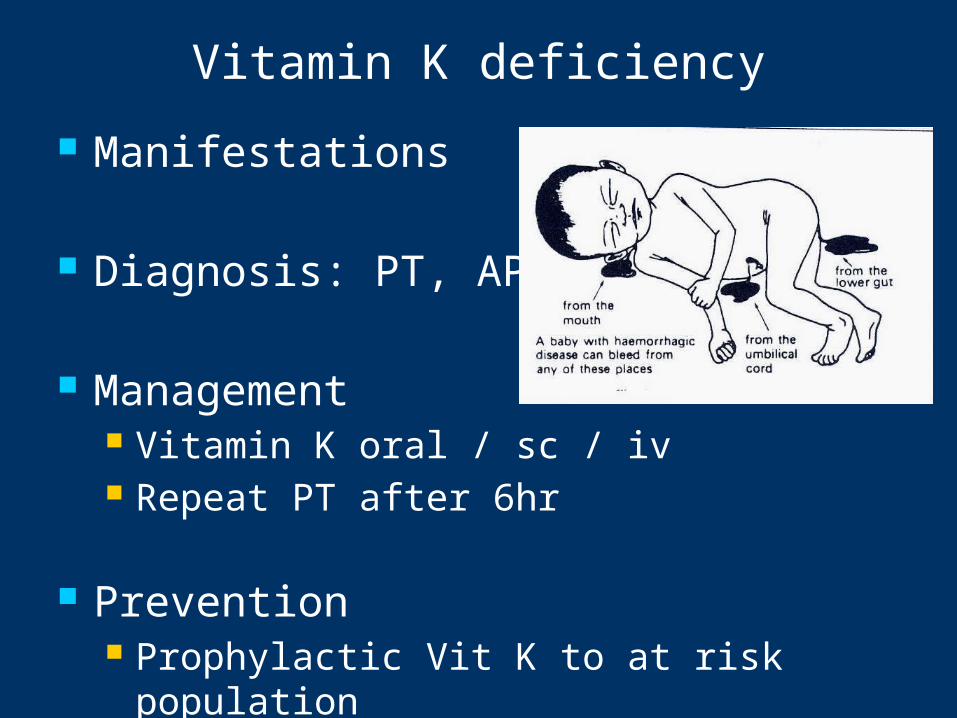
Manifestations
Diagnosis: PT, APTT
Management Vitamin K oral / sc / iv Repeat PT after 6hr
Prevention Prophylactic Vit K to at risk population
Vitamin K deficiency
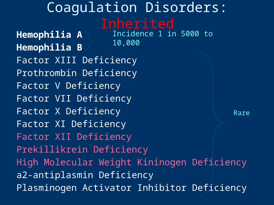
Coagulation Disorders: InheritedHemophilia AHemophilia BFactor XIII DeficiencyProthrombin DeficiencyFactor V DeficiencyFactor VII DeficiencyFactor X DeficiencyFactor XI DeficiencyFactor XII DeficiencyPrekillikrein DeficiencyHigh Molecular Weight Kininogen Deficiencya2-antiplasmin DeficiencyPlasminogen Activator Inhibitor Deficiency
Incidence 1 in 5000 to 10,000
Rare
27
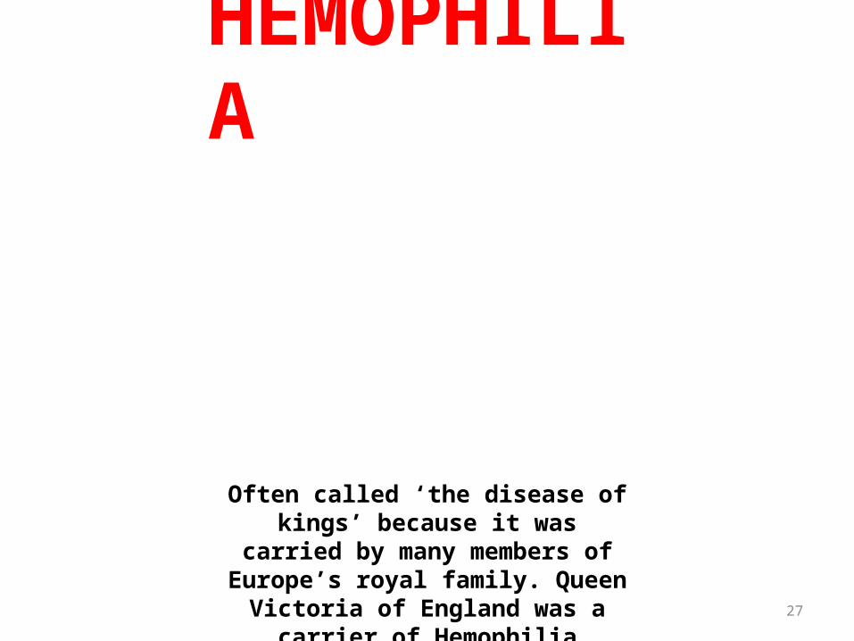
Often called ‘the disease of kings’ because it was carried by many members of Europe’s royal family. Queen Victoria of England was a carrier of Hemophilia
HEMOPHILIA

• X-linked recessive
• Hemophilia A (FVIII def): 80-85%
Hemophilia B (FIX def)
• Mutations of the clotting factor gene
• Family H/O bleeding common,
- generally affects males on the maternal side
- 1/3 no family history – due to new mutations
29
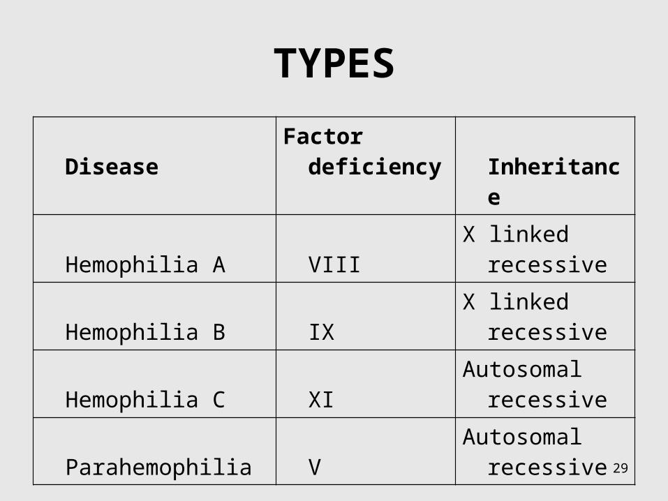
TYPES
Disease Factor deficiency Inheritance
Hemophilia A VIII X linked recessive
Hemophilia B IX X linked recessive
Hemophilia C XI Autosomal recessive
Parahemophilia V Autosomal recessive

30
Distribution Clotting factor activity
Severe hemophilia 50% <1%
Moderate hemophilia 10% 1-5%
Mild hemophilia 30-40% 5-40%
Severity of Hemophilia is defined by measured level of clotting factor activity
HEMOPHILIA

32
• Bleeding can happen anywhere in the body.
• Following an injury / surgery or rarely spontaneous.
CLINICAL MANIFESTATIONS
33
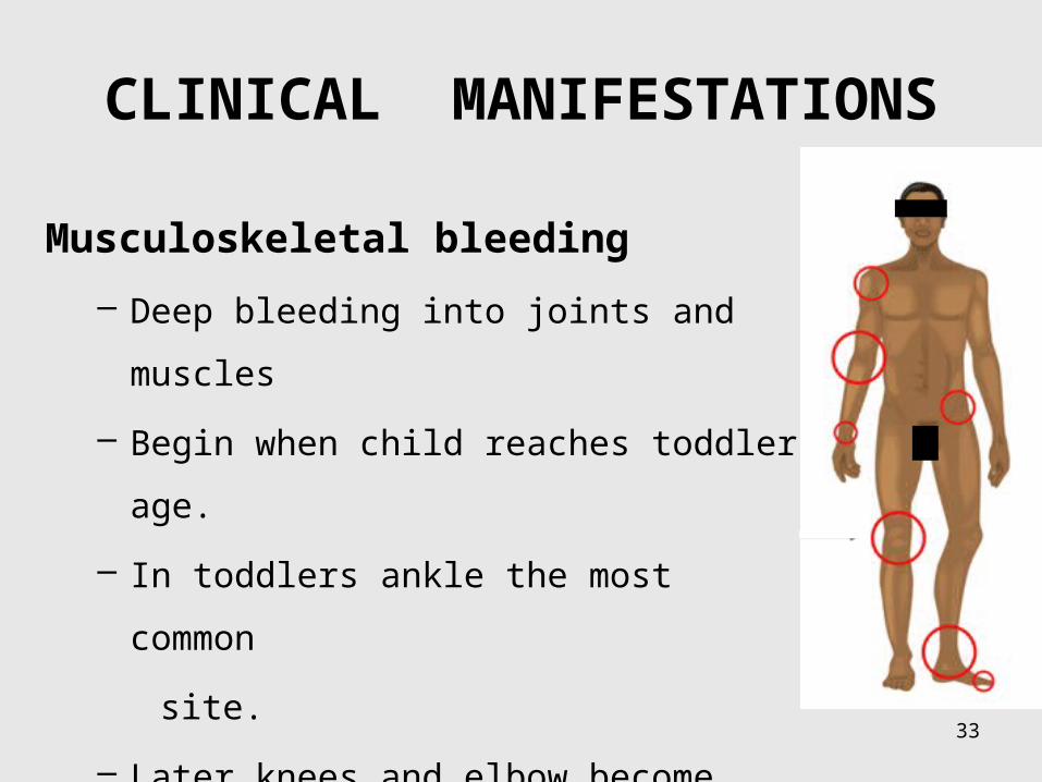
CLINICAL MANIFESTATIONS
Musculoskeletal bleeding
– Deep bleeding into joints and muscles
– Begin when child reaches toddler age.
– In toddlers ankle the most common
site.
– Later knees and elbow become common
sites.

34
• Target joint – Repeated bleeds
Hemophilic arthropathy

35
Other manifestations
• Intracranial haemorrhage
• Hematuria
• Traumatic bleeding
• Venipuncture
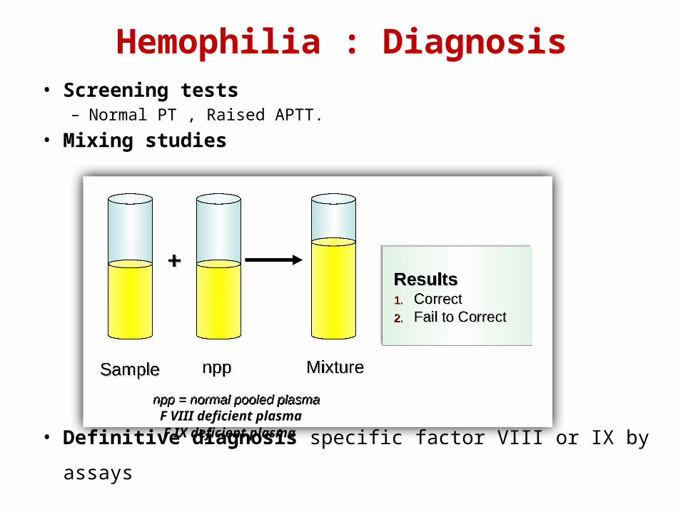
Hemophilia : Diagnosis• Screening tests
– Normal PT , Raised APTT.
• Mixing studies
• Definitive diagnosis specific factor VIII or IX by assays
F VIII deficient plasmaF IX deficient plasma

37
Carrier state and Genetic testing
Three approaches: 1. Patient and family history
2. Coagulation-based assays: unreliable
3. DNA testing: GOLD standard
Prenatal diagnosis
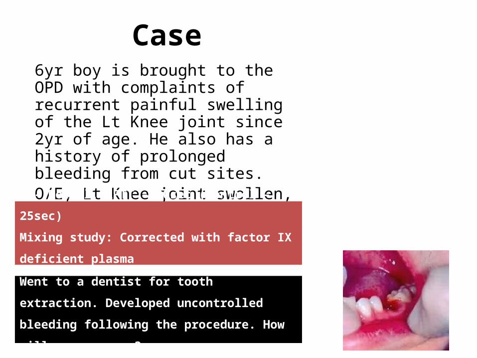
Case 6yr boy is brought to the OPD with complaints of recurrent painful swelling of the Lt Knee joint since 2yr of age. He also has a history of prolonged bleeding from cut sites. O/E, Lt Knee joint swollen, tenderInvestigations ???
PT- Normal, APTT – 90sec (Control – 25sec)
Mixing study: Corrected with factor IX deficient plasma
Factor VIII assay: < 1% activity
Went to a dentist for tooth extraction. Developed
uncontrolled bleeding following the procedure. How
will you manage ?
Hemophilia: Management
Issues to be considered
• Lifestyle modifications• Available therapeutic options• Inhibitors complicating Hemophilia• Prophylactic factor therapy• Transfusion transmitted infections
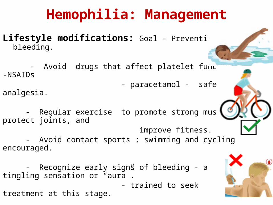
Hemophilia: Management
Lifestyle modifications: Goal - Prevention of bleeding.
- Avoid drugs that affect platelet function -NSAIDs - paracetamol - safe for analgesia.
- Regular exercise to promote strong muscles, protect joints, and improve fitness. - Avoid contact sports ; swimming and cycling encouraged.
- Recognize early signs of bleeding - a tingling sensation or “aura”. - trained to seek treatment at this stage.
- Carry identification indicating the diagnosis, severity, and contact information .

Hemophilia: Management
Available pharmacological agents
• Factor concentrates
• Cryoprecipitate
• Fresh Frozen Plasma and Cryo-Poor Plasma
• Adjuvant Pharmacological Options– Desmopressin (DDAVP)– Tranexamic acid– Epsilon aminocaproic acid (EACA)
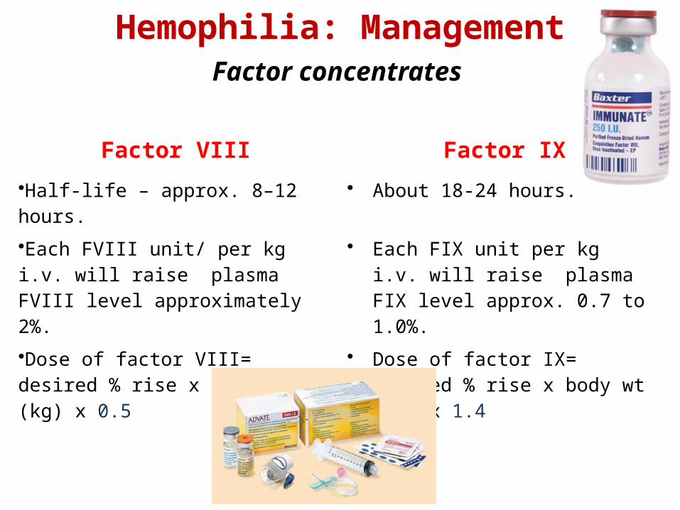
Hemophilia: Management
Factor VIII Factor IX
•Half-life – approx. 8–12 hours. • About 18-24 hours.
•Each FVIII unit/ per kg i.v. will raise plasma FVIII level approximately 2%.
• Each FIX unit per kg i.v. will raise plasma FIX level approx. 0.7 to 1.0%.
•Dose of factor VIII= desired % rise x body wt (kg) x 0.5
• Dose of factor IX= desired % rise x body wt (kg) x 1.4
Factor concentrates

Type of Hemorrhage Desired factor level Duration (days) (longer if indicated)
Hemophilia A Hemophilia B
Joint 10%–20% 10%–20% 1–2
Muscle (except iliopsoas) 10%–20% 10%–20% 2–3
Iliopsoas• initial•maintenance
20%–40%10%–20%
15%–30%10%–20%
1-23-5
CNS/head•initial•maintenance
50%–80%30%–50% 20%–40%
50%–80%30%–50% 20%–40%
1-34-7
8-14
Throat and neck• initial•maintenance
30%–50%10%–20%
30%–50%10%–20%
1-34-7
Gastrointestinal• initial• maintenance
30%–50%10%–20%
30%–50%10%–20%
1–34–7
Renal 20%–40% 15%–30% 3–5
Deep laceration 20%–40% 15%–30% 5-7
Surgery (major)• Pre-op• Post-op
60%–80%30%–40%20%–30%10%–20%
50%–70%30%–40%20%–30%10%–20%
1–34–6
7–14
WHF Recommendations
for target factor levels

Hemophilia: Management
Cryoprecipitate - Prepared by slow thawing of FFP at 4°C for 10–24 hours. - Contains – FVIII, vWF, fibrinogen, & FXIII (not FIX or XI). - supernatant - cryo-poor plasma and contains other coagulation
factors VII, IX, X, and XI. - FVIII /bag of cryoppt is 60-100 units (avg-80 units) in a 30-40 ml
vol. -does not contain factor IX, so no use in Haemophilia B
Concerns : - factor content of individual packs variable. - not subjected to viral inactivation procedures

Hemophilia: Management
Fresh Frozen Plasma• FFP can be used to treat both hemophilia A &B• 1 U FFP contains about 160-250ml plasma with activity of ~80%.• Rate and total dose limited by the risk of acute or chronic circulatory
overload.• How to use
– Thaw.– Transfuse over how many minutes.– Reusing after thawing
• Disadvantages: – No viral inactivation– F level >20-25% difficult to achieve

Hemophilia: Management
Desmopressin
• Only effective in mild hemophilia A - single i.v. infusion of 0.3 mg/kg expected to boost FVIII level 3-6 fold
• Ineffective in severe hemophilia A
• No value in hemophilia B - does not affect FIX levels
• Nasal spray available - useful for home treatment of minor bleeding problems.

Hemophilia: Management Tranexamic acid / EACA
• Antifibrinolytic agent, competitively inhibits activation of plasminogen to plasmin.
• Valuable in controlling bleeding from mucosal surfaces (e.g., oral bleeding, epistaxis, menorrhagia) - dental surgery - eruption of teeth
• Tranexa dose for children - 25 mg/kg up to three times daily - 500 mg tablet can be crushed, dissolved in water for topical use on bleeding mucosal lesions.

48
Management of Hemophilic arthropathy
• Analgesics, ice packs ( 5 minutes on, 10 minutes off, for as long as the joint feels hot), avoidance of weight bearing and immobilisation.
• Factor replacement- most important
• Physiotherapy

Hemophilia: Management
Inhibitors:• Suspected - when no / inadequate response to factor
replacement.
• Detected by: – Measuring factor levels after factor replacement– Mixing studies
• Treatment: – low-responders - specific factor at a much higher dose– High responders - alternative agents like bypassing agents : as
recombinant factor VIIa and prothrombin complex concentrates.

Hemophilia: Management
Prophylactic Therapy• Administration of clotting factors at regular intervals to prevent
bleeding - Patients with clotting factor level > 1% seldom have spontaneous bleeding
• 25-40 IU/kg of clotting factor concentrates - 3 times/week for hemophilia A - twice a week for hemophilia B• Expensive but preservation of joint function & improved QOL
• Administered by subcutaneous access port of central line
51
Comprehensive care
• Comprehensive team including hemophilia specialist, nurse coordinator, social worker, psychologist, physiotherapist, orthopaedic surgeon, primary care physician, financial counsellor and sometimes infectious disease specialist
• Provided primarily through comprehensive hemophilia treatment centres

Case 6yr boy is brought to the OPD with complaints of recurrent painful swelling of the Lt Knee joint since 2yr of age. He also has a history of prolonged bleeding from cut sites. O/E, Lt Knee joint swollen, tenderInvestigations ???
PT- Normal, APTT – 90sec (Control – 25sec)
Mixing study: Corrected with factor IX deficient plasma
Factor VIII assay: < 1% activity
Went to a dentist for tooth extraction. Developed
uncontrolled bleeding following the procedure. How
will you manage ?

Classification of bleeding disorders
• Primary Hemostatic defect– Platelet disorder
• Qualitative• Quantitative
– Von Willebrand Disease• Coagulation Disorders (Clotting factor deficiency)
– Acquired– Inherited
• Vascular

Classification of bleeding disorders
• Primary Hemostatic defect– Platelet disorder
• Qualitative• Quantitative
BERNARD- SOULIER SYNDROME
GLANZMANN’s Thrombasthenia
Diagnosis•BT•Platelet counts•Failure to agglutinate by Ristocetin•PFA•Flowcytometry•Genetic testing
Classification of bleeding disorders
• Primary Hemostatic defect– Platelet disorder
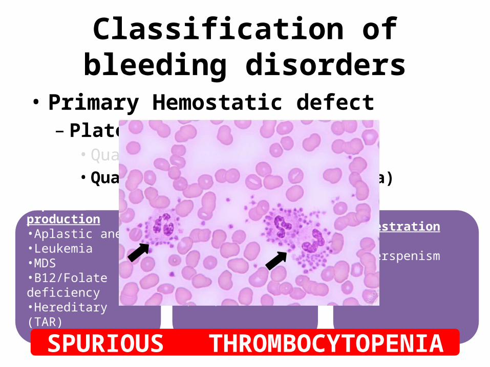
• Qualitative• Quantitative (Thrombocytopenia)
Impaired production•Aplastic anemia•Leukemia•MDS•B12/Folate deficiency•Hereditary (TAR)
Increased destruction•ITP•SLE•Thrombotic microangiopathy (HUS)
Sequestration
•Hyperspenism
SPURIOUS THROMBOCYTOPENIA
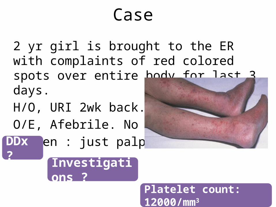
Case
2 yr girl is brought to the ER with complaints of red colored spots over entire body for last 3 days.H/O, URI 2wk back.O/E, Afebrile. No PallorSpleen : just palpable
DDx ?
Investigations ?
Platelet count: 12000/mm3
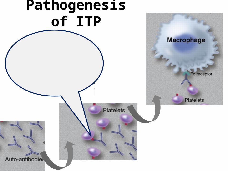
Pathogenesis of ITP

Definitions
• Newly diagnosed ITP: diagnosis to 3 months
• Persistent ITP: 3 to 12 months from diagnosis
• Chronic ITP: lasting for more than 12 months
Immune Thrombocytopenia (ITP)Platelet count less than 100 × 109/L in absence of other causes or
disorders that may be associated with thrombocytopenia
Source: The American Society of Hematology 2011 evidence-based practice guideline for immune thrombocytopenia

Investigations
• Complete blood count
• Peripheral smear
• Bone marrow aspiration ???
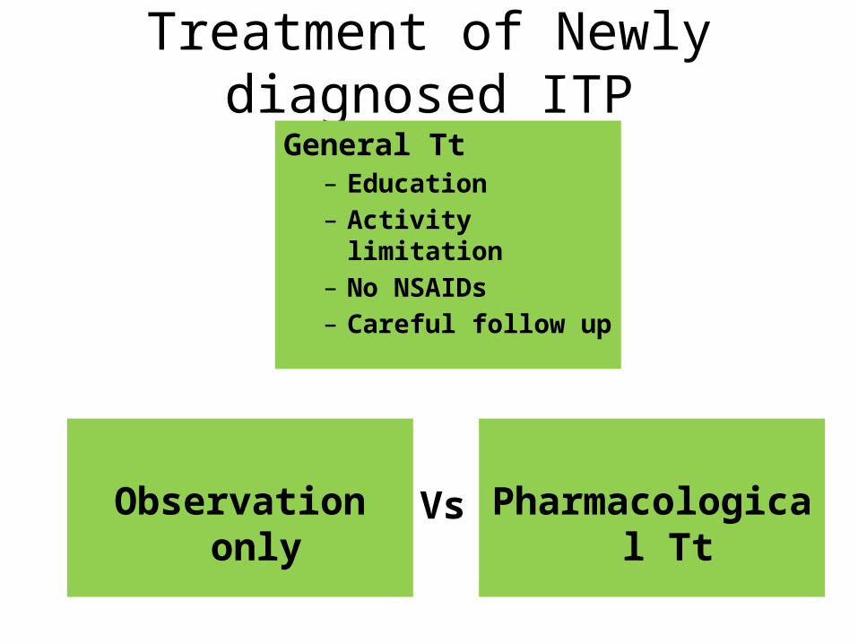
Treatment of Newly diagnosed ITPGeneral Tt
– Education– Activity limitation– No NSAIDs– Careful follow up
Observation only Vs Pharmacological Tt

Case
2 yr girl is brought to the ER with complaints of red colored spots over entire body for last 3 days.H/O, URI 2wk back.O/E, Afebrile. No PallorSpleen : just palpable
Observation only
Daily follow up advised.Comes next day with Epistaxis and gum bleeding.
What next???

Treatment of Newly diagnosed ITPGeneral Tt
– Education– Activity limitation– No NSAIDs– Careful follow up
Observation only
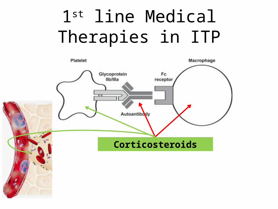
1st line Pharmacological Tt• Corticosteroids• IVIG• Anti D
Vs
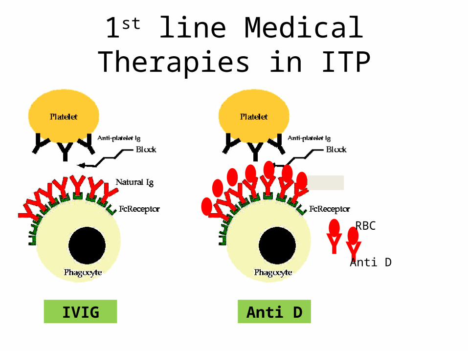
1st line Medical Therapies in ITP
IVIG
Y YAnti D
RBC
Anti D
1st line Medical Therapies in ITP
Corticosteroids
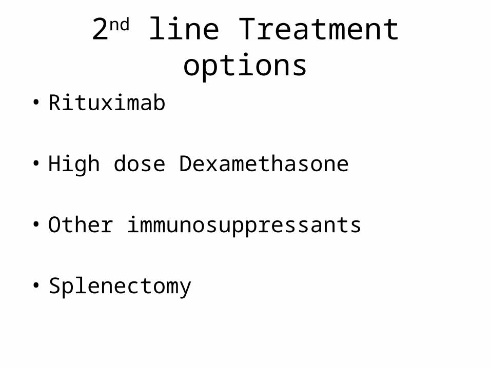
2nd line Treatment options
• Rituximab
• High dose Dexamethasone
• Other immunosuppressants
• Splenectomy

Classification of bleeding disorders
• Primary Hemostatic defect– Platelet disorder
• Qualitative• Quantitative
– von Willebrand Disease• Coagulation Disorders (Clotting factor deficiency)
– Acquired– Inherited
• Vascular
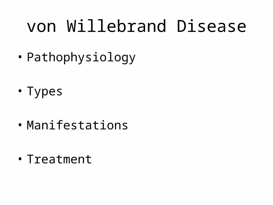
von Willebrand Disease
• Pathophysiology
• Types
• Manifestations
• Treatment
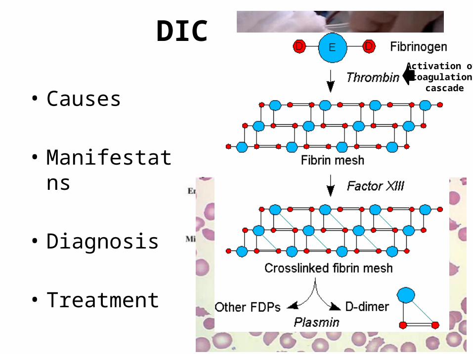
DIC
• Causes
• Manifestations
• Diagnosis
• Treatment
Activation of coagulation
cascade

Thank You