care and maintenance to reduce vascular … · nursing best practice guideline shaping the future...
TRANSCRIPT

Nursing Best Practice GuidelineShaping the future of Nursing
April 2005
Care and Maintenance to ReduceVascular Access Complications
2008

Greetings from Doris Grinspun
Executive Director
Registered Nurses’ Association of Ontario
It is with great excitement that the Registered Nurses’ Association of Ontario (RNAO)
disseminates this nursing best practice guideline to you. Evidence-based practice supports
the excellence in service that nurses are committed to deliver in our day-to-day practice.
We offer our endless thanks to the many institutions and individuals that are making
RNAO’s vision for Nursing Best Practice Guidelines (NBPGs) a reality. The Government
of Ontario recognized RNAO’s ability to lead this program and is providing multi-year funding. Tazim
Virani – NBPG program director – with her fearless determination and skills, is moving the program
forward faster and stronger than ever imagined. The nursing community, with its commitment and passion
for excellence in nursing care, is providing the knowledge and countless hours essential to the creation and
evaluation of each guideline. Employers have responded enthusiastically to the request for proposals
(RFP), and are opening their organizations to pilot test the NBPGs.
Now comes the true test in this phenomenal journey: Will nurses utilize the guidelines in their day-to-day practice?
Successful uptake of these NBPGs requires a concerted effort of four groups: nurses themselves, other
healthcare colleagues, nurse educators in academic and practice settings, and employers. After lodging
these guidelines into their minds and hearts, knowledgeable and skillful nurses and nursing students need
healthy and supportive work environments to help bring these guidelines to life.
We ask that you share this NBPG, and others, with members of the interdisciplinary team. There is much
to learn from one another. Together, we can ensure that Ontarians receive the best possible care every time
they come in contact with us. Let’s make them the real winners of this important effort!
RNAO will continue to work hard at developing and evaluating future guidelines. We wish you the best for
a successful implementation!
Doris Grinspun, RN, MSN, PhD (cand), OOnt
Executive Director
Registered Nurses’ Association of Ontario

1
Nursing Best Practice Guideline
How to Use this Document
This nursing best practice guideline is a comprehensive document providing resources necessaryfor the support of evidence-based nursing practice. The document needs to be reviewed and applied,based on the specific needs of the organization or practice setting/environment, as well as the needs andwishes of the client. Guidelines should not be applied in a “cookbook” fashion but used as a tool to assist indecision making for individualized client care, as well as ensuring that appropriate structures andsupports are in place to provide the best possible care.
Nurses, other healthcare professionals and administrators who are leading and facilitating practice changeswill find this document valuable for the development of policies, procedures, protocols, educationalprograms, assessments and documentation tools. It is recommended that the nursing best practiceguidelines be used as a resource tool. Nurses providing direct client care will benefit from reviewing therecommendations, the evidence in support of the recommendations and the process that was used todevelop the guidelines. However, it is highly recommended that practice settings/environments adaptthese guidelines in formats that would be user-friendly for daily use. This guideline has some suggestedformats for such local adaptation and tailoring.
Organizations wishing to use the guideline may decide to do so in a number of ways:■ Assess current nursing and healthcare practices using the recommendations in the guideline.■ Identify recommendations that will address identified needs or gaps in services.■ Systematically develop a plan to implement the recommendations using associated tools and resources.
RNAO is interested in hearing how you have implemented this guideline. Please contact us to share your
story. Implementation resources will be made available through the RNAO website to assist individuals and
organizations to implement best practice guidelines.
Registered Nurses’ Association of Ontario
Nursing Best Practice Guidelines Program
111 Richmond Street West, Suite 1100
Toronto, Ontario M5H 2G4
Website: www.rnao.org/bestpractices
Care and Maintenance to Reduce Vascular Access Complications
2
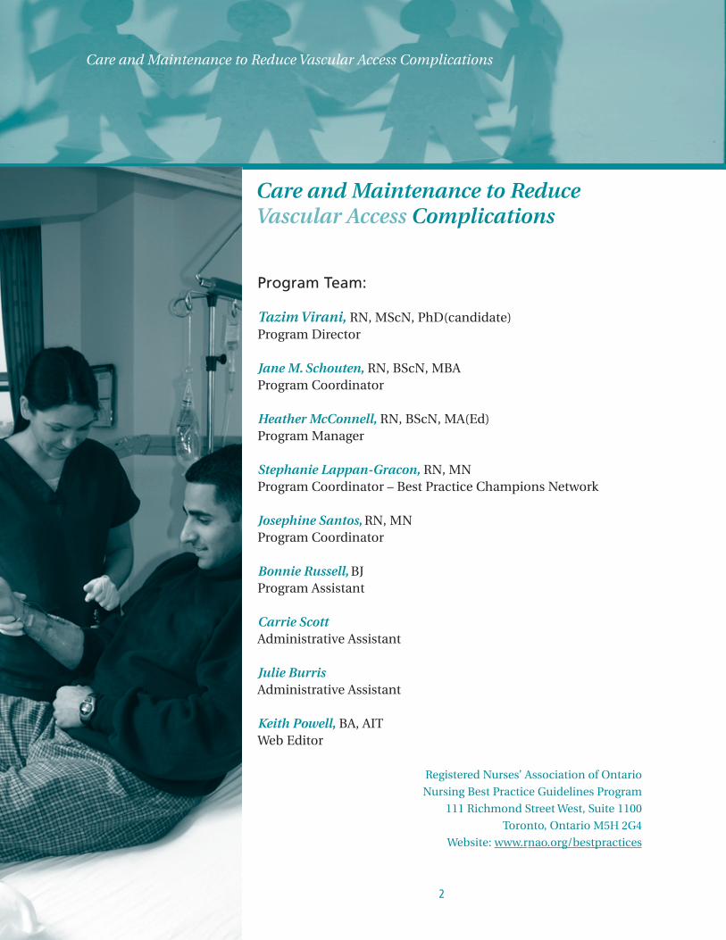
Program Team:
Tazim Virani, RN, MScN, PhD(candidate)Program Director
Jane M. Schouten, RN, BScN, MBAProgram Coordinator
Heather McConnell, RN, BScN, MA(Ed)Program Manager
Stephanie Lappan-Gracon, RN, MNProgram Coordinator – Best Practice Champions Network
Josephine Santos, RN, MNProgram Coordinator
Bonnie Russell, BJProgram Assistant
Carrie ScottAdministrative Assistant
Julie BurrisAdministrative Assistant
Keith Powell, BA, AITWeb Editor
Care and Maintenance to ReduceVascular Access Complications
3
Nursing Best Practice Guideline
Development Panel Members
Susanne Nelson, RN, BScN, MN (C), CINA(C)Team Leader
Nurse Coordinator – Vascular Access
University Health Network
Toronto, Ontario
Lisa Valentine, RN, BScN, MNTeam Facilitator
Practice Consultant
College of Nurses of Ontario
Toronto, Ontario
Sharon Armes, RN, CINA(C)Clinical Education Coordinator
Bard Canada Inc.
Mississauga, Ontario
Adrienne Austin, RN, BScN, CINA(C)Clinical Manger, Vascular Access Therapy
Hamilton Health Sciences Centre
Hamilton, Ontario
Nan Cleator, RN, CINA(C)National Clinical Consultant
Victorian Order of Nurses – Canada
Huntsville, Ontario
Lina D’Onofrio, RN, MNClinical Nurse Specialist
Transfusion Services
University Health Network
Toronto, Ontario
Cynthia Giff, RNNursing Director Medical/Surgical Units
Brockville General Hospital
Brockville, Ontario
Susanne Gomes, RN, BScN,Oncology Nurse
Thunder Bay Regional Health Sciences Centre
Thunder Bay, Ontario
Glenda Hicks, RN, BScNNurse Educator/Clinician
Critical Care Program
St. Joseph’s Heath Centre
Sudbury, Ontario
Kris Paton, RN, CINA(C)Clinical Leader, Vascular Access Therapy
Hamilton Health Sciences Centre
Hamilton, Ontario
Sharon Rodkin, RN, CINA(C)Manager, Clinical Consulting
Baxter Corporation
Mississauga, Ontario
Jane M. Schouten, RN, BScN, MBAProgram Staff – Facilitator
Nursing Best Practice Guidelines Program
Registered Nurses’ Association of Ontario
Toronto, Ontario
RNAO also wishes to acknowledge the following:
Diane Legere, RN, APCCN, BScN, MScN(C) for her
work as a Research Assistant in conducting the
quality appraisal of the literature and preparation
of evidence tables for the development of this
guideline; and
Teresa Harper, RN, MSN (Patient EducationSpecialist) for her expertise on client education and
her contribution to the development of the client
education recommendation of this guideline.
Declarations of interest and confidentiality were made by all members of the guideline development panel. Further details are
available from the Registered Nurses’ Association of Ontario.
Care and Maintenance to Reduce Vascular Access Complications
4
Acknowledgement
Stakeholders representing diverse perspectives were solicited for their feedback and the Registered Nurses’ Association
of Ontario wishes to acknowledge the following for their contribution in reviewing this Nursing Best Practice Guideline.
Charlene Allin, RN, BA, CNCC(C) Charge Nurse ICU/Telemetry, Leamington District Memorial Hospital,Leamington, Ontario
Gina Bagger, RN, BScN Former Vascular Access Resource Nurse, The Hospital for Sick Children, Toronto, Ontario
Nancy A. Bauer, Hon.BA, Professional Practice Leader, Leamington District Memorial Hospital,Hon.Bus. Admin., RN, ET Leamington, Ontario
Michele Bellows, RN, CINA(C) Director of Care and Emergency Room, Stoneridge Manor/Carleton Place andDistrict Memorial, Carleton Place, Ontario
Joan Bennett, RN, CINA(C) RN Surgical Day Care, Peri-operative Services, St. Michael’s Hospital, Toronto, Ontario
Suzanne Benoit, BScN, RN, CINA(C) Nurse Clinician, Systemic Treatment Program, Northeastern Ontario RegionalCancer Centre of the Sudbury Regional Hospital, Sudbury, Ontario
Isobelle Blake, RN, BScN, CINA(C) Director of Clinical Management, Bayshore Home Health, St. Catharines, Ontario
Rosemary Bland, RN, BSN, Nurse Manager, Palliative Care, Oncology, and Ambulatory Care, CON(C), CINA(C), OCN Joseph Brant Memorial Hospital, Burlington, Ontario
Rebecca Brock, RN Program Manager, Victorian Order of Nurses, North Bay, Ontario
Linda Brown, RN, BScN, CINA(C) Nurse Clinician, Chatham-Kent Health Alliance, Chatham, Ontario
Donna Burkart, RN, BN Clinical Educator, Lake of the Woods District Hospital, Kenora, Ontario
Pat Carlson, RN, CINA(C) Senior Skin Health Representative, 3M Canada Company, Health Care Division,London, Ontario
Risa Cashmore, RN, BSc, CIC, CINA(C) Infection Control Specialist, Peel Region Public Health, Brampton, Ontario
Kathleen Cranston, RN, CON(C), CCRP Infection Control Practitioner, Thunder Bay Regional Health Sciences Centre,Thunder Bay, Ontario
Kimberly Dalla Bona, RN, BScN, CINA(C) Clinical Resource Nurse, Saint Elizabeth Health Care, Windsor, Ontario
Catherine Davilmar, BScN, MBA Clinical Consultant, Baxter Corporation, Mount Royal, Quebec
Susanne Dodman, RN, BA Clinical Education Specialist, Baxter Corporation, Mississauga, Ontario
Linda Giesler, RN, BScN Nurse Clinician, North Bay General Hospital, North Bay, Ontario
Jocelyn Grecia Hill, RN, BSN, ONC Clinical Nurse Educator/Clinical Nurse Leader – IV Therapy & Home Infusion, St. Paul’s Hospital-Providence Health Care, Vancouver, British Columbia
Elizabeth Hummel, RN, BScN Nurse Clinician CCU/ER, North Bay General Hospital, North Bay, Ontario

5
Nursing Best Practice Guideline
Dianne Husbands, RN, BA, Acting Nurse Manager, IV Therapy, 5 Chest, 2 Uro/Gyne, BScN, ENC(C), MN(C) St. Joseph’s Healthcare, Hamilton, Ontario
Diana Iaquinto, RN, BScN, CPN(C) Registered Perioperative Nurse, St. Joseph’s Healthcare, Hamilton, Ontario
Tami Jemson, RN Patient Care Coordinator IV Program, Kelowna General Hospital, Kelowna,British Columbia
Elsabeth Jensen, RN, PhD Research Coordinator/Scientist, University of Western Ontario/Lawson HealthResearch Institute, London, Ontario
Sherri Keller, RN, CINA(C) Clinical Education Specialist, Becton Dickinson, and Company, Oakville, Ontario
Trinet Landry, RN, BScN, CINA
Madeleine Larson, RN Nurse Educator, Family Child Program, Sudbury Regional Hospital, Sudbury, Ontario
Mark C. Lepinsky, HBScN, CETN Surgical Services Clinical Educator, Thunder Bay Regional Health SciencesCentre, Thunder Bay, Ontario
June MacDonald-Jenkins, RN, BScN, MN(C) Professor Health Studies Durham/UOIT Collaborative Nursing Program, DurhamCollege and the University of Ontario Institute of Technology, Oshawa, Ontario
Lorraine Mackett, RN, HBScN, CETN Manager 2B (Medical Unit), Thunder Bay Regional Health Sciences Centre,Thunder Bay, Ontario
Joan Maguire, RN, CINA(C) IV Therapy Resource Nurse, Southlake Regional Health Centre, Newmarket, Ontario
Terry Major, RN, BScN, CON(C) Thunder Bay Regional Health Sciences Centre, Thunder Bay, Ontario
Sue Masoorli, RN President/CEO, Perivascular Nurse Consultants, Philadelphia, Pennsylvania
Sherry McKnight, RN, BScN, CINA(C) Nurse Clinician, Brant Community Healthcare System, Brantford, Ontario
Jo-Ann Murphy, RN Team Manager, Near North Community Care Access Centre, North Bay, Ontario
Suzanne Nagy, RN, CINA(C) Director, Pharmaceutical Services, Bayshore Home Health, Mississauga, Ontario
Kylie Nowak, RN, MN Nurse Clinician, Infusion Therapy, Mount Sinai Hospital, Toronto, Ontario
Louise Oak, RN PICC Nurse Insertionist, Sault Area Hospital, Sault Ste. Marie, Ontario
Cherie Pinkerton, RN, BN Nurse Educator, Health Sciences Centre, Winnipeg, Manitoba
Susan Pitalzke, RN, BScN, MPH Director of Clinical Oncology Systems, Thunder Bay Regional Health SciencesCentre, Regional Cancer Program, Thunder Bay, Ontario
Marg Poling, RN, BScN, PHCNP Palliative Care Nurse Practitioner, Palliative Care Advisor, Victorian Order ofNurses, Thunder Bay and District, Thunder Bay, Ontario
Wendy L. Pomponio, RN, BScN Nurse Clinician, Medical & Rehabilitation Services, Brant Community HealthcareSystem, Brantford, Ontario
Donna Prenger, RN, ONA Registered Nurse (Oncology), Thunder Bay Regional Health Science Centre,Thunder Bay, Ontario
Christina Purdon, RN, BScN Clinical Educator, Thunder Bay Regional Health Sciences Centre, Thunder Bay, Ontario
Mary Runde, RN, MN-ACNP, Educator ICU/Critical Care, Sault Area Hospital, CNCC (C), CCN(C), CINA Sault Ste. Marie, Ontario
Gwen Schledewitz, RN Victorian Order of Nurses, North Bay, Ontario
Freda Seddon, RN, RM Staff Nurse, Victorian Order of Nurses, Peterborough, Victoria, HaliburtonBranch, Bobcaygeon, Ontario

Care and Maintenance to Reduce Vascular Access Complications
6
Jill Steele, RN Case Manager, Near North Community Care Access Centre, North Bay, Ontario
Donna Tucker, RN, MScN Project Director, Healthy Work Environments Best Practice Guidelines Project,RNAO, Toronto, Ontario
Colleen Valente, RN(EC), Oncology Nurse Practitioner, Systemic Therapy, Thunder Bay Regional Health MN(C), CON(C), CHPCN(C) Sciences Centre, Integrated Cancer Program, Thunder Bay, Ontario
Susan Watson, RN, CINA (C), NCA Nursing Supervisor, VHA Home Health Care, Chatham, Ontario
Sahar Whelan, BScPhm, MScPhm Director of Pharmacy, Coram Healthcare Ltd., Toronto, Ontario
Sandra Whittle, RN RN, Oncology/Medical Day Clinic, Leamington District Memorial Hospital,Leamington, Ontario
Patti Wolfe, RN Clinical Consultant, Baxter Corporation, Mississauga, Ontario
Anita Woolman, RN Visiting Nurse, Victorian Order of Nurses, Hunstville, Ontario
Lorna Zubrickas, RN CINA (C), CON(C) Clinical Educator, Cambridge Memorial Hospital, Cambridge, Ontario

7
Nursing Best Practice Guideline
Care and Maintenance to Reduce Vascular Access ComplicationsDisclaimer
These best practice guidelines are related only to nursing practice and not intended to take into account
fiscal efficiencies. These guidelines are not binding for nurses and their use should be flexible to
accommodate client/family wishes and local circumstances. They neither constitute a liability or discharge
from liability. While every effort has been made to ensure the accuracy of the contents at the time of
publication, neither the authors nor the Registered Nurses’ Association of Ontario (RNAO) give any
guarantee as to the accuracy of the information contained in them nor accept any liability, with respect to
loss, damage, injury or expense arising from any such errors or omission in the contents of this work. Any
reference throughout the document to specific pharmaceutical products as examples does not imply
endorsement of any of these products.
Copyright
With the exception of those portions of this document for which a specific prohibition or limitation against
copying appears, the balance of this document may be produced, reproduced and published, in any form,
including in electronic form, for educational or non-commercial purposes, without requiring the consent
or permission of the Registered Nurses’ Association of Ontario, provided that an appropriate credit or
citation appears in the copied work as follows:
Registered Nurses’ Association of Ontario. (2005). Care and Maintencance to Reduce Vascular Access
Complications. Toronto, Canada: Registered Nurses’ Association of Ontario.

Care and Maintenance to Reduce Vascular Access Complications
8
Table of Contents
Summary of Recommendations . . . . . . . . . . . . . . . . . . . . . . . . . . . . . . . . . . . . . . . . . . . . . . . . . . . . . . . . . . . . . . . . . 10
Interpretation of Evidence . . . . . . . . . . . . . . . . . . . . . . . . . . . . . . . . . . . . . . . . . . . . . . . . . . . . . . . . . . . . . . . . . . . . . . 12
Responsibility for Development . . . . . . . . . . . . . . . . . . . . . . . . . . . . . . . . . . . . . . . . . . . . . . . . . . . . . . . . . . . . . . . . . 13
Purpose & Scope . . . . . . . . . . . . . . . . . . . . . . . . . . . . . . . . . . . . . . . . . . . . . . . . . . . . . . . . . . . . . . . . . . . . . . . . . . . . . . 13
Development Process . . . . . . . . . . . . . . . . . . . . . . . . . . . . . . . . . . . . . . . . . . . . . . . . . . . . . . . . . . . . . . . . . . . . . . . . . . 14
Definition of Terms. . . . . . . . . . . . . . . . . . . . . . . . . . . . . . . . . . . . . . . . . . . . . . . . . . . . . . . . . . . . . . . . . . . . . . . . . . . . . 16
Background Context . . . . . . . . . . . . . . . . . . . . . . . . . . . . . . . . . . . . . . . . . . . . . . . . . . . . . . . . . . . . . . . . . . . . . . . . . . . 17
Practice Recommendations . . . . . . . . . . . . . . . . . . . . . . . . . . . . . . . . . . . . . . . . . . . . . . . . . . . . . . . . . . . . . . . . . . . . . 19
Education Recommendations . . . . . . . . . . . . . . . . . . . . . . . . . . . . . . . . . . . . . . . . . . . . . . . . . . . . . . . . . . . . . . . . . . . 40
Organization & Policy Recommendations . . . . . . . . . . . . . . . . . . . . . . . . . . . . . . . . . . . . . . . . . . . . . . . . . . . . . . . . 42
Research Gaps & Future Implications. . . . . . . . . . . . . . . . . . . . . . . . . . . . . . . . . . . . . . . . . . . . . . . . . . . . . . . . . . . . 45
Evaluation & Monitoring of Guideline . . . . . . . . . . . . . . . . . . . . . . . . . . . . . . . . . . . . . . . . . . . . . . . . . . . . . . . . . . . 46
Implementation Strategies. . . . . . . . . . . . . . . . . . . . . . . . . . . . . . . . . . . . . . . . . . . . . . . . . . . . . . . . . . . . . . . . . . . . . . 48
Process for Update/Review of Guideline . . . . . . . . . . . . . . . . . . . . . . . . . . . . . . . . . . . . . . . . . . . . . . . . . . . . . . . . . 50
References . . . . . . . . . . . . . . . . . . . . . . . . . . . . . . . . . . . . . . . . . . . . . . . . . . . . . . . . . . . . . . . . . . . . . . . . . . . . . . . . . . . . 51
Bibliography . . . . . . . . . . . . . . . . . . . . . . . . . . . . . . . . . . . . . . . . . . . . . . . . . . . . . . . . . . . . . . . . . . . . . . . . . . . . . . . . . . 55

9
Nursing Best Practice Guideline
Appendix A: Search Strategy for Existing Evidence . . . . . . . . . . . . . . . . . . . . . . . . . . . . . . . . . . . . . . . . . . . . . . . . 60
Appendix B: Glossary of Clinical Terms . . . . . . . . . . . . . . . . . . . . . . . . . . . . . . . . . . . . . . . . . . . . . . . . . . . . . . . . . . 64
Appendix C: Vein Anatomy & Blood Flow Rates for Peripheral Site Selection . . . . . . . . . . . . . . . . . . . . . . . . 67
Appendix D: Tip Placement for Tunneled Catheter . . . . . . . . . . . . . . . . . . . . . . . . . . . . . . . . . . . . . . . . . . . . . . . . 68
Appendix E: Groshong® Valve Function . . . . . . . . . . . . . . . . . . . . . . . . . . . . . . . . . . . . . . . . . . . . . . . . . . . . . . . . . . 69
Appendix F: Catheter Patency. . . . . . . . . . . . . . . . . . . . . . . . . . . . . . . . . . . . . . . . . . . . . . . . . . . . . . . . . . . . . . . . . . . 70
Appendix G: Blood Withdrawal from Central Venous Access Devices . . . . . . . . . . . . . . . . . . . . . . . . . . . . . . . 72
Appendix H: Data Collection Tool for Central Venous Access Devices . . . . . . . . . . . . . . . . . . . . . . . . . . . . . . . 74
Appendix I: Developing Patient Teaching Materials . . . . . . . . . . . . . . . . . . . . . . . . . . . . . . . . . . . . . . . . . . . . . . . 76
Appendix J: Planning Guide for Educational Materials . . . . . . . . . . . . . . . . . . . . . . . . . . . . . . . . . . . . . . . . . . . . 77
Appendix K: Patient Educational Material – Example . . . . . . . . . . . . . . . . . . . . . . . . . . . . . . . . . . . . . . . . . . . . . 78
Appendix L: Educational Resources. . . . . . . . . . . . . . . . . . . . . . . . . . . . . . . . . . . . . . . . . . . . . . . . . . . . . . . . . . . . . . 81
Appendix M: Description of the Toolkit . . . . . . . . . . . . . . . . . . . . . . . . . . . . . . . . . . . . . . . . . . . . . . . . . . . . . . . . . . 82
Care and Maintenance to Reduce Vascular Access Complications
10
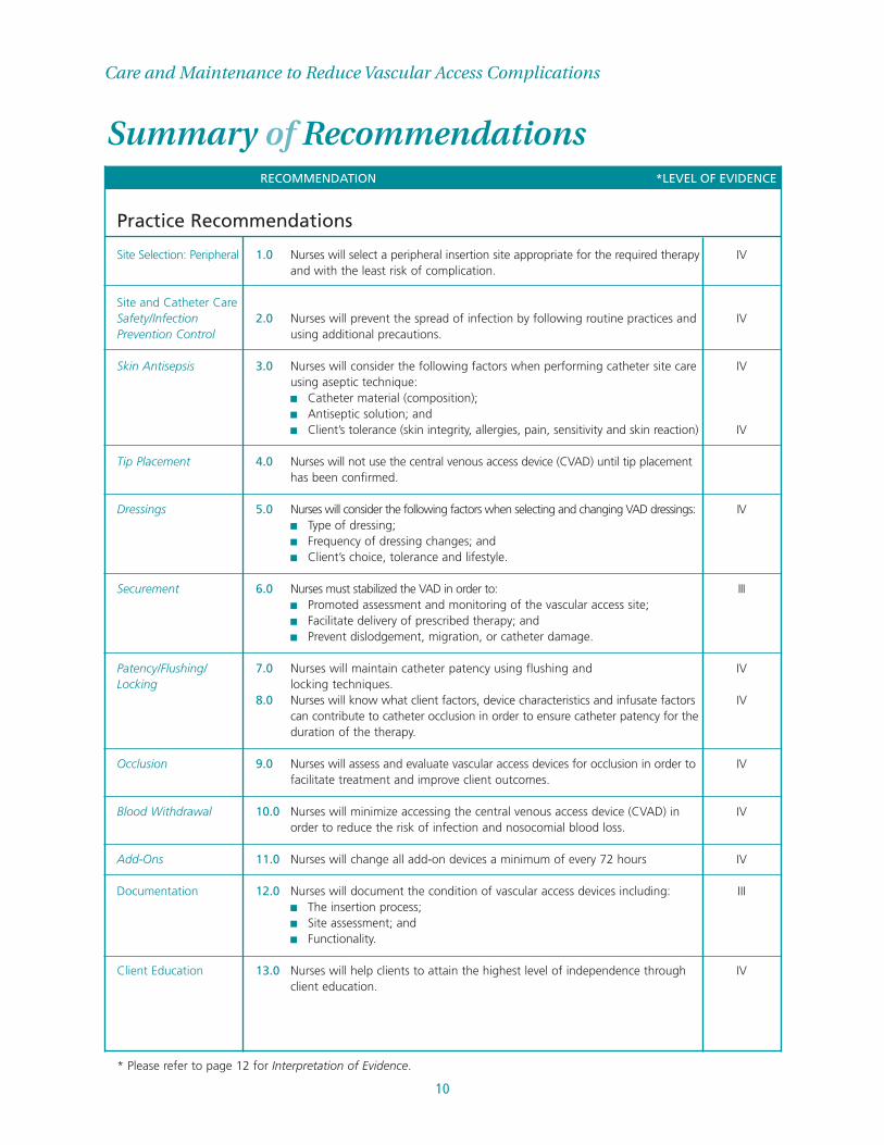
Summary of RecommendationsRECOMMENDATION *LEVEL OF EVIDENCE
Practice Recommendations
Site Selection: Peripheral 1.0 Nurses will select a peripheral insertion site appropriate for the required therapy IVand with the least risk of complication.
Site and Catheter CareSafety/Infection 2.0 Nurses will prevent the spread of infection by following routine practices and IVPrevention Control using additional precautions.
Skin Antisepsis 3.0 Nurses will consider the following factors when performing catheter site care IVusing aseptic technique: ■ Catheter material (composition);■ Antiseptic solution; and ■ Client’s tolerance (skin integrity, allergies, pain, sensitivity and skin reaction) IV
Tip Placement 4.0 Nurses will not use the central venous access device (CVAD) until tip placement has been confirmed.
Dressings 5.0 Nurses will consider the following factors when selecting and changing VAD dressings: IV■ Type of dressing;■ Frequency of dressing changes; and■ Client’s choice, tolerance and lifestyle.
Securement 6.0 Nurses must stabilized the VAD in order to: III■ Promoted assessment and monitoring of the vascular access site; ■ Facilitate delivery of prescribed therapy; and■ Prevent dislodgement, migration, or catheter damage.
Patency/Flushing/ 7.0 Nurses will maintain catheter patency using flushing and IVLocking locking techniques.
8.0 Nurses will know what client factors, device characteristics and infusate factors IVcan contribute to catheter occlusion in order to ensure catheter patency for the duration of the therapy.
Occlusion 9.0 Nurses will assess and evaluate vascular access devices for occlusion in order to IVfacilitate treatment and improve client outcomes.
Blood Withdrawal 10.0 Nurses will minimize accessing the central venous access device (CVAD) in IVorder to reduce the risk of infection and nosocomial blood loss.
Add-Ons 11.0 Nurses will change all add-on devices a minimum of every 72 hours IV
Documentation 12.0 Nurses will document the condition of vascular access devices including: III■ The insertion process;■ Site assessment; and■ Functionality.
Client Education 13.0 Nurses will help clients to attain the highest level of independence through IVclient education.
* Please refer to page 12 for Interpretation of Evidence.
Education Recommendations
14.0 The principles and practice of infusion therapy should be included in the basic IVeducation curriculum, be available as continuing education, be provided in orientation to new employees and be made available through continuing professional development opportunities.
15.0 Schools of Nursing will include RNAO best practice guidelines Assessment IVand Device Selection for Vascular Access and Care and Maintenance to Reduce Vascular Access Complications as reference material for core curricula.
Organization & Policy Recommendations
16.0 Health care organizations will have policies that address components of IVvascular access therapy in order to ensure positive client outcomes.
17.0 Health care organizations, in collaboration with their infection control teams, IVwill monitor complications of infusion therapy and use data to employ risk reduction strategies.
18.0 Health care organizations will implement the use of safety engineered IIIdevices and equipment to reduce the nurse’s risk of sharps injuries that can lead to blood borne diseases. The organization’s risk management program will monitor assessment of these practices and incidents.
19.0 Health care organizations have access to infusion therapy nursing expertise IIIto support optimal vascular access outcomes.
20.0 Nursing best practice guidelines can be successfully implemented only where IVthere are adequate planning, resources, organizational and administrative support, as well as appropriate facilitation. Organizations may wish to develop a plan for implementation that includes:■ An assessment of organizational readiness and barriers to education.■ Involvement of all members (whether in a direct or indirect supportive
function) who will contribute to the implementation process.■ Dedication of a qualified individual to provide the support needed for the
education and implementation process.■ Ongoing opportunities for discussion and education to reinforce the
importance of best practices.■ Opportunities for reflection on personal and organizational experience in
implementing guidelines.In this regard, RNAO (through a panel of nurses, researchers and administrators) has developed the Toolkit: Implementation of Clinical Practice Guidelinesbased on available evidence, theoretical perspectives and consensus. The Toolkit is recommended for guiding the implementation of the RNAO guideline Care and Maintenance to Reduce Vascular Access Complications.
11
Nursing Best Practice Guideline
Summary of RecommendationsRECOMMENDATION *LEVEL OF EVIDENCE

Care and Maintenance to Reduce Vascular Access Complications
12
Interpretation of EvidenceLevels of Evidence
Ia Evidence obtained from meta-analysis or systematic review of randomized controlled trials.
Ib Evidence obtained from at least one randomized controlled trial.
IIa Evidence obtained from at least one well-designed controlled study without randomization.
IIb Evidence obtained from at least one other type of well-designed quasi-experimental study
without randomization.
III Evidence obtained from well-designed non-experimental descriptive studies, such as
comparative studies, correlation studies and case studies.
IV Evidence obtained from expert committee reports or opinions and/or clinical experiences of
respected authorities.
13
Nursing Best Practice Guideline
Responsibility for DevelopmentThe Registered Nurses’ Association of Ontario (RNAO), with funding from the Government
of Ontario, has embarked on a multi-year project of nursing best practice guideline development, pilot
implementation, evaluation and dissemination. In this sixth cycle of the projectprogram, one of the areas
of emphasis is on vascular access care and management to reduce complications for the client. This
guideline was developed by a panel of nurses convened by the RNAO, conducting its work independent of
any bias or influence from the Ontario Government of Ontario.
Purpose & Scope Best practice guidelines (BPG) are systematically developed statements to assist practitioners’
and clients’ decisions about appropriate health care (Field & Lohr, 1990). This best practice guideline focuses
on assisting all nurses in diverse practice settings, both institutional and community, who provide care to
clients requiring vascular access. This guideline incorporates best practices related to the care and
maintenance of vascular access devices applicable to all adult clients requiring this kind of care. It should
be noted that the main focus of this guideline is the care and maintenance of central venous access devices
(CVAD); however, where care and maintenance strategies can be used for both CVAD and peripheral
venous access devices (PVAD), this has been articulated in the specific recommendations. This guideline
does not include recommendations related to the care of clients requiring infusion therapy through the
following devices: arterial lines, hemodialysis catheters, pulmonary artery lines, pheresis lines, epidural
catheters, pressure monitoring devices, umbilical vein, femoral catheters, and/or intraosseous lines.
Nurses working with other types of vascular access devices will require further practice direction from
guidelines in their unique area of practice.
As part of the health care team, nurses caring for clients with indwelling vascular access devices have an
important role in providing safe infusion therapy. This guideline focuses its recommendations on: Practice
Recommendations that assist the nurse to provide appropriate care for the client, including site selection,
site care, and catheter care; Education Recommendations for supporting the knowledge, skills, and
judgment required for nurses; and Organization and Policy Recommendations addressing the importance
of a supportive practice environment as an enabling factor for providing high quality nursing care, which
includes ongoing evaluation of guideline implementation.
The purpose of this guideline is to provide evidence-based support for nurses related to the care and
maintenance of vascular access devices, client education and safety. Specific clinical questions to be
addressed include: ■ How can the risk of complications be minimized through appropriate care and maintenance of
vascular access devices?■ What strategies should be used for client and staff education to address the care and maintenance of
vascular access devices?
It is acknowledged that the individual competencies of nurses varies between nurses and across categories
of nursing professionals (RPNs and RNs) and are based on knowledge, skills, attitudes, critical analysis and

Care and Maintenance to Reduce Vascular Access Complications
14
decision making which are enhanced over time by experience and education. It is expected that individual
nurses will perform only those aspects of care and maintenance for vascular access devices for which they
have received appropriate education and have experience. It is expected that nurses will seek appropriate
consultation in instances where the client’s care needs surpass the nurse’s ability to act independently. It
is acknowledged that effective healthcare depends on a coordinated health care team approach
incorporating ongoing communication between health professionals and clients, ever mindful of the
personal choices and unique needs of each individual client.
Development Process In June of 2004, a panel of nurses with expertise in vascular access from institutional, community,
educational and industry settings (including vendor companies) was convened under the auspices of the
RNAO. At the outset, the panel established the scope of the guideline through a process of discussion and
consensus. It was decided to focus on the care and maintenance of vascular access in order to reduce
complications for the client.
A set of nine published guidelines related to vascular access care and maintenance were identified through
a structured search, the details of which are described in Appendix A. These guidelines were reviewed
according to a set of screening criteria, established through panel consensus, which resulted in the
elimination of two guidelines. The screening criteria used were:
■ Guideline was in English; ■ Guideline was dated no earlier than 2000; ■ Guideline was strictly about the topic area;■ Guideline was evidence-based; and ■ Guideline was available and accessible for retrieval.
Seven guidelines were critically appraised with the intent of identifying existing guidelines that were
current, developed with rigour, evidenced-based and which addressed the scope identified by the panel for
this best practice guideline. A quality appraisal was conducted on these seven clinical practice guidelines using
the Appraisal of Guidelines for Research and Evaluation (AGREE) Instrument (AGREE Collaboration, 2001).
This process yielded a decision to work primarily with five existing guidelines including:
15
Nursing Best Practice Guideline
1. Centers for Disease Control and Prevention (CDC) (2002). Guidelines for the prevention of
intravascular catheter-related infections. Morbidity and Mortality Weekly Report (MMWR) 51
(No. RR-10), 1-29.
2a. Department of Health (DH) (2001a). Guidelines for preventing infections associated with the
insertion and maintenance of central venous catheters: Introduction. Journal of Hospital Infection 47,
S13-S19.
2b. Department of Health (DH) (2001b). Guidelines for preventing infections associated with the
insertion and maintenance of central venous catheters. Journal of Hospital Infection 47, S47-S67.
3a. Evidence-Based Practice in Infection Control (EPIC) (2001a). The EPIC project: Developing national
evidence-based guidelines for preventing hospital-acquired infections. National evidence-based
guidelines for preventing hospital-acquired infections associated with the use of central venous
catheters. Technical report part A. Available: http://www.epic.tvu.ac.uk/epicphase/epic1.html
3b. Evidence-Based Practice in Infection Control (EPIC) (2001b). The EPIC project: Developing national
evidence-based guidelines for preventing hospital-acquired infections. National evidence-based
guidelines for preventing hospital-acquired infections associated with the use of central venous
catheters. Technical report part B. Available: http://www.epic.tvu.ac.uk/epicphase/epic1.html
4. Intravenous Nurses Society (INS) (2000). Infusion nursing: Standards of practice. Journal of
Intravenous Nursing, 23, S1-S88.
5. Royal College of Nursing (RCN) (2003). Standards for infusion therapy. London: Author.
The panel members divided into subgroups to undergo specific activities using the short-listed guidelines,
other literature and additional resources for the purpose of drafting recommendations. This process
yielded a draft set of recommendations. The panel members as a whole reviewed the recommendations,
discussed gaps, available evidence and came to consensus on a draft guideline.
This draft was submitted to a set of external stakeholders for review and feedback. An acknowledgement of
these reviewers is provided at the front of this document. Stakeholders represented various health care
disciplines, clients and families, as well as professional associations. External stakeholders were provided
with specific questions for comment, as well as the opportunity to give overall feedback and general
impressions. The results were compiled and reviewed by the development panel. Discussion and
consensus resulted in revisions to the draft document prior to publication.

Care and Maintenance to Reduce Vascular Access Complications
16
Definition of TermsFor terms not included here, please refer to Appendix B: Glossary of Clinical Terms.
Clinical Practice Guidelines or Best Practice Guidelines: Systematically developed
statements to assist practitioner and client decisions about appropriate health care for specific
clinical (practice) circumstances (Field & Lohr, 1990).
Consensus: A process for making decisions, not a scientific method for creating new knowledge.
At its best, consensus development merely makes the best use of available information, be that
scientific data or the collective wisdom of the participants (Black et al., 1999).
Education Recommendations: Statements of educational requirements and educational
approaches/strategies for the introduction, implementation and sustainability of the best practice
guideline.
Infusion Therapy: The parenteral administration of fluids, medications, nutritional support,
and transfusion of blood and blood products, delivered through a vascular access device (VAD)
inserted into a peripheral or central vein.
Organization & Policy Recommendations: Statements of conditions required for a
practice setting that enable the successful implementation of the best practice guideline. The
conditions for success are largely the responsibility of the organization, although they may have
implications for policy at a broader government or societal level.
Practice Recommendations: Statements of best practice directed at the practice of
healthcare professionals that are ideally evidence-based.
Vascular Access Devices: Catheters placed directly into the venous system for infusion therapy
and/or phlebotomy purposes.

17
Nursing Best Practice Guideline
Background Context Vascular Access Devices (VADs) are a common and important part of clinical practice for the
administration of parenteral fluids, nutrients, medications and blood products. In addition, VADs provide
a route to monitor the hemodynamic status of a client. Over the last two decades vascular access device
(VAD) technology has advanced and new treatment regimens have emerged. These changes bring with
them the desire to support best practice to provide more effective vascular access care. The desired clinical
goal is positive client outcomes as evidenced by completion of therapy, absence of complications and
client satisfaction with care delivery.
Nurses practicing vascular access care require the knowledge, skill, and judgment to manage VADs. The
clinical focus is on the prevention of complications; however if complications develop, nurses must
recognize, report, and intervene appropriately for positive client outcomes.
Nurses with additional education and clinical expertise in infusion therapy serve as clinical champions and
advocates. This specialized role, in concert with supportive organizational structures and processes, leads
to improved overall infusion practice and client outcomes (Centers for Disease Control and Prevention (CDC), 2002;
Farr, 1996; Fridkin et al., 1996; Intravenous Nurses Society (INS), 2000; Miller et al., 1996). As an integral part of the
multidisciplinary team, these nurses create a link between all aspects of care and bridge the gap between
hospital and community.
Healthcare organizations are challenged by nursing shortages. In particular the role of the infusion nurse
specialists in Ontario has been reduced due to downsizing and constrained resources. Clients are more
acutely ill, and have more complex care needs than ever before. Therefore, a nursing assessment of risk
factors, device selection, and catheter maintenance to minimize complications is a critical factor to client
recovery and survival (Mezey & Schoder, 2003). These skills are required by all nurses in order to ensure the
best possible client outcomes, recognizing limited resources.

Care and Maintenance to Reduce Vascular Access Complications
18
Critical Thinking
In nursing, critical thinking is crucial to the potential impact on client outcomes and is often viewed as
reasonable, purposeful or goal-directed. Critical thinking, although dependent on clinical reasoning that is
creative and intuitive, is contextual and changes depending on circumstances. Critical thinking is defined
as the “intellectually disciplined process of actively and skillfully conceptualizing, applying, synthesizing,
or evaluating information” (Zunkel, et al., 2004, p. 162).
Scheffer and Rubenfeld (2000) recently reported the results of an attempt to define the concept of critical
thinking in nursing. Fifty-five nursing experts from nine countries participated in the investigation. This
panel of experts identified the characteristics and skills essential for critical thinking in nursing. Although
complete consensus was not achieved, 45 of the 51 participating experts agreed on the following definition
of critical thinking: “Critical thinking in nursing is an essential component of professional accountability
and quality nursing care…. Critical thinkers in nursing practice the cognitive skills of analyzing, applying
standards, discriminating, information seeking, logical reasoning, predicting and transforming knowledge”
(Scheffer & Rubenfeld, 2000, p. 357).
It is important for nurses to apply critical thinking skills when performing comprehensive assessments
related to appropriate device selection. These would include but are not limited to prescribed therapy,
support system/resources, client preference, language/cultural barriers, lifestyle and occupation, and
other variables as outlined in the Assessment and Device Selection for Vascular Access nursing best practice
guideline (RNAO, 2004). These same assessment criteria are also essential when providing care and
maintenance of vascular access devices.
To decrease the risk of complications associated with the use of vascular access devices, it is essential not
only to develop guidelines but to have a process for the implementation of the guidelines. While the
content of this guideline provides practical direction to practitioners, the need to apply critical thinking
skills in problem solving and decision making is recognized as key to the successful implementation of this
guideline into direct practice.

19
Nursing Best Practice Guideline
Practice RecommendationsThis best practice guideline is developed on the foundation provided by the following standards of
practice of the College of Nurses of Ontario (CNO):
Practice guideline: Consent (CNO, 2004a), which provides an overview of the major features of the legislation,
pertinent definitions and the steps nurses need to take to obtain consent. It does not address consent
under the Mental Health Act.
Practice guideline: Culturally sensitive care (CNO, 2004b), which guides nurses in problem solving for
commonly encountered culturally sensitive situations.
Practice guideline: Disagreeing with the multidisciplinary plan of care: Fact sheet (CNO, 2004c), which was
developed to provide direction to nurses when they disagree with other care providers regarding the plan
of care or when they believe the client has not been given informed consent to the plan. It provides the
nurse with a tool for advocacy.
Practice guideline: Guide to decide (CNO, 2004d), which was designed to assist nurses to understand their
accountability in performing procedures and provides a framework for decision making.
Practice guideline: Medical directives – Revised 2000 (CNO, 2004e), which may be implemented for a number
of clients when specific conditions are met, and when specific circumstances exist.
Practice standard: Confidentiality and privacy – personal health information (CNO, 2004f), which provides an
overview of current Ontario legislation, and clarifies nursing standards for confidentiality and of privacy
health information.
Practice standard: Documentation (CNO, 2004g), which outlines nurses’ professional accountability in record
keeping, and the expectations for documentation for all nurses in direct practice.
Practice standard: Ethics (CNO, 2004h), which describes the ethical values that are most important to the
nursing profession in Ontario.
Practice standard: Infection prevention and control (CNO, 2004i), which describes a nurse’s role in infection
prevention and control, recognizing that some client care situations may require consultation with an
Infection Control Practitioner.
Practice standard: Medication (CNO, 2004j) which provides standards for nurses to administer medications
safely and effectively in all practice settings.

Care and Maintenance to Reduce Vascular Access Complications
20
Practice standard: Professional standards – Revised 2002 (CNO, 2004k) which outlines professional
expectations for all Ontario nurses in the areas of service to the public, knowledge, application of
knowledge, ethics, continued competence and accountability/responsibility.
Practice standard: Therapeutic nurse-client relationship (CNO, 2004l), which provides direction, regarding
establishing therapeutic nurse-client relationships.
In addition to the College of Nurses of Ontario documents identified above, it is expected that nurses will
have knowledge of workplace policies and procedures that support infusion therapy and management
within their organization. The nurse should also consider current legislation in the province or country in
which they practice.
Site Selection: PeripheralRecommendation 1.0Nurses will select a peripheral insertion site appropriate for the required therapy and with the least
risk of complication. Level IV
Discussion of EvidenceWhen selecting a site for a peripheral venous access device (PVAD) insertion, the nurse should:■ Avoid areas of flexion (INS, 2000); ■ Avoid the inner aspect of the wrist in order to decrease the risk of damage to the radial, ulnar, and/or
median nerves which are located within a five centimetre (cm) radius of the inner aspect of the wrist
(Masoorli, 1998; Thrush, 1995);■ Avoid the routine use of the veins of the lower extremities due to the increased risk of embolism,
thrombophlebitis of and infection (CDC, 2002; INS, 2000; Tagalakis, 2002);■ Choose the smallest gauge catheter to accommodate the prescribed therapy (INS, 2000);■ Choose a vein with a diameter and blood flow rate for adequate hemodilution of prescribed therapy.
For example: a one time dose of dilantin which has a pH of 12, should be infused using a 22 gauge
PVAD into a vein with a high blood flow rate for greatest hemodilution. Venous blood flow rates to be
considered are as follows: ● Dorsal arch for metacarpal veins (hand veins) diameter varies, with flow rate 10ml/min; ● Cephalic vein: 6mm diameter with flow rate 40ml/min at the level of the antecubital fossa and above; and● Basilic vein: 8mm diameter with flow rate of 95ml/min at the level of the antecubital fossa and above. See
Appendix C: Vein Anatomy & Blood Flow Rates, for a visual depiction of site location.■ Avoid the the antecubital fossa and metacarpal veins if a vesicant has to be peripherally delivered due
to the difficulty in detecting infiltration at these sites of flexion (Oncology Nursing Society (ONS), 2004).■ Assess the type of solution, pH, osmolarity, estimated volume of the infusate and vein condition prior
to selecting an alternative site due to infiltration/extravasation (INS, 2000).
For CVAD site selection, refer to RNAO (2004) best practice guideline: Assessment and Device Selection for
Vascular Access.
Alert: Clients receiving vascular access therapy longer than six days should be assessed forintermediate to long-term dwelling devices. Refer to the RNAO (2004) best practice guidelineAssessment and Device Selection for Vascular Access.
21
Nursing Best Practice Guideline
Site & Catheter Care
Safety/Infection Prevention and Control Recommendation 2.0Nurses will prevent the spread of infection by following routine practices and using additional
precautions. Level IV
Discussion of Evidence:In Ontario, nurses are expected to follow infection control standards of practice as outlined by the College
of Nurses of Ontario (CNO, 2004I) and by Health Canada (Public Health Agency of Canada (PHAC), 1998, 1999, 2003).
Infection prevention and control is based on two levels of precautions:■ Routine practices; and ■ Additional precautions.
Routine practices, prevention strategies used with all clients at all times, are based on the assumption that
all blood and certain body fluids have the potential to harbour infectious organisms (e.g., bacteria, virus or
fungus). Routine practices reduce the risk of exposure to: ■ Blood, including blood products, and materials soiled with blood;■ All body fluids (secretions and excretions) except sweat, regardless of whether they contain blood (e.g.,
urine, feces, semen, vaginal and respiratory secretions, cerebral spinal fluid); ■ Non-intact skin, weeping or draining lesions or wounds; and ■ Mucous membranes: eyes, nose, mouth, rectum or vagina (PHAC, 1999).
Routine practices include:■ Hand hygiene;■ Assessment of client risk factors;■ Screening;■ Hazard or risk reduction; and ■ Application of personal protective equipment (PPE).
Hand Hygiene Hand hygiene reduces the transmission of infection (e.g., micro-organism) by the hands and includes
cleaning hands with soap and water or with alcohol-based hand cleansers before and after, and as required
during all client care.
Hand hygiene also includes maintaining hand health, avoiding nail polish, artificial nails or jewellery and
keeping nails trimmed and clean. The fingernail area has been associated with a major portion of hand
flora as it can harbour micro-organisms (CDC, 2002; Health Canada, 2003).
Alert: Hand hygiene is the single most important infection prevention and control practice.

Care and Maintenance to Reduce Vascular Access Complications
22
Assessment of Client Risk FactorsOther routine practices include screening and assessment of client risk factors in order to reduce hazards
associated with infectious diseases. It is beyond the scope of this guideline to present a comprehensive,
inclusive discussion on infection control practices, nor replace governmental recommendations around
the use of protective equipment. Rather this guideline aims to alert the nurse to the importance of
following these practices.
Assessing client risk factors helps the nurse to determine the level of protection required by the client.
Factors to consider include: ■ Immunosuppression;■ Coagulopathies;■ Signs and symptoms of infection;■ History of exposure to infectious disease; and■ Client response during site assessment.
ScreeningIn addition to assessing clients for potential infection, the nurse should also screen families and visitors for
illness in order to protect the client from potential risk. Client screening for infectious diseases is ongoing
and encompasses client feedback (e.g., verbalized concerns regarding discomfort from IV site, chills, etc.)
(CNO, 2004; Ministry of Health and Long-Term Care (MOHLTC), 2002).
Hazard or Risk ReductionRisk reduction or hazard control strategies begin with an analysis by the nurse in order to make an
informed decision as to what strategies to employ to protect both the client and the staff. Risk reduction
strategies begin at the source (e.g., having the client who is coughing wear a mask during CVAD care); and
can include procedural changes to the care performed by the nurse to reduce the risk (e.g., using safety
engineered devices, proper disposal of infectious waste) (CNO, 2004i; PHAC, 2002). Nurses can further reduce
risk of hazard to themselves with the use of personal protective equipment (PPE).
Personal Protective Equipment (PPE)Application of personal protective equipment (PPE) is used to reduce or control any of the risks identified
as a result of the screening and risk assessment. PPE includes, but is not limited to: gloves; eye protection;
gowns; and masks. PPE is applied before direct contact with the client and prior to starting a procedure. For
example, the nurse applies gloves before removing a soiled dressing from the exit site of the CVAD (PHAC,
1999). Nurses should refer to Ministry of Health directives and organizational policies and procedures for
direction regarding to the use of PPE.
Alert: Reusable medical equipment will be cleaned, disinfected or sterilized between client use inadherence to national and provincial regulations and requirements (e.g., Health Canada orProvincial Ministries of Health).Any single use item (e.g., tourniquets, vacutainers) will remainwith the client (PHAC, 1998).

23
Nursing Best Practice Guideline
Skin Antisepsis Recommendation 3.0Nurses will consider the following factors when performing catheter site care using aseptic
technique:■ Catheter material (composition);■ Antiseptic solution; and■ Client tolerance (skin integrity, allergies, pain, sensitivity and skin reaction). Level IV
Discussion of EvidenceSkin cleansing and antisepsis of the insertion site is considered one of the most important measures for
preventing infections associated with vascular access devices (Evidence-Based Practice in Infection Control (EPIC),
2001a, 2001b; LeBlanc & Cobbett, 2000; Pearson, 1996a, 1996b). Skin must be clean; that is, free of soil, dust, and
organic material prior to applying the antiseptic (CDC, 2002; Health Canada, 2003). Organisms responsible for
catheter-related infections originate mainly from the client’s own skin flora (Crow, 1996; Jackson, 2001; RCN,
2003) or from the hands of the health care professional inserting or handling the device (Hadaway, 2003b;
Jackson, 2001). These organisms can be introduced along with the catheter or can gain access while the
catheter is in place. Catheter movement in or out of the insertion site (known as “pistoning”) can also allow
for skin organisms to migrate into the tract and potentially cause infections (Hadaway, 2003b).
Catheter Material (composition)Nurses must be knowledgeable about the type of device (central or peripheral) in order to make
appropriate care decisions around the skin antiseptic to be used during catheter care. Nurses will disinfect
clean skin with an appropriate antiseptic before catheter insertion and with each dressing change. The
antiseptic solution must be compatible with the catheter material (Hadaway, 2003a). Acetone products
should be avoided as they may cause irritation and affect the integrity of the catheter (O’Grady, et al., 2002;
Pearson, 1996a, 1996b) and alcohol-based solutions are not recommended for certain devices. Therefore, the
nurse must be aware of the health setting’s procedures around specific devices in order to protect the client
from harm.
Antiseptic SolutionStudies have shown that 2% chlorhexidine gluconate solution significantly lowers catheter-related
bloodstream infection rates when compared with 10% povidone-iodine and 70% isopropyl alcohol (LeBlanc &
Cobbett, 2000; Maki, Ringer & Alvarado, 1991; Mimoz, et al., 1996; Rosenthal, 2003; Zitella, 2004). Chlorhexidine gluconate
offers a broad spectrum of antimicrobial activity and long-term microbacteriocidal action after application
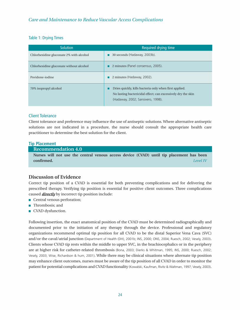
(Hadaway, 2003a). Antiseptics should remain on the insertion site and be allowed to air dry before catheter
insertion and/or dressing change. Table 1 describes the required drying time needed for particular solutions
in order to prevent skin breakdown as a result of chemical reaction between the solution and the dressing.
Care and Maintenance to Reduce Vascular Access Complications
24
Table 1: Drying Times
Client ToleranceClient tolerance and preference may influence the use of antiseptic solutions. Where alternative antiseptic
solutions are not indicated in a procedure, the nurse should consult the appropriate health care
practitioner to determine the best solution for the client.
Tip PlacementRecommendation 4.0Nurses will not use the central venous access device (CVAD) until tip placement has been
confirmed. Level IV
Discussion of EvidenceCorrect tip position of a CVAD is essential for both preventing complications and for delivering the
prescribed therapy. Verifying tip position is essential for positive client outcomes. Three complications
caused directly by incorrect tip position include:■ Central venous perforation;■ Thrombosis; and■ CVAD dysfunction.
Following insertion, the exact anatomical position of the CVAD must be determined radiographically and
documented prior to the initiation of any therapy through the device. Professional and regulatory
organizations recommend optimal tip position for all CVAD to be the distal Superior Vena Cava (SVC)
and/or the caval/atrial junction (Department of Health (DH), 2001b; INS, 2000; ONS, 2004; Ruesch, 2002; Vesely, 2003).
Clients whose CVAD tip rests within the middle to upper SVC, in the brachiocephalics or in the periphery
are at higher risk for catheter-related thrombosis (Bona, 2003; Dierks & Whitman, 1995; INS, 2000; Ruesch, 2002;
Vesely, 2003; Wise, Richardson & hum, 2001). While there may be clinical situations where alternate tip position
may enhance client outcomes, nurses must be aware of the tip position of all CVAD in order to monitor the
patient for potential complications and CVAD functionality (Kowalski, Kaufman, Rivitz & Waltman, 1997; Vesely, 2003).
Chlorhexidine gluconate 2% with alcohol
Chlorhexidine gluconate without alcohol
Povidone-iodine
70% isopropyl alcohol
■ 30 seconds (Hadaway, 2003b).
■ 2 minutes (Panel consensus, 2005).
■ 2 minutes (Hadaway, 2002).
■ Dries quickly, kills bacteria only when first applied.
No lasting bactericidal effect; can excessively dry the skin
(Hadaway, 2002; Sansivero, 1998).
Solution Required drying time
25
Nursing Best Practice Guideline
Although the tip position is identified immediately post insertion, it is critical to understand that there are
significant changes in the position of a catheter tip when the client changes position. On average, all
peripherally inserted central catheters (PICCs) will move at least two centimetres (cm) caudal (away from
the head) with arm movement. Catheters inserted via the subclavian or jugular veins will move on average
two to three cm cephalad (toward the head). A catheter, whose initial post insertion x-ray shows the tip to
be in the distal SVC, may in fact have a final tip position (once the patient sits up) in the high SVC. This
position could lead to an increased risk of complications as outlined above.
Although there is discussion that tip position should be “routinely” checked, an optimal time frame has not
been identified. At a minimum, the tip position should be checked radiographically if the CVAD
functionality changes and/or signs and symptoms of complications are observed (INS, 2000; ONS, 2004).
Nurses need to seek expert advice and advocate on the client’s behalf for other appropriate tests in order
to troubleshoot CVAD functionality. Some of these procedures include: ■ X-ray to verify tip position;■ Dye study as indicated;■ Ultrasound and/or Doppler ultrasound; and■ Fluoroscopy.
Appendix D contains a visual representation of the correct tip position of a tunneled CVAD.
Dressings Recommendation 5.0Nurses will consider the following factors when selecting and changing VAD dressings.■ Type of dressing;■ Frequency of dressing changes; and■ Client choice, tolerance and lifestyle. Level IV
Discussion of EvidenceTypeThe type of dressing used on the VAD has been recognized as one of the variables which affect complication
rates associated with these devices (Larwood, 2000). In addition, dressings offer securement of the VAD. Most
studies support and recommend the use of dressings (Larwood, 2000); however, the type of dressing remains
controversial (CDC, 2002). Dressings may be sterile transparent semi-permeable membrane (TSM), colloid
or sterile gauze (Hadaway, 2003). Sterile gauze dressings are more appropriate than transparent dressings
when insertion sites are bleeding, oozing or if the client is diaphoretic (CDC, 2002; Hadaway, 2003b; Rosenthal, 2003).
Care and Maintenance to Reduce Vascular Access Complications
26
Frequency of Dressing ChangeFactors pertaining to recommended dressing changes include moisture vapour permeability and the type
of product used (dry sterile gauze versus transparent dressing). Dressing changes using aseptic technique
should be completed every 48 hours for gauze and every seven days for TSM dressings or sooner if
contaminated, non-adherent, damp, loose, or visibly soiled (CDC, 2002; Hadaway, 2003b; O’Grady et al., 2002;
Pearson, 1996a, 1996b; Rosenthal, 2003; Ross & Orr, 1997). For accessed implanted VAD, the non-coring needle is
replaced every five to seven days, in concert with the dressing change (INS, 2000; Karamanoglu et al., 2003).
Sterile gauze under a transparent dressing is considered a gauze dressing and should be changed at a
minimum of every 48 hours (Jackson, 2001; INS, 2000; Rosenthal, 2003). For newly inserted CVAD, dressings
should be changed 24 hours post insertion (Cook, 1999).
Client ToleranceThe type of dressing may be a matter of client preference (CDC, 2002; Gillies et al., 2003). A meta-analysis
comparing the risk for catheter-related blood stream infections (CRBSIs) for groups using transparent
dressings versus groups using gauze dressing was reviewed by the Centers for Disease Control and
Prevention (CDC, 2002). The risk for CRBSIs did not differ between the groups and thus the choice of dressing
was a matter of preference (CDC, 2002).
Alert: The use of sterile versus non-sterile clean gloves during dressing changes remains anunresolved issue (CDC, 2002; Pearson, 1996a, 1996b). Therefore, either type can be used whenperforming catheter site care (O’Grady et al., 2002).
Practice Considerations■ Antimicrobial ointments should not be applied to insertions sites as they promote fungal infections
and antimicrobial resistance (CDC, 2002; EPIC, 2001; Hadaway, 2003a; Larwood, 2000; O’Grady et al., 2002).
■ Transparent dressings, should be placed on the skin (avoid stretching) and smoothed from the center
out to the edge and molded around the catheter. The edges of the transparent dressing should not be
sealed with tape (Jackson, 2001).
■ Transparent dressings require less frequent changes than standard gauze and tape and are reported to
save nursing time (CDC, 2002). A reduction in unscheduled restarts as well as an increase in dwell time
were noted when transparent dressings were used. A trend towards lower frequencies of phlebitis and
infiltration was reported in clients with transparent dressings (Tripepi-Bova et al., 1997).
■ Tunneled vascular access devices that are well healed may not require a dressing (CDC, 2002; O’Grady et
al., 2003). Implanted vascular access devices, which are healed and not accessed, do not require a
dressing. If the device is accessed, a sterile transparent semi-permeable dressing should be applied
(INS, 2000).

27
Nursing Best Practice Guideline
Securement Recommendation 5.0Nurses must stabilized the VAD in order to:■ Promote assessment and monitoring of the vascular access site;■ Facilitate delivery of prescribed therapy; and■ Prevent dislodgement, migration, and/or catheter damage. Level III
Discussion of EvidenceIn addition to securement using dressings, the following adjuncts can be used to further secure the VAD:■ Tape and/or sterile surgical strips;■ Sutures;■ Securement devices; and■ Stabilization dressings: specially designed securement and dressing products.
TapeWithout obscuring the insertion site, the catheter hub can be secured with tape or sterile surgical strips as
long as the tape is not applied directly to the catheter-skin junction site (INS, 2000). Other products may be
appropriate for clients whose skin integrity may predispose the use of tape as a means of securement.
Similar to choosing an appropriate antiseptic solution the nurse should also consider the client’s tolerance
for various types of tape, providing one which minimizes client discomfort.
Alert: Do not use pins of any kind to secure a device as this can damage the device andsubsequently interfere with therapeutic outcomes for the client.
SuturesSutures may be used to secure the hub of the catheter to the client’s skin. However, in the event that sutures
become loose or are no longer intact, the nurse should notify the physician and use sterile surgical tape or
a securement device as a temporary measure to prevent dislodgement (INS, 2000).
Both taping and suturing can allow micro-movement of the catheter that can result in complications
including but not limited to: phlebitis; infiltration; extravasation; dislodgement; disconnection; and
infection. Additionally, suturing the catheter increases the risk of needle stick injuries to the health care
provider. The Occupational Safety and Health Administration (Occupational Health and Safety and Health
Administration (OSHA), 2001), recommends using engineering controls as one option to secure medical
catheters. Engineering controls are designed to reduce the potential for needle sticks by eliminating the
need to suture medical catheters.
Securement DevicesThere are a number of commercially available securement devices that can be used for central and
peripheral devices. These devices are designed to reduce complications associated with suturing including
needle stick injuries and client infection. A study by Crinch and Maki (2002) compared a sutureless
securement device with sutures for the securement of PICCs. In this study (p<0.01), catheter-related blood
stream infections were significantly lower in the group of clients that received the sutureless securement device.
Care and Maintenance to Reduce Vascular Access Complications
28
A prospective, controlled study by Sheppard et al. (1999) determined that the peripheral vascular access
dwell time associated with a securement device resulted in significantly longer average dwell times and
significantly fewer local complications. The securement device also reduced clinical time in managing
vascular access. McMahon (2002) found that securement devices used with PICCs reduced catheter
migration and thus reduced the rate of catheter repair and exchange.
Alert: Securement devices must be changed at least every seven days (CDC, 2002).
Transparent dressings also assist in the securement of the device, allow for continuous visual inspection of
the catheter and permit the client to bathe and shower without saturating the dressing. The frequency of
peripheral vascular access catheter dislodgement by the client was significantly less with a transparent
dressing (EPIC, 2001; Larwood, 2000; Pearson, 1996a, 1996b).
Practice ConsiderationsTubing can be looped to relieve tension and is secured with tape independent of catheter tape, thus
preventing dislodgement of the catheter by an accidental pull on the tubing (Weinstein, 2001).
Patency/Flushing/LockingRecommendation 7.0Nurses will maintain catheter patency using flushing and locking techniques. Level IV
Discussion of Evidence Maintaining catheter patency is an important measure for all types of VADs. Regardless of the frequency,
type or volume, the majority of literature on maintaining patency recommends the use of correct flushing
and locking techniques (RCN, 2003). Flushing prevents the mixing of incompatible medications or solutions
and/or cleans the catheter lumen of blood or fibrin buildup. Locking prevents blood from backing up into
the catheter lumen when the device is not in use (ONS, 2004).
Flushing Turbulent Flush Technique
Although the issue of turbulent flush technique is addressed in the literature, there are no randomized
controlled trials (RCT) to support this technique. Despite the lack of RCT, the panel recommends the
turbulent flush technique as the best practice at this time to help prevent VAD occlusion.
All VADs (peripheral and central) should be flushed using turbulent flush technique to prevent the mixing
of incompatible medications or solutions and to reduce complications such as fibrin buildup or
accumulation of medication precipitate inside the catheter lumen. While flushing is meant to prevent fibrin
buildup, it is important to recognize that all VADs will accumulate fibrin coating to some extent (ONS, 2004).
In order to perform a flush with turbulence, the nurse should use a push-pause (stop-start) method. This
allows the solution to “scrub or clean” the inside of the device wall to promote removal of blood/fibrin and
to help prevent buildup of medication precipitate on the internal lumen of the device. (Dougherty, 1997; RCN, 2003).

29
Nursing Best Practice Guideline
Alert: Excessive flushing pressure can cause clots to be dislodged, catheter separation and/orcatheter rupture. In order to reduce the potential of excessive pressure, it is generallyrecommended that a 10 mL (or larger) syringe be used for flushing (RCN 2003). Larger syringescreate less pressure when flushing and more pressure when withdrawing oraspirating. Smaller syringes on the other hand produce more pressure when flushing andless pressure when withdrawing.
When using the turbulent flush technique, it is important to assess device function as highpressures could be generated in devices that have occlusion complications (e.g., fibrinbuildup) or are constricted in any way (e.g., kinked or clamped). Appendix F containsinformation for assessment of blood withdrawal and management of withdrawal occlusion.
Locking Positive pressure locking technique
Positive pressure locking techniques maintain positive pressure inside the lumen in order to prevent blood
reflux from the vein into the lumen of the VAD, thus preventing fibrin buildup, clots and thrombotic device
occlusions (INS, 2000; RCN, 2003). Nurses must know how to achieve positive pressure locking with the VAD
and infusion equipment they are using. Hadaway (2001) reports that positive pressure locking techniques
are rarely employed consistently and correctly, leading to a slowly building thrombus in the device lumen.
When using open ended VADs without systems to achieve fluid displacement (see the discussion that
follows on valve technology), the correct technique involves maintaining positive pressure on the syringe
plunger while closing the clamp and before removing the syringe from the cap of the device (INS, 2000;
Macklin, 1997).
Valve technology – Positive pressure caps
Devices with positive fluid displacement reduce or eliminate the variable of inconsistent flushing
technique. Several positive pressure cap products consistently achieve positive fluid displacement and
positive end-pressure. Positive pressure caps work by redirecting a small amount of fluid into the internal
catheter tip when the tubing or syringe is disconnected from the device hub. This prevents blood reflux into
the lumen. Catheters using positive pressure valves should not be clamped until after disconnection of the
flush syringe. Early studies are reporting a decrease in catheter occlusion with use of this type of valve (INS
2000; RCN, 2003).
Valve technology – Vascular access devices
A closed or open-ended valved CVAD is designed to resist blood reflux into the catheter lumen from the
vein. The closed-ended valved CVAD (e.g., Groshong® – see Appendix E) has an internal three way valve at
the tip of the catheter. Alternately, the open-ended valved CVAD (e.g., Pressure Activated Safety Valve
(PASV®)) has an open ended tip, and a pressure valve in the hub of the catheter. When the syringe is
disconnected the valve for both of these technologies is in a neutral position preventing flow in either
direction. The nurse needs to maintain positive pressure on the syringe plunger when disconnecting the
syringe from the cap or hub. Positive pressure caps can also be used with valved catheters (INS, 2000; ONS,
2004, RCN, 2003).

Care and Maintenance to Reduce Vascular Access Complications
30
The Four Elements of Flushing and Locking
Flushing and locking interventions involve four elements that need to be described in client specific orders
and/or in established medical directives. It is important that flushing/locking interventions are based on
standards of practice, evidence-based practice guidelines and current research (EPIC, 2001b; INS, 2000; RCN 2003).
These four elements include:■ Type of solution;■ Concentration of solution;■ Volume of solution; and ■ Frequency of administration.
1. Type of solution Flushing: The nurse should choose normal saline or other compatible solutions to flush the VAD in order
to prevent incompatibilities between two infusates.
Locking: Locking solutions used with positive pressure technique maintain catheter patency by preventing
blood reflux and by reducing the risk of blood clotting in the device lumen when blood reflux occurs.
Locking solutions include:■ Normal saline; and ■ Heparin.
Positive Pressure Saline Locking
There is a significant amount of literature which supports normal saline locking to maintain
patency of CVADs when using valved CVADs (e.g. PASV® or Groshong®) and/or when using positive
pressure caps (EPIC, 2001b; INS, 2000; RCN 2003).
Positive Pressure Heparin Locking
When open-ended devices are used without positive pressure technology (i.e., positive pressure
caps), blood can reflux into the device lumen. To reduce the incidence of the refluxed blood clotting
heparin can be used to lock the device. Concentrations of heparin reported for use in CVADs range
from 10-1000 IU/mL of which the most commonly reported heparin concentration is 100 IU/mL.
A meta-analysis of randomized controlled trials focusing on CVADs (CDC, 2002) concluded that
heparin significantly reduced bacterial colonization and showed a strong but non-significant trend
towards reductions of catheter-related bacteremia. Because thrombi and fibrin deposits on catheters
might serve as a nidus for microbial colonization of intravascular catheters the use of anticoagulants
might have a role to play in the prevention of catheter related blood stream infections (CRBSI) (CDC,
2002). Despite this benefit, heparin should be used with caution because it poses the risk of serious
complications even in small doses. Heparin has been associated with iatrogenic hemorrhage (a life-
threatening reaction to heparin), heparin induced thrombocytopenia (HIT), drug interactions and
inaccurate blood results (Dougherty, 1997; EPIC 2001b; Hadaway, 2001). Therefore, it should be used only
when necessary in order to reduce heparin-related complications.

31
Nursing Best Practice Guideline
Critical Thinking
Remember to heparinize the device not the client. Some clients that require a significantvolume or concentration of Heparin locking solutions may experience complications. Considerwithdrawal of the heparin lock prior to flushing to reduce the amount of heparin a client receives.While this is practiced in some settings, there is no scientific evidence to support this recommendation.The panel offers this as an option that could be considered based on the risks and benefits for someclients at high risk of device occlusion.
2. Concentration of SolutionThe concentration of the flushing or locking solution relates to the use of heparin. The heparin used should
be the lowest therapeutic concentration (e.g., 10 IU/mL) and have the smallest volume that will maintain
patency relative to the internal volume of the device (DH, 2001b; INS, 2000; RCN, 2003).
3. Volume of Solution Flushing: Nurses will use sufficient volumes of flush solutions to clean the internal lumen of the device
(3-5 mL for PVAD and 10-20 mL for CVAD). The volume after blood withdrawal and medication
administration should be at least 20 mL for all VADs. Macklin (1997) concludes that problems may occur
with too little flush solution but not with too much.
Locking: The volume should be at least twice the volume capacity of the catheter lumen (usually between
3-10 mL for all devices (Macklin, 1997)) plus the priming volume of all add-on devices of the infusion system
(e.g., extension tubing) (INS, 2000).
4. Frequency of AdministrationGenerally, flushing shall be performed:■ After blood sampling;■ When converting from continuous to intermittent therapies;■ Before and after medication administration;■ Before and after administration of blood components; ■ Before and after intermittent therapy; and■ For maintenance of a dormant device (INS, 2000; RCN, 2003).
Frequency of device use often determines the frequency of flushing and locking. Devices used
intermittently are flushed before administration and are flushed and locked at a minimum after every
infusion or medication administration. The schedule of catheter flushes varies among practice settings and
among indwelling devices (Ray, 1999). Despite this, VADs should be flushed and locked at established
intervals to maintain patency and to prevent occlusion.
Table 2 summarizes the recommended flushing and locking interventions related to VAD tip position and
technology.

Care and Maintenance to Reduce Vascular Access Complications
32
■ Text
Structure Process Outcome
Table 2: Flushing & Locking Interventions
Device Flush Solution, Concentration & Frequency Locking Solution, Concentration & Freuency
■ Routinely flush intermittent devices with 3-5 mL of 0.9% sodium chloride solution orcompatible solution with each use
■ Flush every 12 hours if frequency of use is>12 hours.
■ Routinely flush midlines with 5-10 mL of 0.9%sodium chloride solutions.
■ Routinely flush midlines weekly when dormant
■ Routinely flush valved or closed-ended CVADswith 10-20mL of normal saline.
■ Flush devices used intermittently with each use ■ Flush dormant devices at the same schedule as
locking according to the manufacturer unlessocclusive problems indicate otherwise
■ Routinely flush open ended CVADs with 10-20mL normal saline or compatible solutions
■ Flush dormant open ended CVADs weekly withnormal saline
■ Flush CVADs with normal saline when using apositive pressure cap
■ Routinely flush IVADs when dormant or whenused intermittently with normal saline
PeripheralVADs
Midline (a peripheral device
with the tip below
the level of the axilla)
ValvedCVADs (PASV®, Groshong®)
Open-ended non-valvedCVADs
CVADs utilizing positive
pressure flush caps
Ports –ImplantedVAD (IVAD)
■ Routinely lock peripheral devices using the last 1-2 mL of normal saline flush solution
■ Lock the device using at least twice the volumecapacity of the catheter plus the priming volumeof add-on devices. (e.g., extension tubing –commonly from 3-10 mL)
■ Routinely lock midlines using the last 1-2 mL ofnormal saline flush solution
■ Lock midlines using at least twice the internalvolume of the catheter plus priming volume ofadd-on devices
■ Routinely lock valved CVADs with the normalsaline flush solution maintaining positivepressure on the syringe plunger whendisconnecting the syringe.
■ Routinely lock dormant CVADs or lumens everyseven days or according to the manufacturerunless occlusive problems indicate otherwise
■ Lock the device using at least twice the volumecapacity of the catheter plus the priming volumeof add-on devices
■ Adhere to the manufacturer’s recommendationsfor locking solutions
■ Routinely lock open ended CVADs once weeklywith heparin when not in use (e.g., dormantCVADs or used intermittently)
■ Lock dormant (not in use) CVADs or lumensusing 3 to 5ml of 10-100 units/mL heparin unlessocclusive problems indicate otherwise
■ Lock using at least twice the volume of thecatheter lumen volume plus the priming volumeof add-on devices
■ Lock CVADs with normal saline when usingpositive pressure flush caps
■ Lock the IVAD using at least twice the volumecapacity of the device plus the priming volume ofadd-on devices
■ Frequently reported heparin lock volumes forIVADs are between 5mL to 10mL
■ Lock closed ended IVADs with normal saline■ Follow the manufacturer’s recommendation for
locking solutions
(DH, 2001b; Hadaway, 2001; INS, 2000; Krzywda, 1998; ONS, 2004; RCN, 2003; Wade & Bush, 1998)

33
Nursing Best Practice Guideline
Recommendation 8.0Nurses will know what client factors, device characteristics and infusate factors can contribute to
catheter occlusion in order to ensure catheter patency for the duration of the therapy. Level IV
Discussion of Evidence:Nurses need to be knowledgeable about client and device factors that increase the risk of VAD occlusion.
Ongoing assessment and evaluation of contributing factors and advocacy with the multidisciplinary health
care team will lead to appropriate treatment and improved client outcomes.
Client FactorsSeveral client factors can play a role in contributing to increased risk of thrombosis and subsequent device
occlusion. The nurse should assess clients for the following factors: ■ Disease processes and/or medications that may alter circulation and/or coagulation status; ■ History of clots such as Deep Vein Thrombosis (DVT) and pulmonary embolus;■ Prior history of device occlusion(s);■ Client adherence to catheter care protocols; and■ Changes in intrathoracic pressure caused by persistent coughing, retching or vomiting, excessive
crying, heavy lifting and vigorous exercise that can cause blood reflux into the CVAD (Haire & Herbst, 2000).
Device Factors (catheter characteristics)In order to reduce the risk of occlusion and maintain catheter patency, the nurse, in concert with the health
care team should:■ Choose the appropriate device for the therapy considering the size of device (smaller lumens have
increased chance of intra-luminal clots and less chance of extra-luminal clots whereas large lumen
devices have less chance of intra-luminal clots and greater chance of extra-luminal clots)■ Choose valve technologies that are designed to minimize blood reflux and reduce the occurrence of
occlusion (e.g., Groshong®, PASV®, and positive pressure caps);■ Minimize blood sampling through CVADs due to the increased risk of fibrin deposits and thrombus
device occlusions (Gorski & Czaplewski, 2004; Haire & Herbst, 2000; Krzywda 1998); ■ Monitor clients regularly for altered tip position of CVADs. Tip position outside the superior vena cava
increases the risk of vein thrombosis and subsequent device blockage; and■ Be knowledgeable of incompatibilities between infusates in order to choose locking and flushing
techniques that will prevent/reduce occlusions.
Infusate FactorsCertain intravenous solutions can also cause thrombus formation and device occlusion. These include: ■ Irritants whose pH lies outside the normal range (7.35 – 7.45 pH). The greater the deviation from
normal pH, the more irritating the infusate and the more risk of luminal thrombosis;■ Vesicants with pH less than 5 and greater than 9, and/or osmolarity greater than 500 Osmol/L increase
the risk of luminal thrombosis and subsequent device blockage; and■ Medications that have the potential to precipitate and cause occlusion. The nurse should flush the
VAD with sufficient amounts of normal saline following administration prior to locking, administering
another medication or resuming infusion in order to clear the lumen of any residual buildup.

Care and Maintenance to Reduce Vascular Access Complications
34
OcclusionRecommendaton 9.0Nurses will assess and evaluate vascular access devices for occlusion in order to facilitate treatment
and improve client outcomes. Level IV
Discussion of Evidence Catheter occlusion is a common complication of CVADs with 60% of occlusions caused by thrombosis (Haire
& Herbst, 2000). Occlusions are identified by the inability to infuse solutions or withdraw blood. “Nurses
should aspirate the catheter to confirm patency prior to [the] administration of medications and/or
solutions. If resistance is met or no evidence of blood return is noted, the [nurse] should take further steps
to assess patency of the catheter prior to administration of medications and/or solutions” (INS, 2000, p. 56).
Attempts to force a flush into a catheter that presents resistance especially with the inability to withdraw
blood may potentially pose a risk to the client (Cook, 1999). Restoring catheter patency is often an urgent
matter because loss of patency means loss of venous access in many patients.
Loss of patency occurs for many reasons and may include kinking or malposition, fibrin sheath buildup,
lipid deposition, device rupture or breakage and drug precipitates. The most common reasons for occlusion
are fibrin sheaths or clots.
“Determining the most probable cause of a catheter occlusion requires first assessing whether the problem
is mechanical, non-thrombotic, or thrombotic. The initial check of the catheter should include the
assessment for mechanical obstruction within the tubing, pumps, catheter, clamps, catheter insertion site
or sutures. Assessment must also include identification of the last medication infusion and the type of
flush. A sluggish flow or inability to infuse fluids or withdraw blood is the first indication of an occlusion.
The entire infusion plan should be assessed to determine whether drugs and fluids are being given
appropriately and that the device is being maintained correctly” (Haire & Herbst, 2000, p. 35).
Treatment of an occluded catheter is dependent upon the cause of the occlusion. Time is a factor in the
success of catheter clearance. The longer the catheter remains occluded the lower the success rate of
clearance. Protocols for the use and contraindications of thrombolytic/precipitate clearance agents to
restore catheter clearance patency should be established in organizational policies and procedures and are
supported by manufacturer’s guidelines (INS, 2000).
Methods to clear non-thrombotic precipitate occlusions are: ■ 70% ethanol and sodium hydroxide (0.1N) for lipid occlusions: ■ 0.1N hydrochloric acid (HCl) for medication precipitates with low ph; and ■ 1.0 mEq/mL sodium bicarbonate for high pH precipitates.
Methods to clear a thrombotic occlusion include using a thrombolitic agent early to reduce the incidence
of complete occlusion, saving time and money. Early identification and treatment of problematic catheters
with thrombolytics can reduce the incidence of catheter occlusion and subsequent thrombosis (ONS, 2004).
The use of an algorithm for identification and management of thrombotic catheter occlusions may lead to
improved patient outcomes and resource utilization (Barton et al., 1998). Appendix F: Catheter Patency
contains a trouble shooting algorithm and describes further actions for managing occlusions.

35
Nursing Best Practice Guideline
Blood Withdrawal Recommendation 10.0
Nurses will minimize accessing the central venous access device (CVAD) in order to reduce the
risk of infection and nosocomial blood loss. Level IV
Discussion of Evidence Where frequent blood sampling from a client is needed and using the CVAD for blood sampling is
indicated, it is important to minimize the number of entries into the central venous system in order to
prevent complications.
Nosocomial blood loss (blood loss occurring as a result of blood testing or treatment) is a potential risk for
clients whose CVADs are used frequently for blood sampling. Obtaining blood samples from a CVAD is a
potential source of infection to the patient (CDC, 2002). Minimal entries into the CVAD should be part of the
assessment and plan of care for the patient and should be scheduled to conserve blood and to minimize
manipulation of the hub/device. Aseptic technique is used for all entries into the CVAD (CDC, 2002).
Methods described in the literature for obtaining blood samples from CVADs include: ■ Re-infusion (Cosca et al, 1998; Hinds 1991); ■ Push-pull (Holmes, 1998); and ■ Discard (Frey, 2003).
See Appendix G for a description of these methods.
Consensus on a preferred single method for blood sampling through a CVAD has not been reached due to
the potential complications that can occur with any of these techniques. The re-infusion method may
introduce blood clots back into the client’s venous system (Cosca et al., 1998) while frequent blood sampling
using the discard method can cause a significant loss of blood over time resulting in negative outcomes
(e.g., anemia). To reduce the total amount of blood loss, nurses can incorporate blood conservation
strategies including:■ Peripheral sampling whenever possible and/or when the clinical situation does not preclude the use of
peripheral sampling; and■ Flushing prior to withdrawing;
The amounts of discard recorded in the literature varies; however, the discard amount should be sufficient
to yield a non-contaminated sample. Weinstein (2001) recommends three to six millilitres (mL) discard and
McCall, et al. (2003) states two times the internal volume for coagulation tests. The INS (2002) describes the
amount of discard as “appropriate”.
The literature does not identify specific volumes that a CVAD should be flushed with post blood
withdrawal. The Royal College of Nursing Standards of Infusion Therapy (2003) describes the amount of
flush as the “amount needed to clear the catheter of all residual blood” (p. 33). There are no standard
amounts supported by the literature. In actual practice flushing volumes may vary from five to 30 mL, and
nurses need to be aware of protocols in their particular practice settings.

Care and Maintenance to Reduce Vascular Access Complications
36
Add-OnsRecommendation 11.0Nurses will change all add-on devices at a minimum of every 72 hours. Level IV
Discussion of Evidence The intravenous (IV) system includes: ■ Parenteral fluids;■ Administration sets; ■ Add-ons; and■ In the case of PVAD, the peripheral short catheter.
When vascular access add-on devices are used, they are changed at least as frequently as with each catheter
or administration set replacement, or whenever the integrity of either product is compromised. The system
should be assessed prior to medication administration, ensuring all components are intact. The integrity
of the injection/access ports should be confirmed before and immediately after each use and if
compromised, be replaced immediately (INS, 2000; RCN 2003).
Administration SetsThe administration set starts at the spike and terminates at the hub with a Luer-Lok™ connection to the
vascular device. All primary and secondary continuous IV administration sets should be replaced at a
minimum every 72 hours or changed immediately if contamination is suspected or the integrity of the
system has been compromised (CDC, 2002; DH, 2001b; INS, 2000). Further, the IV administration set should be
replaced whenever the vascular access device is re-sited (DH, 2001b). The type of solution administered can
dictate the frequency of administration set changes (CDC, 2002; INS, 2000; RCN, 2003). Intermittent
administration sets should be changed every 24 hours or immediately upon suspected contamination or
when the integrity of the product or system has been compromised (Dougherty, 1997; INS, 2000; RCN, 2003).
Exceptions to the 72 hour rule include lipids, blood and blood products. Table 3 gives change times for each
of these.
Table 3: Change Times According to Solution Type
(Canadian Standards Association, 2004; CDC, 2002; DH, 2001b; EPIC, 2001b; RCN, 2003)
Solution Type Set Change Frequency
Parenteral Nutrition (Amino Acids)
Lipids, Parenteral Nutrition Solutions containing Lipids
Whole blood & components (platelets, red blood cellconcentrate, plasma, cryoprecipitate)
Fractionated products( IvIG, Clotting factors, Albumin)
q 72 hours or when integrity compromised
q 24 hours or when integrity compromised
4 hours or 2 units or immediately upon suspectedcontamination
Upon completion of infusion

37
Nursing Best Practice Guideline
Prior to changing any add-on component and before accessing the system, the nurse must disinfect
external surfaces of the catheter hub and connection ports with an appropriate solution (Recommendation 3.0)
to prevent the migration of microorganisms (DH, 2001b). Tape should not be used as a method of junction
securement (INS, 2000; RCN, 2003) because it can cause the migration of micro-organisms.
Electronic Infusion DevicesWhile not a true add-on device, the electronic infusion device, an adjunct for the administration of
prescribed infusates is not intended to alleviate the nurse’s responsibility for monitoring the prescribed
rate of infusion. The device should have anti-free flow protection and have a lock-out feature to prevent
tampering. The nurse will have working knowledge of the devices, including how to operate and
troubleshoot alerts and alarms of the infusion pump. Organizational policies should outline criteria for
pump use, and should include routine preventive maintenance programs which adhere to manufacturer
guidelines (INS, 2000; RCN, 2003).
Peripheral Short Catheters Site RotationDespite the focus on CVAD in this guideline, complications related to peripheral IV sites require
consideration. In order to prevent complications associated with prolonged dwell time much of the
literature supports removing and rotating peripheral IV sites at least every 72 hours or immediately if
contamination is suspected (CINA, 1999; CDC, 2002; INS, 2000; RCN, 2003). In fact, studies of short peripheral
catheters indicate that the incidence of thrombophlebitis and bacterial colonization of catheters increases
when they are left in place greater than 72 hours (CDC, 2002). While some settings may advocate changing
sites every 96 hours, there are insufficient RCT available to support this claim. As a result, the development
panel recommends 72 hour site rotation as best practice at this time.
DocumentationRecommendation 12.0Nurses will document the condition of the vascular access device including:■ The insertion process;■ Site assessment; and ■ Functionality. Level III
Discussion of EvidenceDocumentation in the client’s health record is an integral part of safe and effective nursing practice (CNO,
2004g). Clear, comprehensive and accurate documentation is a record of the judgment and critical thinking
used in professional practice and provides an account of nursing’s unique contribution to health care (CNO,
2004g, 2004k).
For the purposes of this guideline, the documentation will include recording in the client’s health record all
information reflective of the equipment and supplies being used. Initial VAD insertion documentation
should include, but not be limited to, the following: ■ Insertion;■ Site assessment; and■ Functionality.
Care and Maintenance to Reduce Vascular Access Complications
38
Insertion documentation includes:■ The insertion date and time; ■ The name of the anatomic location (peripheral or central) to describe the insertion site. Do not
identify a device by vein used (e.g., subclavian line); ■ The type of VAD, catheter gauge and length. Use trade names (e.g., Hickman, Angiocath, Insyte) as
appropriate to identify the actual catheter in situ. Do not interchange trade names to describe devices.
If the trade name is not known, use the generic terms such as peripheral vascular access device
(PVAD); implanted vascular access device (IVAD) or implanted port;■ The number of attempts required to insert VAD and type(s) of VADs used in each attempt; and■ The client’s response using his/her own words. Do not use descriptive statements (e.g. patient
tolerated procedure well) (Masoorli, 2002).
Site assessment documentation includes:■ The assessment of the VAD site. Avoid using the statement “dry and intact” as this is unacceptable and
lacks sufficient meaning; and ■ Any complication observed. In the event of no complications document “no complications noted”
(Masoorli, 2002).
Functionality documentation includes:■ The function of the VAD including presence of blood return, amount of blood draw and infusion solution; and ■ The IV solution and/or medications or flushing solution used (Masoorli, 2002).
Table 4 contains a sample of documentation and Appendix H includes a sample documentation tool.
Table 4: Sample Documentation
*Documentation of dressings is not included in this table due to the variety of available products and the differences in practicesetting policies and procedures.
Sample Documentation of PICC Line Insertion* Sample Documentation for PVAD insertion*
25/01/05 1000 hoursDouble lumen, 5Fr., 53cm PICC inserted via basilic vein in theright forearm, on first attempt. External length 2cm. Clientstated he was comfortable during the procedure. PICC linecapped pending X -ray confirmation of tip position. A. Nurse, RN
25/01/05 1115 hoursX – ray for confirmation of tip position post PICC insertioncompleted. Verbal order taken from Dr. Ross: “Catheter tip inlower third of SVC. May start IV therapy”. A. Nurse, RN
25/01/05 1125 hours IV Infusion ringers lactate at 100cc/hr started via infusionpump #12. PICC line patent and infusing at prescribed rate. No complications noted. A. Nurse, RN
01/02/05 1800 hours #22, 1 inch, PVAD inserted on first attempt into leftcephalic vein in the left forearm. Insertion site dressedwith a transparent dressing. D5W infusion started at50mL/hr by gravity. Client stated no significantdiscomfort during and after PVAD insertion. Client states“that did not hurt much”. A. Nurse, RN.
01/02/05 2110 hoursClient receiving IV therapy via peripheral VAD, leftcephalic vein in the left forearm. D5W infusing at50ml/hr by gravity. No complications noted frominsertion site. Client states that he has no pain at thistime. A. Nurse, RN

39
Nursing Best Practice Guideline
Client EducationRecommendation 13.0Nurses will help clients to attain the highest level of independence through client education.
Level IV
Discussion of EvidenceWhile the foundation of client education is communication between the nurse and the client/caregiver, in
the absence of providing culturally sensitive care, optimal patient outcomes cannot be achieved (CNO,
2004b). The care provider’s ability to communicate effectively with clients and caregivers can also affect
health outcomes (Schillinger, et al., 2004; Wilson, 2003). The importance of communicating effectively is evident
when faced with these facts: ■ According to Statistics Canada (1996) almost 1/2 of Canadian adults and 80% of seniors have limited
literacy skills. Low literacy occurs in all demographic groups of the population. ■ Low literacy skills and poor health are clearly related (Wilson, 2003).■ Health literacy has been defined as one’s ability to access, understand and use health information.
People with limited literacy skills have low health literacy (Wilson, 2003).■ Poor literacy may be a marker for other communication problems, beyond reading. People with low
health literacy may have difficulty speaking with health care providers and understanding what their
health care providers tell them (Roter, Stashefsky-Margalit & Rudd, 2001).
To enable clients to become as independent as possible, nurses need to not only communicate effectively
but they also need to apply the principles of adult learning within their practice. Freda (2004) has adapted
the foundation of adult learning principles by Malcolm Knowles, for care providers who teach clients and
caregivers. Adult learning principles are as follows (Freda, 2004):■ Adults learn best when they perceive a need to learn;■ Adults learn best using active participation;■ Adults require opportunities to practice new skills;■ Adults need the behaviour reinforced; ■ Teaching of adults should progress from the known to the unknown;■ Teaching of adults should progress from simpler concepts to more complex topics; and■ Immediate feedback and correction of misconceptions increases learning.
To foster independence in a client with a vascular access device, nurses can frame questions around the
general goals of client education to determine what the client is ready to learn.
The general goals of client education include:
1. To make informed decisions;
2. To develop basic survival self-care skills;
3. To recognize problems and know what to do in response; and
4. To get questions answered and find resources for answers.

Care and Maintenance to Reduce Vascular Access Complications
40
Strategies to help the nurse engage in effective client education include:■ Being aware that clients may have low or limited literacy skills. This affects how well they can
understand verbal and written communication; ■ Speaking and writing clearly, using familiar words without jargon, using simple definitions of medical
terms that the client needs to understand, and using examples that relate to the client’s experiences; ■ Repeating key messages several times, and using different ways to present the information (e.g.,
talking, looking at pictures, watching a video, using models, or using the computer); ■ Interacting with printed information to reinforce what is being taught including writing the client’s
name on the material, underlining or highlighting important information or the answers to the client’s
questions; and sending printed information home with the client; ■ Including a family member or support person (with the client’s permission) when teaching;■ Assessing the client’s recall and comprehension at the end of the encounter by asking the client to
restate the main concepts and what he or she will do; ■ Developing client “plain language” education materials that are specific to the client’s needs. Nurses
can consult with other members of the health care team to contribute to and/or review the material.
Clients and families can also evaluate the material to make sure it meets their needs. The following
appendices contain resources for developing patient education materials:● Appendix I: Developing Patient Teaching Materials● Appendix J: Planning Guide for Educational Materials● Appendix K: Patient Education Materials – Example
■ Providing a contact number in the event the client or caregiver has further questions; and■ Documenting what you taught, what materials you gave, and the client’s response.
Education RecommendationsRecommendation 14.0The principles and practice of infusion therapy should be included in the basic education
curriculum, be available as continuing education, be provided in orientation to new employees
and be made available through continuing professional development opportunities. Level IV
Discussion of EvidenceNurses are responsible for ensuring that they have the requisite knowledge, skill and judgment necessary
to provide safe and effective care and maintenance of VADs (CNO, 2004k).
Nurses have a professional responsibility to participate in continuing education to sustain and advance
nursing practice. Active participation in infusion-related continuing education programs is essential to
ensure current knowledge of infusion care and improved client outcome through collaboration and
sharing of current information. Organizations should provide and facilitate opportunities for nurses to
continually expand their knowledge and skills in this clinical area (INS, 2000).
41
Nursing Best Practice Guideline
The Food and Drug Administration noted in a study conducted in the United States over a two year period,
that 55% of vascular access device infections were related to healthcare professionals not having sufficient
education and training in this area (Food and Drug Administration (FDA), 1994).
The Centers for Disease Control and Prevention (CDC, 2002) highlighted the need for well-organized didactic
and interactive education programs for health care providers who insert and maintain catheters to prevent
intravascular catheter infections. Other training recommendations include education regarding the
indications for intravascular catheter use, proper procedures for the insertion and maintenance of devices,
evaluation using performance indicators and appropriate infection control measures to prevent
intravascular catheter infections.
Nursing knowledge and expertise relative to care and maintenance of VADs spans the continuum from
generalist nurse to specialist infusion nurse practicing in an expanded role (INS, 2000). The Canadian
Intravenous Nurses Association (CINA, 1999) and the Intravenous Nurses Society (INS, 2000) support a
curriculum for VAD education that addresses both theoretical knowledge and clinical application. Ryder
(1993) reflected not only on the need to master a skill but in addition the requirement to practice in such a
manner that skill, knowledge, competency and accountability are demonstrated. Recommended content
areas for formalized instruction programs to address theories and principles of infusion therapy include
(but are not limited to) (CINA, 1999: Dugger, 1997; INS, 2002): ■ Principles and practices of vascular access planning, including client assessment and device selection; ■ Anatomy and physiology;■ Technology and clinical application (e.g., devices, delivery systems);■ Fluid and electrolyte balance;■ Pharmacology;■ Infection control;■ Recognition of specialty populations (e.g., pediatrics, hematology, geriatrics, oncology);■ Transfusion therapy;■ Parenteral nutrition; ■ Approaches to prevent, detect and minimize complications; ■ Communication and advocacy; and■ Quality Assurance/Performance Improvement
Appendix L contains a list of educational resources designed to support nursing professional development.

Care and Maintenance to Reduce Vascular Access Complications
42
Recommendation 15.0Schools of Nursing will include the RNAO best practice guidelines Assessment and Device Selection
for Vascular Access and Care and Maintenance to Reduce Vascular Access Complications as
reference material for core curricula. Level IV
Discussion of evidence There is significant support in the literature acknowledging the need for the implementation of more
infusion therapy content, both didactic and practical application, in nursing school curricula (Verweyst
Kaufman, 1992). Faculty in baccalaureate programs must adjust their curricula to meet the needs of students
entering health care settings where rapid changes mean increased responsibility for the performance of all basic
vascular access procedures (Barry Frame & Chrystal, 1999). Some of the expectations of staff nurses’ performance
include assessment and device selection, care and maintenance of VADs, PVAD insertion, transfusion of
blood/blood components, and the collection of blood specimens.
The recommendations contained in the RNAO best practice guidelines related to vascular access can be
used to guide curriculum development in order to meet student needs and to facilitate knowledge transfer
of evidence-based practice.
Organization & Policy RecommendationsRecommendation 16.0Health care organizations will have policies that address components of vascular access therapy in
order to ensure positive client outcomes. Level IV
Discussion of Evidence Policies and procedures are the fundamental basis for clinical applications of infusion therapies and serve
as a guide to provide the clinician with information necessary to provide quality care (INS, 2002).
Mismanaged treatment related to vascular access can lead to increased morbidity, disability, and increased
length of stay. Polices and procedures that address mismanagement serve to benefit the organization in
several ways. In a quasi-experimental study, Barton, Danek, Johns & Coons (1998) (as cited in McConnell, Nelson
& Virani, 2004) studied the impact of nursing assessment at the onset of infusion therapy and found improved
client outcomes, fewer delays in therapy related to loss of the VAD, fewer device complications, shorter
length of stay (LOS) and decreased costs. Policies describe the course and purpose of the action(s) to be
taken. Procedures detail the particular steps to be undertaken with expected outcome(s) with additional
steps to be taken if adverse events occur. Both should be developed with the input of the multidisciplinary
team in order to ensure client safety and positive client outcomes.

43
Nursing Best Practice Guideline
Recommendation 17.0Health care organizations, in collaboration with their infection control teams, will monitor
complications of infusion therapy and use data to employ risk reduction strategies. Level IV
Discussion of Evidence Individual organizations need to base their decisions on how to apply infection prevention and control
principles not only on published data but also on their own infection surveillance statistics. Those with
relatively high infection rates should implement conservative practices until acceptable rates are achieved.
Sound epidemiologic judgment based on accurate data is required to plan risk reduction interventions
which are most likely to have an impact on a given infection problem (PHAC, 1998).
Recommendation 18.0Health care organizations will implement the use of safety engineered devices and equipment to
reduce the nurse’s risk of sharps injuries that can lead to blood borne diseases. The organization’s
risk management program will monitor assessment of these practices and incidents. Level III
Discussion of EvidenceNurses exposed to sharps are at an increased risk of needle stick injuries. This type of injury can lead to
serious or fatal blood borne diseases such as Hepatitis B, Hepatitis C, and Human Immunodeficiency Virus
(HIV). Safety engineered devices decrease injuries by 62-88% (National Institute for Occupational Safety & Health
(NIOSH), 1999).
An environmental scan of the Ontario health regulations found no actual legislation to enforce such
implementation (Ministry of Ontario, Occupational Health and Safety Act, Regulation 67/93); however, workplace safety
legislation directs provincial intervention by the Ministry of Labour to audit employers for both
implementation of risk management programs and health care setting policies and procedures designed
to increase staff and client safety (CDC, 2002; NIOSH, 1999).
Recommendation 19.0Health care organizations have access to infusion therapy nursing expertise to support optimal
vascular access outcomes. Level III
Discussion of EvidenceExcellence in practice, ongoing education and research can best be attained when infusion therapy nursing
experts supported by their health care organizations, work to improve vascular access outcomes for their clients.
Staff nurses are expected to perform all basic vascular access procedures and acquire an expanded set of
skills in vascular access maintenance (Barry Frame & Chrystal, 1999). In addition, ongoing changes and new
technology designed to improve vascular access outcomes continue to challenge the staff nurse to remain
current in practice. Infusion nurse specialists provide relevant and timely nursing education reflecting
evidence-based best practice, improve overall infusion practice by sharing their knowledge and expertise,
as well as by participating in the implementation of risk management strategies to monitor practice.

Care and Maintenance to Reduce Vascular Access Complications
44
Recommendation 20.0Nursing best practice guidelines can be successfully implemented only where there are adequate
planning, resources, organizational and administrative support, as well as appropriate facilitation.
Organizations may wish to develop a plan for implementation that includes:■ An assessment of organizational readiness and barriers to education.■ Involvement of all members (whether in a direct or indirect supportive function) who will
contribute to the implementation process.■ Dedication of a qualified individual to provide the support needed for the education and
implementation process.■ Ongoing opportunities for discussion and education to reinforce the importance of best practices.■ Opportunities for reflection on personal and organizational experience in implementing guidelines.
In this regard, RNAO (through a panel of nurses, researchers and administrators) has developed the
Toolkit: Implementation of Clinical Practice Guidelines based on available evidence, theoretical
perspectives and consensus. The Toolkit is recommended for guiding the implementation of the
RNAO guideline Care and Maintenance to Reduce Vascular Access Complications. Level IV
Discussion of EvidenceGraham et al. (2002) indicate that in order for guidelines to be implemented successfully, a critical step
must be the formal adoption of the guidelines by the organization. One way this can be accomplished is by
incorporating recommendations into policy and procedures with respect to care and maintenance of vascular
access devices. This key step helps to provide direction regarding the expectation of the organization and
facilitates integration of the guideline into such systems as the quality management process.
New initiatives such as the implementation of best practice guidelines require strong leadership from
nurses who are able to transform the evidence-based recommendations into useful tools that will assist in
directing practice. It is suggested that the RNAO Toolkit (2002) be considered to assist organizations to
develop the leadership required for successful implementation. Refer to Appendix M for a description of
the RNAO Toolkit: Implementation of Clinical Practice Guidelines.
45
Nursing Best Practice Guideline
Research Gaps & Future Implications There is limited Canadian clinical research in terms of randomized controlled trials and/or cohort studies
to support current practices regarding vascular access care and maintenance. Information is not
consolidated in one area or guideline and the development panel were challenged in gathering current
relevant information for the development of this guideline.
The development panel, in reviewing the evidence, has identified several gaps in the research literature
related to VAD care and maintenance. In considering these gaps, the following research priorities were
identified that would benefit client outcomes:
Interventions■ Effectiveness of various cleansing solutions for reducing VAD complications.
● Which solutions are most effective for which VAD?● Which solutions are most effective for clients with factors that predispose them to VAD
complications?■ Effectiveness of various types of dressings on reducing VAD complications.■ Effectiveness of various securement methods for preventing VAD migration.■ Effectiveness of scheduled thrombolysis treatments to reduce CVAD thrombolytic occlusions in clients
with hypercoagulation. ■ Effectiveness of various flushing techniques and solutions and their impact on VAD complications.■ Effectiveness of heparin versus 0.9% normal saline for locking.■ Effectiveness of changing IV administration sets every 96 hours versus every 72 hours.
Education■ Effectiveness of targeted client education on reducing VAD complications.■ Effectiveness of various nursing education techniques in academic and clinical settings to foster
critical thinking around care and maintenance of vascular access devices.■ Effectiveness of increasing nursing knowledge of patient types and disease process on improving
client outcomes.
Monitoring/EvaluationDevelopment and validation testing of audit and surveillance tools for complications of VAD.
AdvocacyDevelopment of Ontario legislation mandating the use of safety engineered devices.

Care and Maintenance to Reduce Vascular Access Complications
46
Objectives
Evaluation/Monitoring of GuidelineOrganizations implementing the recommendations in this nursing best practice guideline
are recommended to consider how the implementation and its impact will be monitored and evaluated.
The following table, based on a framework outlined in the RNAO Toolkit: Implementation of Clinical
Practice Guidelines (2002), illustrates some indicators for monitoring and evaluation:
Objectives
Organization/Unit
To evaluate the supportsavailable in the organizationthat allow for nurses to carefor and maintain vascularaccess devices.
Review of best practiceguideline recommendationsby organizationalcommittee(s) responsible forpolicies or procedures thatrelate to the recommendationsin this guideline.
Availability of, and access to,infusion therapy specialists.
Structures to facilitatecontinuity of care betweenprovider organizations.
Structures to support nursesto attend educational sessionsrelated to VAD care andmaintenance.
Opportunities for nurses toparticipate in committeemeetings related to VAD careand maintenatnce.
To evaluate the changes inpractice that lead towardsimproved care andmaintenance practices.
Monitoring of commoncomplications such as:■ Phlebitis rate;■ Line occlusion;■ Infection; and■ Number of failed attempts.
Number of admissions toemergency department forcomplications/restarts.
Availability of structuredvascular access assessmenttools/algorithms.
To evaluate the impact ofimplementation of therecommendations.
Existence of policies related to VADCare and Maintenance that includeVAD complications.
Existence of policies that addresssafety and reduce the risk of injury.
Appropriate VAD care andmaintenance that includeprevention of VAD complications.
Decrease in readmission ratesrelated to central vascular accessdevice complications.
Increased data collection.
Existence of supports and resources.
Access to infusion therapy specialistat each facility.
Structure Process Outcome
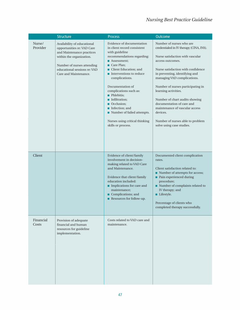
47
Nursing Best Practice Guideline
Objectives ■ Text ■ Text ■ Text
Structure Process Outcome
Nurse/Provider
Client
FinancialCosts
Availability of educationalopportunities re: VAD Careand Maintenance practiceswithin the organization.
Number of nurses attendingeducational sessions re: VADCare and Maintenance.
Provision of adequatefinancial and humanresources for guidelineimplementation.
Evidence of documentationin client record consistentwith guidelinerecommendations regarding:■ Assessment;■ Care Plan;■ Client Education; and■ Interventions to reduce
complications.
Documentation ofcomplications such as:■ Phlebitis;■ Infiltration;■ Occlusion;■ Infection; and■ Number of failed attempts.
Nurses using critical thinkingskills or process.
Evidence of client/familyinvolvement in decision-making related to VAD Careand Maintenance.
Evidence that client/familyeducation included:■ Implications for care and
maintenance;■ Complications; and■ Resources for follow-up.
Costs related to VAD care andmaintenance.
Number of nurses who arecredentialed in IV therapy (CINA, INS).
Nurse satisfaction with vascularaccess outcomes.
Nurse satisfaction with confidencein preventing, identifying andmanaging VAD complications.
Number of nurses participating inlearning activities.
Number of chart audits showingdocumentation of care andmaintenance of vascular accessdevices.
Number of nurses able to problemsolve using case studies.
Documented client complicationrates.
Client satisfaction related to:■ Number of attempts for access;■ Pain experienced during
procedure;■ Number of complaints related to
IV therapy; and■ Lifestyle.
Percentage of clients whocompleted therapy successfully.
Structure Process Outcome

Care and Maintenance to Reduce Vascular Access Complications
48
Implementation StrategiesThe Registered Nurses’ Association of Ontario, and the guideline development panel has
compiled a list of implementation strategies to assist healthcare organizations or healthcare disciplines
that are interested in implementing this guideline. A summary of these strategies follows:
■ Have a dedicated person such as an advanced practice nurse or a clinical resource nurse who will
provide support, clinical expertise and leadership. The individual should also have good interpersonal,
facilitation and project management skills.
■ Establish a steering committee comprised of key stakeholders and members committed to leading the
initiative. Keep a work plan to track activities, responsibilities and timelines.
■ Provide educational sessions and ongoing support for implementation. The education sessions may
consist of presentations, facilitator’s guide, handouts and case studies. Binders, posters and pocket
cards may be used as ongoing reminders of the training. Plan education sessions that are interactive,
include problem solving, address issues of immediate concern and offer opportunities to practice new
skills (Davies & Edwards, 2004).
■ Provide organizational support such as having the structures in place to facilitate the implementation.
For example, hiring replacement staff so participants will not be distracted by concerns about work and
having an organizational philosophy that reflects the value of best practices through policies and
procedures. Develop new assessment and documentation tools (Davies & Edwards, 2004).
■ Identify and support designated champions on each unit to promote and support implementation.
Celebrate milestones and achievements, acknowledging work well done (Davies & Edwards, 2004).
■ Organizations implementing this guideline should look at a range of self-learning, group learning,
mentorship and reinforcement strategies that will over time, build the knowledge and confidence of
nurses in implementing this guideline.
■ Beyond skilled nurses, the infrastructure required to implement this guideline includes access to
specialized equipment and treatment materials. Orientation of the staff to the use of specific products
must be provided and regular refresher training planned.
49
Nursing Best Practice Guideline
■ Teamwork, collaborative assessment and treatment planning with the client and family and through
interdisciplinary work are beneficial in implementing guidelines successfully.
■ RNAO’s Advanced/Clinical Practice Fellowships (ACPF) Project is another way that registered nurses’ in
Ontario may apply for a fellowship and have an opportunity to work with a mentor who has expertise in
vascular access management. With the ACPF, the nurse fellow will have the opportunity to hone their
skills in this topic area.
■ Identify, develop and support Best Practice Champions and include people who have expertise in the
topic area, facilitation skills and knowledge of adult education principles in order to support, develop,
mentor, and train other nurses within organizations to ensure knowledge transfer.
In addition to the tips mentioned above, the RNAO has developed resources that are available on the
website. A Toolkit for implementing guidelines can be helpful if used appropriately. A brief description of
this Toolkit can be found in Appendix M.
A complete version of the document is available at: http://www.rnao.org/bestpractices.
Care and Maintenance to Reduce Vascular Access Complications
50
Process for Update/Review of GuidelineThe Registered Nurses’ Association of Ontario proposes to update the best practice
guidelines as follows:
1 Each nursing best practice guideline will be reviewed by a team of specialists (Review Team) in the
topic area every three years following the last set of revisions.
2 During the three-year period between development and revision, RNAO Nursing Best Practice
Guidelines program staff will regularly monitor for relevant literature in the field.
3 Based on the results of the monitor, program staff will recommend an earlier revision period.
Appropriate consultation with a team of members comprising original panel members and other
specialists in the field will help inform the decision to review and revise the Guideline earlier than the
three-year milestone.
4 Three months prior to the three year review milestone, the program staff will commence the planning
of the review process by:
a Inviting specialists in the field to participate in the Review Team. The Review Team will be
comprised of members from the original panel as well as other recommended specialists.
b Compiling feedback received, questions encountered during the dissemination phase as well as
other comments and experiences of implementation sites.
c Compiling new clinical practice guidelines in the field, systematic reviews, meta-analysis papers,
technical reviews and randomized controlled trial research and other relevant literature.
d Developing detailed work plan with target dates and deliverables.
The revised Guideline will undergo dissemination based on established structures and processes.

51
Nursing Best Practice Guideline
ReferencesAppraisal of Guidelines for Research and Evaluation (AGREE) Instrument (2001). Available: http://www.agreecollaboration.org
Barry Frame, K. & Chrystal, C. (1999). Faculty and clinicians collaborate to teach basic intravenous skills to senior baccalaureatenursing students, Journal of IV Nursing, 22(5), 253-258.
Barton, A., Danek, G., Johns, P., & Coons, M. (1998) Improving patient outcomes through CQI: Vascular access planning. Journalof Nursing Quality, 13(2), 77-85.
Black, N., Murphy, M., Lamping, D., McKee, M., Sanderson, C., Askham, J., et al. (1999). Consensus development methods:Review of the best practice in creating clinical guidelines. Journal of Health Services Research & Policy, 4(4), 236-248.
Bona, R. D. (2003) Central line thrombosis in patients with cancer. Current Opinion in Pulmonary Medicine, 9, 362-366.
Canadian Intravenous Nurses Association. (1999). Intravenous therapy guidelines. (2nd ed.). Toronto: Author.
Canadian Standards Association. (2004). Blood and blood components. Ottawa: Author.
Centers for Disease Control and Prevention. (2002). Guidelines for the prevention of intravascular catheter-related infections.Morbidity and Mortality Weekly Report (MMWR), 51(No. RR-10), 1-29.
College of Nurses of Ontario. (2004a). Practice guideline: Consent. Toronto: Author.
College of Nurses of Ontario. (2004b). Practice guideline: Culturally sensitive care. Toronto: Author.
College of Nurses of Ontario. (2004c). Practice guidelines disagreeing with the multidisciplinary plan of care - Fact sheet. Toronto: Author.
College of Nurses of Ontario. (2004d). Practice guideline: Guide to decide. Toronto: Author.
College of Nurses of Ontario. (2004e). Practice guideline: Medical directives - Revised 2000. Toronto: Author.
College of Nurses of Ontario. (2004f). Practice standard: Confidentiality and privacy - personal health information. Toronto: Author.
College of Nurses of Ontario. (2004g). Practice standard: Documentation. Toronto: Author.
College of Nurses of Ontario. (2004h). Practice standard: Ethics. Toronto: Author.
College of Nurses of Ontario. (2004i). Practice standard: Infection prevention and control. Toronto: Author.
College of Nurses of Ontario. (2004j). Practice standard: Medication. Toronto: Author.
College of Nurses of Ontario. (2004k). Practice standard: Professional standards – Revised 2002. Toronto: Author.
College of Nurses of Ontario. (2004l). Practice standard: Therapeutic nurse-client relationship. Toronto: Author.
Cook, N. (1999). Central venous catheters: preventing infection and occlusion. British Journal of Nursing, 8(15), 980.
Cosca, P. A., Smith, S., Chatfield, S. et al. (1998). Reinfusion of discard blood from venous access devices. Oncology NursingForum, 25(6), 73-76.
Crinch, C. L., & Maki, D. G. (2002). The promise of novel technology for the prevention of intravascular device-relatedbloodstream infection II: Long-term devices. Clinical Infectious Diseases, 34(9), 1232-1242.
Crow, S. (1996). Infection risks in IV therapy. Official Journal of the National Intravenous Therapy Association (NITA), 10, 101-105.
Davies, B., & Edwards, N. (2004). RNs measure effectiveness of best practice guidelines. Registered Nurse Journal, 16(1), 21-23.
Department of Health (2001a). Guidelines for preventing infections associated with the insertion and maintenance of centralvenous catheters: Introduction. Journal of Hospital Infection, 47, S13-S19.
Department of Health (2001b). Guidelines for preventing infections associated with the insertion and maintenance of centralvenous catheters. Journal of Hospital Infection, 47, S47-S67.

Care and Maintenance to Reduce Vascular Access Complications
52
Dierks, M. M., & Whitman E. D. (1995). Catheter tip position in the analysis of central venous access device outcome. Journal ofVascular Access Devices, 1(3), 11-14.
Dougherty, L. (1997). Reducing the risk of complications in IV therapy. Nursing Standard, 12(5), 40-42.
Dugger, B. (1997). Intravenous nursing competency: Why is it important? Journal of Intravenous Nursing, 20(6), 287-297.
Evidence-Based Practice in Infection Control (EPIC). (2001a). The EPIC project: Developing national evidence-based guidelines forpreventing hospital-acquired infections. National evidence-based guidelines for preventing hospital-acquired infections associatedwith the use of central venous catheters. Technical report part A. Available: http://www.epic.tvu.ac.uk/epicphase/epic1.html
Evidence-Based Practice in Infection Control (EPIC). (2001b). The EPIC project: Developing national evidence-based guidelines forpreventing hospital-acquired infections. National evidence-based guidelines for preventing hospital-acquired infections associatedwith the use of central venous catheters. Technical report part B. Available: http://www.epic.tvu.ac.uk/epicphase/epic1.html
Farr, B. (1996). Understaffing: A risk factor for infection in the era of downsizing? Infection Control Hospital Epidemiology 17(3),147-150.
Field, M. J. & Lohr, K. N. (1990). Guidelines for clinical practice: Directions for a new program. Washington DC: National Academy Press.
Food and Drug Administration, Center for Devices and Radiological Health, Office of Training and Assistance and Central venousCatheter Working Group (1994). Central venous catheter complications (3 videocassettes and guide). Springfield: NationalTechnical Information Services.
Freda, M. C. (2004). Issues in patient education. Journal of Midwifery and Women’s Health, 49, 203-209.
Fridkin, S. K., Pear, S. M., Williamson, T. H., Galgiani, J. N., & Jarvis, W. R. (1996). The role of understaffing in central Venouscatheter-associated bloodstream infection. Infection Control & Hospital Epidemiology, 17(3), 150-158.
Frey, A. M. (2003). Drawing blood samples from vascular access devices. Evidence-based practice. Journal of Infusion Nursing,26(5), 285-293.
Gillies, D., O’Riordan, E., Carr, D., O’Brien, I., & Gunning, R. (2003). Central venous catheter dressings; a systemic review. Journalof Advanced Nursing, 44(6), 623-632.
Graham, I., Harrison, M., Brouwers, M., Davies, B., & Dunn, S. (2002). Facilitating the use of evidence in practice: Evaluating andadapting clinical practice guidelines for local use by health care organizations. Journal of Obstetric, Gynecological and NeonatalNursing, 31(5,) 599-611.
Gorski, L., & Czaplewski, L. M. (2004). Peripherally inserted catheters and midline catheters for the home care nurse. Home HealthCare Nurse, 22(11), 758-771.
Hadaway, L. C. (2001). IV risk reducers. Nursing Management, 32(12), 57-59.
Hadaway, L. C. (2002). What you can do to decrease catheter-related infections: Meticulous cleaning of skin and insertion sitecan keep bad bugs out of your patient’s bloodstream. Nursing, 32(9), 46-48.
Hadaway, L. C. (2003a). Infusing without infecting. Nursing, 33(10), 58-64.
Hadaway, L. C. (2003b). Skin flora and infection. Journal of Infusion Nursing, 26(1), 44-48.
Haire, W. D., & Herbst, S. F. (2000). Consensus conference on the use of alteplase (t-PA) for the management of thromboticcatheter dysfunction. Journal of Vascular Access Devices, 5(2), 28–35.
Hamilton Health Sciences Centre IV Team. (2002). Algorithm for troubleshooting catheter occlusions. (Unpublished document).Hamilton: Author.
Hamilton Health Sciences Centre IV Team. (2002). Data collection tool for central venous access devices. (Unpublished document).Hamilton: Author.
Hamilton Health Sciences Centre – Patient Education. (2002). Going home with your PICC. (Unpublished document). Hamilton: Author.
Health Canada (2003). Infection control guidance in a non-outbreak setting (in the absence of SARS) when an individual presentsto a health care institution with a respiratory infection (Draft). Available: http://www.phac-aspc.gc.ca/sars-sras/pdf/sars-icg-nonoutbreak_e.pdf
Hinds, P. S. (1991). An investigation of the safety of the blood reinfusion step used with tunneled access devices in children withcancer. Journal of Pediatric Oncology Nursing, 8(4), 159-164.

53
Nursing Best Practice Guideline
Holmes, K. (1998). Comparison of push-pull vs. discard method from central venous catheters for blood testing. Journal ofIntravenous Nursing, 21(5), 282-285.
Intravenous Nurses Society (2000). Infusion nursing: Standards of practice. Journal of Intravenous Nursing, 23(6S), S1-S88.
Intravenous Nurses Society (2002). Policies and procedures for infusion nursing (2nd ed.). Norwood: Author.
Jackson, D. (2001). Infection control principles and practices in the care and management of vascular access devices in thealternate care setting. Journal of Intravenous Nursing, 24(3S), S28-34, S5-8.
Karamanoglu, A., Yumuk, P. F., Gumus, M., Aliustaglu, M., Selimen, D. et al. (2003). Port needles: Do they need to be removedas frequently in infusional chemotherapy? Journal of Infusion Nursing, 26(4), 239-242.
Kowalski, C. M., Kaufman, J. A., Rivitz, J. A., & Waltman, A. C. (1997). Migration of central venous catheters: Implications forinitial catheter tip positioning. Journal of Vascular & Interventional Radiology, 8(3), 443-447.
Krzywda, E. A. (1998). Central venous access – catheters, technology, and philosophy. MEDSURG Nursing, 7(3), 132-141.
Larwood, K. A. (2000). Reducing central venous catheter infections. Australian Critical Care, 13(3), 107-112.
LeBlanc, A. & Cobbett, S. (2000). Traditional practice versus evidence-based practice for IV skin preparation. Canadian Journal ofInfection Control, 15(1), 9-14.
Macklin, D. (1997). How to manage PICCs. American Journal of Nursing, 97(9), 26-33.
Maki, D., Ringer, M., & Alvarado, C. J. (1991). Prospective randomized trial of povidone-iodine, alcohol, and chlorhexidine forprevention of infection associated with central venous and arterial catheters. The Lancet, 338, 339-343.
Masoorli, S. (2002). How to accurately document I.V. insertion. Nursing 2002, June, 65.
Masoorli, S., Angeles, T., BarBone, M., et al. (1998). Danger points. How to prevent nerve injuries from venipuncture. Nursing 98September, 35-39.
McCall, R. E., & Tankersley, C. M. (2003). Blood collection variables, complications and procedural errors. In Phlebotomy Essentials.(3rd ed.). Philadelphia: Lippincott, Williams & Wilkins.
McConnell, H., Nelson, S., & Virani, T. (2004). Nursing best practice guideline: Assessment and device selection for vascular access.CINA 2004, 34-35.
McMahon, D. (2002). Evaluating new technology to improve patient outcomes: A quality improvement approach. Journal ofInfusion Nursing, 25(4), 250-255.
Mezey, M., & Schoder, J. (2003). Editorial. Journal of Infusion Nursing, 26(3), 125-126.
Miller, J., Goetz, A., Squier, C., et al. (1996). Reduction in nosocomial intravenous device-related bacteremias after institution ofan intravenous therapy team. Journal of Intravenous Nursing, 19, 103-106.
Mimoz, O., Laurence, P., Lawrence, C., Edouard, A., Costa, Y., Samii, K., & Brun-Buisson, C. (1996). Prospective, randomized trialof two antiseptic solutions for prevention of central venous or arterial catheter colonization and infection in intensive care unitpatients. Critical Care Medicine, 24, 1818-1823.
Ministry of Health and Long-Term Care (MOHLTC) (2002). Final report of the Infection Control and Surveillance Standards TaskForce: Community health providers preventing respiratory illnesses in community settings in non outbreak conditions. Available: http://www.health.gov.on.ca/english/providers/program/pubhealth/sars/docs/docs3/report_taskforce_community_031104.pdf
Ministry of Ontario, Occupational Health and Safety Act, Regulation 67/93: Health Care and Residential Facilities. Available:http://www.e-laws.gov.on.ca:81/ISYSquery/IRL5891.tmp/1/doc
Mosby’s Medical, Nursing, and Allied Health Dictionary (1990) (3rd ed.). St. Louis: The C.V. Mosby Company.
National Institute for Occupational Safety and Health (NIOSH) (1999). Preventing needle stick injuries in health care settings.Available: http://www.cdc.gov/niosh/pdfs/2000-108.pdf
Occupational Safety and Health Administration (2001). Securing medical catheters: Fact sheet. Available:http://www.osha.gov/SLTC/bloodbornepathogens/factsheet_catheters.pdf
O’Grady, N. P., Alexander, M., Dellinger, E. P., Gerberding, J. L., Heard, S. O., Maki, D. G. et al. (2002). Guidelines for theprevention of intravascular catheter-related infections. American Journal of Infection Control, 30(8), 476-489.
Oncology Nursing Society (2004). Access Device Guidelines (2nd ed.). Pittsburgh: Author.

Care and Maintenance to Reduce Vascular Access Complications
54
Pearson, M. L. (1996a). Guideline for prevention of intravascular-device-related infections. Infection Control & HospitalEpidemiology, 17(7), 438-473.
Pearson, M. L. (1996b). Special communication: HICPAC guideline for prevention of intravascular device-related infections: PartsI & II. American Journal of Infection Control, 24(4), 262-293.
Public Health Agency of Canada. (1997). Preventing infections associated with indwelling intravascular devices. CanadaCommunicable Disease Report, 23S8. Available: http://www.phac-aspc.gc.ca/publicat/ccdr-rmtc/97vol23/23s8/iiadinde_e.html
Public Health Agency of Canada. (1998). Infection control guidelines – hand washing, cleaning, disinfection and sterilization inhealth care. Canada Communicable Disease Report, 24S8. Available: http://www.phac-aspc.gc.ca/publicat/ccdr-rmtc/98pdf/cdr24s8e.pdf
Public Health Agency of Canada. (1999). Routine Practices and Additional Precautions for Preventing the Transmission of Infectionin Health Care (Revision of Isolation and Precaution Techniques). Canada Communicable Disease Report (CCDR), 25S4. Available:http://www.phac-aspc.gc.ca/publicat/ccdr-rmtc/99pdf/cdr25s4e.pdf
Public Health Agency of Canada. (2002). Prevention and control of occupational infections in health care. Canada CommunicableDisease Report (CCDR), 28S1. Available:http://www.phac-aspc.gc.ca/publicat/ccdr-rmtc/02pdf/28s1e.pdf
Public Health Agency of Canada (2003). Alcohol for hand hygiene: New comparative studies Add to the Evidence Base. CanadaCommunicable Disease Report (CCDR), 29-01. Available: http://www.phac-aspc.gc.ca/publicat/ccdr-rmtc/03vol29/dr2901eb.html
Ray, C. E., Jr. (1999). Infection control principles and practices in the care and management of central venous access devices.Journal of Intravenous Nursing, 22(6S), S18-25, S4-7.
Registered Nurses’ Association of Ontario. (2004). Assessment and device selection for vascular access. Toronto: Author.
Registered Nurses’ Association of Ontario. (2005). Care and maintenance to reduce vascular access complications. Toronto: Author.
Registered Nurses’ Association of Ontario. (2002). Toolkit: Implementation of clinical practice guidelines. Toronto: Author.
Rosenthal, K. (2003). Pinpointing intravascular device infections. Nursing Management, 34(6), 35-43.
Ross, V. M. & Orr, P. A. (1997). Prevention of infections related to central venous catheters. Critical Care Nursing Quarterly, 20(3), 79-88.
Roter, D. L., Stashefsky-Margalit, R., & Rudd, R. (2001). Current perspectives on patient education in the U.S. Patient Educationand Counseling, 44, 79-86.
Royal College of Nursing (2003). Standards for infusion therapy. London: Author.
Ruesch, S, Walder, B., & Tramer, M. (2002). Complications of central venous catheters: Internal jugular versus subclavian access –A systemic review. Critical Care Medicine, 30(2), 454-460.
Ryder, M. (1993). Peripherally Inserted Central Catheters. Nursing Clinics of North America, 28(4), 938-971.
Sansivero, G. E. (1998). Antimicrobial products: Good infection fighters? Nursing, 28(8), 25.
Scheffer, B. K. & Rubenfeld, M.G. (2000). A consensus statement of critical thinking in nursing. Journal of Nursing Education,29(8), 352-359.
Schillinger, D., Bindman, A., Wang, F., Stewart, A., & Piette, J. (2004). Functional health literacy and the quality of physician-patient communication among diabetic patients. Patient Education and Counseling, 52, 315-323.
Schmid, M. W. (2000). Risks and complications of peripherally and centrally inserted intravenous catheters. Critical Care NursingClinics of North America, 12(2),165-174.
Sheppard, K., LeDesma. M., Morris, N., & O’Connor, K. (1999). A prospective study of two intravenous catheter securementtechniques in a skilled nursing facility. Journal of Intravenous Nursing, 22(3), 151-153.
Tagalakis, V., Kahn, S. R., Libman, M., & Blostein, M. (2002). The epidemiology of peripheral vein infusion thrombophlebitis: Acritical review. The American Journal of Medicine, 113, 146-151.
Thrush, D. N., & Belsole, R. (1995). Radial nerve injury after routine peripheral vein cannulation. Journal of Clinical Anaesthesia,7, 160-162.

55
Nursing Best Practice Guideline
Tripepi-Bova, K. A., Woods, K. D., & Loach, M. C. (1997). A comparison of transparent polyurethane and dry gauze dressings forperipheral IV catheter sites: Rates of phlebitis, infiltration, and dislodgement by patients. American Journal of Critical Care, 6(5),377-381.
Umstead, G. (1984). Tobramycin blood levels from an indwelling right atrial catheter. Drug Intelligence and Clinical Pharmacy, 18,815-817.
Verweyst Kaufman, M. E. (1992). Intravenous therapy education in associate degree nursing programs. Journal of IntravenousTherapy, 15, 238-243.
Vesely, T. (2003). Central venous catheter tip position: A continuing controversy. Journal of Vascular & Interventional Radiology,14,527-534
Wade, B. H. & Bush, S. E. (1998). Infection control and outpatient parenteral antibiotic therapy. Infectious Disease Clinics of NorthAmerica, 12(4), 979-994.
Weinstein, S. (2001). Plumer’s Principles & Practice of Intravenous Therapy. (7th Ed.) Philadelphia: Lippincott, Williams & Wilkins.
Wilson, J. F. (2003). The crucial link between literacy and health. Annals of Internal Medicine, 139(10), 875-878.
Wise, M., Richardson, D., Lum, P. (2001). Catheter tip position a sign of things to come. Journal of Vascular Access Devices(summer), 18-27.
Wizowski, L., Harper, T. & Hutchings T. (2002). Writing health information for patients and families. Hamilton: Hamilton Health Sciences.
Zitella, L. (2004). Central venous catheter site care for blood and marrow transplant recipients. Clinical Journal of OncologyNursing, 7(3), 289-298.
Zunkel, G., Cesarotti, E., Rosdahl, D. & McGrath, J. (2004). Enhancing diagnostic reasoning skills in nurse practitioner students.Nurse Educator, 29 (4), 161-165
BibliographyAlvarado-Ramy, F., Beltrami, E. M., Short, L. J., Srivastava, P. U., Henry, K., Mendelson, M. et al. (2003). A comprehensiveapproach to percutaneous injury prevention during phlebotomy: Results of a multicenter study, 1993-1995. Infection Control &Hospital Epidemiology, 24(2), 97-104.
Andes, D. R., Urban, A. W., Acher, C. W., & Maki, D. G. (1998). Septic thrombosis of the basilic, axillary, and subclavian veinscaused by a peripherally inserted central venous catheter. American Journal of Medicine, 105(5), 446-450.
Arduino, M. J., Bland, L. A., Danzig, L. E., McAllister, S. K., & Aguero, S. M. (1997). Microbiologic evaluation of needleless andneedle-access devices. American Journal of Infection Control, 25(5), 377-380.
Baker, G. R., Norton, P. G., Flintoft, V., Blais, R., Brown, A., Cox, J. et al. (2004). The Canadian Adverse Events Study: Theincidence of adverse events among hospital patients in Canada. Canadian Medical Association Journal, 170(11), 1678-1686.
Bantar, C., Bustos, J. L., Vesco, E., & Morera, G. (2002). Concise communication. Central venous catheter-related infection: A prospective, observational study to assess the incidence rate at a teaching hospital in Argentina. Infection Control & HospitalEpidemiology, 23(12), 757-758.
Besarab, A. (2004). Vascular access: Issues and management. Contributions to Nephrology., 142, 29-46.
Bessoud, B., De Baere, T., Kuoch, V., Desruennes, E., Cosset, M.-F., Lassau, N. et al. (2003). Experience at a single institutionwith endovascular treatment of mechanical complications caused by implanted central venous access devices in pediatric andadult patients. American Journal of Roentgenology, 180(2), 527-532.
Bijma, R., Girbes, A. R., Kleijer, D. J., & Zwaveling, J. H. (1999). Concise communications. Preventing central venous catheter-related infection in a surgical intensive-care unit. Infection Control & Hospital Epidemiology, 20(9), 618-620.
Bong, J. J., Kite, P., Ammori, B. J., Wilcox, M. H., & McMahon, M. J. (2003). The use of a rapid in situ test in the detection of centralvenous catheter-related bloodstream infection: A prospective study. Journal of Parenteral & Enteral Nutrition, 27(2), 146-150.

Care and Maintenance to Reduce Vascular Access Complications
56
Brown, J. D., Moss, H. A., & Elliott, T. S. J. (1997). The potential for catheter microbial contamination from a needlelessconnector. Journal of Hospital Infection, 36(3), 181-189.
Buswell, L. & Beyea, S. C. (1998). Flushing protocols for tunneled central venous catheters: An integrative review of theliterature. Online Journal of Knowledge Synthesis for Nursing, 5(3).
Capka, M. B., Carey, S., Marks, D., Wilson, M., & Benard, J. (1991). Nursing observations of central venous catheters: Theeffect on patient outcome. Journal of Intravenous Nursing, 14(4), 243-246.
Cauley, J. A., Robbins, J., Chen, Z., Cummings, S. R., Jackson, R. D., LaCroix, A. Z. et al. (2003). Effects of estrogen plusprogestin on risk of fracture and bone mineral density. Journal of the American Medical Association, 290(13), 1729-1739.
Chaiyakunapruk, N., Veenstra, D. L., Lipsky, B. A., & Saint, S. (2002). Chlorhexidine compared with povidone-iodine solution forvascular catheter-site care: A meta-analysis. Annals of Internal Medicine., 136(11), 792-801.
Cheesman, D. (290). Clinical. Intravenous care: The benefits of closed-system connectors. British Journal of Nursing, 10(5),287-288.
Chlebicki, M. P. & Teo, E. K. (2003). Review of peripherally inserted central catheters in the Singapore acute-care hospital.Singapore Medical Journal, 44(10), 531-535.
Coopersmith, C. M., Zack, J. E., Ward, M. R., Sona, C. S., Schallom, M. E., Everett, S. J. et al. (2004). The impact of bedsidebehavior on catheter-related bacteremia in the intensive care unit. Archives of Surgery, 139(2), 131-136.
Curchoe, R. M., Powers, J., & El Daher, N. (2002). Weekly transparent dressing changes linked to increased bacteremia rates.Infection Control & Hospital Epidemiology, 23(12), 730-732.
Dawson, S., Irwin, L., Aaron, E., Willan, A., & Barr, R. (1995). Minimizing blood loss in the indwelling central venous cathetersin children with cancer. Canadian Journal of Medical Technology, 57, 102-107.
Deacon, V. L. (2004). The Safe Medical Device Act and its impact on clinical practice. Journal of Infusion Nursing, 27(1), 31-36.
Dezfulian, C., Lavelle, J., Nallamothu, B. K., Kaufman, S. R., & Saint, S. (2003). Rates of infection for single-lumen versusmultilumen central venous catheters: A meta-analysis. Critical Care Medicine, 31(9), 2385-2390.
Dhesi, J. K., Jackson, S. H. D., Bearne, L. M., Moniz, C., Hurley, M. V., Swift, C. G. et al. (2004). Vitamin D supplementationimproves neuromuscular function in older people who fall. Age and Ageing, 33, 589-595.
Dierdorf, B., Elgin, K., & Rea, K. (2001). Examining large bore central line use in acute care settings. MEDSURG Nursing, 10(6),313-314.
Do, A. N., Ray, B. J., Banerjee, S. N., Illian, A. F., Barnett, B. J., Pham, M. H. et al. (1999). Bloodstream infection associated withneedleless device use and the importance of infection-control practices in the home health care setting. Journal of InfectiousDiseases, 179(2), 442-448.
Docherty, B. (2001). Clinical practice review: A review of core nursing practice developments, utilising clinical research,effectiveness and audit information. Central line management. Professional Nurse, 16(7), 1206.
Dunning, J. (2003). Ultrasonic guidance and the complications of central line placement in the emergency department.Emergency Medicine Journal, 20(6), 551-552.
Eggimann, P., Harbarth, S., Constantin, M., Touveneau, S., Chevrolet, J., & Pittet, D. (2000). Impact of a prevention strategytargeted at vascular-access care on incidence of infections acquired in intensive care. Lancet (North American Edition),355(9218), 1864-1868.
Eggimann, P. & Pittet, D. (2002). Overview of catheter-related infections with special emphasis on prevention based oneducational programs. Clinical Microbiology & Infection, 8(5), 295-309.
Finn, L. (1997). Clinical. Nurses’ documentation of infection control precautions: 1. British Journal of Nursing, 6(11), 607-611.
Forauer, A. R. & Alonzo, M. (2000). Change in peripherally inserted central catheter tip position with abduction and adductionof the upper extremity. Journal of Vascular & Interventional Radiology, 11(10), 1315-1318.
Galway, R., Harrod, M. E., Crisp, J., Donnellan, R., Hardy, J., Harvey, A. et al. (2003). Central venous access and handwashing:Variability in policies and practices. Paediatric Nursing, 15(10), 14-18.
Gillies, D., O’Riordan, L., Carr, D., Frost, J., Gunning, R., & O’Brien, I. (2003). Gauze and tape and transparent polyurethanedressings for central venous catheters. (Cochrane Review). The Cochrane Library. Oxford: John Wiley & Sons, Ltd.

57
Nursing Best Practice Guideline
Gillies, D., O’Riordan, L., Wallen, M., Rankin, K., Morrison, A., & Nagy, S. (2004). Timing of intravenous administration setchanges: A systematic review. Infection Control & Hospital Epidemiology, 25(3), 240-250.
Gonsalves, C. F., Eschelman, D. J., Sullivan, K. L., DuBois, N., & Bonn, J. (2003). Incidence of central vein stenosis and occlusionfollowing upper extremity PICC and port placement. Cardiovascular & Interventional Radiology., 26(3), 123-127.
Gorski, L. & Czaplewski, L. M. (2004). Peripherally inserted catheters and midline catheters for the home care nurse. HomeHealth Care Nurse, 22(11), 758-771.
Graeber, B. D. & Muder, R. R. (2001). Frequency of intravenous administration set changes and bacteremia: Defining the risk.Infection Control & Hospital Epidemiology, 22(8), 476.
Grove, J. R. & Pevec, W. C. (2000). Venous thrombosis related to peripherally inserted central catheters. Journal of Vascular &Interventional Radiology, 11(7), 837-840.
Grune, F., Schrappe, M., Basten, J., Wenchel, H. M., Tual, E., Stutzer, H. et al. (2004). Phlebitis rate and time kinetics of shortperipheral intravenous catheters. Infection, 32(1), 30-32.
Hallquist Viale, P., Yamamoto, D. S., & Geyton, J. E. (180). Extravasation of infusate via implanted ports: Two case studies.Clinical Journal of Oncology Nursing, 3(4), 145-151.
Halstead, S. (2001). Audit to improve use of central venous catheters. Professional Nurse, 16(8), 1287-1290.
Hankins, J., Waldman-Lonsway, R. A., Hedrick, C., & Perdue, M. B. (2001). Infusion therapy in clinical practice. (2nd ed.)Philadelphia: W. B. Saunders Company.
Harako, M. E. & Nguyen, T. H. (2004). Optimizing the patient positioning for PICC line tip determination. Emergency Radiology,10, 186-189.
Hibbard, J., Mulberry, G. K., & Brady, A. R. (2002). A clinical study comparing the skin antisepsis and safety of chloraprep, 70%isopropyl alcohol, and 2% aqueous chlorhexidine. Journal of Infusion Nursing, 25(4), 244-249.
Himberger, J. R. & Himberger, L. C. (2001). Issues in accuracy of laboratory results. Accuracy of drawing blood through infusingintravenous lines. Heart & Lung: Journal of Acute & Critical Care, 30(1), 66-73.
Hind, D., Calvert, N., McWilliams, R., Davidson, A., Paisley, S., Beverley, C. et al. (2003). Ultrasonic locating devices for centralvenous cannulation: Meta-analysis. British Medical Journal, 327(7411), 361.
Hugonnet, S., Sax, H., Eggimann, P., Chevrolet, J.– C., & Pittet, D. (2004). Nosocomial Bloodstream Infection and ClinicalSepsis. Emerging Infectious Diseases, 10(1), 76-81.
Kaufman, M. E. (1992). Intravenous therapy education in associate degree nursing programs. Journal of Intravenous Therapy,15(14), 238-243.
Keenan, S. P. (2002). Use of ultrasound to place central lines. Journal of Critical Care, 17(2), 126-137.
Klerk, C. P., Smorenburg, S. M., & Buller, H. R. (2003). Thrombosis prophylaxis in patient populations with a central venouscatheter: A systematic review. Archives of Internal Medicine, 163(16), 1913-1921.
Lai, K. K. (1998). Safety of prolonging peripheral cannula and IV tubing use from 72 hours to 96 hours presented in part at the4th Annual SHEA Meeting, New Orleans, Louisiana, March 20-22, 1994. American Journal of Infection Control, 26(1), 66-70.
Larwood, K. A., Anstey, C. M., & Dunn, S. V. (2000). Managing central venous catheters: A prospective randomised trial of twomethods. Australian Critical Care, 13(2), 44-50.
Luebke, M. A., Arduino, M. J., Duda, D. L., Dudar, T. E., McAllister, S. K., Bland, L. A. et al. (1998). Comparison of the microbialbarrier properties of a needleless and a conventional needle-based intravenous access system. American Journal of InfectionControl, 26(4), 437-441.
MacGeorge, L., Steeves, L., & Steeves, R. H. (1998). Comparison of the mixing and reinfusion methods of drawing blood froma hickman catheter. Oncology Nursing Forum, 15(3), 335-338.
Macklin, D. (1997). How to manage PICCs. American Journal of Nursing, 97(9), 26-33.
Maki, D., Ringer, M., & Alvarado, C. J. (1991). Prospective randomized trial of povidone-iodine, alcohol and chlorhexidine forprevention of infection associated with central venous and arterial catheters. The Lancet, 338(8763), 338-343.
Mayo, D. J., Diamond, E. P., Framer, W., & McDonald, K. H. (1996). Discard volumes necessary for clinically useful coagulationstudies from heparinized hickman catheters. Oncology Nursing Forum, 23(4), 671-675.

Care and Maintenance to Reduce Vascular Access Complications
58
McCall, R. E. & Tankersley, C. M. (2003). Blood collection variables, complications and procedural errors in phlebotomyessentials. (3rd ed.). Philadelphia: Lippincott, Williams & Wilkins.
Memish, Z. A., Arabi, Y., Cunningham, G., Kritchevsky, S., Braun, B., Richards, C. et al. (2003). Comparison of US and non-UScentral venous catheter infection rates: Evaluation of processes and indicators in infection control study. AJIC: American Journalof Infection Control, 31(4), 237-242.
Mermel, L. A. (2000). Prevention of intravascular catheter-related infections. Annals of Internal Medicine, 132(5), 391-402.
Mermel, L. A., Farr, B. M., Sherertz, R. J., Raad, I. I., O’Grady, N., Harris, J. S. et al. (2001). Guidelines for the management ofintravascular catheter-related infections. Journal of Intravenous Nursing, 24(3), 180-205.
Millam, D. A. (1985). A study of IV therapy education. NITA Journal, 8(5), 393-407.
Mimoz, O., Laurence, P., Lawrence, C., Edouard, A., Costa, Y., Samii, K. et al. (2003). Catheter securement: Trends inperformance and complications associated with the use of either traditional methods or an adhesive anchor device. Journal ofVascular Access, 8(1), 29-33.
Montgomery, L. A. & Budreau, G. K. (1996). Implementing a clinical practice guideline to improve pediatric intravenousinfiltration outcomes. AACN Clinical Issues: Advanced Practice in Acute & Critical Care, 7(3), 411-424.
Moureau, N. & Iannucci, A. L. (2003). Catheter securement: Trends in performance and complications associated with the useof either traditional methods or an adhesive anchor device. Journal of Vascular Access Devices, 8(1), 29-33.
Nazarian, G. K., Bjarnason, H., Dietz, C. A. Jr., & Bernada, C. A. (1997). Changes in tunneled catheter tip position when apatient is upright. Journal of Vascular & Interventional Radiology, 8, 437-441.
Nikoletti, S., Leslie, G., Gandossi, S., Coombs, G., & Wilson, R. (1999). A prospective, randomized, controlled trial comparingtransparent polyurethane and hydrocolloid dressings for central venous catheters. American Journal of Infection Control, 27(6),488-496.
O’Connell, F. M. & Heard, S. O. (576). Central venous catheter infections: Practical bedside measures to reduce risk:mechanisms, microorganisms, methods of insertion, maintenance. Journal of Critical Illness, 13(9), 569-573.
Ouwendyk, M. & Helferty, M. (1996). Central venous catheter management: How to prevent complications. Anna Journal,23(6), 572-579.
Parker, L. (1999). IV devices and related infections: Causes and complications. British Journal of Nursing, 8(22), 1491-1492.
Parker, L. (2002). Management of intravascular devices to prevent infection. British Journal of Nursing, 11(4), 240.
Peter, D. A. & Saxman, C. (2003). Preventing air embolism when removing CVCs: an evidence-based approach to changingpractice. MEDSURG Nursing, 12(4), 223-229.
Petersen, C. (2001). Clinical issues. Heparin v saline locks; “hosing”; reading in the OR; sterility of dropped packages; trays asprep table covers. AORN Journal, 74(6), 900.
Petersen, J., Brakstad, M. T., Rowbotham, R. K., & Bagley, C. M. Jr. (1999). Silicone venous access devices positioned with theirtips high in the superior vena cava are more likely to malfunction. American Journal of Surgery, 180(1), 78-79.
Phillips, L. D. (2001). Manual of I.V. therapeutics. (3rd ed.). Philadelphia: F. A. Davis Company.
Pinto, K. M. (1994). Accuracy of coagulation values obtained from a heparinized central venous catheter. Oncology NursingForum, 21(3), 573-575.
Puel, V., Caudry, M., Le Matayer, P., Baste, J. C., Midy, D., Marsault, C. et al. (1993). Superior vena cava thrombosis related tocatheter malposition in cancer chemotherapy given through implanted ports. Cancer, 72(7), 2248-2252.
Raad, I., Hanna, H. A., Awad, A., Alrahwan, A., Bivins, C., Khan A. et al. (2001). Optimal frequency of changing intravenousadministration sets: Is it safe to prolong use beyond 72 hours? Infection Control & Hospital Epidemiology, 22(8), 475-476.
Rigdon, R. O. (2001). Protocols for the prevention of intravascular device-related infections. Critical Care Nursing Quarterly,24(2), 39-47.
Ritter, J. (2000). Using invasive devices safely. Nursing, 30(6), Hosp-30.
Roach, H., Larson, E., & Bartlett, D. B. (1996). Intravascular site care: Are critical care nurses practicing according to writtenprotocols? Heart & Lung: Journal of Acute & Critical Care, 25(5), 401-408.
59
Nursing Best Practice Guideline
Rosenthal, K. (2003). Pinpointing intravascular device infections. Nursing Management, 34(6), 35-43.
Ross, V. M. & Orr, P. A. (1997). Prevention of infections related to central venous catheters. Critical Care Nursing Quarterly,20(3), 79-88.
Rudy, E. B., Lucke, J. F., Whitman, G. R., & Davidson, L. J. (2001). Benchmarking patient outcomes. Journal of NursingScholarship, 33(2), 185-189.
Schmid, M. W. (2000). Risks and complications of peripherally and centrally inserted intravenous catheters. Critical Care NursingClinics of North America, 12(2), 165-174.
Sheppard, K., LeDesma, M., Morris, N., & O’Connor, K. (1999). A prospective study of two intravenous catheter securementtechniques in a skilled nursing facility. Journal of Intravenous Nursing, 22(3), 151-153.
Sinclair, R. C., Maxfield, A., Marks, E. L., Thompson, D. R., & Gershon, R. R. M. (2002). Prevalence of safer needle devices andfactors associated with their adoption: Results of a national hospital survey. Public Health Reports, 117(4), 340-349.
Sitges-Serra, A., Pi-Suner, T., Garces, JM., & Segura, M. (1995). Pathogenesis and prevention of catheter-related septicemia.American Journal of Infection Control, 23(5), 310-316.
Sterba, K. G. (2001). Controversial issues in the care and maintenance of vascular access devices in the long-term/subacute careclient. Journal of Infusion Nursing, 24(4), 249-254.
Taylor, M. J. (2000). A fascination with phlebitis. Journal of Vascular Access Device (Fall 2000), 24-27.
Tezak, B. (2003). Leading change: Validating central line practices with research. Canadian Intravenous Nurses AssociationJournal, 1934-1937.
Timsit, J. F. (2000). Scheduled replacement of central venous catheters is not necessary. Infection Control & HospitalEpidemiology, 21(6), 371-374.
van de Wetering, M. D. & van Woensel, J. B. M. (2003). Prophylactic antibiotics for preventing early central venous cathetergram-positive infections in oncology patients. (Cochrane Review). The Cochrane Library. Oxford: John Wiley & Sons, Ltd.
Vost, J. & Longstaff, V. (1997). Clinical. Infection control and related issues in intravascular therapy. British Journal of Nursing,6(15), 846.
Walder, B., Pittet, D., & Tramer, M. R. (2002). Prevention of bloodstream infections with central venous catheters treated withanti-infective agents depends on catheter type and insertion time: Evidence from a meta-analysis. Infection Control & HospitalEpidemiology, 23(12), 748-756.
Ward, D. (2003). Improving patient hand hygiene. Nursing Standard, 17(35), 39-42.
Wilson, J. F. (2003). The crucial link between literacy and health. Annals of Internal Medicine, 139(10), 875-878.
Yoo, S., Ha, M., Choi, D., & Pai, H. (2001). Effectiveness of surveillance of central catheter-related bloodstream infection in anICU in Korea. Infection Control & Hospital Epidemiology, 22(7), 433-436.

Care and Maintenance to Reduce Vascular Access Complications
60
Appendix A: Search Strategy for Existing EvidenceSTEP 1 – Database SearchA database search for existing infusion therapy guidelines was conducted by a university health sciences
library. After the scope of the guideline was established, a search of the MEDLINE, Embase and CINAHL
databases for guidelines and articles published from January 1996 to November 2004 was conducted using
the following search terms: “catheterization, peripheral/ or catheterization, peripheral central venous”,
“central venous catheters/or peripherally inserted central catheters”, “nursing role”, “nursing care”,
“vascular access devices/or catheters”, “vascular/or vascular access devices”, “implantable”, “catheter-
related complications”, “equipment contamination”, “equipment safety” “catheter care, vascular”, “catheter
occlusion”, “catheter-related infections”, “nursing assessment”, “practice guideline(s)”, “clinical practice
guideline(s)”, “standards”, “consensus statement(s)”, “consensus”, “evidence based guidelines” and “best
practice guidelines”. This search generated numerous abstracts which were then reviewed by a masters
prepared Research Assistant assigned to the project for the purposes of selecting articles based on
inclusion criteria that related to the clinical questions. The Research Assistant conducted a quality
appraisal of the selected articles and summarized the studies according to the following:■ Study type;■ Sample (number of subjects/characteristics);■ Intervention used in the study;■ Measures used in the study;■ Findings; and■ Limitations.
This summary was distributed to the panel.
STEP 2 – Structured Web Site SearchOne individual searched an established list of 53 web sites for content related to the topic area. This list of
sites, reviewed and updated in July 2003, was compiled based on existing knowledge of evidence-based
practice websites, known guideline developers and recommendations from the literature. Presence or
absence of guidelines was noted for each site searched as well as date searched. The websites at times did
not house a guideline but directed to another web site or source for guideline retrieval. Guidelines were
either downloaded if full versions were available or were ordered by phone/email.
■ Agency for Healthcare Research and Quality: http://www.ahcpr.gov
■ Alberta Heritage Foundation for Medical Research – Health Technology Assessment: http://www.ahfmr.ab.ca//hta
■ Alberta Medical Association – Clinical Practice Guidelines: http://www.albertadoctors.org/
■ American College of Chest Physicians: http://www.chestnet.org/guidelines
■ American Medical Association: http://www.ama-assn.org
■ Association of Women’s Health, Obstetric and Neonatal Nursing: http://www.awhonn.org
■ British Columbia Council on Clinical Practice Guidelines: http://www.hlth.gov.bc.ca/msp/protoguides/index.html
■ BC Office of Health Technology Assessment: http://www.chspr.ubc.ca
■ Canadian Centre for Health Evidence: http://www.cche.net/che/home.asp
■ Canadian Coordinating Office for Health Technology Assessment: http://www.ccohta.ca

61
Nursing Best Practice Guideline
■ Canadian Institute for Health Information: http://secure.cihi.ca/cihiweb/home_e.html
■ Centers for Disease Control and Prevention: http://www.cdc.gov
■ Centre for Evidence-Based Mental Health: http://cebmh.com
■ Centre for Evidence-Based Pharmacotherapy: http://www.aston.ac.uk/lhs/teaching/pharmacy/cebp
■ CMA Infobase: Clinical Practice Guidelines: http://mdm.ca/cpgsnew/cpgs/index.asp
■ CREST: http://www.crestni.org.uk
■ Cochrane Database of Systematic Reviews: http://www3.interscience.wiley.com/cgi-bin/mrwhome/106568753/HOME
■ Core Library for Evidence-based Practice: http://www.shef.ac.uk/scharr/ir/core.html
■ Database of Abstracts of Reviews of Effectiveness (DARE): http://www.york.ac.uk/inst/crd/darehp.htm
■ Evidence-based On-Call: http://www.eboncall.org
■ Guideline Advisory Council: http://gacguidelines.ca
■ Guideline International Network: http://www.g-i-n.net
■ Institute of Child Health: http://www.ich.ucl.ac.uk/ich
■ Institute for Clinical Systems Improvement: http://www.icsi.org/index.asp
■ Joanna Briggs Institute: http://www.joannabriggs.edu.au/about/home.php
■ Medic8.com: http://www.medic8.com/ClinicalGuidelines.htm
■ Medscape Women’s Health: http://www.medscape.com/womenshealthhome
■ Monash University Centre for Clinical Effectiveness: http://www.med.monash.edu.au/healthservices/cce/evidence
■ National Guideline Clearinghouse: http://www.guidelines.gov
■ National Institute for Clinical Excellence: http://www.nice.org.uk
■ National Library of Medicine Health Services/Technology Assessment:
http://hstat.nlm.nih.gov/hq/Hquest/screen/HquestHome/s/64139
■ Netting the Evidence: A ScHARR [School of Health & Related Research, University of Sheffield, UK]
Introduction to Evidence Based Practice on the Internet http://www.shef.ac.uk/scharr/ir/netting
■ New Zealand Guidelines Group: http://www.nzgg.org.nz
■ NLM Health Services/Technology Assessment: http://hstat.nlm.nih.gov/hq/Hquest/screen/Contents/s/43466
■ NHS R & D Health Technology Assessment Programme: http://www.hta.nhsweb.nhs.uk/htapubs.htm
■ Nursing & Midwifery Practice Development Unit: http://www.nmpdu.org
■ Oregon State Board of Nursing: http://www.oregon.gov/OSBN
■ Periodic Task Force on Preventive Health Care: http://www.ctfphc.org
■ PEDro: The Physiotherapy Evidence Database: http://www.pedro.fhs.usyd.edu.au/index.html
■ Queen’s University at Kingston: http://post.queensu.ca/~bhc/gim/cpgs.html
■ Royal College of General Practitioners: http://www.rcgp.org.uk
■ Royal College of Nursing: http://www.rcn.org.uk/index.php
■ Royal College of Physicians: http://www.rcplondon.ac.uk
■ Sarah Cole Hirsh Institute: http://fpb.cwru.edu/HirshInstitute
■ Scottish Intercollegiate Guidelines Network: http://www.sign.ac.uk
■ Society of Obstetricians and Gynecologists of Canada Clinical Practice Guidelines:
http://www.sogc.medical.org/sogcnet/index_e.shtml
■ The Canadian Cochrane Network and Centre: http://cochrane.mcmaster.ca
■ The Qualitative Report: http://www.nova.edu/ssss/QR
■ Trent Research Information Access Gateway: http://www.shef.ac.uk/scharr/triage/TRIAGEindex.htm
■ TRIP Database: http://www.tripdatabase.com
■ University of California, San Francisco: http://medicine.ucsf.edu/resources/guidelines/index.html

Care and Maintenance to Reduce Vascular Access Complications
62
STEP 3 – Search Engine Web SearchA website search for existing guidelines was conducted via the search engine “Google”, using the search
terms identified above. One individual conducted this search, noting the search term results, the websites
reviewed, date and a summary of the findings. The search results were further critiqued by a second
individual who identified guidelines and literature not previously retrieved.
STEP 4 – Hand Search/Panel Contributions Additionally, panel members were already in possession of a few of the identified guidelines. In some
instances, a guideline was identified by panel members and not found through the previous search
strategies. These were guidelines that were developed by local groups or specific professional associations.
STEP 5 – Core Screening CriteriaThis above search method revealed nine guidelines, several systematic reviews and numerous articles
related to care and maintenance of vascular access devices.
The final step in determining whether the clinical practice guideline would be critically appraised was to
have two individuals screen the guidelines based on the following criteria. These criteria were determined
by panel consensus:
■ Guideline was in English;■ Guideline was dated no earlier than 2000;■ Guideline was strictly about the topic area;■ Guideline was evidence based, and ■ Guideline was available and accessible for retrieval.
RESULTS OF THE SEARCH STRATEGYThe results of the search strategy and the decision to critically appraise identified guidelines are itemized
below. Nine guidelines met the screening criteria and were critically appraised using the Appraisal of
Guidelines for Research and Evaluation (AGREE) Instrument (AGREE Collaboration, 2001).

63
Nursing Best Practice Guideline
PRACTICE GUIDELINES RETRIEVED AND CRITICALLY APPRAISED
■ Centers for Disease Control and Prevention. (2002). Guidelines for the prevention of intravascular
catheter-related infections. Morbidity and Mortality Weekly Report (MMWR), 51 (No. RR-10), 1-29
■ Department of Health. (2001a). Guidelines for preventing infections associated with the insertion
and maintenance of central venous catheters: Introduction. Journal of Hospital Infection, 47, S13-S19
■ Department of Health. (2001b). Guidelines for preventing infections associated with the insertion
and maintenance of central venous catheters. Journal of Hospital Infection, 47, S47-S67
■ Evidence Based Practice in Infection Control. (2001a). The EPIC project: Developing national
evidence-based guidelines for preventing hospital-acquired infections. National evidence-based
guidelines for preventing hospital-acquired infections associated with the use of central venous
catheters. Technical report part A. Available: http://www.epic.tvu.ca.uk/epicphase/epic1.html
■ Evidence Based Practice in Infection Control. (2001a). The EPIC project: Developing national
evidence-based guidelines for preventing hospital-acquired infections. National evidence-based
guidelines for preventing hospital-acquired infections associated with the use of central venous
catheters. Technical report part B. Available: http://www.epic.tvu.ca.uk/epicphase/epic1.html
■ Intravenous Nurses Society (2000). Infusion nursing: Standards of practice. Journal of Intravenous
Nursing, 23, S1-S88
■ Oncology Nursing Society. (2001). Chemotherapy and biotherapy guidelines and
recommendations for practice. Pittsburgh: Author.
■ Oncology Nursing Society. (2004). Access device guidelines: Recommendations for nursing practice
and education. Pittsburgh: Author.
■ Royal College of Nursing. (2003). Standards for infusion therapy. London: Author.

Care and Maintenance to Reduce Vascular Access Complications
64
Appendix B: Glossary of Clinical TermsAdditional Precautions: Extra precautions in addition to routine practices that are based on the way
organisms are transmitted. These modes include:■ Contact;■ Droplet; and ■ Airborne transmission.
Add-ons: These are devices that include stopcocks, extension sets, manifold sets, extension loops, solid
cannula caps, injection/access ports, needles or needleless systems and filters.
Antiseptic: A substance that destroys or stops the growth of micro-organisms on living tissue (i.e., skin) (CNO, 2004).
Caps:■ Provide access to the vascular system for the purpose of medication administration, and/ or
connectivity of administration sets;■ May be used to cap unused lumen, or as a part of a saline lock; and■ May be of needle free or needle access design.
(CDC, 2002; INS, 2000; RCN, 2003).
Catheter dislodgement: Movement of the catheter into and out of the insertion site. Causes of catheter
dislodgement include inappropriate securement of the catheter, and motion of the extremity, neck or
shoulder. Catheter dislodgement may cause occlusion of the catheter and lead to a change in the catheter
tip location. Signs and symptoms of catheter dislodgement include changes in the external length of the
catheter, clinical signs of local catheter infection and inability to flush or infuse via the catheter (RCN, 2003).
Caudal: Toward the tail or end of the body, away from the head (Mosby, 1990).
Cephalad: Toward the head (Mosby, 1990).
Chemical incompatibility: Change in the molecular structure or pharmacological properties of a substance
that may or may not be visually observed (RCN, 2003).
Compatibility: Capability to be mixed and administered without undergoing undesirable chemical and/or
physical changes or loss of therapeutic action (RCN, 2003).
Erythema: Redness of skin along vein track that results from vascular irritation or capillary congestion in
response to irritation; may be a precursor to phlebitis (RCN, 2003).

65
Nursing Best Practice Guideline
Extension Sets:■ Are customized IV tubing which adds length to the administration set, or alternatively may be capped
and added to PVAD to create a saline lock;■ May be added to CVAD for line maintenance/access; and■ Are considered part of the device if added under sterile conditions at time of CVAD insertion.
Extravasation: Inadvertent infiltration of vesicant solution of mediation into surrounding tissue; rated by a
standard scale (RCN, 2003).
Hypertonic: Solution of higher osmotic concentration than that of a reference solution or of an isotonic
solution; having a concentration greater than the normal tonicity of plasma (RCN, 2003).
Hypotonic: Solution of lower osmotic concentration than that of a reference solution or of an isotonic
solution; having a concentration less than the normal tonicity of plasma (RCN, 2003).
Implanted Vascular Access Device or Implanted Port: A catheter surgically placed into a vessel or body cavity
and attached to a reservoir located under the skin (RCN, 2003).
Incompatible: Incapable of being mixed or used simultaneously without undergoing chemical or physical
changes or producing undesirable effects (RCN, 2003).
Infiltration: Inadvertent administration of a non-vesicant solution or medication into surrounding tissue;
rated by a standard scale (RCN, 2003).
Irritant: Agent capable of producing discomfort or pain at the venipuncture site or along the internal lumen
of the vein (RCN, 2003).
Isotonic: Having the same osmotic concentration as the solution with which it is compared (i.e., plasma)
(RCN, 2003).
Midline catheter: A midline catheter is a device that is inserted via the antecubital veins and advanced into
the veins of the upper arm but not extending past the axilla (usually about 20cm in length (RCN, 2003).
Milliosmoles (mOsm): One-thousandth of an osmole; osmotic pressure equal to one-thousandth of the
molecular weight of a substance divided by the number of ions that the substance forms in a litre of
solution (RCN, 2003).
Occluded: Blocked because of precipitation of infusate, clot formation or anatomic compression (RCN, 2003).
Osmolality: Characteristic of a solution determined by the iconic concentration of the dissolved substances
per unit of solvent; measured in milliosmoles per kilogram (RCN, 2003).
Osmolarity: Number of osmotically active particles in a solution (RCN, 2003).
Care and Maintenance to Reduce Vascular Access Complications
66
pH: Degree of acidity or alkalinity of a substance (RCN, 2003).
Phlebitis: Inflammation of a vein; may be accompanied by pain, erythema, edema, streak formation and/or
palpable cord; rated by a standard scale (RCN, 2003).
Peripherally inserted central catheter (PICC): Soft, flexible, central venous catheter inserted into an extremity
and advanced until the tip is positioned in the lower third of the superior vena cava (RCN, 2003).
Personal protective equipment (PPE): Specialized clothing or equipment worn by an employee for protection
against an infectious hazard (e.g., gloves, masks, protective eyewear, gowns). General work clothes (e.g.,
uniforms, pants, shirts or blouses) not intended to function as protection against a hazard and are not
considered to be personal protective equipment (CNO, 2004).
Post-infusion phlebitis: Inflammation of the vein occurring after the infusion has been terminated and the
catheter removed, usually identified within 48 hours after removal.
Proximal: Closest to the centre of midline of the body or trunk, or nearer to the point of attachment; the
opposite of distal (RCN, 2003).
PSI: Pounds per square inch; a measurement of pressure; 1 PSI = 50 mm/Hg (Weinstein, 2001).
Routine Practices: Chosen by Health Canada to emphasize that it is the level of care that should be provided
for all clients at all times. It incorporates previous precautions against blood borne pathogens (Universal
Precautions) but with the addition of body fluid precautions. The focus is on prevention.
Safety device system/Safety engineered device: Engineered physical attribute of a device that effectively
reduces the risk of blood borne pathogen exposure (RCN, 2003).
Standard Precautions: Is the term used by the U.S. Centers for Disease Control and Prevention and is used for what
in Canada is known as Routine Practices. The two don’t differ in principle, however; “Standard Precautions”
were written for Acute Care Settings and may not be appropriate for community care practice settings.
Tunneled catheter: Vascular access device whose proximal end is tunneled subcutaneously from the
insertion site and brought out through the skin at an exit site (see Appendix D) (INS, 2000).
Thrombolytic agent: Pharmacological agent capable of dissolving blood clots (RCN, 2003).
Vesicant: Agent capable of causing injury when it escapes from the intended vascular pathway into
surrounding tissue (RCN, 2003).
Universal Precaution: An outdated term used for precautions against blood borne pathogens. Refer to
Routine Practices.

Nursing Best Practice Guideline
Cephalic Vein(250 mL/min) Basilic Vein
(250 mL/min)
Accessory Cephalic Vein(95 mL/min) Median Cubital Vein
(95 mL/min)
Median Vein(40 mL/min)
Cephalic Vein(40 mL/min)
Basilic VeinMetacarpal Veins
(10mL/min)
Appendix C: Vein Anatomy & Blood FlowRates for Peripheral Site SelectionVenous blood flow rates nurses will consider when selecting a peripheral site include:
■ Dorsal arch (metacarpal veins) diameter varies, with flow rate 10mL/minute■ Flow rate in upper arm, just below the axilla, is approximately 250 mL/minute■ Flow rate in hand and lower forearm ranges from 10-95 mL/minute
Illustrated by: Nancy A. Bauer, BA, Bus. Admin, RN, ET.67

Care and Maintenance to Reduce Vascular Access Complications
68
Subclavian Vein
Superior Vena Cava (SVC)2-3 L/min
Dacron Cuff
Exit Site
Tunneled portion of Catheter
Appendix D: Tip Placement for Tunneled Catheter
Illustrated by: Nancy A. Bauer, BA, Bus. Admin, RN, ET.

69
Nursing Best Practice Guideline
Appendix E: Groshong® Valve FunctionThe Groshong® catheter has a rounded, closed catheter tip that incorporates a three-way valve. This
pressure sensitive valve remains closed during normal vena cava pressure. Applying negative pressure
(aspirating) allows the valve to open inward and infusing fluids creates positive pressure into the catheter
and forces the valve to open outward.
Blood Flows into catheterfrom venous system
��
Infusate Flows to venous system
��
Catheter not in use(locked, capped)
��
Aspirating Infusing Closed (Neutral)
Illustrated by: Nancy A. Bauer, BA, Bus. Admin, RN, ET.

Care and Maintenance to Reduce Vascular Access Complications
70
Appendix F: Catheter PatencyTo check patency the nurse should aspirate the CVAD and check for the presence of blood return
without resistance.
If resistance is met or there is absence of blood return, the nurse will take further actions to assess the
device and take or seek appropriate intervention(s) to restore patency before administration of
medications or solutions. These actions include: ■ Ruling out the presence of mechanical obstruction or problems (e.g., closed clamps, kinks, device damage);■ Attempting to flush the device – if resistance is met, no attempt should be made to continue flushing
and/or using additional pressure as a thrombus may be dislodged, causing a pulmonary embolism (Haire
& Herbst, 2000; INS, 2000; RCN, 2003);■ Flushing the device (if no resistance is noted) with normal saline in a 10 mL syringe using the turbulent
flush technique; ■ Checking for blood return again – a smaller barrel syringe exerts less negative pressure when
withdrawing blood and may result in more success. If blood withdrawal is not obtained, do not infuse
medications or solutions through the device; and ■ Documenting the assessment and actions taken in the client’s chart to provide continuity of care. When
documenting blood return, indicate “no complications noted” or specify the complication (e.g., no
blood return obtained; or blood return obtained with resistance). Thrombus formation may be present
as a fibrin sleeve outside the device or as a partial or complete obstruction of the internal lumen of the
device progressing to central venous obstruction (RCN, 2003).
Managing withdrawal occlusionsWithdrawal occlusions are defined as the ability to flush a CVAD and infuse fluids and medications but an
inability to withdraw blood. If a withdrawal occlusion persists, it is expected that the individual nurse will
perform only those aspects of the assessment and resolution of the withdrawal occlusion for which they
have experience. It is expected that nurses seek appropriate consultation in instances where the client’s care
needs surpass their ability to act independently. Nurses are expected to report the withdrawal occlusion to
physician, advocate for appropriate medical interventions to determine the cause of the occlusion and for
resolution of the occlusion before using the device for medication or fluid administration. Solutions such as
irritants, vesicants with a pH less than 5.0 or greater than 9.0 or osmolarity greater than 500 should not be
administered through a device where blood return is not obtained because if the device is malpositioned it
can result in complications (e.g., infiltration and extravasation).
Critical Thinking
In some situations (e.g., palliation, access is severely limited, unblocking interventions limited or clientchoice), it may be necessary to analyze the risk and benefits of using a device with a withdrawalocclusion. Involvement of the other members of the health care team as well as the client in thedecision is important to ensure appropriate decision-making. The client/caregivers should be assistedto make an informed consent by understanding the risk and benefits of receiving their specificinfusion through a device with a withdrawal occlusion. Solutions that are non-irritating, non-vesicantand with a pH greater than 5.0 and less than 9.0, and osmolality <500, will generally result in lesssevere complications if infiltration occurs. Ensure safety measures are in place to reduce the risk (e.g., careful assessment for complications during the infusion).
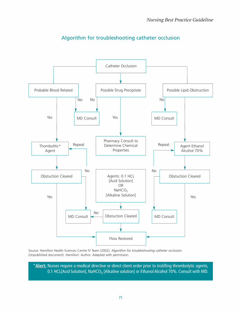
71
Nursing Best Practice Guideline
Diabetic Foot Ulcer
Treat the Cause Client Centered CareLocal Wound Care
Edge of Wound
Algorithm for troubleshooting catheter occlusion
Thombolitic*Agent
Pharmacy Consult toDetermine Chemical
Properties
Agents: 0.1 HCL[Acid Solution]
ORNaHCO3
[Alkaline Solution]
Obstuction Cleared
MDConsultant
No No No
YesYes
Flow Restored
Agent EthanolAlcohol 70%
No No
MD ConsultNo
Yes Yes
RepeatRepeat
Possible Lipid Obstruction
Obstuction Cleared
Source: Hamilton Health Sciences Centre IV Team (2002). Algorithm for troubleshooting catheter occlusion. (Unpublished document). Hamilton: Author. Adapted with permission.
*Alert: Nurses require a medical directive or direct client order prior to instilling thrombolytic agents,0.1 HCL[Acid Solution], NaHCO3 [Alkaline solution] or Ethanol Alcohol 70%. Consult with MD.
MD Consult
MD Consult MD Consult
Possible Drug Precipitate
Obstuction Cleared
Probable Blood Related
Catheter Occlusion

Care and Maintenance to Reduce Vascular Access Complications
72
Appendix G: Blood Withdrawal fromCentral Venous Access Devices
Discard■ Removes potential
contaminate from thecatheter (CVAD)
Push – Pull■ Requires mixing the blood
back and forth in a syringeseveral times to eliminatecontaminates from thecatheter (CVAD)
Re-infusion■ Involves returning the discard
specimen after obtainingblood samples
(Cosca et al., 1998; Holmes, 1998)
■ Remove a specific amount of blood fromcatheter (CVAD) via syringe or vacutainer
■ Use a “new” syringe or vacutainer for theblood sample
■ Flush CVAD with 0.9% saline
■ Using a 10 ml syringe, flush catheter(CVAD) with 5 ml 0.9% saline
■ Without removing syringe, 6 ml of blood isaspirated, then pushed back into catheter(CVAD)
■ Repeat this process x3■ Remove the empty syringe and attach new
syringe/vacutainer to obtain blood sample■ Flush CVAD with 0.9% saline
■ Aspirate 6 ml of blood into a syringe andcap with a sterile cap
■ Obtain blood specimen(s) via a syringe orvacutainer
■ Re-infuse the discard from the 1st syringe.
■ Potential nosocomical blood loss■ Potential to confuse a discard
syringe with blood samplesyringe
■ May be difficult to obtain enoughblood for 3 – 4 push-pullsequences
■ Risk of haemolysis with theagitation of blood
■ Potential to re-infuse clot(s)■ Potential to re-infuse
contaminated discard■ Potential for error including the
possibility of confusing thediscard syringe with the bloodsample
Blood WithdrawalMethod of Blood Withdrawal Description Potential Complication

73
Nursing Best Practice Guideline
Objectives
Lumen
Amount of Discard
Equipment
■ Use the largest Lumen for blood draws ■ Dedicate a lumen for blood draws when CVAD has multiple lumens■ Generally not recommended to use heparinized catheter (CVAD) for coagulation
studies■ Use of lumens for drug levels if the drug was infused via that lumen:
● Does not support the use of silastic catheters for sampling drug levels if the drugis given through the same catheter
● Aminoglycoside drug levels should be obtained by standard venicollection (inleukemia patients with indwelling right atrial catheters)
● If venipuncture is contraindicated or difficult to perform, the drug level shouldbe obtained from the catheter only after flushing
● One method describes to infuse a solution a minimum of 30 min after the doseand then obtain peak levels by standard procedure
● Another method is to administer a 10 mL IV push flush with 0.9% NaCI throughthe catheter, which would ensure delivery of the full dose of the aminoglycosideto the patient and then obtain the peak level by standard procedures
● Regardless of methods, one should view all drug levels obtained from thesecatheters with skepticism since the system has several possible sources forcontamination
■ Appropriate amount to ensure accuracy of lab results■ Discard 3-6 mL■ Consider dead space volume of CVAD
● 2x dead space volume – non coagulation tests● 6x dead space volume – non coagulation tests
■ Use the vacutainer or syringe or a combination of both to obtain blood samples■ Use safety engineered devices whenever possible (e.g., transfer devices) to reduce
the potential for negative outcomes (e.g., needle stick injury)■ Smaller syringes exert less negative pressure when withdrawing blood samples from
catheters (CVAD)
Blood Withdrawal – Additional Considerations
(Frey, 2003; INS, 2000; McCall et al., 2003; ONS, 2004)
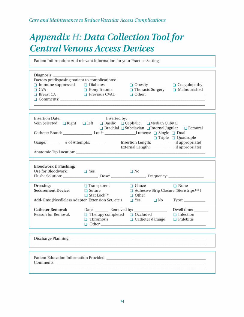
Care and Maintenance to Reduce Vascular Access Complications
74
Appendix H: Data Collection Tool forCentral Venous Access Devices
Patient Information: Add relevant information for your Practice Setting
Diagnosis: ______________________Factors predisposing patient to complications:❏ Immune suppressed ❏ Diabetes ❏ Obesity ❏ Coagulopathy❏ CVA ❏ Bony Trauma ❏ Thoracic Surgery ❏ Malnourished❏ Breast CA ❏ Previous CVAD ❏ Other: _____________________________❏ Comments: _____________________________________________________________________________________________________________________________________________________________
Insertion Date: ________________ Inserted by: ___________________Vein Selected: ❏ Right ❏ Left ❏ Basilic ❏ Cephalic ❏Median Cubital
❏ Brachial ❏ Subclavian ❏ Internal Jugular ❏ FemoralCatheter Brand: _______________ Lot #: ________________Lumens: ❏ Single ❏ Dual
❏ Triple ❏ QuadrupleGauge: ______ # of Attempts: _______ Insertion Length: ________ (if appropriate)
External Length: ________ (if appropriate)Anatomic Tip Location: _________________________________________________________________
Bloodwork & Flushing:Use for Bloodwork: ❏ Yes ❏ NoFlush: Solution: __________________ Dose: __________________ Frequency: __________________
Dressing: ❏ Transparent ❏ Gauze ❏ NoneSecurement Device: ❏ Suture ❏ Adhesive Strip Closure (Steristrips™ )
❏ Stat Lock™ ❏ OtherAdd-Ons: (Needleless Adapter, Extension Set, etc.) ❏ Yes ❏ No Type: ___________
Catheter Removal: Date: _______ Removed by: _________________ Dwell time: _______Reason for Removal: ❏ Therapy completed ❏ Occluded ❏ Infection
❏ Thrombus ❏ Catheter damage ❏ Phlebitis❏ Other ______________________________________________________
Discharge Planning: _____________________________________________________________________________________________________________________________________________________________
Patient Education Information Provided: ___________________________________________________Comments: ____________________________________________________________________________________________________________________________________________________________________

75
Nursing Best Practice Guideline
Post Insertion Complications
❏ Early stage mechanical phlebitis:Intervention: ______________________________________________________________________________________________________________________________________________________________
❏ Phlebitis:0 pain at site, no erythema, swelling, induration or palpable venous cord at or around the site
+1 pain at site, no erythema, swelling, induration or palpable venous cord+2 some erythema or swelling or both at site, no induration or palpable venous cord+3 erythema and swelling at site, palpable venous cord < 7.5 cm. above site+4 erythema and swelling at site, induration and palpable venous cord > 7.5 cm. above site
Intervention: _______________________________________________________________________
❏ Occlusion: ❏ Thrombotic ❏ Precipitate ❏ LipidsUnblocking procedure performed: ❏ Yes ❏ No # of times _______Occlusion resolved ❏ Yes ❏ No
❏ Infection: ❏ Insertion site (drainage, erythema, tender) ____________________❏ Catheter sepsis (positive blood culture, positive catheter tip culture,
same organism, may be febrile) ______________________________❏ Catheter sepsis suspected (no positive blood culture, fever resolves
after removal) ______________________________________________❏ Infection resolved __________________________________________
❏ Thrombus (validated radiographically) Location: _____________________________
❏ Embolus Type: ❏ Air ❏ Catheter ❏ Thrombus
❏ Catheter Damage: (describe) ____________________________________________________Successful repair: (date) ____________________________________________________
❏ Pain: (describe) ________________________________________________________________________________________________________
❏ Leaking at Insertion Site: ____________________________________________________
❏ Other: _______________________________________________________________________________
Comments:__________________________________________________________________________________________________________________________________________________________________________________________________________________________________________________________________________________________________________________________________________________________________________________________________________________________________________________________________________________________________________________________________________
Source: Hamilton Health Sciences Centre IV Team (2002). Data collection tool for central venous access devices. (Unpublisheddocument). Hamilton: Author. Reprinted with permission.

Care and Maintenance to Reduce Vascular Access Complications
76
Appendix I: Developing Patient Teaching Materials
The process of developing patient education materials
1. Starting the process: establishing a planning group
�
2. Assess available patient education materials
�
3. Identify your purpose and the patient population
�
4. Decide on the content
�
5. Write the first draft in plain language
�
6. Apply a clear design
�
7. Get feedback from the clinical team
�
8. Get feedback from patients and families
�
9. Get approval from the clinical team
�
10. Produce the material
�
11. Distribute and use the material
�
12. Evaluate the effectiveness of the material
Source: Wizowski, L., Harper, T., & Hutchings T. (2002) Writing health information for patients and families. Hamilton, ON:Hamilton Health Sciences. Reprinted with permission.

77
Nursing Best Practice Guideline
Appendix J: Planning guide for Educational MaterialsPurpose What knowledge or attitudes are you trying to enhance?
What specific behaviour(s) are you trying to change?
What “message” do you wish to convey?
How will this material be different from existing materials?
How will this material help/benefit your practice/program?
How will this material be helpful to the learner?
Target audience Who is the target audience? Students, patients, staff?
What are the characteristics of the target audience?
What is their previous experience?
What do they already know?
What motivates the audience?
What are their interests and habits related to the topic?
Are members of the target audience available to evaluate materials?
Content Has a literature search been completed?
Have you identified and assessed existing materials?
Has the target audience validated the learning need, content and format?
Have content experts validated the accuracy and relevance of the content?
Does the content support the hospital’s mission, vision and values?
Theory What theoretical framework guides the knowledge, value or behaviour change?
Is this framework appropriate and relevant to the learner?
Format How will this material be used by the learner?
Is the material easy to access and use?
How will this material be displayed and distributed?
Does the format support learning (self-directed, group)?
Is the material user-friendly?
If computer-based, is it compatible with existing systems?
If printed, do table of contents, index and bibliography contribute to ease of use?
Budget What source(s) of funding will be used to acquire or develop this material?
What quantity is needed?
Can production costs be estimated from printing specifications? (# of pages, paper
size, paper weight, artwork, binding, # of copies)
Validity Is the material up-to-date?
Does it replace existing material?
Does it add additional information?
Are the producer, editor and author reputable?
Source: Wizowski, L., Harper, T., & Hutchings T. (2002) Writing health information for patients and families. Hamilton, ON:Hamilton Health Sciences. Reprinted with permission

Care and Maintenance to Reduce Vascular Access Complications
78
Appendix K: Patient Education Material – Example
Name: __________________________
Learning about…
Going home with your PICC
Information about your PICC for your community nurse:
Your PICC was put in by _________________________on _________________
doctor or nurse date
At:
McMaster University Medical Centre ❏ Other
❏ Hamilton General Hospital _______________________
❏ Henderson General Hospital _______________________
❏ St. Joseph’s Healthcare _______________________
If you have problems with you PICC, call _________________________________
Brand name of PICC _________________________________
Size or gauge of the PICC _________________________________
❏ Valved ❏ Non-valved ❏ Single Lumen ❏ Double Lumen
Distal tip location: ❏ SVC ❏ Right Atrium Junction ❏ Right Atrium
Trimmed: ❏ Yes ❏ No
Length of internal catheter _________________________________cm
Length of external catheter _________________________________cm
Comments: ____________________________________________________________________________________
The above information was completed by _________________________________________________________
Nurse
On _______________________________________________________ Date _______________________________
unit or ward

79
Nursing Best Practice Guideline
You are going home with a PICC. PICC stands for Peripherally Inserted Central Catheter. The catheter is a
long thin tube and may stay in your arm for several weeks or months. Your PICC may be used to give you
fluids, medications, nutrition, blood products and/or to take blood samples for testing.
Your PICC will need routine care, such as dressing changes and flushing. Your community nurse will
discuss this with you.
When you first get your PICCWhen you first get home, it is normal for the area around your PICC to be sore. This can last a few days. It
may bleed or ooze a little. When the community nurse comes to your home, he or she will change the
dressing and check the area around your PICC. Please let the nurse know your concerns. Your nurse’s first
visit is about 12 to 24 hours after you get home.
Warm compressOn the arm where the PICC is, place a warm and moist compress. A compress can be a folded washcloth,
small towel or a warm pack. Wet the washcloth, ring out the excess water, wrap in plastic and then place
over the PICC. Do this when you get home and for the next 3 days. Do this 4 times a day for 10 minutes.
Before your nurse visitsTry not to move the area too much as you do your normal activities. Move your arm as you feel comfortable.
You can shower or bathe, but keep the area dry. Wrapping the PICC in plastic wrap is a way to keep it dry.
Your PICC should always be covered with at dressing. This helps to keep it free of germs.
When you have a PICC do not:■ Lift heavy objects (such as a 10 pound sack of potatoes) with the PICC arm ■ Use scissors near the PICC■ Swim with the PICC■ Play contact sports■ Shovel or garden with the PICC arm
Problem What to do
■ The dressing is soaked with blood
■ The dressing is soaked with clear fluid
■ The area around your PICC gets more puffy and swollen
■ The PICC breaks or falls out
■ Irregular or strange heart beat
■ If you have trouble breathing
■ Do not remove the dressing.Apply pressure with another dressing or clean cloth
■ Call the community Nurse■ If you bleed a lot or the bleeding does not stop –
go to Emergency
■ Cover with a clean dressing or cloth■ Call the community nurse
■ Call the community nurse
■ Cover with a clean dressing or cloth■ Call the community nurse
■ Call the community nurse or go to Emergency
■ Call 911 or go to Emergancy

Care and Maintenance to Reduce Vascular Access Complications
80
SuppliesBefore leaving the hospital you may be given a few supplies. More supplies will be sent to your home in a
big box. In the box there may be a bag with medications. Check the label on the bag to see if the
medications need to go into the fridge. There are supplies in the box for dressings, medications and taking
care of the PICC. When you are ready, your nurse will help you learn about these.
Keep your supply box in a clean dry safe place and away from children or pets.
Your supplies are provided by the CCAC. Talk with your nurse about making sure that you have the right
supplies. If you run out of supplies – call your community nurse.
You will need to have these other supplies:■ Liquid soap in a pump, or bottle of waterless soap ■ Paper towels■ Garbage bag ■ Clean towel
Living with your PICCYour PICC should always be covered. Do not have your blood pressure taken, blood work taken or IV’s
started with the PICC arm. When you return to your normal routine such as household chores, cooking,
work or sexual activity, you can use your arm as you feel comfortable. Do not overuse your arm with the
PICC. (Please review the section on the bottom of page 79.)
Your PICC needs special care to keep it clean and working well. This care may be done by you, a family
member, a friend or a nurse. Your nurse will help you learn about the care of your PICC.
When you have a PICC, it is a good idea to:■ Have your emergency telephone numbers close by■ Have your community nurse telephone number close by■ Keep a list of your questions, problems, notes in a book or diary■ Keep a calendar for clinic appointments and follow-up appointments with your community nurse
Setting up your homeYou will need a work area or space to lay out the supplies. When choosing the best place to do your PICC
care, keep these things in mind:■ Good lighting■ A room that is easy to keep clean and free of dust■ A comfortable spot in the room to sit or lie down■ A safe area away from children and pets■ A place free of drafts away from open windows, heating ducts and fans
When your community nurse comes or when you do your PICC care:■ Set aside 20 to 45 minutes■ Limit distractions: such as not answering your phone■ Try to do it the same time each day: tell your friends and family this time so they do not bother you■ Clean your work area before and after the PICC care
Source: Hamilton Health Sciences Centre – Patient Education. (2002). Going home with your PICC. (Unpublished document).Hamilton: Author. Reprinted with permission.

81
Nursing Best Practice Guideline
Appendix L: Education ResourcesThe following is a list of resources the development panel thought to be useful for education purposes.
It should not be considered a complete or comprehensive list.
Websites
Canadian Institute for Health Information (CIHI)
www.cihi.ca
Institute for Safe Medication Practices (ISMP)
www.ismp.org
Public Health Agency of Canada (PHAC)
www.phac-aspc.gc.ca
Registered Nurses’ Association of Ontario. (2004.) Assessment and Device Selection for Vascular Access.
Toronto: Author.
Available: http://www.rnao.org/bestpractices/completed_guidelines/BPG_Guide_C4_iv_therap.asp
The National Institute for Occupational Safety and Health (NIOSH)
www.cdc.gov/niosh/homepage.html
Professional Practice Network of Ontario (PPNO)
www.ppno.ca
Other resources
Registered Nurses’ Association of Ontario. (2004). User Guide: Implementation of the Home Health Care
Orientation Program for Nurses. Toronto: Author. Available: www.rnao.org
Registered Nurses’ Association of Ontario. (2004). Preceptorship Resource Kit. Toronto: Author.
Available: www.rnao.org

Care and Maintenance to Reduce Vascular Access Complications
82
Appendix M: Description of the ToolkitToolkit: Implementing Clinical Practice Guidelines
Best practice guidelines can only be successfully implemented if there are: adequate planning, resources,
organizational and administrative support as well as appropriate facilitation. RNAO, through a panel of nurses,
researchers and administrators, has developed the Toolkit: Implementation of Clinical Practice Guidelines
based on available evidence, theoretical perspectives and consensus. The Toolkit is recommended for
guiding the implementation of any clinical practice guideline in a healthcare organization.
The Toolkit provides step-by-step directions to individuals and groups involved in planning,
coordinating,and facilitating the guideline implementation. Specifically, the Toolkit addresses the
following key steps in implementing a guideline:
1. Identifying a well-developed, evidence-based clinical practice guideline
2. Identification, assessment and engagement of stakeholders
3. Assessment of environmental readiness for guideline implementation
4. Identifying and planning evidence-based implementation strategies
5. Planning and implementing evaluation
6. Identifying and securing required resources for implementation
Implementing guidelines in practice that result in successful practice changes and positive clinical impact
is a complex undertaking. The Toolkit is one key resource for managing this process.
The Toolkit is available through the Registered Nurses’ Association of Ontario.
The document is available in a bound format for a nominal fee, and is also
available free of charge from the RNAO website. For more information, an
order form or to download the Toolkit, please visit the RNAO website at
www.rnao.org/bestpractices.
83
Nursing Best Practice Guideline
Notes:

Care and Maintenance to Reduce Vascular Access Complications
84
Notes:
85
Nursing Best Practice Guideline
Notes:

Care and Maintenance to Reduce Vascular Access Complications
86
Notes:

87
Nursing Best Practice Guideline
Notes:
Care and Maintenance to Reduce Vascular Access Complications
88
Notes:
1
Review 2008
Nursing BestPractice GuidelineShaping the future of Nursing
Review Panel Members
Susanne Nelson, RN, BScN, MN, CVAA(C)Review ChairCNS – Vascular AccessUniversity Health NetworkToronto, Ontario
Sharon Armes, RN, CVAA(C)Senior Clinical Education CoordinatorBard Canada Inc.Mississauga, Ontario
Adrienne Austin, RN, BScN, CVAA(C)Clinical Manager, Vascular Access TherapyHamilton Health Sciences CentreHamilton, Ontario
Nan Clark, RN, CVAA(C), CON(C), CCHN(C)International Community ConsultantSaint Elizabeth Health CareMarkham, Ontario
Glenda Hicks, RN, BScNNurse ClinicianCritical Care ProgramSudbury Regional HospitalSudbury, Ontario
Julia Johnston, RN, BScN, MNAdvanced Practice Nurse, Palliative Care ProgramTrillium Health CentreMississauga, Ontario
Jenny Oey Chung, RN, BScN, MNProgram ManagerInternational Affairs and BestPractice Guidelines ProgramsRegistered Nurses’ Association of OntarioToronto, Ontario
Kris Paton, RN, CVAA(C)Clinical Leader, Vascular Access TherapyHamilton Health Sciences CentreHamilton, Ontario
Sharon Rodkin, RN, CVAA(C)Manager, Clinical ConsultingBaxter CorporationMississauga, Ontario
Lisa Valentine, RN, BScN, MNClinical Nurse Specialist/Case ManagerRegional Stroke Program – North East GTASunnybrook Health Sciences CentreToronto, Ontario
Supplement IntegrationThis supplement to the nursing best practice guideline Care and Maintenance to Reduce Vascular Access Complications is the result of a scheduled review of the guideline. As part of its commitment to ensure consistency with the best available evidence, the Registered Nurses’ Association of Ontario (RNAO) has established a monitoring and review pro-cess, which involves a full review of each guideline every 3 years.
As part of the health care team, nurses caring for clients with indwelling vascu-lar access devices have an important and continued role in providing safe infusion therapy, across the continuum of care. Therefore, they have an important role in helping clients understand and reduce their risk for complications. Importantly, though the main focus of this guideline review re-mains the care and maintenance of central venous access devices (CVAD), some rec-ommendations apply to both CVAD and peripheral venous access devices (PVAD) as per the original guideline. Please note that to ensure consistency with the scope of the original guideline, this review has not ad-dressed recommendations related to the care of clients requiring infusion therapy through the following devices: arterial lines, hemo-dialysis catheters, pulmonary artery lines, pheresis lines, epidural catheters, pressure monitoring devices, umbilical vein, femoral catheters, and/or intraosseous lines. Nurses working with these devices will require fur-ther practice direction from guidelines in their areas of practice.
Review ProcessA panel of specialists was assembled for this review, comprised of members from the original development panels of the Assessment and Device Selection for Vascular Access and Care and Maintenance to Reduce Vascular Access Complications guidelines, as well as other recommended individuals with particular expertise in this practice area. A structured evidence review based on the scope of the original guideline was conduct-ed to capture the relevant literature. Initial findings regarding the impact of the current evidence base on the original guideline were summarized for the review panel. The re-view panel members were given a mandate to review the original guideline in light of the new evidence, specifically to ensure the validity, appropriateness and safety of the guideline recommendations as published in 2005. In December 2007, the panel met to achieve consensus on the impact of this new evidence on the existing recommendations.
Care and Maintenance to ReduceVascular Access Complications
Guideline supplement

�
Literature ReviewConcurrent with the review of existing guidelines, a search for recent literature relevant to the scope of the guideline was conducted with guidance from the Review Chair. The search of electronic databases, including CINAHL, Medline and EMBASE, was conducted by a health sciences librarian. Articles identified and recommended by panel members were also considered. A Master’s prepared nurse completed the inclusion/exclusion review, quality appraisal and data extraction of the retrieved studies, and the summary of the literature findings. The comprehensive data tables and reference lists were provided to all panel members.
A summary of the evidence review is provided in the flow chart below.
Panel ReviewAfter a review of the current evidence, no substantive changes were made to the recommendations; however, several inac-curacies were noted in the original publi-cation, and will be addressed in this review document. Additional resources have also been identified and are listed below as Appendix A.
Review FindingsA review of the most recent research studies and relevant guidelines published since the development of the original guideline does not support substantive changes to the recommendations. Howev-er, the panel has identified several research gaps in the available evidence, which will be outlined below.
Review of Existing GuidelinesOne individual searched an established list of websites for published guidelines and other relevant content. This list was compiled based on existing knowledge of evidence-based practice websites and recommendations from the literature. Six international guidelines on infusion therapy were critically appraised using the Appraisal of Guidelines for Research and Evaluation Instrument (AGREE, �001). From this appraisal, two guidelines were identi-fied to inform the review process and were circulated to all panel members:
Central Venous Access Device Guideline Panel. (�006).
Managing central venous access devices in cancer patients:
A clinical practice guideline. Toronto (ON): Cancer Care
Ontario (CCO).
Infusion Nurses Society (INS). (�006). Infusion Nursing
Standards of Practice. Journal of Infusion Nursing, 29(1S),
S1-S9�.
Review Process Flow Chart
New Evidence
Literature Searched
Yield 195 abstracts
Yield 6 guidelines that met inclusion criteria
18 studies that met inclusion criteria
Quality appraisal of studies
� guidelines included after AGREE review
Develop evidence summary table
Review of �005 guideline based on new evidence
Supplement published
Dissemination
Yield 6 guidelines to be screened for consistency with inclusion/exclusion
criteria.
�
3.0 Nurses will consider the following factors when per-forming catheter site care using aseptic technique:
• Catheter material (composition); • Antiseptic solution; and • Client’s tolerance (skin integrity,
allergies, pain, sensitivity and skin reaction)
(Level IV)
Original recommendations identified by the panel for update Review Findings 2008
Although the evidence supports the original recommendation, the panel would like to include an alert:
Aseptic technique must include choice of solution, use of friction, and adequate contact time in order to be considered an effective use of technique. Please consult your institutional policy or infection control policies for more details. The panel also recognizes there has been discussion around circular vs. east-west application techniques; although evidence does not exist at present to support a recommendation for one technique over the other, this topic will be noted for future cycles of guideline review.
4.0 Nurses will not use the central venous access device (CVAD) until tip placement has been confirmed.
(Level IV)
Although the evidence supports the original recommendation, the panel would like to include an alert:
Anatomical tip location must be documented by a radiologist/attending physician following insertion, and this documentation must be accessible to all the client’s health care providers throughout the continuum of care.
Please note: the original discussion of the evidence referred to an illustration (Appendix D of the original guideline) which is inaccurate; please refer to Appendix B below for a revised visual representation of correct tip placement.
7.0 Nurses will maintain catheter patency using flushing and locking techniques.
(Level IV)
11.0 Nurses will change all add-on devices at a minimum of every 7� hours.
(Level IV)
The panel has identified some inaccuracies in Table � (p. ��) of the original guideline. These have been updated below; please refer to Appendix C.
The current review of the evidence has resulted in the recognition of a research gap around the rates of catheter-related bloodstream infections and phlebitis related to increasing the time interval for replacement of add-on devices. As sufficient evidence does not exist at present to change the original recommen-dation, this topic will be noted for future cycles of guideline review.
19.0 Health care organizations have access to infusion therapy nursing expertise to support optimal vascular access outcomes.
(Level III)
Although the evidence supports the original recommendation, the panel would like to emphasize the importance of health care organizations having access to credentialed infusion therapy nurses to support optimal vascular access outcomes. Please refer to the corresponding recommendation and discussion in the RNAO best practice guideline Assessment and Device Selection for Vascular Access (Recommendation 6).

�
Appendices
Appendix A: Additional Resources
Registered Nurses’ Association of Ontario (RNAO)
Vascular Access self-directed e-learning modules: Caring for Your Patients Receiving Intravenous Therapy www.RNAO.org/intravenous/
Health Education Fact Sheet – You and Your IV
Canadian Vascular Access Association (CVAA) http://www.cvaa.info/
Government of Ontario
Infection Control Today
Legislation �7�/07 of the Occupational Health and Safety Act – Needle Safety www.e-laws.gov.on.ca
www.infectioncontroltoday.com
Institute for Safe Medication Practices Canada
www.ismp-canada.org
Ontario Ministry of Health and Long-Term Care
www.health.gov.on.ca
Safer Healthcare Now www.saferhealthcarenow.ca
Appendix B: CVAD Tip Placement
Illustrated by: Nancy A. Bauer, BA, Bus.
Admin, RN, ET (�005)
Illustrated by: Nancy A. Bauer, BA, Bus.
Admin, RN, ET (�008)
Used with permission. Bard Canada (�007). CVAD tip
position. Mississauga, Ontario: Bard Canada.
INCORRECT PLACEMENT CORRECT PLACEMENT CORRECT PLACEMENT (from original guideline; *(see larger scale (X-RAY) tip too high) image on page 5)
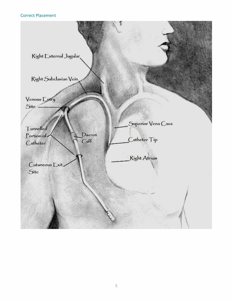
5
Correct Placement

6
Heparin (commonly used concentrations are 10 or 100 units/mL)
Heparin (commonly used concentrations are 10 or 100 units/mL)
Heparin (commonly used concentrations are 10 or 100 units/mL)
As per manufacturers’ recommendations
Flush and lock with � mL 0.9% sodium chloride
5 – 10 mL0.9% sodium chloride
Flush and lock with 10 – �0 mL 0.9% sodium chloride
10 – �0 mL 0.9% sodium chloride
Flush and lock with 10 – �0 mL 0.9% sodium chloride
10 – �0 mL 0.9% sodium chloride
10 – �0 mL 0.9% sodium chloride
After each access or daily if not in use
After each access or weekly if not in use
After each access or weekly if not in use
After each access or weekly if not in use
After each access or weekly if not in use
After each access or every four weeks if not in use As per manufacturers’ recommendations
Peripheral Short-Catheter
Peripheral Midline-Catheter (non-valved)
Peripheral Midline-Catheter (valved technology)
Central Vascular Access Device (CVAD), non-valved (e.g. Percutaneous, Tunneled, PICC)
CVAD with valved technology(e.g., Groshong®, PASV®) Implanted Vascular Access Devices (IVAD), non-valved
IVAD with valved technology (e.g. Groshong®, PASV®)
Appendix C: Flushing and Locking Interventions
Organizations may choose to modify Appendix C based on current clinical practice, device technology, and/or patient assessment.
Ensure that the catheter lumen is flushed using Sodium Chloride 0.9% prior to locking the lumen.
VASCULAR ACCESS DEVICE FLUSHING LOCK SOLUTION FREQUENCY SOLUTION
Note: Heparin is absolutely contraindicated in patients with Heparin-induced Thrombocytopenia (HIT), consult with physician re: alternative measures. For more details about flushing and locking solutions, please refer to the manufacturer’s recommendations.

7
References / Bibliography:AGREE Collaboration. (�001). Appraisal of Guidelines for Research and Evaluation (AGREE) Instrument. [Online]. Available: www.agreetrust.org.
Borschel, D.M., Chenoweth, C.E., Kaufman, S.R., Vander Hyde, K., VanDerElzen, K.A., Raghunathan, T.E., et al. (�006). Are antiseptic-coated central venous catheters effective in a
real-world setting? American Journal of Infection Control, 34(6), �88-9�.
Cadman, A., Lawrance, J.A.L., Fitzsimmons, L., Spencer-Shaw, A., & Swindell, R. (�00�). To clot or not to clot? That is the question in central venous catheters. Clinical Radiology,
59(�), ��9-�55.
Caers, J., Fontaine, C., Vinh-Hung, V., De Mey, J., Ponnet, G., Oost, C., et al. (�005). Catheter tip position as a risk factor for thrombosis associated with the use of subcutaneous
infusion ports. Supportive Care in Cancer, 13(5), ��5-��1.
Carrer, S., Bocchi, A., Bortolotti, M., Braga, N., Gilli, G., Candini, M., et al. (�005). Effect of different sterile barrier precautions and central venous catheter dressing on the skin
colonization around the insertion site. Minerva Anestesiologica, 71(5), 197-�06.
Central Venous Access Device Guideline Panel. (�006). Managing central venous access devices in cancer patients: A clinical practice guideline. Toronto (ON): Cancer Care Ontario (CCO).
Erbay, A., Ergonul, O., Stoddard, G.J., & Samore, M.H. (�006). Recurrent catheter-related bloodstream infections: Risk factors and outcome. International Journal of Infectious
Diseases, 10(5), �96-�00.
Gillies, D., O’Riordan, L., Carr, D., Frost, J., Gunning, R., & O’Brien, I. (�00�). Gauze and tape and transparent polyurethane dressings for central venous catheters. Cochrane Database
of Systematic Reviews, Issue �. Art. No.: CD00�8�7. DOI: 10.100�/1�651858.CD00�8�7.
Gillies, D., O’Riordan, L., Wallen, M., Morrison, A., Rankin, K., & Nagy, S. (�005). Optimal timing for intravenous administration set replacement. Cochrane Database of Systematic
Reviews, Issue �. Art. No.: CD00�588. DOI: 10.100�/1�651858. CD00�588.pub�.
Gorski, L.A. (�00�). Central venous access device outcomes in a homecare agency: A 7-year study. Journal of Infusion Nursing, 27(�), 10�-111.
Hamilton, H.C., & Foxcroft, D.R. (�007). Central venous access sites for the prevention of venous thrombosis, stenosis and infection in patients requiring long-term intravenous
therapy. Cochrane Database of Systematic Reviews, Issue �. Art. No.: CD00�08�. DOI: 10.100�/1�651858.CD00�08�.pub�.
Infusion Nurses Society (INS). (�006). Infusion Nursing Standards of Practice. Journal of Infusion Nursing, 29(1S), S1-S9�.
Macklin, D., Chernecky, C., Nugent, K., & Waller, J. (�00�). A collaborative approach to improving patient care associated with vascular access devices. Journal of Vascular Access
Devices, 8(�), 8-1�.
Maki, D.G., Kluger, D.M., & Crnich, C.J. (�006). The risk of bloodstream infection in adults with different intravascular devices. Mayo Clinic Proceedings, 81(9), 1159-1171.
Moureau, N., Mlodzik, L., & Markel Pool, S. (�005). The use of alteplase for treatment of occluded central venous catheters in home care. Journal of Vascular Access Devices,
10(�), 1��-1�9.
Ngo, A., & Murphy, S. (�005). A theory-based intervention to improve nurses’ knowledge, self-efficacy, and skills to reduce PICC occlusion. Journal of Infusion Nursing, 28(�), 17�-181.
Niel-Weise, B.S., Daha, T.J., & van den Broek, P.J. (�006). Is there evidence for recommending needleless closed catheter access systems in guidelines? A systematic review of
randomized controlled trials. Journal of Hospital Infection, 62(�), �06-�1�.
Pronovost, P., Needham, D., Berenholtz, S., Sinopoli, D., Chu, H., Cosgrove, S., et al. (�006). An intervention to decrease catheter-related bloodstream infections in the ICU. New
England Journal of Medicine, 355(�6), �7�5-�7��.
Render, M.L., Brungs, S., Kotagal, U., Nicholson, M., Burns, P., Ellis, D., et al. (�006). Evidence-based practice to reduce central line infections. Joint Commission Journal on Quality
and Patient Safety, 32(5), �5�-�60.
Waagen, G.L., & Zimmaro Bliss, D. (�00�). Development of evidence-based protocols for evaluation and management of dysfunctional vascular access devices in interventional
radiology. Journal of Vascular Nursing, 21(�), 50-6�.
Warren, D.K., Zack, J.E., Mayfield, J.L., Chen, A., Prentice, D., Fraser, V.J., et al. (�00�). The effect of an education program on the incidence of central venous catheter-associated
bloodstream infection in a medical ICU. Chest, 126(5), 161�-1618.

Nursing Best Practice Guideline
Care and Maintenance to ReduceVascular Access Complications
This program is funded by the Government of Ontario
April 2005
ISBN# 0-920166-67-9