cardiovascular system by dr. godara motilal
TRANSCRIPT


KARAGANDA STATE MEDICAL
UNIVERCITY
STUDENT INDIVIDUAL WORK
SUBMITTED BY – GODARA MOTI LAL
GROUP NO. 3003
SUBJECT- Propaedeutics
Topic – Aortic Insufficiency


49 WM sent for “transplant” evaluation from local cardiologistHPI – DOE x 6mos-1year, insidious onset, also
with L sided Chest tightness when tired/stressed and occasionally awakens him at night, last 1-2hrs and relieved with anxiolytics. Occasional lightheadedness after taking Coreg. Denies PND, Orthopnea, cough, pre/syncope. Former maintenance worker, now on medical leave. Pt states trying to remain active (walking/ swimming) but limited by dyspnea

PMHx: • Chronic LBP• Obesity• OSA/ CPAP• Depression/Anxiety• Basal Cell CarcinomaSocial:• no alcohol, cocaine, tobacco or drugs; married with 1
teenage son Meds (at presentation)• Carvedilol 50 bid, Celebrex, Lasix, ASA, anxiolytic,
hydrocodone, Combivent MDI

PE: HR 65 BP 170/67
HNT: jvp est 10cm,
CV: nl S1/2, + S3, no S4; PMI displaced
laterally to ant ax; 3/6 diastolic
decrescendo m USB
Resp: basilar rales
Abd: obese, no ascites/masses
Ext: tr edema, distal pulses brisk

Lab: • Chem 142 \ 110 / 14 H/H 16.6/ 46
4.4 / 27 \ 1.4
TSH, LFT, FLP, WBC/PPC, Coags nl
ECG: NSR, PRWP, nl axis, no ischemia
Etiology
Physical Examination
Assessing Severity
Natural History
Prognosis
Timing of Surgery
© Continuing Medical Implementation
…...bridging the care gap

Any conditions resulting in incompetent aortic leafletsCongenital• Bicuspid valve
Aortopathy• Cystic medial necrosis
• Collagen disorders (e.g. Marfan’s)
• Ehler-Danlos
• Osteogenesis imperfecta
• Pseudoxanthoma elasticum
Acquired• Rheumatic heart disease
• Dilated aorta (e.g. hypertension..)
• Degenerative
• Connective tissue disorders
E.g. ankylosing spondylitis, rheumatoid arthritis, Reiter’s syndrome, Giant-cell arteritis )
• Syphilis (chronic aortitis)
Acute AI: aortic dissection, infective endocarditis, trauma
© Continuing Medical Implementation
…...bridging the care gap

Dyspnea, orthopnea, PND
Chest pain.
• Nocturnal angina >> exertional angina
• ( diastolic aortic pressure and increased LVEDP thus
coronary artery diastolic flow)
With extreme reductions in diastolic pressures
(e.g. < 40) may see angina
© Continuing Medical Implementation
…...bridging the care gap

Quincke’s sign: capillary
pulsation
Corrigan’s sign: water
hammer pulse
Bisferiens pulse (AS/AR >
AR)
De Musset’s sign: systolic
head bobbing
Mueller’s sign: systolic
pulsation of uvula
Durosier’s sign: femoral
retrograde bruits
Traube’s sign: pistol shot
femorals
Hill’s sign:BP Lower
extremity >BP Upper
extremity by
• > 20 mm Hg - mild AR
• > 40 mm Hg – mod AR
• > 60 mm Hg – severe AR
© Continuing Medical Implementation
…...bridging the care gap

Widened pulse pressure
• Systolic – diastolic =
pulse pressure
High pitched, blowing,
decrescendo diastolic
murmur at LSB
Best heard at end-
expiration & leaning
forward
Hands & Knee position
© Continuing Medical Implementation …...bridging the care gap
S1 S2 S1

Apex:
• Enlarged
• Displaced
• Hyper-dynamic
• Palpable S3
• Austin-Flint murmur
Aortic diastolic
murmur• length correlates with
severity (chronic AR)
• in acute AR murmur
shortens as
Aortic DP=LVEDP
• in acute AR - mitral pre-
closure
© Continuing Medical Implementation
…...bridging the care gap

Assess severity by impact on peripheral signs and LV• peripheral signs = severity
• LV = severity
• S3
• Austin -Flint
• LVH
• radiological cardiomegaly
© Continuing Medical Implementation
…...bridging the care gap


Asymptomatic %/YNormal LV function (~good prognosis)• Progression to symptoms or LV dysfunction <
6• Progression to asymptomatic LV dysfunction <
3.5• 75% 5-year survival• Sudden death < 0.2
Abnormal LV function• Progression to cardiac symptoms 25
Symptomatic (Poor prognosis)• Mortality > 10
© Continuing Medical Implementation
…...bridging the care gap Bonow RO, et al, JACC. 1998;32:1486.
TX: Medical Surgery BEFORE LV dysfunction

ACC/AHA Class I• Symptomatic patients with preserved LVF (LVEF
>50%)
• Asymptomatic patients with mild to moderate LV dysfunction (EF 25-49%)
• Patients undergoing CABG, aortic or other valvular surgery
ACC/AHA Class II a• Asymptomatic patients with preserved LVEF but
severe LV dilatation (EDD>75 mm or ESD > 55mm)
© Continuing Medical Implementation
…...bridging the care gap

ACC/AHA Class II b• Patients with severe LV dysfunction (EF < 25%)
• Asymptomatic patients with normal systolic func-tion at rest (EF >0.50) and progressi ve LV dilata-tion when the degree of dilatation is moderatelysevere (EDD 70 to 75 mm, ESD 50 to 55 mm).
ACC/AHA Class III • Asymptomatic patients with normal systolicf unction at
rest (EF >0.50) and LV dilatation when the degree of dilatation is not severe (EDD <70 mm, ESD <50 mm).
© Continuing Medical Implementation
…...bridging the care gap

Etiology
Pathophysiology
History / Physical Findings
Natural History
Diagnosis
Management

Aortic Root• Age related dilatation
• Medial degeneration/ Marfans
• Dissection
• HTN
• Other ( osteogenesis imperfecta, Reiters, syphilitic aortitis, Bechet, psoriatic arthritis, relapsing polychondritis, UC arthritis, AS, giant cell arteritis
Aortic Valve• Rheumatic
• Calcific degeneration
• Congenital (Bicuspid, VSD)
• Myxomatous degeneration
• Endocarditis
• Structural degeneration of Bioprosthetic valve
• Other (SLE, AS, Takayasu, Whipple, Crohns)

increased LV EDVaddition of new sarcomeres in series/ elongation of myocytes and myocardial fibers (Eccentric Hypertrophy)enlarged chamber/ increased wall stress is stimulus for concentric hypertrophydilatation and hypertrophy with resultant recruitment of preload reserve allow compensation and maintenance of LV systolic functionmay be asymptomatic for decades until decompensated state develops, wall thickening unable to keep pace with hemodynamic load, increased interstitial fibrosis and decreased compliance symptoms of CHF ensue

Braunwald 6th ed
CO at rest may approach 25 L/min in severe AI with little increase in EDPvery large EDV (Cor Bovinum)
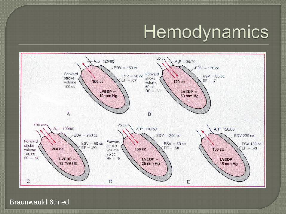
Braunwauld 6th ed

Braunwauld 6th ed

DOE, Orthopnea, PND• usually after 4th / 5th decade and significant
cardiomegaly and LV dysfxnAngina pectoris• develops later, nocturnal sxs prominent; often with
diaphoresis due to HR slowing with arterial DBP falling to low levels
Palpitations / Head pounding• especially in supine position, pounding of heart
against chest wall• tachycardia from stress/exertion may precipitate and
cause extreme discomfort for pt
From Braunwauld. Cardiovascular Dz, 6th ed.

de Musset sign – head bobbing with heartbeat
Corrigan pulse – “water hammer” pulse
Bisferiens pulse – brach/ fem arteries
Hill sign – popliteal > brachial by 60mmHg
Traube sign – “pistol shot sounds” over fem artery
Duroziez sign – sys m when femoral artery compressed proximally
and diastolic m when compressed distally
Quincke sign – capillary pulsations
Apical impulse - diffuse, hyperdynamic and displaced inf/lat
systolic thrill – base/suprasternal notch / carotid arteries

Diastolic murmur• high frequency, sitting up, leaning forward• duration > intensity correlates with severity • mild AR – early diastole, hi pitched blowing• severe AR – holodiastolic, rough• musical (“cooing dove”) – eversion/perforation of Ao cusp• Primary valve dz – heard best LSB 3-4 intercostal• Ao Root dz – heard best RSB
Austin Flint murmur• mid-late diastolic apical rumble – severe AR
Wide Pulse PressureSystolic flow murmur (/thrill)

Mortality rate for severe AI+CHF sxs > 20-50%/yr2
Bonow, et al. JACC Nov 1988 2Aronow , et a. Am J Cardiol 1994; 74: 286. l


2D/ M-Mode• AV/ Ao Root anatomic abnormalities• LV dimension / sphericity• AMVL – fluttering, reverse doming• increased EPSSDoppler• Color Flow Mapping• Continuous Wave• Flow reversal in desc Ao (100% sens 97% spec for
severe AI)Limitations – What is severe AI?

AMVL fluttering
Color Flow – top mild, bottom moderate

Vasodilators• goal is to reduce SBP, improve forward SV, reduce regurgitant
volumeUses• severe AR + sxs/ LV dysfxn• short term hemodynamic improvement in pt with symptomatic AR
before AVR• prolong compensated phase of asymptomatic patients• No indication for asymptomatic pt with mild AI and normal LV fxn
Studied in AI• Nifedipine, Hydralizine, ACEI, Nipride, Prazosin• Children/ severe AR – ACEI reversed LV dilatation/wall stress• avoid (-) inotrope in LV dysfxn
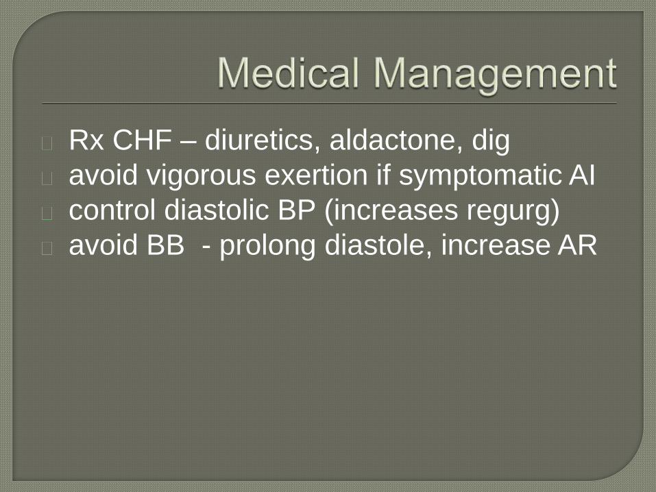
Rx CHF – diuretics, aldactone, dig
avoid vigorous exertion if symptomatic AI
control diastolic BP (increases regurg)
avoid BB - prolong diastole, increase AR

Goal is to intervene before irreversible LV
systolic dysfxn ensues• initially reversible, mainly due to afterload excess
– full recovery in LV size/fxn possible
• with progressive chamber dilatation, decreased
myocardial contractility >> afterload excess as
cause of LV dysfxn.
• associated with worse recovery of LV fxn and
increased mortality
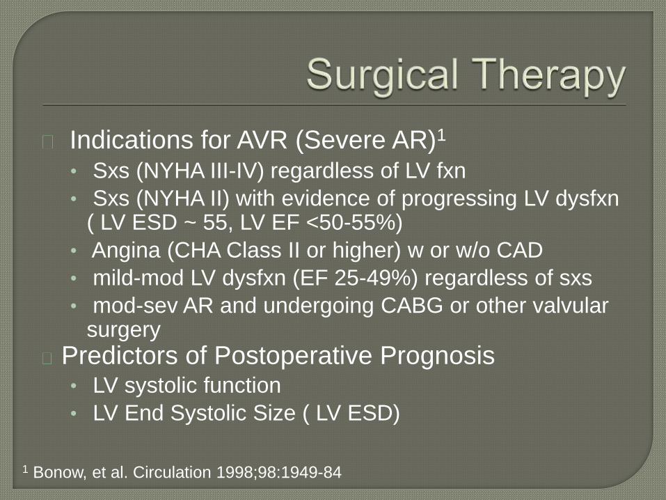
Indications for AVR (Severe AR)1
• Sxs (NYHA III-IV) regardless of LV fxn
• Sxs (NYHA II) with evidence of progressing LV dysfxn ( LV ESD ~ 55, LV EF <50-55%)
• Angina (CHA Class II or higher) w or w/o CAD
• mild-mod LV dysfxn (EF 25-49%) regardless of sxs
• mod-sev AR and undergoing CABG or other valvular surgery
Predictors of Postoperative Prognosis• LV systolic function
• LV End Systolic Size ( LV ESD)
1 Bonow, et al. Circulation 1998;98:1949-84
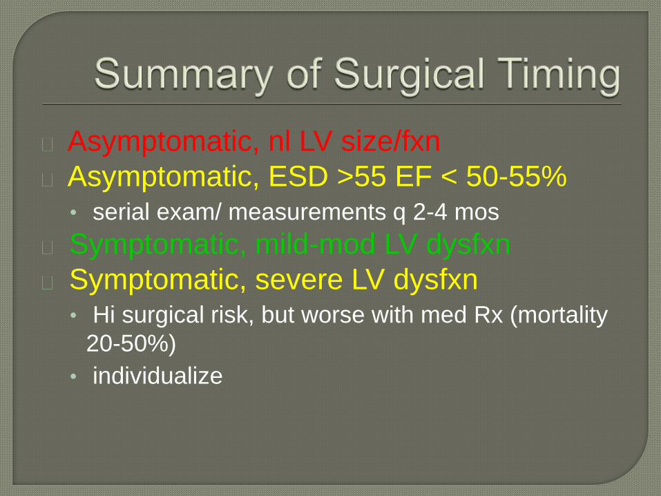
Asymptomatic, nl LV size/fxn
Asymptomatic, ESD >55 EF < 50-55%• serial exam/ measurements q 2-4 mos
Symptomatic, mild-mod LV dysfxn
Symptomatic, severe LV dysfxn• Hi surgical risk, but worse with med Rx (mortality
20-50%)
• individualize

Preload kept high immediate postop
period to fill dilated LV
temporary IABP use may be necessary
until LV fxn improves early post op

Ao Root disease• annuloplasty or other valve sparing surgery
possible if pure Ao Root dz
Primary AV disease• valve replacement
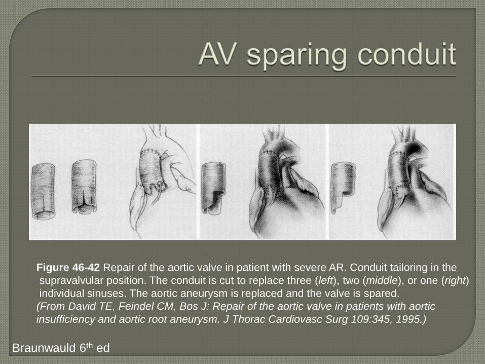
Figure 46-42 Repair of the aortic valve in patient with severe AR. Conduit tailoring in the
supravalvular position. The conduit is cut to replace three (left), two (middle), or one (right)
individual sinuses. The aortic aneurysm is replaced and the valve is spared.
(From David TE, Feindel CM, Bos J: Repair of the aortic valve in patients with aortic
insufficiency and aortic root aneurysm. J Thorac Cardiovasc Surg 109:345, 1995.)
Braunwauld 6th ed

Figure 29-15 A. Björk-Shiley Monostrut
mechanical prosthesis. B. Sorin Allcarbon
monoleaflet mechanical prosthesis.
C. Medtronic-Hall mechanical prosthesis.
D. Omnicarbon mechanical prosthesis.
Edmunds. Cardiac Surgery in the Adult. Ch 29
Figure 29-16 A. Carpentier-Edwards Supra-
annular porcine bioprosthesis. B. Hancock II
porcine bioprosthesis. C. Hancock modified
orifice porcine bioprosthesis. D. St. Jude
Medical Bioimplant porcine bioprosthesis.
