cardiovascular system - 1 file download
TRANSCRIPT

CARDIOVASCULAR SYSTEM Chest pain
Assessment of patients with suspected cardiac chest pain
NICE issued guidelines in 2010 on the 'Assessment and diagnosis of recent onset chest pain or discomfort of suspected cardiac origin'. Patients presenting with acute chest pain Immediate management of suspected acute coronary syndrome (ACS)
• Glyceryl trinitrate • Aspirin 300mg. Nice do not recommend giving other antiplatelet agents (i.e.
Clopidogrel) outside of hospital • Do not routinely give oxygen, only give if sats < 94%* • Perform an ecg as soon as possible but do not delay transfer to hospital. A normal ecg
does not exclude ACS
Referral
• Current chest pain or chest pain in the last 12 hours with an abnormal ECG: emergency admission
• Chest pain 12-72 hours ago: refer to hospital the same-day for assessment • Chest pain > 72 hours ago: perform full assessment with ECG and troponin
measurement before deciding upon further action
*NICE suggest the following in terms of oxygen therapy:
• Do not routinely administer oxygen, but monitor oxygen saturation using pulse oximetry as soon as possible, ideally before hospital admission. Only offer supplemental oxygen to:
v People with oxygen saturation (spo2) of less than 94% who are not at risk of hypercapnic respiratory failure, aiming for spo2 of 94-98%
v People with chronic obstructive pulmonary disease who are at risk of hypercapnic respiratory failure, to achieve a target spo2 of 88-92% until blood gas analysis is available.

Source: PassMedicine
2
Patients presenting with stable chest pain With all due respect to NICE the guidelines for assessment of patients with stable chest pain are rather complicated. They suggest an approach where the risk of a patient having coronary artery disease (CAD) is calculated based on their symptoms (whether they have typical angina, atypical angina or non-anginal chest pain), age, gender and risk factors. NICE define anginal pain as the following:
1. Constricting discomfort in the front of the chest, neck, shoulders, jaw or arms
2. Precipitated by physical exertion
3. Relieved by rest or GTN in about 5 minutes
v Patients with all 3 features have typical angina v Patients with 2 of the above features have atypical angina v Patients with 1 or none of the above features have non-anginal chest pain
If patients have typical anginal symptoms and a risk of CAD is greater than 90% then no further diagnostic testing is required. It should be noted that all men over the age of 70 years who have typical anginal symptoms fall into this category. For patients with an estimated risk of 10-90% the following investigations are recommended. Note the absence of the exercise tolerance test:
Estimated likelihood of CAD
Diagnostic testing
61-90% Coronary angiography 30-60% Functional imaging, for example:
• Myocardial perfusion scan with SPECT • Stress echocardiography • First-pass contrast-enhanced magnetic resonance (MR)
perfusion • MR imaging for stress-induced wall motion abnormalities.
10-29% CT calcium scoring

Source: PassMedicine
3
Pulses
Pulsus parodoxus
• Greater than the normal (10 mmHg) fall in systolic blood pressure during inspiration → faint or absent pulse in inspiration
• Severe asthma, cardiac tamponade
Slow-rising/plateau
• Aortic stenosis
Collapsing
• Aortic regurgitation • Patent ductus arteriosus • Hyperkinetic (anaemia, thyrotoxic, fever, exercise/pregnancy)
Pulsus alternans
• Regular alternation of the force of the arterial pulse • Severe LVF
Bisferiens pulse
• 'Double pulse' - two systolic peaks • Mixed aortic valve disease
'Jerky' pulse
• Hypertrophic obstructive cardiomyopathy*
*HOCM may occasionally be associated with a bisferiens pulse

Source: PassMedicine
4
Heart sounds
The first heart sound (S1) is caused by closure of the mitral and tricuspid valves whilst the second heart sound (S2) is due to aortic and pulmonary valve closure S1
• Closure of mitral and tricuspid valves • Soft if long PR or mitral regurgitation • Loud in mitral stenosis
S2
• Closure of aortic and pulmonary valves • Soft in aortic stenosis • Splitting during inspiration is normal
S3 (third heart sound)
• Caused by diastolic filling of the ventricle • Considered normal if < 30 years old (may persist in women up to 50 years old) • Heard in left ventricular failure (e.g. Dilated cardiomyopathy), constrictive pericarditis
(called a pericardial knock)
S4 (fourth heart sound)
• May be heard in aortic stenosis, HOCM, hypertension • Caused by atrial contraction against a stiff ventricle • In HOCM a double apical impulse may be felt as a result of a palpable S4

Source: PassMedicine
5
Heart sounds: S2
S2 is caused by the closure of the aortic valve (A2) closely followed by that of the pulmonary valve (P2)
Causes of a loud S2
• Hypertension: systemic (loud A2) or pulmonary (loud P2)
• Hyperdynamic states • Atrial septal defect without
pulmonary hypertension
Causes of a soft S2
• Aortic stenosis
Causes of fixed split S2
• Atrial septal defect
Causes of a widely split S2
• Deep inspiration
Causesofareversed(paradoxical)splitS2(P2occursbeforeA2)
• LBBB • Severe aortic stenosis • Right ventricular pacing • WPW type B (causes early P2) • Patent ductus arteriosus

Source: PassMedicine
6
• RBBB • Pulmonary stenosis • Severe mitral regurgitation
Murmurs
Ejection systolic
• Aortic stenosis • Pulmonary stenosis, HOCM • ASD, Fallot's
Holosystolic (pansystolic)
• Mitral/tricuspid regurgitation (high-pitched and 'blowing' in character)
• VSD ('harsh' in character)
Late systolic
• Mitral valve prolapse • Coarctation of aorta
Early diastolic
• Aortic regurgitation (high-pitched and 'blowing' in character) • Graham-Steel murmur (pulmonary regurgitation, again high-pitched and 'blowing' in
character)
Mid-late diastolic
• Mitral stenosis ('rumbling' in character) • Austin-Flint murmur (severe aortic regurgitation, again is 'rumbling' in character)
Continuous machine-like mumur

Source: PassMedicine
7
• Patent ductus arteriosus
Adult advanced life support
The joint European Resuscitation Council and Resuscitation Council (UK) 2010 guidelines do not alter significantly from the 2005 guidelines. Please see the link for more details, below is only a very brief summary of key points / changes. Major points include:
• Ratio of chest compressions to ventilation is 30:2 • Chest compressions are now continued while a defibrillator is charged • During a VF/VT cardiac arrest, adrenaline 1 mg is given once chest compressions have
restarted after the third shock and then every 3-5 minutes (during alternate cycles of CPR). In the 2005 guidelines, adrenaline was given just before the third shock. Amiodarone 300 mg is also given after the third shock
• Atropine is no longer recommended for routine use in asystole or pulseless
Electrical activity (PEA).
• A single shock for VF/pulseless VT followed by 2 minutes of CPR, rather than a series of 3 shocks followed by 1 minute of CPR
• Asystole/pulseless-electrical activity should be treated with 2 minutes of CPR, rather than 3, prior to reassessment of the rhythm
• Delivery of drugs via a tracheal tube is no longer recommended • Following successful resuscitation oxygen should be titrated to achieve saturations of
94-98%. This is to address the potential harm caused by hyperoxaemia
***The absence of a carotid pulse in the presence of sinus tachycardia indicates that this is a non-shockable rhythm, and the appropriate algorithm should be followed as explained below. The only shockable rhythms are ventricular fibrillation and ventricular tachycardia.

Source: PassMedicine
8
Syncope
Syncope may be defined as a transient loss of consciousness due to global cerebral hypoperfusion with rapid onset, short duration and spontaneous complete recovery. Note how this definition excludes other causes of collapse such as epilepsy. The European Society of Cardiology published guidelines in 2009 on the investigation and management of syncope. They suggested the following classification: Reflex syncope (neurally mediated)
• Vasovagal: triggered by emotion, pain or stress. Often referred to as 'fainting' • Situational: cough, micturition, gastrointestinal • Carotid sinus syncope
Orthostatic syncope
• Primary autonomic failure: Parkinson's disease, Lewy body dementia • Secondary autonomic failure: e.g. Diabetic neuropathy, amyloidosis, uraemia • Drug-induced: diuretics, alcohol, vasodilators • Volume depletion: haemorrhage, diarrhoea
Cardiac syncope
• Arrhythmias: bradycardias (sinus node dysfunction, AV conduction disorders) or tachycardias (supraventricular, ventricular)
• Structural: valvular, myocardial infarction, hypertrophic obstructive cardiomyopathy • Others: pulmonary embolism
Reflex syncope is the most common cause in all age groups although orthostatic and cardiac causes become progressively more common in older patients. Evaluation
• Cardiovascular examination • Postural blood pressure readings: a symptomatic fall in systolic BP > 20 mmHg or
diastolic BP > 10 mmhg or decrease in systolic BP < 90 mmHg is considered diagnostic • ECG • Carotid sinus massage • Tilt table test • 24 hour ECG

Source: PassMedicine
9
Peri-arrest rhythms: tachycardia
The 2010 Resuscitation Council (UK) guidelines have simplified the advice given for the management of peri-arrest tachycardias. Separate algorithms for the management of broad-complex tachycardia, narrow complex tachycardia and atrial fibrillation have been replaced by one unified treatment algorithm Following basic ABC assessment, patients are classified as being stable or unstable according to the presence of any adverse signs:
• Shock: hypotension (systolic blood pressure < 90 mmhg), pallor, sweating,
Cold, clammy extremities, confusion or impaired consciousness
• Syncope • Myocardial ischaemia • Heart failure
If any of the above adverse signs are present then synchronised DC shocks should be given Treatment following this is given according to whether the QRS complex is narrow or broad and whether the rhythm is regular or irregular. The full treatment algorithm can be found at the Resuscitation Council website, below is a very limited summary: Broad-complex tachycardia Regular
§ Assume ventricular tachycardia (unless previously confirmed SVT with bundle branch block)
§ Loading dose of amiodarone followed by 24 hour infusion Irregular
§ AF with bundle branch block - treat as for narrow complex tachycardia § Polymorphic VT (e.g. Torsade de pointes) - IV magnesium
Narrow-complex tachycardia Regular
§ Vagal manoeuvres followed by IV adenosine § If above unsuccessful consider diagnosis of atrial flutter and control rate (e.g. Beta-
blockers) Irregular
§ Probable atrial fibrillation § If onset < 48 hr consider electrical or chemical cardioversion § Rate control (e.g. Beta-blocker or digoxin) and anticoagulation

Source: PassMedicine
10
Peri-arrest rhythms: bradycardia
The 2010 Resuscitation Council (UK) guidelines emphasise that the management of bradycardia depends on:
1. Identifying the presence of signs indicating haemodynamic compromise - 'adverse signs'
2. Identifying the potential risk of asystole
Adverse signs The following factors indicate haemodynamic compromise and hence the need for treatment:
• Shock: hypotension (systolic blood pressure < 90 mmhg), pallor, sweating,
Cold, clammy extremities, confusion or impaired consciousness
• Syncope • Myocardial ischaemia • Heart failure
Atropine is the first line treatment in this situation. If this fails to work, or there is the potential risk of asystole then transvenous pacing is indicated Potential risk of asystole The following indicate a potential risk of asystole and hence the need for treatment with transvenous pacing:
• Complete heart block with broad complex QRS • Recent asystole • Mobitz type II AV block • Ventricular pause > 3 seconds
If there is a delay in the provision of transvenous pacing the following interventions may be used:
• Atropine, up to maximum of 3mg • Transcutaneous pacing • Adrenaline infusion titrated to response

Source: PassMedicine
11
Ventricular tachycardia
Ventricular tachycardia (VT) is broad-complex tachycardia originating from a ventricular ectopic focus. It has the potential to precipitate ventricular fibrillation and hence requires urgent treatment. There are two main types of VT:
v Monomorphic VT: most commonly caused by myocardial infarction v Polymorphic VT: A subtype of polymorphic VT is torsades de pointes which is
precipitated by prolongation of the QT interval. The causes of a long QT interval are listed below
Causes of a prolonged QT interval
Congenital Drugs Other § Jervell-Lange-Nielsen
syndrome (includes deafness and is due to an abnormal potassium channel)
§ Romano-Ward syndrome (no deafness)
§ amiodarone, sotalol, class 1a antiarrhythmic drugs
§ tricyclic antidepressants, fluoxetine
§ chloroquine § terfenadine § erythromycin
§ electrolyte: hypocalcaemia, hypokalaemia, hypomagnesaemia
§ acute myocardial infarction
§ myocarditis § hypothermia § � subarachnoid
haemorrhage Management If the patient has adverse signs (systolic BP < 90 mmhg, chest pain, heart failure or rate > 150 beats/min) then immediate cardioversion is indicated. In the absence of such signs antiarrhythmics may be used. If these fail, then electrical cardioversion may be needed with synchronised DC shocks Drug therapy
• Amiodarone: ideally administered through a central line • Lidocaine: use with caution in severe left ventricular impairment • Procainamide

Source: PassMedicine
12
Verapamil should NOT be used in VT
If drug therapy fails
• Electrophysiological study (EPS) • Implant able cardioverter-defibrillator (ICD) - this is particularly indicated in patients
with significantly impaired LV function
***Hypokalemia is the most important cause of ventricular tachycardia (VT) clinically, followed by hypomagnesaemia. Severe hyperkalaemia may cause VT in certain circumstances, for example in patients with structural heart disease, but it is not as common a cause as hypokalemia.
Torsades de pointes
Torsades de pointes ('twisting of the points') is a rare arrhythmia associated with a long QT interval. It may deteriorate into ventricular fibrillation and hence lead to sudden death Causes of long QT interval
• Congenital: Jervell-Lange-Nielsen syndrome, Romano-Ward syndrome • Antiarrhythmics: amiodarone, sotalol, class 1a antiarrhythmic drugs • Tricyclic antidepressants • Antipsychotics • Chloroquine • Terfenadine • Erythromycin • Electrolyte: hypocalcaemia, hypokalaemia, hypomagnesaemia • Myocarditis • Hypothermia • Subarachnoid haemorrhage
Management
• IV magnesium sulphate

Source: PassMedicine
13
Supraventricular tachycardia
Whilst strictly speaking the term supraventricular tachycardia (SVT) refers to any tachycardia that is not ventricular in origin the term is generally used in the context of paroxysmal SVT. Episodes are characterised by the sudden onset of a narrow complex tachycardia, typically an atrioventricular nodal re-entry tachycardia (AVNRT). Other causes include atrioventricular re-entry tachycardias (AVRT) and junctional tachycardias. Acute management
• Vagal manoeuvres: e.g. Valsalva manoeuvre • Intravenous adenosine 6mg → 12mg → 12mg: contraindicated in asthmatics - verapamil
is a preferable option • Electrical cardioversion
Prevention of episodes
• Beta-blockers • Radio-frequency ablation

Source: PassMedicine
14
Atrial fibrillation:rate control and maintenance of sinus rhythm
The Royal College of Physicians and NICE published guidelines on the management of atrial fibrillation (AF) in 2006. The following is also based on the joint American Heart Association (AHA), American College of Cardiology (ACC) and European Society of Cardiology (ESC) 2012 guidelines Agents used to control rate in patients with atrial fibrillation
• Beta-blockers • Calcium channel blockers • Digoxin (not considered first-line anymore as they are less effective at controlling the
heart rate during exercise. However, they are the preferred choice if the patient has coexistent heart failure)
Agents used to maintain sinus rhythm in patients with a history of atrial fibrillation
• Sotalol • Amiodarone • Flecainide • Others (less commonly used in UK): disopyramide, dofetilide, procainamide,
propafenone, quinidine
The table below indicates some of the factors which may be considered when considering either a rate control or rhythm control strategy
Factors favouring rate control Factors favouring rhythm control Older than 65 years History of ischaemic heart disease With contraindications to antiarrhythmic drugs. Unstable for cardioversion.
Younger than 65 years Symptomatic First presentation Lone AF or AF secondary to a corrected precipitant (e.g. Alcohol) Congestive heart failure

Source: PassMedicine
15
The first thing is to determine the type of AF, so here are some important (simplified) definitions.
• Acute - onset within the previous 48 hours. • Paroxysmal - spontaneous termination within seven days and most often within 48
hours. • Recurrent - two or more episodes. • Persistent - not self-terminating; lasting longer than seven days, or prior cardioversion. • Permanent - long-standing AF (defined as over a year) that is not successfully
terminated by
Atrial fibrillation: anticoagulation
NICE updated their guidelines on the management of atrial fibrillation (AF) in 2014. They suggest using the CHA2DS2-vasc score to determine the most appropriate anticoagulation strategy. This scoring system superceded the CHADS2 score.
Risk factor Points
C Congestive heart failure 1 H Hypertension (or treated hypertension) 1 A2 Age >= 75 years 2
Age 65-74 years 1 D Diabetes 1 S2 Prior Stroke or TIA 2 V Vascular disease (including ischaemic heart disease and peripheral arterial
disease) 1
S Sex (female) 1 The table below shows a suggested anticoagulation strategy based on the score:
Score Anticoagulation 0 No treatment 1 Males: Consider anticoagulation
Females: No treatment (this is because their score of 1 is only reached due to their gender)
2 or more
Offer anticoagulation
NICE recommend that we offer patients a choice of anticoagulation, including warfarin and the novel oral anticoagulants (noacs). There are complicated rules surrounding which NOAC is licensed for which risk factor - these can be found in the NICE guidelines. Aspirin is no longer recommended for reducing stroke risk in patients with AF

Source: PassMedicine
16
Doctors have always thought carefully about the risk/benefit profile of starting someone on warfarin. A history of falls, old age, alcohol excess and a history of previous bleeding are common things that make us consider whether warfarinisation is in the best interests of the patient. NICE now recommend we formalise this risk assessment using the HASBLED scoring system.
There are no formal rules on how we act on the HAS-BLED score although a score of >= 3 indicates a 'high risk' of bleeding, defined as intracranial haemorrhage, hospitalisation, haemoglobin decrease >2 g/L, and/or transfusion.
Atrial fibrillation: pharmacological cardioversion
The Royal College of Physicians and NICE published guidelines on the management of atrial fibrillation (AF) in 2006. The following is also based on the joint American Heart Association (AHA), American College of Cardiology (ACC) and European Society of Cardiology (ESC) 2012 guidelines Agents with proven efficacy in the pharmacological cardioversion of atrial fibrillation
• Amiodarone • Flecainide (if no structural heart disease) • Others (less commonly used in UK): quinidine, dofetilide, ibutilide, propafenone
Less effective agents
Risk factor Points
H Hypertension, uncontrolled, systolic BP > 160 mmhg 1 A Abnormal renal function (dialysis or creatinine > 200)
Or Abnormal liver function (cirrhosis, bilirubin > 2 times normal, ALT/AST/ALP > 3 times normal
1 for any renal abnormalities 1 for any liver abnormalities
S Stroke, history of 1 B Bleeding, history of bleeding or tendency to bleed 1 L Labile inrs (unstable/high inrs, time in therapeutic range < 60%) 1 E Elderly (> 65 years) 1 D Drugs Predisposing to Bleeding (Antiplatelet agents, nsaids)
Or Alcohol Use (>8 drinks/week)
1 for drugs 1 for alcohol
Atrial fibrillation - cardioversion: amiodarone + flecainide

Source: PassMedicine
17
• Beta-blockers (including sotalol) • Calcium channel blockers • Digoxin • Disopyramide • Procainamide
Atrial fibrillation: cardioversion
There are two scenarios where cardioversion may be used in atrial fibrillation:
• Electrical cardioversion as an emergency if the patient is haemodynamically unstable • Electrical or pharmacological cardioversion as an elective procedure where a rhythm
control strategy is preferred.
The notes below refer to cardioversion being used in the elective scenario for rhythm control. The wording of the 2014 NICE guidelines is as follows:
Offer rate or rhythm control if the onset of the arrhythmia is less than 48 hours, and start rate control if it is more than 48 hours or is uncertain Onset < 48 hours If the atrial fibrillation (AF) is definitely of less than 48 hours onset patients should be heparinised. Patients who have risk factors for ischaemic stroke should be put on lifelong oral anticoagulation. Otherwise, patients may be cardioverted using either:
• Electrical - 'DC cardioversion' • Pharmacology - amiodarone if structural heart disease, flecainide or amiodarone in
those without structural heart disease
Following electrical cardioversion if AF is confirmed as being less than 48 hours duration then further anticoagulation is unnecessary Onset > 48 hours If the patient has been in AF for more than 48 hours then anticoagulation should be given for at least 3 weeks prior to cardioversion. An alternative strategy is to perform a transoesophageal

Source: PassMedicine
18
echo (TOE) to exclude a left atrial appendage (LAA) thrombus. If excluded patients may be heparinised and cardioverted immediately. NICE recommend electrical cardioversion in this scenario, rather than pharmacological. If there is a high risk of cardioversion failure (e.g. Previous failure or AF recurrence) then it is recommend to have at least 4 weeks amiodarone or sotalol prior to electrical cardioversion Following electrical cardioversion patients should be anticoagulated for at least 4 weeks. After this time decisions about anticoagulation should be taken on an individual basis depending on the risk of recurrence
Atrial fibrillation: post-stroke
NICE issued guidelines on atrial fibrillation (AF) in 2006. They included advice on the management of patients with AF who develop a stroke or transient-ischaemic attack (TIA). Recommendations include:
• Following a stroke or TIA warfarin should be given as the anticoagulant of choice. Aspirin/dipyridamole should only be given if needed for the treatment of other comorbidities
In acute stroke patients, in the absence of haemorrhage, anticoagulation therapy should be commenced after 2 weeks. If imaging shows a very large cerebral infarction then the initiation of anticoagulation should be delayed

Source: PassMedicine
19
Key Points
Rate control algorithm § step 1: administer thromboprophylaxis;
Side effects of amiodarone has some important side effects such as hypo/hyperthyroidism due to looking similar to thyroxine and causes thrombophlebitis.
A calcium channel blocker (CCB) i.e. Verapamil) for rate control, with digoxin only being indicated in sedentary patients. These are correct but NOT first line. For further reading
If the presentation is atrial fibrillation (as seen by palpitations and absent P waves on ECG), the options for management include rate or rhythm control, with the possibility of thromboprophylaxis.
§ However, as he has an unmeasurable BP and signs of haemodynamic instability, patient is acutely unwell and so his arrhythmia must be treated as soon as possible with DC cardioversion without delaying for thromboprophylaxis. Anticoagulation should be continued for 4 weeks after cardioversion however.
§ If patient is clinically stable and had atrial fibrillation of over 48 hours duration, the risk of stroke would mean parenteral anticoagulation would take precedence over cardioversion. (source: NICE, https://www.nice.org.uk/guidance/cg180)
Asthma is a contraindication to the prescription of a beta-blocker. NICE therefore recommend a rate-limiting calcium channel blocker.Consideration should also be given to antithrombotic therapy.
'If pharmacological cardioversion has been agreed on clinical and resource grounds for new-onset atrial fibrillation, offer:
• Flecainide or amiodarone if there is no evidence of structural or ischaemic heart disease or
• Amiodarone if there is evidence of structural heart disease.'

Source: PassMedicine
20
Hypertension:
DIAGNOSIS AND MANAGEMENT
NICE published updated guidelines for the management of hypertension in 2011. Some of the key changes include:
• Classifying hypertension into stages • Recommending the use of ambulatory blood pressure monitoring (ABPM) and home
blood pressure monitoring (HBPM) • Calcium channel blockers are now considered superior to thiazides • Bendroflumethiazide is no longer the thiazide of choice
Diagnosing hypertension
Firstly, NICE recommend measuring blood pressure in both arms when considering a diagnosis of hypertension. If the difference in readings between arms is more than 20 mmhg then the measurements should be repeated. If the difference remains > 20 mmhg then subsequent blood pressures should be recorded from the arm with the higher reading. It should of course be remember that there are pathological causes of unequal blood pressure readings from the arms, such as supravalvular aortic stenosis. It is therefore prudent to listen to the heart sounds if a difference exists and further investigation if a very large difference is noted. NICE also recommend taking a second reading during the consultation, if the first reading is > 140/90 mmhg. The lower reading of the two should determine further management. NICE suggest offering ABPM or HBPM to any patient with a blood pressure >= 140/90 mmhg. If however the blood pressure is >= 180/110 mmhg:
• Immediate treatment should be considered • If there are signs of papilloedema or retinal haemorrhages NICE recommend same day
assessment by a specialist • NICE also recommend referral if a phaeochromocytoma is suspected (labile or postural
hypotension, headache, palpitations, pallor and diaphoresis)

Source: PassMedicine
21
Ambulatory blood pressure monitoring (ABPM)
• At least 2 measurements per hour during the person's usual waking hours (for example, between 08:00 and 22:00)
• Use the average value of at least 14 measurements
If ABPM is not tolerated or declined HBPM should be offered. Home blood pressure monitoring (HBPM)
• For each BP recording, two consecutive measurements need to be taken, at least 1 minute apart and with the person seated
• BP should be recorded twice daily, ideally in the morning and evening • BP should be recorded for at least 4 days, ideally for 7 days • Discard the measurements taken on the first day and use the average value of all the
remaining measurements
Blood pressure classification
Stage Criteria Stage 1 hypertension
Clinic BP >= 140/90 mmhg and subsequent ABPM daytime average or HBPM average BP >= 135/85 mmhg
Stage 2 hypertension
Clinic BP >= 160/100 mmhg and subsequent ABPM daytime average or HBPM average BP >= 150/95 mmhg
Severe hypertension
Clinic systolic BP >= 180 mmhg, or clinic diastolic BP >= 110 mmhg
Managing hypertension
• A low salt diet is recommended, aiming for less than 6g/day, ideally 3g/day. The average adult in the UK consumes around 8-12g/day of salt. A recent BMJ paper* showed that lowering salt intake can have a significant effect on blood pressure. For example, reducing salt intake by 6g/day can lower systolic blood pressure by 10mmhg
• Caffeine intake should be reduced • The other general bits of advice remain: stop smoking, drink less alcohol, eat a balanced
diet rich in fruit and vegetables, exercise more, lose weight

Source: PassMedicine
22
ABPM/HBPM >= 135/85 mmHg (i.e. Stage 1 hypertension)
• Treat if < 80 years of age AND any of the following apply; target organ damage, established cardiovascular disease, renal disease, diabetes or a 10-year cardiovascular risk equivalent to 20% or greater
ABPM/HBPM >= 150/95 mmhg (i.e. Stage 2 hypertension)
• Offer drug treatment regardless of age
For patients < 40 years consider specialist referral to exclude secondary causes. Step 1 treatment
• Patients < 55-years-old: ACE inhibitor (A) • Patients > 55-years-old or of Afro-Caribbean origin: calcium channel blocker
Step 2 treatment
• ACE inhibitor + calcium channel blocker (A + C)
Step 3 treatment
• Add a thiazide diuretic (D, i.e. A + C + D) • NICE now advocate using either chlorthalidone (12.5-25.0 mg once daily) or indapamide
(1.5 mg modified-release once daily or 2.5 mg once daily) in preference to a conventional thiazide diuretic such as bendroflumethiazide
NICE define a clinic BP >= 140/90 mmhg after step 3 treatment with optimal or best tolerated doses as resistant hypertension. They suggest step 4 treatment or seeking expert advice Step 4 treatment
• Consider further diuretic treatment • If potassium < 4.5 mmol/l add spironolactone 25mg od • If potassium > 4.5 mmol/l add higher-dose thiazide-like diuretic treatment • If further diuretic therapy is not tolerated, or is contraindicated or ineffective, consider
an alpha- or beta-blocker

Source: PassMedicine
23
Patients who fail to respond to step 4 measures should be referred to a specialist. NICE recommend: If blood pressure remains uncontrolled with the optimal or maximum tolerated doses of four drugs, seek expert advice if it has not yet been obtained. Blood pressure targets
Clinic BP ABPM / HBPM
Age < 80 years 140/90 mmhg 135/85 mmhg Age > 80 years 150/90 mmhg 145/85 mmhg
New drugs Direct renin inhibitors
• E.g. Aliskiren (branded as Rasilez) • By inhibiting renin blocks the conversion of angiotensinogen to angiotensin I • No trials have looked at mortality data yet. Trials have only investigated fall in blood
pressure. Initial trials suggest aliskiren reduces blood pressure to a similar extent as angiotensin converting enzyme (ACE) inhibitors or angiotensin-II receptor antagonists
• Adverse effects were uncommon in trials although diarrhoea was occasionally seen • Only current role would seem to be in patients who are intolerant of more established
antihypertensive drugs
Isolated systolic hypertension
v Isolated systolic hypertension (ISH) is common in the elderly, affecting around 50% of people older than 70 years old
v . The Systolic Hypertension in the Elderly Program (SHEP) back in 1991 established that treating ISH reduced both strokes and ischaemic heart disease.
v Drugs such as thiazides were recommended as first line agents. This approach is contradicated by the 2011 NICE guidelines which recommends treating ISH in the same stepwise fashion as standard hypertension.
Diabetes mellitus: hypertension management
NICE recommend the following blood pressure targets for type 2 diabetics:
• If end-organ damage (e.g. Renal disease, retinopathy) < 130/80 mmhg • Otherwise < 140/80 mmhg
Source: PassMedicine
24
Key Points: § The 2011 NICE guidelines recognise that in the past there was overtreatment of 'white
coat' hypertension. The use of ambulatory blood pressure monitoring (ABPM) aims to reduce this. There is also good evidence that ABPM is a better predictor of cardiovascular risk than clinic blood pressure readings. See the following study for more details:
§ NICE now only recommend diagnosing people over the age of 80 years as hypertensive if they have stage 2 hypertension (ABPM daytime average or HBPM average BP >= 150/95 mmhg).
§ A 2012 BMJ article (BMJ 2012;345:e7473) on resistant hypertension highlighted the importance of correcting lifestyle factors in patients with resistant hypertension. Only around 10% of patients with resistant hypertension have a secondary case, e.g. Conn's syndrome.
§ Isolated hypertension perspective+ > 55 years+ ejection fraction is 44% = An ACE inhibitor rather than a CCB. A beta-blocker should also be added due to the evidence of heart failure.
§ The use B-blocker in the treatment of HTN has declined recently as Less likely to prevent stroke + potential impairment of glucose tolerance
§ If a calcium channel blocker is not suitable, for example because of oedema or intolerance, or if there is evidence of heart failure or a high risk of heart failure, offer a thiazide-like diuretic
§ Tight blood pressure remains a key management aim in patients with diabetic nephropathy. ACE inhibitors are clearly the most evidence based management in this arena.
§ Persistently high BP+Diabetic retinopathy= thiazide based diuretic (e.g. Indapamide) § If the egfr is less than 30 ml/min/1.73m² then thiazides should be avoided as the BNF
states:Thiazides and related diuretics are ineffective if egfr is less than 30 ml/minute/1.73 m2 and should be avoided; metolazone remains effective but with a risk of excessive dieresis
§ Spironolactone and angiotensin II receptor blockers may risk precipitating hyperkalaemia.
§ A 2013 Cochrane review casted doubt on the wisdom of lower blood pressure targets for patients with diabetes. It compared patients who had tight blood pressure control (targets < 130/85 mmhg) with more relaxed control (< 140-160/90-100 mmhg). Patients who were more tightly controlled had a slightly reduced rate of stroke but otherwise outcomes were not significantly different. Because ACE-inhibitors have a renoprotective effect in diabetes they are the first-line antihypertensives recommended for NICE. Patients of African or Caribbean family origin should be offered an ACE-inhibitor plus either a thiazide diuretic or calcium channel blocker. Further management then reverts to that of non-diabetic patients. Autonomic neuropathy may result in more postural symptoms in patients taking antihypertensive therapy.

Source: PassMedicine
25
Primary hyperaldosteronism
Primary hyperaldosteronism was previously thought to be most commonly caused by an adrenal adenoma, termed Conn's syndrome. However, recent studies have shown that bilateral idiopathic adrenal hyperplasia is the cause in up to 70% of cases. Differentiating between the two is important as this determines treatment. Adrenal carcinoma is an extremely rare cause of primary hyperaldosteronism Features
• Hypertension • Hypokalaemia (e.g. Muscle weakness) • Alkalosis
Investigations
• High serum aldosterone • Low serum renin • High-resolution CT abdomen • Adrenal vein sampling
Management
• Adrenal adenoma: surgery • Bilateral adrenocortical hyperplasia: aldosterone antagonist e.g. Spironolactone
CT abdomen showing a right-sided adrenal adenoma in a patient who presented with hypertension and hypokalaemia. The adenoma can be seen 'next to' or 'below' the liver.

Source: PassMedicine
26
Phaeochromocytoma
Phaeochromocytoma is a rare catecholamine secreting tumour. About 10% are familial and may be associated with MEN type II, neurofibromatosis and von Hippel-Lindau syndrome Basics
• Bilateral in 10% • Malignant in 10% • Extra-adrenal in 10% (most common site = organ of Zuckerkandl, adjacent to the
bifurcation of the aorta)
Features are typically episodic
• Hypertension (around 90% of cases, may be sustained) • Headaches • Palpitations • Sweating • Anxiety
Tests
• 24 hr urinary collection of metanephrines (sensitivity 97%*) • This has replaced a 24 hr urinary collection of catecholamines (sensitivity 86%)
Surgery is the definitive management. The patient must first however be stabilized with medical management:
• Alpha-blocker (e.g. Phenoxybenzamine), given before a • Beta-blocker (e.g. Propranolol)
*BMJ 2012; 344 doi: http://dx.doi.org/10.1136/bmj.e1042 (Published 20 February 2012)

Source: PassMedicine
27
Acute Coronary Syndrome:Angina Pectoris
Angina pectoris: drug management
The management of stable angina comprises lifestyle changes, medication, percutaneous coronary intervention and surgery. NICE produced guidelines in 2011 covering the management of stable angina Medication
• All patients should receive aspirin and a statin in the absence of any contraindication • Sublingual glyceryl trinitrate to abort angina attacks • NICE recommend using either a beta-blocker or a calicum channel blocker first-
line based on 'comorbidities, contraindications and the person's preference' • If a calcium channel blocker is used as monotherapy a rate-limiting one such as
verapamil or diltiazem should be used. If used in combination with a beta-blocker then use a long-acting dihydropyridine calcium-channel blocker (e.g. Modified-release nifedipine). Beta-blockers should not be prescribed concurrently with verapamil (risk of complete heart block)
• If there is a poor response to initial treatment then medication should be increased to the maximum tolerated dose (e.g. For atenolol 100mg od)
• If a patient is still symptomatic after monotherapy with a beta-blocker add a calcium channel blocker and vice versa
• If a patient is on monotherapy and cannot tolerate the addition of a calcium channel blocker or a beta-blocker then consider one of the following drugs: a long-acting nitrate, ivabradine, nicorandil or ranolazine
• If a patient is taking both a beta-blocker and a calcium-channel blocker then only add a third drug whilst a patient is awaiting assessment for PCI or CABG
Nitrate tolerance
• Many patients who take nitrates develop tolerance and experience reduced efficacy • The BNF advises that patients who develop tolerance should take the second dose of
isosorbide mononitrate after 8 hours, rather than after 12 hours. This allows blood-nitrate levels to fall for 4 hours and maintains effectiveness
• This effect is not seen in patients who take modified release isosorbide mononitrate
Ivabradine
• A new class of anti-anginal drug which works by reducing the heart rate

Source: PassMedicine
28
• Acts on the If ('funny') ion current which is highly expressed in the sinoatrial node, reducing cardiac pacemaker activity
• Adverse effects: visual effects, particular luminous phenomena, are common. Bradycardia, due to the mechanism of action, may also be seen
• There is no evidence currently of superiority over existing treatments of stable angina
Nitrates
Nitrates are a group of drugs which have vasodilating effects. The main indications for their use is in the management of angina and the acute treatment of heart failure. Sublingual glyceryl trinitrate is the most common drug used in patients with ischaemic heart disease to relieve angina attacks. Mechanism of action
• Cause release of nitric oxide in smooth muscle, increasing cgmp which leads to a fall in intracellular calcium levels
• In angina they both dilate the coronary arteries and also reduce venous return which in turn reduces left ventricular work, reducing myocardial oxygen demand
Side-effects
• Hypotension • Tachycardia • Headaches • Flushing
Exercise tolerance tests
Exercise tolerance tests (ETT, also exercise ECG) are used for a variety of indications:
• Assessing patients with suspected angina - however the 2010 NICE Chest pain of recent onset guidelines do not support the use of etts for all patients
• Risk stratifying patients following a myocardial infarction • Assessing exercise tolerance • Risk stratifying patients with hypertrophic cardiomyopathy

Source: PassMedicine
29
ETT has a sensitivity of around 80% and a specificity of 70% for ischaemic heart disease. Heart rate:
• Maximum predicted heart rate = 220 - patient's age • The target heart rate is at least 85% of maximum predicted to allow reasonable
interpretation of
A test as low-risk or negative Contraindications
• Myocardial infarction less than 7 days ago • Unstable angina • Uncontrolled hypertension (systolic BP > 180 mmhg) or hypotension (systolic BP < 90
mmhg) • Aortic stenosis • Left bundle branch block: this would make the ECG very difficult to interpret
Stop if:
• Exhaustion / patient request • 'Severe', 'limiting' chest pain • > 3mm ST depression • > 2mm ST elevation.Stop if rapid ST elevation and pain • Systolic blood pressure > 230 mmhg • Systolic blood pressure falling > 20 mmhg • Attainment of maximum predicted heart rate • Heart rate falling > 20% of starting rate • Arrhythmia develops

Source: PassMedicine
30
Acute Coronary Syndrome: Myocardial infarction(MI)
MANAGEMENT OF STEMI A number of studies over the past 10 years have provided an evidence for the management of ST-elevation myocardial infarction (STEMI)
In the absence of contraindications, all patients should be given
• Aspirin • Clopidogrel: the two major studies (CLARITY and COMMIT) both confirmed benefit but
used different loading doses (300mg and 75mg respectively) • Low molecular weight heparin
NICE suggest the following in terms of oxygen therapy:
• Do not routinely administer oxygen, but monitor oxygen saturation using pulse oximetry as soon as possible, ideally before hospital admission. Only offer supplemental oxygen to:
• People with oxygen saturation (spo2) of less than 94% who are not at risk of hypercapnic respiratory failure, aiming for spo2 of 94-98%
• People with chronic obstructive pulmonary disease who are at risk of hypercapnic respiratory failure, to achieve a target spo2 of 88-92% until blood gas analysis is available.
Primary percutaneous coronary intervention (PCI) has emerged as the gold-standard treatment for STEMI but is not available in all centres. Thrombolysis should be performed in patients without access to primary PCI

Source: PassMedicine
31
With regards to thrombolysis:
• Tissue plasminogen activator (tpa) has been shown to offer clear mortality benefits over streptokinase
• Tenecteplase is easier to administer and has been shown to have non-inferior efficacy to alteplase with a similar adverse effect profile
An ECG should be performed 90 minutes following thrombolysis to assess whether there has been a greater than 50% resolution in the ST elevation
• If there has not been adequate resolution then rescue PCI is superior to repeat thrombolysis
• For patients successfully treated with thrombolysis PCI has been shown to be beneficial. The optimal timing of this is still under investigation
Glycaemic control in patients with diabetes mellitus
• In 2011 NICE issued guidance on the management of hyperglycaemia in acute coronary syndromes
• It recommends using a dose-adjusted insulin infusion with regular monitoring of blood glucose levels to glucose below 11.0 mmol/l
• Intensive insulin therapy (an intravenous infusion of insulin and glucose with or without potassium, sometimes referred to as 'DIGAMI') regimes are not recommended routinely
*** Deep ST depression in V1-V3 with tall T waves is a sign of a severe posterior myocardial
infarction. These patients should be treated as a STEMI. If posterior leads are placed on the back of the patient and ECG redone ST elevation will be seen. Therefore the patient should be treated with primary percutaneous coronary intervention. NICE guidelines on STEMI care 2013:
'Offer coronary angiography, with follow-on primary PCI if indicated, as the preferred coronary reperfusion strategy for people with acute STEMI if:
• Presentation is within 12 hours of onset of symptoms and • Primary PCI can be delivered within 120 minutes of the time when fibrinolysis could have
been given'

Source: PassMedicine
32
*** ST elevation of 1mm in leads II, III and avf reflects significant cardiac ischaemia due to the right
coronary artery occlusion. The medical registrar should be contacted to urgently assess the patient. Note right coronary artery occlusions puts the patient at risk of cardiac arrhythmias (due to blood supply to the sino atrial node).
Thrombolysis or percutaneous intervention in myocardial infarction
Thrombolytic drugs activate plasminogen to form plasmin. This in turn degrades fibrin and help breaks up thrombi. They in primarily used in patients who present with a ST elevation myocardial infarction. Other indications include acute ischaemic stroke and pulmonary embolism, although strict inclusion criteria apply. Examples
• Alteplase • Tenecteplase • Streptokinase
Contraindications to thrombolysis
• Active internal bleeding • Recent haemorrhage, trauma or surgery (including dental extraction) • Coagulation and bleeding disorders • Intracranial neoplasm • Stroke < 3 months • Aortic dissection • Recent head injury • Pregnancy • Severe hypertension
Side-effects
• Haemorrhage • Hypotension - more common with streptokinase • Allergic reactions may occur with streptokinase

Source: PassMedicine
33
Acute coronary syndrome: management of NSTEMI
NICE produced guidelines in 2013 on the Secondary prevention in primary and secondary care for patients following a myocardial infarction management of unstable angina and non-ST elevation myocardial infarction (NSTEMI). These superceded the 2010 guidelines which advocated a risk-based approach to management which determined whether drugs such as clopidogrel were given. All patients should receive
• Aspirin 300mg • Nitrates or morphine to relieve chest pain if required
Whilst it is common that non-hypoxic patients receive oxygen therapy there is little evidence to support this approach. The 2008 British Thoracic Society oxygen therapy guidelines advise not giving oxygen unless the patient is hypoxic. Antithrombin treatment. Fondaparinux should be offered to patients who are not at a high risk of bleeding and who are not having angiography within the next 24 hours. If angiography is likely within 24 hours or a patients creatinine is > 265 µmol/l unfractionated heparin should be given. Clopidogrel 300mg should be given to all patients and continued for 12 months. Intravenous glycoprotein iib/iiia receptor antagonists (eptifibatide or tirofiban) should be given to patients who have an intermediate or higher risk of adverse cardiovascular events (predicted 6-month mortality above 3.0%), and who are scheduled to undergo angiography within 96 hours of hospital admission. Coronary angiography should be considered within 96 hours of first admission to hospital to patients who have a predicted 6-month mortality above 3.0%. It should also be performed as soon as possible in patients who are clinically unstable.

Source: PassMedicine
34
The table below summaries the mechanism of action of drugs commonly used in the management of acute coronary syndrome:
Medication Mechanism of action Aspirin Antiplatelet - inhibits the production of thromboxane A2 Clopidogrel Antiplatelet - inhibits ADP binding to its platelet receptor Enoxaparin Activates antithrombin III, which in turn potentiates the inhibition of
coagulation factors Xa Fondaparinux Activates antithrombin III, which in turn potentiates the inhibition of
coagulation factors Xa Bivalirudin Reversible direct thrombin inhibitor
Key Points A modest rise in troponin is seen in around one-third of patients with acute pericarditis. The widespread nature of the ECG changes (across coronary territories) points away from an ischaemic cause. It would also be very unusual for a 30-year-old woman to suffer an acute coronary syndrome.
The 2013 NICE myocardial infarction guidelines replaced the 2010 advice - risk scores are no longer needed to determine whether clopidogrel is given.
NICE NSTEMI/unstable angina guidelines are based on 6 month mortality risk:
• If > 1.5% clopidogrel for 12 months • If > 3% angiography within 96 hours

Source: PassMedicine
35
Myocardial infarction: complications
Patients are at risk of a number of immediate, early and late complications following a myocardial infarction (MI). Cardiac arrest This most commonly occurs due to patients developing ventricular fibrillation and is the most common cause of death following a MI. Patients are managed as per the ALS protocol with defibrillation. Cardiogenic shock If a large part of the ventricular myocardium is damaged in the infarction the ejection fraction of the heart may decrease to the point that the patient develops cardiogenic shock. This is difficult to treat. Other causes of cardiogenic shock include the 'mechanical' complications such as left ventricular free wall rupture as listed below. Patients may require inotropic support and/or an intra-aortic balloon pump. Chronic heart failure As described above, if the patient survives the acute phase their ventricular myocardium may be dysfunctional resulting in chronic heart failure. Loop diuretics such as furosemide will decrease fluid overload. Both ACE-inhibitors and beta-blockers have been shown to improve the long-term prognosis of patients with chronic heart failure. Tachyarrhythmias Ventricular fibrillation, as mentioned above, is the most common cause of death following a MI. Other common arrhythmias including ventricular tachycardia. Bradyarrhythmias Atrioventricular block is more common following inferior myocardial infarctions.

Source: PassMedicine
36
Pericarditis Pericarditis in the first 48 hours following a transmural MI is common (c. 10% of patients). The pain is typical for pericarditis (worse on lying flat etc), a pericardial rub may be heard and a pericardial effusion may be demonstrated with an echocardiogram. Dressler's syndrome tends to occur around 2-6 weeks following a MI. The underlying pathophysiology is thought to be an autoimmune reaction against antigenic proteins formed as the myocardium recovers. It is characterised by a combination of fever, pleuritic pain, pericardial effusion and a raised ESR. It is treated with nsaids. Left ventricular aneurysm The ischaemic damage sustained may weaken the myocardium resulting in aneurysm formation. This is typically associated with persistent ST elevation and left ventricular failure. Thrombus may form within the aneurysm increasing the risk of stroke. Patients are therefore anticoagulated. Left ventricular free wall rupture This is seen in around 3% of mis and occurs around 1-2 weeks afterwards. Patients present with acute heart failure secondary to cardiac tamponade (raised JVP, pulsus paradoxus, diminished heart sounds). Urgent pericardiocentesis and thoracotomy are required. Ventricular septal defect Rupture of the interventricular septum usually occurs in the first week and is seen in around 1-2% of patients. Features: acute heart failure associated with a pan-systolic murmur. An echocardiogram is diagnostic and will exclude acute mitral regurgitation which presents in a similar fashion. Urgent surgical correction is needed. Acute mitral regurgitation More common with infero-posterior infarction and may be due to ischaemia or rupture of the papillary muscle. An early-to-mid systolic murmur is typically heard. Patients are treated with vasodilator therapy but often require emergency surgical repair.

Source: PassMedicine
37
Myocardial infarction: secondary prevention
NICE produced guidelines on the management of patients following a myocardial infarction (MI) in 2013. Some key points are listed below All patients should be offered the following drugs:
• Dual antiplatelet therapy (aspirin plus a second antiplatelet agent) • ACE inhibitor • Beta-blocker • Statin
Some selected lifestyle points:
• Diet: advise a Mediterranean style diet, switch butter and cheese for plant oil based products. Do not recommend omega-3 supplements or eating oily fish
• Exercise: advise 20-30 mins a day until patients are 'slightly breathless' • Sexual activity may resume 4 weeks after an uncomplicated MI. Reassure patients that
sex does not increase their likelihood of a further MI. PDE5 inhibitors (e.g, sildenafil) may be used 6 months after a MI. They should however be avoided in patient prescribed either nitrates or nicorandil
Clopidogrel
• Since clopidogrel came off patent it is now much more widely used post-MI • STEMI: the European Society of Cardiology recommend dual antiplatelets for 12 months.
In the UK this means aspirin + clopidogrel • Non-ST segment elevation myocardial infarction (NSTEMI): following the NICE 2013
Secondary prevention in primary and secondary care for patients following a myocardial infarction guidelines clopidogrel should be given for the first 12 months
Aldosterone antagonists
• Patients who have had an acute MI and who have symptoms and/or signs of heart failure and left ventricular systolic dysfunction, treatment with an aldosterone antagonist licensed for post-MI treatment (e.g. Eplerenone) should be initiated within 3-14 days of the MI, preferably after ACE inhibitor therapy

Source: PassMedicine
38
Acute pericarditis
Pericarditis is one of the differentials of any patient presenting with chest pain. Features
• Chest pain: may be pleuritic. Is often relieved by sitting forwards • Other symptoms include non-productive cough, dyspnoea and flu-like symptoms • Pericardial rub • Tachypnoea • Tachycardia
Causes
• Viral infections (Coxsackie) • Tuberculosis • Uraemia (causes 'fibrinous' pericarditis) • Trauma • Post-myocardial infarction, Dressler's syndrome • Connective tissue disease • Hypothyroidism
ECG changes
• Widespread 'saddle-shaped' ST elevation • PR depression: most specific ECG marker for pericarditis

Source: PassMedicine
39
ECG showing pericarditis. Note the widespread nature of the ST elevation and the PR depression
Myocarditis
Myocarditis is an acute inflammatory condition of the heart which often occurs in patients with no underlying cardiac disease. Clinical features include chest pain (usually due to co-existant pericarditis), acute heart failure and arrhythmias. While the most common aetiology in Europe is viral, the most common cause worldwide is Trypanosoma cruzi (Chaga's disease).
Causes
• Viral: coxsackie, HIV • Bacteria: diphtheria, clostridia • Spirochaetes: Lyme disease • Protozoa: Chagas' disease, toxoplasmosis • Autoimmune • Drugs: doxorubicin
Presentation
• Usually young patient with acute history • Chest pain, SOB

Source: PassMedicine
40
Aortic dissection: management
Stanford classification
• Type A - ascending aorta, 2/3 of cases • Type B - descending aorta, distal to left subclavian origin, 1/3 of cases
Debakey classification
• Type I - originates in ascending aorta, propagates to at least the aortic arch and possibly beyond it distally
• Type II - originates in and is confined to the ascending aorta • Type III - originates in descending aorta, rarely extends proximally but will extend
distally
Type A
• Surgical management, but blood pressure should be controlled to a target systolic of 100-120 mmhg whilst awaiting intervention
Type B*
• Conservative management • Bed rest • Reduce blood pressure IV labetalol to prevent progression
An intraluminal tear has formed a 'flap' that can be clearly seen in the ascending aorta. This is a Stanford type A dissection
Stanford type B dissection, seen in the descending aorta *endovascular repair of type B aortic dissection may have a role in the future

Source: PassMedicine
41
Heart block
Types of heart block First degree heart block PR interval > 0.2 seconds Second degree heart block
v Type 1 (Mobitz I, Wenckebach): progressive prolongation of the PR interval until a dropped beat occurs
v Type 2 (Mobitz II): PR interval is constant but the P wave is often not followed by a QRS complex
Third degree (complete) heart block There is no association between the P waves and QRS complexes Features
§ Syncope § Heart failure § Regular bradycardia (30-50 bpm) § Wide pulse pressure § JVP: cannon waves in neck § Variable intensity of S1
o o
ECG showing third degree (complete) heart block
***Complete heart block secondary to a right coronary artery (RCA) infarction. The atrioventricular node is supplied by the posterior interventricular artery, which in the majority of patients is a branch of the right coronary artery. In the remainder of patients the posterior interventricular artery is supplied by the left circumflex artery.

Source: PassMedicine
42
Cardiac tamponade
Features
• Dyspnoea • Raised JVP, with an absent Y descent - this is due to the limited right ventricular filling • Tachycardia • Hypotension • Muffled heart sounds • Pulsus paradoxus • Kussmaul's sign (much debate about this) • ECG: electrical alternans
The key differences between constrictive pericarditis and cardiac tamponade are summarised in the table below:
Cardiac tamponade Constrictive pericarditis
JVP Absent Y descent X + Y present Pulsus paradoxus Present Absent Kussmaul's sign Rare Present Characteristic features
Pericardial calcification on CXR
A commonly used mnemonic to remember the absent Y descent in cardiac tamponade is tamponade = tampax

Source: PassMedicine
43
Rheumatic fever: criteria
Rheumatic fever develops following an immunological reaction to recent (2-6 weeks ago) Streptococcus pyogenes infection. Diagnosis is based on evidence of recent streptococcal infection accompanied by:
• 2 major criteria • 1 major with 2 minor criteria
Evidence of recent streptococcal infection
• ASOT > 200iu/ml • History of scarlet fever • Positive throat swab • Increase in dnase B titre
Major criteria
• Erythema marginatum • Sydenham's chorea • Polyarthritis • Carditis (endo-, myo- or peri-) • Subcutaneous nodules
Minor criteria
• Raised ESR or CRP • Pyrexia • Arthralgia (not if arthritis a major criteria) • Prolonged PR interval
Erythema marginatum is seen in around 10% of children with rheumatic fever. It is rare in adults

Source: PassMedicine
44
Infective endocarditis:
Modified Duke criteria
Infective endocarditis diagnosed if
• Pathological criteria positive, or • 2 major criteria, or • 1 major and 3 minor criteria, or • 5 minor criteria
Pathological criteria Positive histology or microbiology of pathological material obtained at autopsy or cardiac surgery (valve tissue, vegetations, embolic fragments or intracardiac abscess content) Major criteria Positive blood cultures
• Two positive blood cultures showing typical organisms consistent with infective endocarditis, such as Streptococcus viridans and the HACEK group, or
• Persistent bacteraemia from two blood cultures taken > 12 hours apart or three or more positive blood cultures where the pathogen is less specific such as Staph aureus and Staph epidermidis, or
• Positive serology for Coxiella burnetii, Bartonella species or Chlamydia psittaci, or • Positive molecular assays for specific gene targets
Evidence of endocardial involvement
• Positive echocardiogram (oscillating structures, abscess formation, new valvular regurgitation or dehiscence of prosthetic valves), or
• New valvular regurgitation
Minor criteria
• Predisposing heart condition or intravenous drug use • Microbiological evidence does not meet major criteria • Fever > 38ºc • Vascular phenomena: major emboli, splenomegaly, clubbing, splinter haemorrhages,
Janeway lesions, petechiae or purpura • Immunological phenomena: glomerulonephritis, Osler's nodes, Roth spots

Source: PassMedicine
45
Infective endocarditis: prophylaxis
The 2008 guidelines from NICE have radically changed the list of procedures for which antibiotic prophylaxis is recommended NICE recommends the following procedures do not require prophylaxis:
• Dental procedures • Upper and lower gastrointestinal tract procedures • Genitourinary tract; this includes urological, gynaecological and obstetric procedures
and childbirth • Upper and lower respiratory tract; this includes ear, nose and throat procedures and
bronchoscopy
The guidelines do however suggest:
• Any episodes of infection in people at risk of infective endocarditis should be investigated and treated promptly to reduce the risk of endocarditis developing
• If a person at risk of infective endocarditis is receiving antimicrobial therapy because they are undergoing a gastrointestinal or genitourinary procedure at a site where there is a suspected infection they should be given an antibiotic that covers organisms that cause infective endocarditis
Infective endocarditis: prognosis and management
Poor prognostic factors
• Staph aureus infection (see below) • Prosthetic valve (especially 'early', acquired during surgery) • Culture negative endocarditis • Low complement levels
Mortality according to organism
• Staphylococci - 30% • Bowel organisms - 15% • Streptococci - 5%

Source: PassMedicine
46
Current antibiotic guidelines (source: British National Formulary)
Scenario Suggested antibiotic therapy Initial blind therapy Native valve
Amoxicillin, consider adding low-dose gentamicin If penicillin allergic, MRSA or severe sepsis Vancomycin + low-dose gentamicin If prosthetic valve Vancomycin + rifampicin + low-dose gentamicin
Native valve endocarditis caused by staphylococci Flucloxacillin If penicillin allergic or MRSA Vancomycin + rifampicin
Prosthetic valve endocarditis caused by staphylococci Flucloxacillin + rifampicin + low-dose gentamicin If penicillin allergic or MRSA Vancomycin + rifampicin + low-dose gentamicin
Endocarditis caused by fully-sensitive streptococci (e.g. Viridans)
Benzylpenicillin If penicillin allergic Vancomycin + low-dose gentamicin
Endocarditis caused by less sensitive streptococci Benzylpenicillin + low-dose gentamicin If penicillin allergic Vancomycin + low-dose gentamicin
Indications for surgery
• Severe valvular incompetence • Aortic abscess (often indicated by a lengthening PR interval) • Infections resistant to antibiotics/fungal infections • Cardiac failure refractory to standard medical treatment • Recurrent emboli after antibiotic therapy

Source: PassMedicine
47
Prosthetic heart valves
The most common valves which need replacing are the aortic and mitral valve. There are two main options for replacement: biological (bioprosthetic) or mechanical.
Biological (bioprosthetic) valves Mechanical valves Usually bovine or porcine in origin Major disadvantage is structural deterioration and calcification over time. Most older patients ( > 65 years for aortic valves and > 70 years for mitral valves) receive a bioprosthetic valve Long-term anticoagulation not usually needed. Warfarin may be given for the first 3 months depending on patient factors. Low-dose aspirin is given long-term.
The most common type now implanted is the bileaflet valve. Ball-and-cage valves are rarely used nowadays Mechanical valves have a low failure rate Major disadvantage is the increased risk of thrombosis meaning long-term anticoagulation is needed. Aspirin is normally given in addition unless there is a contraindication. Target INR
• Aortic: 2.0-3.0 • Mitral: 2.5-3.5
Following the 2008 NICE guidelines for prophylaxis of endocarditis antibiotics are no longer recommended for common procedures such as dental work.
Source: PassMedicine
48
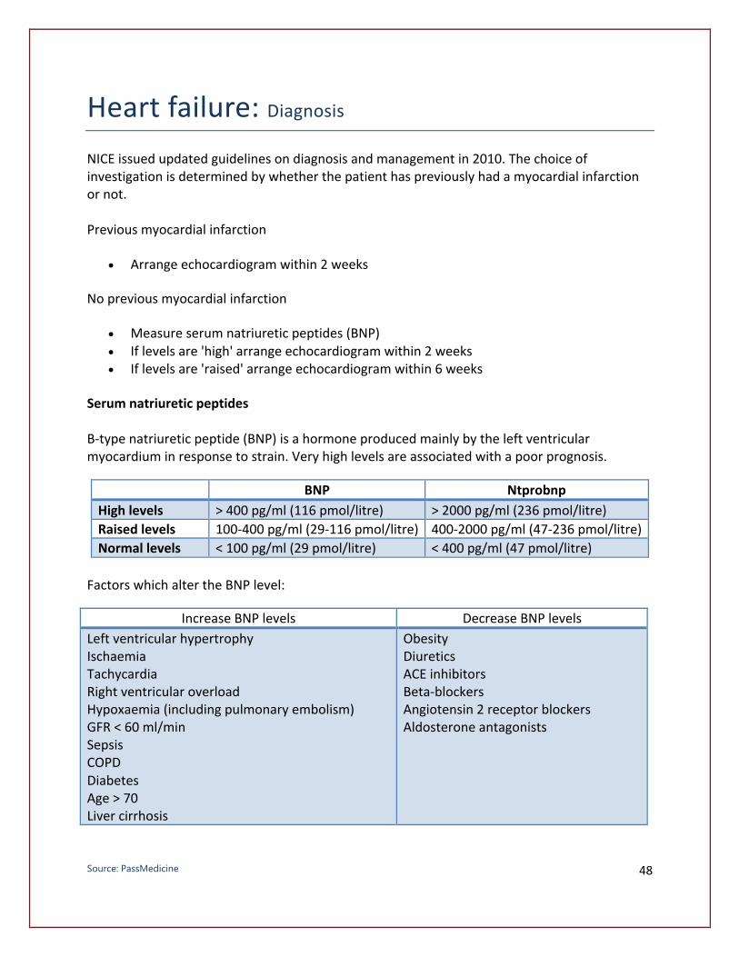
Heart failure: Diagnosis NICE issued updated guidelines on diagnosis and management in 2010. The choice of investigation is determined by whether the patient has previously had a myocardial infarction or not. Previous myocardial infarction
• Arrange echocardiogram within 2 weeks
No previous myocardial infarction
• Measure serum natriuretic peptides (BNP) • If levels are 'high' arrange echocardiogram within 2 weeks • If levels are 'raised' arrange echocardiogram within 6 weeks
Serum natriuretic peptides B-type natriuretic peptide (BNP) is a hormone produced mainly by the left ventricular myocardium in response to strain. Very high levels are associated with a poor prognosis.
BNP Ntprobnp
High levels > 400 pg/ml (116 pmol/litre) > 2000 pg/ml (236 pmol/litre) Raised levels 100-400 pg/ml (29-116 pmol/litre) 400-2000 pg/ml (47-236 pmol/litre) Normal levels < 100 pg/ml (29 pmol/litre) < 400 pg/ml (47 pmol/litre)
Factors which alter the BNP level:
Increase BNP levels Decrease BNP levels Left ventricular hypertrophy Ischaemia Tachycardia Right ventricular overload Hypoxaemia (including pulmonary embolism) GFR < 60 ml/min Sepsis COPD Diabetes Age > 70 Liver cirrhosis
Obesity Diuretics ACE inhibitors Beta-blockers Angiotensin 2 receptor blockers Aldosterone antagonists

Source: PassMedicine
49
Heart failure: NYHA classification
The New York Heart Association (NYHA) classification is widely used to classify the severity of heart failure: NYHA Class I
• No symptoms • No limitation: ordinary physical exercise does not cause undue fatigue, dyspnoea or
palpitations
NYHA Class II
• Mild symptoms • Slight limitation of physical activity: comfortable at rest but ordinary activity results in
fatigue, palpitations or dyspnoea
NYHA Class III
• Moderate symptoms • Marked limitation of physical activity: comfortable at rest but less than ordinary activity
results in symptoms
NYHA Class IV
• Severe symptoms • Unable to carry out any physical activity without discomfort: symptoms of heart failure
are present even at rest with increased discomfort with any physical activity

Source: PassMedicine
50
Heart failure: drug management
A number of drugs have been shown to improve mortality in patients with chronic heart failure:
• ACE inhibitors (SAVE, SOLVD, CONSENSUS) • Spironolactone (RALES) • Beta-blockers (CIBIS) • Hydralazine with nitrates (VHEFT-1)
No long-term reduction in mortality has been demonstrated for loop diuretics such as furosemide. NICE issued updated guidelines on management in 2010, key points include:
• First-line treatment for all patients is both an ACE-inhibitor and a beta-blocker • Second-line treatment is now either an aldosterone antagonist, angiotensin II receptor
blocker or a hydralazine in combination with a nitrate • If symptoms persist cardiac resynchronisation therapy or digoxin* should be
considered • Diuretics should be given for fluid overload • Offer annual influenza vaccine • Offer one-off** pneumococcal vaccine
*** Pulmonary oedema with bilateral course crackles and cough productive of white sputum. The patient has signs of right sided heart failure with raised JVP and peripheral oedema. They also have a history of MI and hypertension that are two risk factors for heart failure. NICE guidance on Acute Heart Failure 2014 states that a patient who has failed medical management of pulmonary oedema with severe dyspnoea should be considered for CPAP. BIPAP is not used in acute pulmonary oedema.
Source: PassMedicine
51
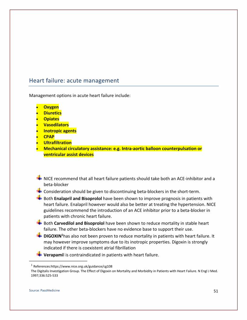
Heart failure: acute management
Management options in acute heart failure include:
• Oxygen • Diuretics • Opiates • Vasodilators • Inotropic agents • CPAP • Ultrafiltration • Mechanical circulatory assistance: e.g. Intra-aortic balloon counterpulsation or
ventricular assist devices
NICE recommend that all heart failure patients should take both an ACE-inhibitor and a beta-blocker
Consideration should be given to discontinuing beta-blockers in the short-term. Both Enalapril and Bisoprolol have been shown to improve prognosis in patients with heart failure. Enalapril however would also be better at treating the hypertension. NICE guidelines recommend the introduction of an ACE inhibitor prior to a beta-blocker in patients with chronic heart failure.
Both Carvedilol and Bisoprolol have been shown to reduce mortality in stable heart failure. The other beta-blockers have no evidence base to support their use.
DIGOXIN1has also not been proven to reduce mortality in patients with heart failure. It may however improve symptoms due to its inotropic properties. Digoxin is strongly indicated if there is coexistent atrial fibrillation
Verapamil is contraindicated in patients with heart failure.
1 References:https://www.nice.org.uk/guidance/cg108 The Digitalis Investigation Group. The Effect of Digoxin on Mortality and Morbidity in Patients with Heart Failure. N Engl J Med. 1997;336:525-533

Source: PassMedicine
52
The following drugs have all been shown to reduce mortality in patients with left ventricular failure:
• ACE-inhibitors • Beta-blockers • Angiotensin receptor blockers • Aldosterone antagonists • Hydralazine and nitrates •
Heart failure: non-drug management
Cardiac resynchronisation therapy
• For patients with heart failure and wide QRS • Biventricular pacing • Improved symptoms and reduced hospitalisation in NYHA class III patients
Exercise training
• Improves symptoms but not hospitalisation/mortality
***In symptomatic heart failure patients who are on optimal medical therapy, there are several treatment options to improve mortality. Cardiac resynchronisation therapy is indicated in patients with left ventricular dysfunction, ejection fracture <35% and QRS duration >120ms. An Implantable cardiac defibrillator (ICD) is indicated in patients with previous sustained ventricular tachycardia, ejection fraction <35% and symptoms no worse than class III of of the New York Heart Association functional classification. Digoxin reduces hospitalisation but not mortality in heart failure. Increasing the furosemide dose may help with symptomatic relief from fluid overload but has no effect on mortality. Heart transplantation and ventricular assist devices would not be an option in a patient of this age. Http://www.nice.org.uk/guidance/ta314/resources/guidance-implantable-cardioverter-defibrillators-and-cardiac-resynchronisation-therapy-for-arrhythmias-and-heart-failure-review-of-ta95-and-ta120-pdf

Source: PassMedicine
53
http://www.nice.org.uk/guidance/ta314/resources/guidance-implantable-cardioverter-defibrillators-and-cardiac-resynchronisation-therapy-for-arrhythmias-and-heart-failure-review-of-ta95-and-ta120-pdf
Valvular Heart Disease
Aortic stenosis
Most common cause:
• Younger patients < 65 years: bicuspid aortic valve • Older patients > 65 years: calcification
Features of severe aortic stenosis
• Narrow pulse pressure • Slow rising pulse • Delayed ESM • Soft/absent S2 • S4 • Thrill • Duration of murmur • Left ventricular hypertrophy or failure
Causes of aortic stenosis
• Degenerative calcification (most common cause in older patients > 65 years)
• Bicuspid aortic valve (most common cause in younger patients < 65 years) • William's syndrome (supravalvular aortic stenosis) • Post-rheumatic disease • Subvalvular: HOCM

Source: PassMedicine
54
Management
• If asymptomatic then observe the patient is general rule • If symptomatic then valve replacement • If asymptomatic but valvular gradient > 50 mmhg and with features such as left
ventricular systolic dysfunction then consider surgery • Balloon valvuloplasty is limited to patients with critical aortic stenosis who are not fit for
valve replacement
Aortic stenosis management: AVR if symptomatic, otherwise cut-off is gradient of 50 mmhg.
No action should be taken at present as if patient is currently asymptomatic. If the aortic valve gradient > 50 mmhg or there is evidence of significant left ventricular dysfunction then surgery is sometimes considered in selected asymptomatic patients
Left ventricular systolic dysfunction will result in a decreased flow-rate across the aortic valve and hence a quieter murmur.
Dilated cardiomyopathy is associated with the development of mitral regurgitation, not aortic regurgitation
In general, aortic valve replacement is indicated in symptomatic patients with severe aortic stenosis. The presence of symptoms is associated with a mortality of 2-3 years. The triad of symptoms is dyspnoea, chest pain and syncope. Valve replacement in asymptomatic patients is more controversial.
Aortic regurgitation
Features
• Early diastolic murmur • Collapsing pulse • Wide pulse pressure • Mid-diastolic Austin-Flint murmur in severe AR - due to partial closure of the anterior
mitral valve cusps caused by the regurgitation streams
Causes (due to valve disease)
• Rheumatic fever • Infective endocarditis • Connective tissue diseases e.g. RA/SLE • Bicuspid aortic valve

Source: PassMedicine
55
Causes (due to aortic root disease)
• Aortic dissection • Spondylarthropathies (e.g. Ankylosing spondylitis) • Hypertension • Syphilis • Marfan's, Ehler-Danlos syndrome
Whilst some patients with acromegaly have mitral valve prolapse (MVP) it is not a common association. It should be remembered that the prevalence of MVP in a standard population is around 5-10%
Mitral valve prolapse
Mitral valve prolapse is common, occurring in around 5-10 % of the population. It is usually idiopathic but may be associated with a wide variety of cardiovascular disease and other conditions Associations
• Congenital heart disease: PDA, ASD • Cardiomyopathy • Turner's syndrome • Marfan's syndrome, Fragile X • Osteogenesis imperfecta • Pseudoxanthoma elasticum • Wolff-Parkinson White syndrome • Long-QT syndrome • Ehlers-Danlos Syndrome • Polycystic kidney disease
Features
• Patients may complain of atypical chest pain or palpitations • Mid-systolic click (occurs later if patient squatting) • Late systolic murmur (longer if patient standing) • Complications: mitral regurgitation, arrhythmias (including long QT), emboli, sudden
death

Source: PassMedicine
56
In this age group hypertrophic obstructive cardiomyopathy would be a more common cause of the murmur/recurrent collapse than aortic stenosis.
HOCM: features
Hypertrophic obstructive cardiomyopathy (HOCM) is an autosomal dominant disorder of muscle tissue caused by defects in the genes encoding contractile proteins. The most common defects involve a mutation in the gene encoding β-myosin heavy chain protein or myosin binding protein C. The estimated prevalence is 1 in 500. Features
• Often asymptomatic • Dyspnoea, angina, syncope • Sudden death (most commonly due to ventricular arrhythmias), arrhythmias, heart
failure • Jerky pulse, large 'a' waves, double apex beat • Ejection systolic murmur: increases with Valsalva manoeuvre and decreases on
squatting
Associations
• Friedreich's ataxia • Wolff-Parkinson White
Echo - mnemonic - MR SAM ASH
• Mitral regurgitation (MR) • Systolic anterior motion (SAM) of the anterior mitral valve leaflet • Asymmetric hypertrophy (ASH)

Source: PassMedicine
57
ECG
• Left ventricular hypertrophy • Progressive T wave inversion • Deep Q waves • Atrial fibrillation may occasionally be seen
ECG showing typical changes of HOCM including LVH and T wave inversion
Dilated cardiomyopathy
Dilated cardiomyopathy (DCM) basics
• Dilated heart leading to systolic (+/- diastolic) dysfunction • All 4 chambers affected but LV more so than RV • Features include arrhythmias, emboli, mitral regurgitation • Absence of congenital, valvular or ischaemic heart disease
Causes often considered separate entities
• Alcohol: may improve with thiamine • Postpartum

Source: PassMedicine
58
• Hypertension
Other causes
• Inherited (see below) • Infections e.g. Coxsackie B, HIV, diphtheria, parasitic • Endocrine e.g. Hyperthyroidism • Infiltrative* e.g. Haemochromatosis, sarcoidosis • Neuromuscular e.g. Duchenne muscular dystrophy • Nutritional e.g. Kwashiorkor, pellagra, thiamine/selenium deficiency • Drugs e.g. Doxorubicin
Inherited dilated cardiomyopathy
• Around a third of patients with DCM are thought to have a genetic predisposition • A large number of heterogeneous defects have been identified • The majority of defects are inherited in an autosomal dominant fashion although other
patterns of inheritance are seen
*these causes may also lead to restrictive cardiomyopathy
*** Haemochromatosis is more commonly associated with restrictive cardiomyopathy but a dilated pattern may also be seen. There is a known association between Wilson's disease and cardiomyopathy but this is extremely rare and not often clinically significant
Wolff-Parkinson White
Wolff-Parkinson White (WPW) syndrome is caused by a congenital accessory conducting pathway between the atria and ventricles leading to a atrioventricular re-entry tachycardia (AVRT). As the accessory pathway does not slow conduction AF can degenerate rapidly to VF Possible ECG features include:
• Short PR interval • Wide QRS complexes with a slurred upstroke - 'delta wave' • Left axis deviation if right-sided accessory pathway* • Right axis deviation if left-sided accessory pathway*

Source: PassMedicine
59
ECG showing short PR interval associated with a slurred upstroke (delta wave). Note the non-specific ST-T changes which are common in WPW and may be mistaken for ischaemia. The left axis deviation means that this is type B WPW, implying a right-sided pathway
Further example showing a characteristic delta wave Differentiating between type A and type B
• Type A (left-sided pathway): dominant R wave in V1
• Type B (right-sided pathway): no dominant R wave in V1

Source: PassMedicine
60
Associations of WPW
• HOCM • Mitral valve prolapse • Ebstein's anomaly • Thyrotoxicosis • Secundum ASD
Management
• Definitive treatment: radiofrequency ablation of the accessory pathway • Medical therapy: sotalol**, amiodarone, flecainide
*in the majority of cases, or in a question without qualification, Wolff-Parkinson-White syndrome is associated with left axis deviation **sotalol should be avoided if there is coexistent atrial fibrillation as prolonging the refractory period at the AV node may increase the rate of transmission through the accessory pathway, increasing the ventricular rate and potentially deteriorating into ventricular fibrillation

Source: PassMedicine
61
Pulmonary embolism: investigation
We know from experience that few patients (around 10%) present with the medical student textbook triad of pleuritic chest pain, dyspnoea and haemoptysis. Pulmonary embolism can be difficult to diagnose as it can present with virtually any cardiorespiratory symptom/sign depending on it's location and size. So which features make pulmonary embolism more likely? The PIOPED study1 in 2007 looked at the frequency of different symptoms and signs in patients who were diagnosed with pulmonary embolism. The relative frequency of common clinical signs is shown below:
• Tachypnea (respiratory rate >16/min) - 96% • Crackles - 58% • Tachycardia (heart rate >100/min) - 44% • Fever (temperature >37.8°C) - 43%
It is interesting to note that the Well's criteria for diagnosing a PE use tachycardia rather than tachypnoea. 2012 NICE guidelines All patients with symptoms or signs suggestive of a PE should have a history taken, examination performed and a chest x-ray to exclude other pathology. If a PE is still suspected a two-level PE Wells score should be performed:
Clinical probability simplified scores
§ PE likely - more than 4 points § PE unlikely - 4 points or less
Clinical feature Points Clinical signs and symptoms of DVT (minimum of leg swelling and pain with palpation of the deep veins)
3
An alternative diagnosis is less likely than PE 3 Heart rate > 100 beats per minute 1.5 Immobilisation for more than 3 days or surgery in the previous 4 weeks 1.5 Previous DVT/PE 1.5 Haemoptysis 1 Malignancy (on treatment, treated in the last 6 months, or palliative) 1

Source: PassMedicine
62
If a PE is 'likely' (more than 4 points) arrange an immediate computed tomography pulmonary angiogram (CTPA). If there is a delay in getting the CTPA then give low-molecular weight heparin until the scan is performed. If a PE is 'unlikely' (4 points or less) arranged a D-dimer test. If this is positive arrange an immediate computed tomography pulmonary angiogram (CTPA). If there is a delay in getting the CTPA then give low-molecular weight heparin until the scan is performed. If the patient has an allergy to contrast media or renal impairment a V/Q scan should be used instead of a CTPA. CTPA or V/Q scan? The consensus view from the British Thoracic Society and NICE guidelines is as follows:
• Computed tomographic pulmonary angiography (CTPA) is now the recommended initial lung-imaging modality for non-massive PE. Advantages compared to V/Q scans include speed, easier to perform out-of-hours, a reduced need for further imaging and the possibility of providing an alternative diagnosis if PE is excluded
• If the CTPA is negative then patients do not need further investigations or treatment for PE
• Ventilation-perfusion scanning may be used initially if appropriate facilities exist, the chest x-ray is normal, and there is no significant symptomatic concurrent cardiopulmonary disease.
Labelled CTPA showing a large saddle embolus
Further CTPA again showing a saddle embolus

Source: PassMedicine
63
ECG from a patient with a PE. Shows a sinus tachycardia and a partial S1Q3T3 - the S wave is not particularly convincing.
ECG of a patient with a PE. It shows some of the ECG features that may be associated with PE (sinus tachycardia, S1, T3 and T wave inversion in the precordial leads). Other features such as the left axis deviation are atypical.

Source: PassMedicine
64
Some other points D-dimers
• Sensitivity = 95-98%, but poor specificity
ECG
• The classic ECG changes seen in PE are a large S wave in lead I, a large Q wave in lead III and an inverted T wave in lead III - 'S1Q3T3'. However this change is seen in no more than 20% of patients
• Right bundle branch block and right axis deviation are also associated with PE • Sinus tachycardia may also be seen
V/Q scan
• Sensitivity = 98%; specificity = 40% - high negative predictive value, i.e. If normal virtually excludes PE
• Other causes of mismatch in V/Q include old pulmonary embolisms, AV malformations, vasculitis, previous radiotherapy
• COPD gives matched defects
CTPA
• Peripheral emboli affecting subsegmental arteries may be missed
Pulmonary angiography
• The gold standard • Significant complication rate compared to other investigations
1. Clinical Characteristics of Patients with Acute Pulmonary Embolism(Data from PIOPED II) Am J Med. Oct 2007; 120(10): 871879.

Source: PassMedicine
65
PulmonaryEmbolism:management
The NICE guidelines of 2012 provided some clarity on how long patients should be anticoagulated for after a pulmonary embolism (PE). Selected points are listed below. Low molecular weight heparin (LMWH) or fondaparinux should be given initially after a PE is diagnosed. An exception to this is for patients with a massive PE where thrombolysis is being considered. In such a situation unfractionated heparin should be used.
• A vitamin K antagonist (i.e. Warfarin) should be given within 24 hours of the diagnosis • The LMWH or fondaparinux should be continued for at least 5 days or until the
international normalised ratio (INR) is 2.0 or above for at least 24 hours, whichever is longer, i.e. LMWH or fondaparinux is given at the same time as warfarin until the INR is in the therapeutic range
• Warfarin should be continued for at least 3 months. At 3 months, NICE advise that clinicians should 'assess the risks and benefits of extending treatment'
• NICE advise extending warfarin beyond 3 months for patients with unprovoked PE. This essentially means that if there was no obvious cause or provoking factor (surgery, trauma, significant immobility) it may imply the patient has a tendency to thrombosis and should be given treatment longer than the norm of 3 months
• For patients with active cancer NICE recommend using LMWH for 6 months
Thrombolysis
• Thrombolysis is now recommended as the first-line treatment for massive PE where there is circulatory failure (e.g. Hypotension). Other invasive approaches should be considered where appropriate facilities exist

Source: PassMedicine
66
Deep vein thrombosis: diagnosis and management
Diagnosis NICE published guidelines in 2012 relating to the investigation and management of deep vein thrombosis (DVT). If a patient is suspected of having a DVT a two-level DVT Wells score should be performed: Two-level DVT Wells score
Clinical feature Points Active cancer (treatment ongoing, within 6 months, or palliative) 1 Paralysis, paresis or recent plaster immobilisation of the lower extremities 1 Recently bedridden for 3 days or more or major surgery within 12 weeks requiring general or regional anaesthesia
1
Localised tenderness along the distribution of the deep venous system 1 Entire leg swollen 1 Calf swelling at least 3 cm larger than asymptomatic side 1 Pitting oedema confined to the symptomatic leg 1 Collateral superficial veins (non-varicose) 1 Previously documented DVT 1 An alternative diagnosis is at least as likely as DVT -2 Clinical probability simplified score
§ DVT likely: 2 points or more § DVT unlikely: 1 point or less
If a DVT is 'likely' (2 points or more)
§ A proximal leg vein ultrasound scan should be carried out within 4 hours and, if the result is negative, a D-dimer test
§ If a proximal leg vein ultrasound scan cannot be carried out within 4 hours a D-dimer test should be performed and low-molecular weight heparin administered whilst waiting for the proximal leg vein ultrasound scan (which should be performed within 24 hours)
If a DVT is 'unlikely' (1 point or less)
§ Perform a D-dimer test and if it is positive arrange: § A proximal leg vein ultrasound scan within 4 hours
If a proximal leg vein ultrasound scan cannot be carried out within 4 hours low-molecular weight heparin should be administered whilst waiting for the proximal leg vein ultrasound scan (which should be performed within 24 hours)

Source: PassMedicine
67
Management Low molecular weight heparin (LMWH) or fondaparinux should be given initially after a DVT is diagnosed.
• A vitamin K antagonist (i.e. Warfarin) should be given within 24 hours of the diagnosis • The LMWH or fondaparinux should be continued for at least 5 days or until the
international normalised ratio (INR) is 2.0 or above for at least 24 hours, whichever is longer, i.e. LMWH or fondaparinux is given at the same time as warfarin until the INR is in the therapeutic range
• Warfarin should be continued for at least 3 months. At 3 months, NICE advise that clinicians should 'assess the risks and benefits of extending treatment'
• NICE add 'consider extending warfarin beyond 3 months for patients with unprovoked proximal DVT if their risk of VTE recurrence is high and there is no additional risk of major bleeding'. This essentially means that if there was no obvious cause or provoking factor (surgery, trauma, significant immobility) it may imply the patient has a tendency to thrombosis and should be given treatment longer than the norm of 3 months. In practice most clinicians give 6 months of warfarin for patients with an unprovoked DVT/PE
• For patients with active cancer NICE recommend using LMWH for 6 months
Further investigations and thrombophilia screening As both malignancy and thrombophilia are obvious risk factors for deep vein thrombosis NICE make recommendations on how to investigate patients with unprovoked clots. Offer all patients diagnosed with unprovoked DVT or PE who are not already known to have cancer the following investigations for cancer:
• A physical examination (guided by the patient's full history) and • A chest X-ray and • Blood tests (full blood count, serum calcium and liver function tests) and urinalysis.
Consider further investigations for cancer with an abdomino-pelvic CT scan (and a mammogram for women) in all patients aged over 40 years with a first unprovoked DVT or PE Thrombophilia screening
• Not offered if patients will be on lifelong warfarin (i.e. Won't alter management) • Consider testing for antiphospholipid antibodies if unprovoked DVT or PE • Consider testing for hereditary thrombophilia in patients who have had unprovoked DVT
or PE and who have a first-degree relative who has had DVT or PE

Source: PassMedicine
68
Angiotensin-converting enzyme inhibitors
Angiotensin-converting enzyme (ACE) inhibitors are now the established first-line treatment in younger patients with hypertension and are also extensively used to treat heart failure. They are known to be less effective in treating hypertensive Afro-Caribbean patients. ACE inhibitors are also used to treat diabetic nephropathy and have a role in secondary prevention of ischaemic heart disease. Mechanism of action:
• Inhibit the conversion angiotensin I to angiotensin II
Side-effects:
• Cough: occurs in around 15% of patients and may occur up to a year after starting treatment. Thought to be due to increased bradykinin levels
• Angioedema: may occur up to a year after starting treatment • Hyperkalaemia • First-dose hypotension: more common in patients taking diuretics
Cautions and contraindications
• Pregnancy and breastfeeding - avoid • Renovascular disease - significant renal impairment may occur in patients who have
undiagnosed bilateral renal artery stenosis • Aortic stenosis - may result in hypotension • Patients receiving high-dose diuretic therapy (more than 80 mg of furosemide a day) -
significantly increases the risk of hypotension • Hereditary of idiopathic angioedema
Monitoring
• Urea and electrolytes should be checked before treatment is initiated and after increasing the dose
• A rise in the creatinine and potassium may be expected after starting ACE inhibitors. Acceptable changes are an increase in serum creatinine, up to 30%* from baseline and an increase in potassium up to 5.5 mmol/l*.
*Renal Association UK, Clinical Knowledge Summaries quote 50% which seems rather high.

Source: PassMedicine
69
SIGN advise that the fall in egfr should be less than 20%. The NICE CKD guidelines suggest that a decrease in egfr of up to 25% or a rise in creatinine of up to 30% is acceptable
** Patients with bilateral renovascular disease (e.g. Renal artery stenosis) may develop an acute kidney injury (AKI) if ACE inhibitors are administered. This is because elevated angiotensin II levels constrict the efferent arteriole more than the afferent arteriole within the kidney, which normally helps to maintain glomerular capillary pressure and filtration. Removing this constriction by blocking angiotensin II formation can cause an abrupt fall in glomerular filtration rate.
NICE advise the following: 'Offer people aged under 55 years an ACE inhibitor or a low-cost ARB. If an ACE inhibitor is prescribed and is not tolerated (for example, because of cough), offer a low-cost ARB.' Up to 15% of patients who are started on an ACE inhibitor develop a dry cough. Her symptoms are interfering with sleep and have persisted now for four weeks. It is therefore appropriate to switch her to an angiotensin II receptor blocker.
Angiotensin II receptor blockers
Angiotensin II receptor blockers are generally used in situations where patients have not tolerated an ACE inhibitor, usually due to the development of a cough. Examples
• Candesartan • Losartan • Irbesartan
Like ACE inhibitors they should be used with caution in patients with renovascular disease. Side-effects include hypotension and hyperkalaemia. Mechanism
• Block effects of angiotensin II at the AT1 receptor
Evidence base
• Shown to reduce progression of renal disease in patients with diabetic nephropathy

Source: PassMedicine
70
• Evidence base that losartan reduces CVA and IHD mortality in hypertensive patients
Patients with aortic stenosis are at risk of profound hypotension with ACE inhibitors. Bendroflumethiazide is a weak diuretic that is commonly co-presecribed with ACE inhibitors. The risk of hypotension relates to high-dose loop diuretics, for example furosemide 80mg bd. Patients with chronic kidney disease stage 2 have, by definition, a glomerular filtration rate of > 60 ml/min/1.73 m². It is unlikely that this will have a significant effect on the risk of side-effects. Tricyclic overdose
Overdose of tricyclic antidepressants is a common presentation to emergency departments. Amitriptyline and dosulepin (dothiepin) are particularly dangerous in overdose. Early features relate to anticholinergic properties: dry mouth, dilated pupils, agitation, sinus tachycardia, blurred vision. Features of severe poisoning include:
• Arrhythmias • Seizures • Metabolic acidosis • Coma
ECG changes include:
• Sinus tachycardia • Widening of QRS • Prolongation of QT interval
Widening of QRS > 100ms is associated with an increased risk of seizures whilst QRS > 160ms is associated with ventricular arrhythmias Management
• IV bicarbonate may reduce the risk of seizures and arrhythmias in severe toxicity • Arrhythmias: class 1a (e.g. Quinidine) and class Ic antiarrhythmics (e.g. Flecainide) are
contraindicated as they prolong depolarisation. Class III drugs such as amiodarone
Source: PassMedicine
71
should also be avoided as they prolong the QT interval. Response to lignocaine is variable and it should be emphasized that correction of acidosis is the first line in management of tricyclic induced arrhythmias
• Intravenous lipid emulsion is increasingly used to bind free drug and reduce toxicity • Dialysis is ineffective in removing tricyclics
Digoxin and digoxin toxicity
Digoxin is a cardiac glycoside now mainly used for rate control in the management of atrial fibrillation. As it has positive inotropic properties it is sometimes used for improving symptoms (but not mortality) in patients with heart failure. Mechanism of action
• Decreases conduction through the atrioventricular node which slows the ventricular rate in atrial fibrillation and flutter
• Increases the force of cardiac muscle contraction due to inhibition of the Na+/K+ atpase pump. Also stimulates vagus nerve
Digoxin toxicity Plasma concentration alone does not determine whether a patient has developed digoxin toxicity. The BNF advises that the likelihood of toxicity increases progressively from 1.5 to 3 mcg/l. Features
• Generally unwell, lethargy, nausea & vomiting, anorexia, confusion, yellow-green vision • Arrhythmias (e.g. AV block, bradycardia)
Precipitating factors
• Classically: hypokalaemia* • Increasing age • Renal failure • Myocardial ischaemia • Hypomagnesaemia, hypercalcaemia, hypernatraemia, acidosis • Hypoalbuminaemia • Hypothermia • Hypothyroidism • Drugs: amiodarone, quinidine, verapamil, diltiazem, spironolactone (competes for
secretion in distal convoluted tubule therefore reduce excretion), ciclosporin. Also drugs which cause hypokalaemia e.g. Thiazides and loop diuretics
Management

Source: PassMedicine
72
• Digibind • Correct arrhythmias • Monitor potassium
*hyperkalaemia may also worsen digoxin toxicity, although this is very small print
Amiodarone
Amiodarone is a class III antiarrhythmic agent used in the treatment of atrial, nodal and ventricular tachycardias. The main mechanism of action is by blocking potassium channels which inhibits repolarisation and hence prolongs the action potential. Amiodarone also has other actions such as blocking sodium channels (a class I effect) The use of amiodarone is limited by a number of factors
• Long half-life (20-100 days) • Should ideally be given into central veins (causes thrombophlebitis) • Has proarrhythmic effects due to lengthening of the QT interval • Interacts with drugs commonly used concurrently e.g. Decreases metabolism of warfarin • Numerous long-term adverse effects (see below)
Monitoring of patients taking amiodarone
• TFT, LFT, U&E, CXR prior to treatment • TFT, LFT every 6 months
Adverse effects of amiodarone use
• Thyroid dysfunction • Corneal deposits • Pulmonary fibrosis/pneumonitis • Liver fibrosis/hepatitis • Peripheral neuropathy, myopathy • Photosensitivity • 'Slate-grey' appearance • Thrombophlebitis and injection site reactions • Bradycardia
Calcium channel blockers

Source: PassMedicine
73
Calcium channel blockers are primarily used in the management of cardiovascular disease. Voltage-gated calcium channels are present in myocardial cells, cells of the conduction system and those of the vascular smooth muscle. The various types of calcium channel blockers have varying effects on these three areas and it is therefore important to differentiate their uses and actions.
Examples Indications & notes Side-effects and cautions
Verapamil Angina, hypertension, arrhythmias Highly negatively inotropic Should not be given with beta-blockers as may cause heart block
Heart failure, constipation, hypotension, bradycardia, flushing
Diltiazem Angina, hypertension Less negatively inotropic than verapamil but caution should still be exercised when patients have heart failure or are taking beta-blockers
Hypotension, bradycardia, heart failure, ankle swelling
Nifedipine, amlodipine, felodipine (dihydropyridines)
Hypertension, angina, Raynaud's Affects the peripheral vascular smooth muscle more than the myocardium and therefore do not result in worsening of heart failure
Flushing, headache, ankle swelling
*** The BNF warns that short-acting formulations of nifedipine are associated with large variations in blood pressure and may cause reflex tachycardia
Beta-blockers
Beta-blockers are an important class of drug used mainly in the management of cardiovascular disorders. Indications
• Angina • Post-myocardial infarction

Source: PassMedicine
74
• Heart failure: beta-blockers were previously avoided in heart failure but there is now strong evidence that certain beta-blockers improve both symptoms and mortality
• Arrhythmias: beta-blockers have now replaced digoxin as the rate-control drug of choice in atrial fibrillation
• Hypertension: the role of beta-blockers has diminished in recent years due to a lack of evidence in terms of reducing stroke and myocardial infarction.
• Thyrotoxicosis • Migraine prophylaxis • Anxiety
Examples
• Atenolol • Propranolol: one of the first beta-blockers to be developed. Lipid soluble therefore
crosses the blood-brain barrier
Side-effects
• Bronchospasm • Cold peripheries • Fatigue • Sleep disturbances, including nightmares
Contraindications
• Uncontrolled heart failure • Asthma • Sick sinus syndrome • Concurrent verapamil use: may precipitate severe bradycardia
*** Beta-blockers combined with verapamil can potentially cause profound bradycardia and asystole.
Beta-blocker overdose
Features • Bradycardia • Hypotension
Heart failure • Syncope • Management
If bradycardic then atropine

Source: PassMedicine
75
In resistant cases glucagon may be used Ø Haemodialysis is not effective in beta-blocker overdose Ø Gastric lavage should only be attempted (if at all) if patients present within 1-2 hours of
taking the overdose.
Prescribing in pregnant patients
Very few drugs are known to be completely safe in pregnancy. The list below largely comprises of those known to be harmful. Some countries have developed a grading system - see the link. Antibiotics
• Tetracyclines • Aminoglycosides • Sulphonamides and trimethoprim • Quinolones: the BNF advises to avoid due to arthropathy in some animal studies
Other drugs
• ACE inhibitors, angiotensin II receptor antagonists • Statins • Warfarin • Sulfonylureas • Retinoids (including topical) • Cytotoxic agents
The majority of antiepileptics including valproate, carbamazepine and phenytoin are known to be potentially harmful. The decision to stop such treatments however is difficult as uncontrolled epilepsy is also a risk
*** NICE are very clear on this point:'Offer antihypertensive drug treatment to women of child-bearing potential in line with the recommendations on Management of pregnancy with chronic hypertension'

Source: PassMedicine
76
Natriuretic peptide
There are two types of natriuretic peptide that can be measured in serum: B-type natriuretic peptide (BNP) and N-terminal pro-BNP (NT-probnp). NT-probnp is the inactive prohormone of BNP and is released from the left ventricle in response to ventricular strain. It acts to increase renal excretion of water and sodium, and relax vascular smooth muscle causing vasodilation. BNP should be measured in patients with suspected heart failure who have not had a previous myocardial infarction. The serum level helps to determine the likelihood of heart failure and thus the need for specialist assessment and echocardiography. The value helps dictate the level of urgency of the referral given high BNF levels (>400) carry poor prognosis. In these cases 2 week wait for urgent echocardiography and specialist assessment is advised. Although elevated levels of BNP do not confirm the diagnosis of heart failure, normal levels rule the diagnosis out (highly sensitivity but varying specificity). NICE suggest that BNP measurements are not necessary in people with suspected heart failure who have had a previous myocardial infarction. These patients require urgent referral, echocardiography and specialist assessment because if heart failure is present this carries a poor prognosis. Increased BNP levels (>400) are not on their own diagnostic of heart failure, and may be elevated as a result of left ventricular hypertrophy, myocardial ischaemia, atrial fibrillation, pulmonary hypertension, hypoxia, pulmonary embolism, right ventricular strain, chronic obstructive pulmonary disease, liver failure, sepsis, diabetes, and renal impairment. In addition, levels tend to be higher in women, and in people older than 70. Heart failure is unlikely if BNP levels are low (<100). However, aldosterone antagonists, ACE inhibitors, angiotensin-II receptor antagonists, beta-blockers and diuretics can all falsely lower BNP levels, as can obesity.
B-type natriuretic peptide
B-type natriuretic peptide (BNP) is a hormone produced mainly by the left ventricular myocardium in response to strain.

Source: PassMedicine
77
Whilst heart failure is the most obvious cause of raised BNP levels any cause of left ventricular dysfunction such as myocardial ischemia or valvular disease may raise levels. Raised levels may also be seen due to reduced excretion in patients with chronic kidney disease. Factors which reduce BNP levels include treatment with ACE inhibitors, angiotensin-2 receptor blockers and diuretics. Effects of BNP
• Vasodilator • Diuretic and natriuretic • Suppresses both sympathetic tone and the renin-angiotensin-aldosterone system
Clinical uses of BNP Diagnosing patients with acute dyspnoea
• A low concentration of BNP(< 100pg/ml) makes a diagnosis of heart failure unlikely, but raised levels should prompt further investigation to confirm the diagnosis
• NICE currently recommends BNP as a helpful test to rule out a diagnosis of heart failure
Prognosis in patients with chronic heart failure
• Initial evidence suggests BNP is an extremely useful marker of prognosis
Guiding treatment in patients with chronic heart failure
• Effective treatment lowers BNP levels
Screening for cardiac dysfunction
• Not currently recommended for population screening

Source: PassMedicine
78
Cardiac imaging: non-invasive techniques excluding echocardiography
The ability to image the heart using non-invasive techniques such as MRI, CT and radionuclides has evolved rapidly over recent years. Nuclear imaging These techniques use radiotracers which are extracted by normal myocardium. Examples include:
• Thallium • Technetium (99mtc) sestamibi: a coordination complex of the radioisotope technetium-
99m with the ligand methoxyisobutyl isonitrile (MIBI), used in 'MIBI' or cardiac Single Photon Emission Computed Tomography (SPECT) scans
• Fluorodeoxyglucose (FDG): used in Positron Emission Tomography (PET) scans
The primary role of SPECT is to assess myocardial perfusion and myocardial viability. Two sets of images are usually acquired. First the myocardium at rest followed by images of the myocardium during stress (either exercise or following adenosine / dipyridamole). By comparing the rest with stress images any areas of ischaemia can classified as reversible or fixed (e.g. Following a myocardial infarction). Cardiac PET is predominately a research tool at the current time MUGA
• Multi Gated Acquisition Scan, also known as radionuclide angiography • Radionuclide (technetium-99m) is injected intravenously • The patient is placed under a gamma camera • May be performed as a stress test • Can accurately measure left ventricular ejection fraction. Typically used before and after
cardiotoxic drugs are used
Cardiac Computed Tomography (CT) Cardiac CT is useful for assessing suspected ischemic heart disease, using two main methods:

Source: PassMedicine
79
• Calcium score: there is known to be a correlation between the amount of atherosclerotic plaque calcium and the risk of future ischemic events. Cardiac CT scan quantify the amount of calcium producing a 'calcium score'
• Contrast enhanced CT: allows visualization of the coronary artery lumen
If these two techniques are combined cardiac CT has a very high negative predictive value for ischaemic heart disease. Cardiac MRI Cardiac MRI (commonly termed CMR) has become the gold standard for providing structural images of the heart. It is particularly useful when assessing congenital heart disease, determining right and left ventricular mass and differentiating forms of cardiomyopathy. Myocardial perfusion can also be assessed following the administration of gadolinium. Currently CMR provides limited data on the extent of coronary artery disease. Please also see the British Heart Foundation link for an excellent summary.
DVLA: cardiovascular disorders
The guidelines below relate to car/motorcycle use unless specifically stated. For obvious reasons, the rules relating to drivers of heavy goods vehicles tend to be much stricter Specific rules
• Hypertension - can drive unless treatment causes unacceptable side effects, no need to notify DVLA. If Group 2 Entitlement the disqualifies from driving if resting BP consistently 180 mmhg systolic or more and/or 100 mm Hg diastolic or more
• Angioplasty (elective) - 1 week off driving • CABG - 4 weeks off driving • Acute coronary syndrome- 4 weeks off driving, 1 week if successfully treated by
angioplasty • Angina - driving must cease if symptoms occur at rest/at the wheel • Pacemaker insertion - 1 week off driving • Implantable cardioverter-defibrillator (ICD): if implanted for sustained ventricular
arrhythmia: cease driving for 6 months. If implanted prophylactically then cease driving for 1 month. Having an ICD results in a permanent bar for Group 2 drivers
• Successful catheter ablation for an arrhythmia- 2 days off driving

Source: PassMedicine
80
• Aortic aneurysm of 6cm or more - notify DVLA. Licensing will be permitted subject to annual review. An aortic diameter of 6.5 cm or more disqualifies patients from driving
• Heart transplant: DVLA do not need to be notified
Heart failure is one of the adverse signs indicating the need for urgent synchronized DC cardioversion