cardiovascular risk assessment using carotid ultrasonography · 3. chambless le, folsom ar, clegg...
TRANSCRIPT

Cardiovascular risk assessment using carotid ultrasonography
The Rotterdam Study

Acknowledgments
The work presented in this thesis was conducted at the department of Epidemiology & Biostatistics of the Erasmus Medical Centre Rotterdam. Financial support came from the NESTOR stimulation program for geriatric research in the Netherlands, the Netherlands Organisation for Scientific Research (NWO), the Netherlands Health Research and Development Council (ZON) and the municipality of Rotterdam.
The author gratefully acknowledges the collaboration with the Julius Centre for General Practice and Patient Oriented Research, University Medical Centre Utrecht, Utrecht (D.E. Grobbee, M.L. Bots, KG.M. Moons) and the Department of Neurology, Erasmus Medical Centre Rotterdam (P.J. Koudstaal).
The printing of this thesis was supported by the department of Epidemiology & Biostatistics, Erasmus Medical Centre Rotterdam; the Julius Centre for General Practice and Patient Oriented Research, University Medical Centre Utrecht, Utrecht; ZON MW; ATL Nederland; Bristol-Myers Squibb; GlaxoSmithKline; Novartis Pharma BV; Servier Nederland BV and Yamanouchi Pharma BV.
Cover Design: A.M. Bijpost, Schagen Printed by: [Optima] Grafische Communicatie, Rotterdam
ISBN 90-77017-09-7
© A. Iglesias del Sol, 2001
No part ofthis book may be reproduced, stored in a retrieval system or transmitted in any form or by any means, without permission of the author, or, when appropriate, of the publishers of the publications.

Cardiovascular risk assessment using carotid ultrasonography
The Rotterdam Study
Cardiovasculaire risicoschatting met behulp van echografie van de halsslagaders
Ret ERGO onderzoek
Proefschrift
ter verkrijging van de graad van doctor aan de Erasmus Universiteit Rotterdam
op gezag van de Rector Magnificus
Prof.dr.ir. I.H. van Bemmel
en volgens besluit van het College voor Promoties.
De openbare verdediging zal plaatsvinden op
woensdag 26 september 2001 om 09.45 uur
door
Antonio Iglesias del Sol
geboren te Amsterdam

Promotiecommissie
Promotoren:
Overige leden:
Co-promotoren:
Prof. dr D.E. Grobbee Prof. dr A. Hofinan
Prof. dr M.G.M. Huillnk
Prof. dr P.J. Koudstaal
Prof. dr J.R.T.C. Roelandt
Prof. dr A.F .H. Stalenhoef Prof. dr C.D.A. Stehouwer
DrM.L. Bots
Dr J.C.M. Witteman
Financial support by the Netherlands Heart Foundation for the publication of this thesis is gratefully acknowledged.

Papers and manuscripts based on the studies described in this thesis
Chapter 2 Bots ML, Iglesias del Sol A, Grobbee DE. Carotid intima-media thickness measurements in observational and intervention studies. Current Res Vascular Dis 1998;3(6):275-83. (modified)
Chapter 3
Iglesias del Sol A, Bots ML, Grobbee DE, Hofman A, Witteman JCM. Carotid intimamedia thickness at different sites: relation to incident myocardial infarction. The Rotterdam Study. European Heart Journal (in press)
Iglesias del Sol A, Bots ML, Hofman A, Grobbee DE, Witteman JCM. Plaques in the carotid artery and risk of myocardial infarction. The Rotterdam Study. Submitted
Hollander M, Bots ML, Iglesias del Sol A, Koudstaal PJ, Grobbee DE, Hofman A, Breteler MME. Carotid plaques increase the risk of stroke and subtypes of cerebral infarction in asymptomatic elderly. The Rotterdam Study. Submitted
Chapter 4
Iglesias del Sol A, Moons KGM, Hollander M, Hofman A, Koudstaal PJ, Grobbee DE, Breteler MME, Witteman JCM, Bots ML. Is carotid intima-media thickness useful in cardiovascular disease risk assessment? The Rotterdam Study. Stroke 2001;32:1532-1538.
Iglesias del Sol A, Bots ML, van der Kuip DAM, Hofman A, Grobbee DE, Witteman JCM. Carotid intima-media thickness and ankle-brachial index: Powerful predictors of myocardial infarction. The Rotterdam Study. Submitted
ChapterS
Iglesias del Sol A, Bots ML, Hollander M, Hofman A, Grobbee DE, Witteman JCM. Progression of atherosclerosis in the carotid artery. The Rotterdam Study. Submitted


Contents
1. Introduction
2. Carotid intima-media thickness measurements in observational and
intervention studies
3. Carotid atherosclerosis and cardiovascular disease
3.1. Carotid intima-media thickness at different sites and risk of incident
myocardial infarction
3.2. Plaques in the carotid artery and risk of myocardial infarction
3.3. Carotid plaques and risk of stroke and subtypes of
cerebral infarction
4. Application in cardiovascular risk assessment
4.1. Is carotid intima-media thickness useful in cardiovascular risk
assessment?
4.2. Carotid intima-media thickness and ankle-brachial index:
Powerful predictors of myocardial infarction
5. Progression of atherosclerosis in the carotid artery
6. General discussion
7. Summary
8. Samenvatting
Dankwoord
About the author
7
29
41
53
67
83
95
109
123
129
135
137


Chapter 1
Introduction

Introduction
Atherosclerosis is the main cause of coronary heart disease, stroke and peripheral arterial disease. These cardiovascular diseases are the most important cause of morbidity and responsible for 50% of all mortality in the United States, Europe and much of Asia. l Since atherosclerosis and cardiovascular diseases are most prominently present in the elderly and the number of elderly people will increase in the coming decades, atherosclerosis-related diseases will put a heavy burden on our health care systems.
Awareness of risk factors associated with atherosclerosis and increased risk of cardiovascular diseases has led to an extensive number of studies on atherosclerosis. Several clinical trials have been initiated to show the effect of treatment of risk factors like hypertension and hyperlipidemia, on cardiovascular risk. Because cardiovascular risk factors so far carmot completely explain atherosclerosis, direct measures of subclinical atherosclerosis may be better tools in explaining and predicting cardiovascular disease.
From this perspective, interest has risen in the non-invasive assessment of atherosclerosis, especially for use in population-based studies to examine determinants and consequences of atherosclerosis. Carotid ultrasonography provides us with the ability to examine both intima-media thickness and carotid plaques as measures of atherosclerosis. Carotid intima-media thickness is now widely used as a measure of atherosclerosis in several population-based studies.'·5 Various cardiovascular risk factors have been related to intima-media thickness and it has been shown to be related to future cardiovascular disease, although studies are sparse. Furthermore, several clinical trials are using carotid intima-media thickness as a proxy endpoint to show benefit of treatment of risk factors. '·8
Data about risk factors for carotid plaques are scarce and studies that investigated the association between carotid plaques and cardiovascular disease focused on cerebrovascular disease only.'·lO
Measurement of carotid intima-media thickness and carotid plaques has provided us an important tool to examine etiology and risk of atherosclerosis and cardiovascular diseases. Therefore, recently questions have been raised about the use
of carotid ultrasonography in clinical practice. More information about the extent of atherosclerosis in the individual patient could enable the general physician to target therapy at subjects with the highest risk that are likely to benefit most.
The aim of this thesis is to provide more information on carotid intima-media thickness and plaques in relation to determinants of atherosclerosis and cardiovascular disease. The studies presented in this thesis are all based on the Rotterdam Study, a population-based cohort study among subjects aged 55 years and older. Chapter 2 provides an overview of the use of carotid intima-media thickness in observational and intervention studies so far and ends with remaining research questions that still had to
3

Chapter I
be elucidated. In chapter 3, the association between carotid atherosclerosis and
cardiovascular disease is investigated. In chapter 3.1 the association between carotid
intima-media thickness at three different sites of the carotid artery and the risk of
incident myocardial infarction is examined, while in chapter 3.2 the association between carotid plaques and risk of incident myocardial infarction is explored. Chapter
3.3 deals with the risk of incident stroke and subtypes of cerebral infarction associated with plaques in the carotid artery. In chapter 4, the clinical application of carotid ultrasonography in cardiovascular risk assessment is examined. In chapter 4.1 the
usefulness of carotid intima-media thickness as compared to other classical cardiovascular risk factors in the assessment of risk of incident myocardial infarction
and stroke is investigated. In chapter 4.2 carotid intima-media thickness is compared to
the ankle-brachial index, another non-invasive measure of atherosclerosis, and we examined the added value of both measurements. The study described in chapter 5
examines which cardiovascular risk factors are of importance in the progression of
plaques in the carotid artery. Finally, chapter 6 describes the main results of the studies
described in this thesis and discusses its limitations. The chapter concludes with
discussing the clinical relevance and provides suggestions for future research on carotid atherosclerosis.
References 1. Ross R. The pathogenesis of atherosclerosis: a perspective for the 19908. Nature.
1993;362:80 I -9. 2. Bats ML, Hofman A, De long PT, Grobbee DE. Common carotid intima-media
thickness as an indicator of atherosclerosis at other sites of the carotid artery. The
Rotterdam Study. Ann Epidemiol. 1996;6:147-53.
3. Chambless LE, Folsom AR, Clegg LX, Sharrett AR, Shahar E, Nieto FJ, Rosamond WD, Evans G. Carotid wall thickness is predictive of incident clinical stroke: the Atherosclerosis Risk in Communities (ARlC) study. Am J Epidemiol. 2000;151:478-
87. 4. Hodis HN, Mack WJ, Barth 1. Carotid intima-media thickness as a surrogate end point
for coronary artery disease [letter; comment]. Circulation. 1996;94:23 I 1-2. 5. 0' Leary D, Polak JF, Kromnal RA, Savage PJ, Borhani NO, Kittner SJ, Tracy R,
Gardin JM, Price TR, Furberg CD. Thickening of the carotid wall. A marker for atherosclerosis in the elderly? Cardiovascular Health Study CoIIaborative Research Group. Stroke. 1996;27:224-31.
6. Mercuri M, Bond MG, Sirtori CR, Veglia F, Crepaldi G, Feruglio FS, Descovich G, Ricci G, Rubba P, Mancini M, Gallus G, Bianchi G, D'Alo G, Ventura A. Pravastatin reduces carotid intima-media thickness progression in an asymptomatic
hypercholesterolemic mediterranean population: the Carotid Atherosclerosis Italian Ultrasound Study. Am J Med. 1996;101 :627-34.
4

Introduction
7. Wendelhag I, Wiklund 0, Wikstrand J. Intima-media thickness after cholesterol
lowering in familial hypercholesterolemia. A three-year ultrasound study of conunon
carotid and femoral arteries. Atherosclerosis. 1995;117:225-36.
8. Byington RP, Evans GW, Espeland MA, Applegate WE, Hunninghake DB, Probstfield J, Furberg CD. Effects of lovastatin and warfarin on early carotid
atherosclerosis: sex-specific analyses. Asymptomatic Carotid Artery Progression
Stndy (ACAPS) Research Group. Circulation. 1999;IOO:eI4-7.
9. Ebrahlln S, Papacosta 0, Whincup P, Wannamethee G, Walker M, Nicolaides AN, Dhanjil S, Griffin M, Be1caro G, Rumley A, Lowe GD. Carotid plaque, intima media
thickness, cardiovascular risk factors, and prevalent cardiovascular disease in men and
women: the British Regional Heart Stndy. Stroke. 1999;30:841-50.
10. Salonen JT, Salonen R. Ultrasonographically assessed carotid morphology and the risk of coronary heart disease. Arterioscler Thromb. 1991; II : 1245-9.
5


Chapter 2
Carotid intima-media thickness in observational and intervention studies


Carotid intima-media thickness in observational and intervention studies
Introduction
In an increasing number of studies carotid intima-media thickness (IMT) measurements are applied to study atherosclerosis in populations at large. I
-8 With
high-resolution B-mode ultrasonography the lumen diameter, intima-media thickness and presence and extent of atherosclerotic lesions can be evaluated. These studies allow for etiologic research into risk factors for atherosclerosis and atherosclerosis as a risk factor for future cardiovascular disease and for progression of atherosclerosis, its determinants and its risks. Furthermore, carotid IMT measurements are of use in intervention studies to assess the efficacy of drug treatment using carotid IMT as a surrogate endpoint for cardiovascular morbidity and mortality.' The use of carotid IMT measurements is conditional on the observation that an increased carotid IMT confers an increased risk of cardiovascular disease. The current chapter provides a brief overview of carotid IMT measurements in observational and intervention studies.
Practice, images, validation and reproducibility For the acquisition of ultrasouod images generally a Duplex ultrasouod machine was used equipped with a 7 to 10 MHz linear array transducer. Images were stored on videotape and in some studies directly on optical disc. In a later stage the stored images were retrieved and carotid IMT was quantified. I
-' Only a few studies used the measurement systems on the ultrasouod machine to measure carotid IMT.8
A typical characteristic longitudinal ultrasouod image of the distal part of the common carotid artery is shown in figure 14 The near (anterior) and far (posterior) wall of the carotid artery are displayed as two bright white lines separated by a hypoechogenic space. For the far wall the distance of the leading edge of the first bright line of the far wall (lumen-intima interface) and the leading edge of the second bright line (media-adventitia interface) indicates the intima-media thickness. For the near wall, the distance between the trailing edge of the first bright line (adventitia-media interface) to the trailing edge of the second bright line (intima-lumen interface) at the near wall provides the best estimate of the near wall intima-media thickness. The inner lumen diameter can be assessed as the distance between the intima-lumen interface at
the near wall and the lumen-intima-interface at the far wall.1O Figure I allows for measurement of common (mean and maximum) carotid IMT of the near wall, far wall and lumen diameter. The image is digitized, and displayed on a screen of a personal computer. The beginning of the carotid bifurcation (widening of the near and far Wall) is the reference point from which the measurements start. With a cursor the interfaces of the near and far wall are marked over a length 10-mm to proximal. Dedicated computer software calculates the mean and maximum carotid IMT and the mean and minimal lumen diameter over that segment. 10 Recently a number of automated edge
9

Chapter 2
detection programs have become available for measuring carotid IMT .11.12 The results
look promising.5
Figure 1. Characteristic longitudinal 2-D ultrasouud image of the distal common carotid artery.
Of approximately 95-99% of the examined participants reliable data on common carotid IMT can be obtained. Carotid IMT measurements of the carotid bifurcation and internal carotid artery are more difficult to obtain, frequencies vary across studies from 75% to 95%.1.1.6 The ability of obtaining information depends to
some extent on the anatomy of the subjects (a low relatively mandible hampers adequate imaging of the internal carotid artery). Data from the Atherosclerosis Risk In
Communities (ARIC) study showed that "missingness" of IMT measurements at the bifurcation and internal carotid artery was a random process and not associated with determinants of an increased carotid IMT. 13
Validation studies, in which ultrasound measurements of carotid IMT were compared with histology, showed that ultrasonic far wall carotid IMT truly and accurately represents intima-media thickness.1O
.I4.I5 In contrast, these studies have
indicated that near wall carotid IMT measurements may considerably underestimate the true intima-media thickness. In addition, the near wall measurement may be affected by the axial resolution and the gain settings of the ultrasound equipment. The
10

Carotid intima-media thid:-ness in observational and intervention studies
question, whether it matters very much should near wall common carotid IMT measurement not accurately represents the true near wall intima-media thickness, will be addressed elsewhere in the article.
In several studies the reproducibility of carotid IMT measurements has been evaluated.5
•l6
.!7 The mean differences in repeated measurements between sonographers, between readers and between visits were small and a good correlation between paired carotid IMT measurements was seen. Measurement variability was small in relation to the biological variability between subjects. The mean difference between visits, i.e., measurement error of carotid IMT was not related to levels of most of the risk factors for atherosclerosis (random misclassification)5.16
Carotid IMT measures and distribution Strict carotid ultrasound protocols were used to quantifY the presence and extent of carotid atherosclerosis using carotid IMT measurements. l
-S These ultrasound protocols
ensure standardized image acquisition, reduce measurement variability and allow for assessment of change in carotid IMT over time. The existing protocols clearly differ across studies. In general, these approaches differ in four aspects, i.e., the length of the segment of the measurement (maximum or mean intima-media thickness); the artery (left or right); the site (common carotid artery, carotid bifurcation and internal carotid artery) and the location of the measurement (near and far wall). Similarly, the individual outcome variable, based on carotid IMT, differs across studies from a (weighted) average of all measurements at all sites and locations to site specific far wall measurements. At present it is not clear which approach is clearly superior for use in observational and intervention studies.
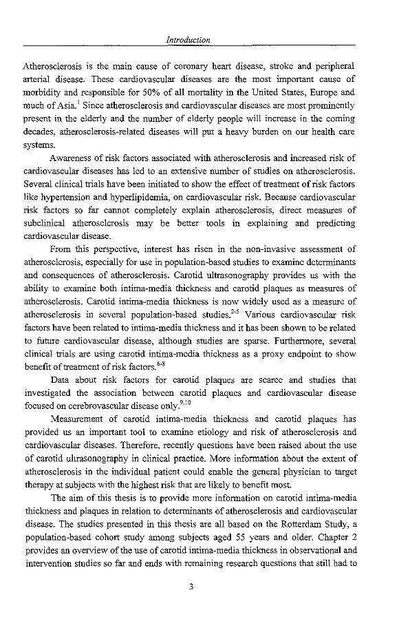
Figure 2 presents the distribution of common carotid IMT measurements as observed among the participants of the Rotterdam Study (mean common carotid IMT was 0.80 mm (SD 0.16»4 Common carotid IMT was nonnally distributed with a small tail to the right. Similar distributions have been described for carotid IMT measurements in the carotid bifurcation and internal carotid artery.IS Importantly, there appears to be no clear cut-off point to indicate at which common carotid IMT level a subject is diseased Or not and thus decisions to dichotomize subjects in groups of subjects with and without abnonnalities are therefore arbitrary.
11
Chapter 2
Figure 2. Distributiou of common CIMT measurements the Rotterdam Study.
15
,-., ~ '" 10 '-'
= Q .--"" Q
~ 5 Q
"" ~
o
Cross sectional findings
Age and sex
Intima-media thickness (mm)
Table I shows general characteristics of a number of large community based cohort studies in which carotid lMT is assessed. With increasing age common carotid IMT increases in both men and women. Based on cross-sectionally obtained data estimates were around 0.009 mm/year for men and 0.009 mm/year for women. 1·8.19·21 Common
carotid lMT was increased in men compared to women for all ages, with a mean difference of around 5 to 10 %. This has been interpreted as differences in presence or extent of atherosclerosis. However, this difference is partly attributable to differences in end-diastolic lumen diameter and may therefore reflect differences in physiology rather than differences in atherosclerosis per se.22
Prevalent cardiovascular disease
Subject with prevalent cardiovascular disease, such as myocardial infarction, angina pectoris, intermittent claudication and stroke, have in general a thicker common carotid intima-media compared to those without symptomatic cardiovascular disease.23-25
12

1'a bleL GeIl~'al~~!Ja!~~~~i~L~L~!,~e popula.tion:~"-!,,d. o.~serv~!~Ials!l'!!ie~ using.<2I.~IJ~!,~~~~lIl~<:!!,!,I Is ... ~_~ ••• ~.~ •.•• , .... Study name Baseline data Men Women Age Outcome variable
collection (N) (N) range
(years)
Atherosclerosis Risk in 1987-1989 4730 6090 45-64 mean of max. lMT CCA, BIF and ICA
Communities
Cardiovascular Health Study 1989-1990 2255 2946 65-102 mean of max. lMT CCA, BlF and lCA
Edinburgh AIielY Study 1992-1994 1156 59-78 maximum of far wall CCA, BIF, lCA
France 1988-1989 none 517 45-54 atherosclerosis based on plaques and
lMT
Kuopio Ischemic Heali Disease 1987-1989 1252 none 40,48,54, mean of max. IMT CCA and BIF
Study 60
Insulin Resistance • 643 798 40-69 see Cardiovascular Health Study
Atherosclerosis Study
Rotterdam Study 1990-1993 3105 4878 55-106 mean of mean lMT at CCA and BlF
IMT = in'li~~-::-~llcdhi thickness~; CCA"-::-~onm;;;--; ca-rotid artery; BiF~~ -;;rotid bifu~atrc;;;;~iCA :;;t:;--;;-;Tc-a-~otid-;rt;;y~~~~~~~~~~~'~'~-~-~'"~-~~~~~~~---- --~~~~~-. -* Not mentioned in the reference

Chapter 2
The magnitude of the difference in common carotid IMT between subjects with and
without symptomatic cardiovascular disease vaties from 6 to 12 % across studies.
Carotid IMT was increased in subjects with intermittent claudication compared to
subjects without intermittent claudication. The magnitude of the increase ranged from
15 % to 20 % and was similar for men and women.' In several studies strong and
graded associations of common carotid IMT to left ventricular mass, measured by echocardiography, have been reported?6-28
Atherosclerosis elsewhere
An increased common carotid IMT has been shown to be associated with presence of
atherosclerosis elsewhere in the arterial system. A clear association between maximum
common carotid IMT and severity of internal carotid atherosclerosis has been reported.5.19.24
Presence of calcifications of the abdominal aorta was associated with 18 %
increase in common carotid IMT .29 A gradual increase in common carotid IMT with
decrease in ankle-arm index, as an indicator of atherosclerosis in the arteries of the
lower extremities, has been reported. Subjects with peripheral arterial disease, defined
as an ankle-arm index < 0.90 has a significantly increased carotid IMT compared to
those without peripheral arterial disease (ankle-arm index 2: 0.90). The age and gender
adjusted difference was 0.107 mm (95% CI, 0.071-0.143), reflecting a 15 % increase.8,30
Cardiovascular risk Jactors
Elevated levels of established cardiovascular risk factors, such as LDL cholesterol,
systolic blood pressure, body mass index, and a decrease in HDL cholesterol were associated with an increased carotid IMTI-J.6.7.9.20-22 An increase in carotid IMT of
around 8 % among subjects with hypertension compared to normotensive subjects has
been reported. For current smoking the increase in carotid IMT was between 5 and 10
% increased relative to non-smokers. The magnitude of the difference in carotid IMT
in subjects with diabetes mellitus compared to subjects without glucose intolerance
ranged from 7 to 12 %.
Longitudinal imdings Intima-media thickness as predictor oj fUture disease
In cross-sectional studies an increased carotid IMT has been associated with
unfavorable levels of established cardiovascular risk factors, with prevalent
cardiovascular and cerebrovascular disease and with atherosclerosis elsewhere in the
arterial system. Based on these findings there is a growing belief that carotid IMT
measurements can be regarded as an indicator of generalized atherosclerosis and may
14

Carotid intima-media thickness in observational and intervention studies
be used as an intermediate endpoint or proxy endpoint as a suitable alternative for cardiovascular morbidity and mortality.' This view is conditional on the observation that increased carotid IMT is related to future cardiovascular events. At present we are aware of only four published reports that explored the possible association between carotid IMT and incident events.
Salonen and co-workers, in a study carried out in a random sample (n = 1,257) of middle-aged Finnish men, reported that an increase of 0.1 mm in maximum far wall common carotid IMT was associated with 11% (95% CI, 6-16) increase in risk of myocardial infarction.30 This analysis was based on 36 coronary heart disease events that occurred after 1 to 3 years of follow-up.
Findings from the ARIC study were recently reported based on 290 coronary heart disease events that occurred after a 4 to 7 years of follow up in 7289 women and 5552 men, aged 45-64 years.31 In this study carotid IMT was based on the mean far wall of the common carotid artery, the bifurcation and the internal carotid artery. The age adjusted risk of coronary heart disease increased gradually with increasing carotid IMT (figure 3).
Figure 3. Risk of coronary heart disease by CIMT. Results from the ARIC study.
14
-I .... Q., 12 "" I
"" 10 "" . -------- ---,....
:... 8 ----------- ----- -+-Men O.l Q., O.l 6 -ll-Women .... '" :...
4 ~ 2 -_.-U
0
<0.60 0.60- 0.70- 0.80- >=1.00 0.69 0.79 0.99
Intima-media thickness (mm)
15

8
Chapter 2
Analyses with carotid IMT as a continuous variable indicated that the risk of CHD
increased by 69% (95% cr, 50%-90%) in middle-aged women and 36% (95% cr, 23%-51%) in middle-aged men per 0.19 mm increase in carotid IMT. Results for the common carotid rMT per 0.19 mm increase (one SD) were 92% (95% cr, 66%-122%)
and 32% (95% cr, 8%-23%), respectively. As expected, adjustment for several cardiovascular risk factors reduced, but not abolished, the magnitude of the
association.
In the Rotterdam Study, a prospective cohort study among 7983 subjects aged
55 years, a nested case-control approach was used to evaluate whether common
carotid IMT is related to future stroke and myocardial infarction32 The analysis was
based on 98 myocardial infarctions and 95 strokes, and a sample of 1373 subjects who remained free from myocardial infarction and stroke during follow-up. The mean
duration of follow-up was 2.7 years. Carotid rMT was based on the average of the near and far wall of the left and right common carotid artery. The risk of stroke and of myocardial infarction gradually with increasing IMT (figure 4).
Figure 4. Association between common CIMT and risk of first stroke and myocardial infarction. Resnlts from the Rotterdam Study.
Myocardial infarction
<iJ.75 0.75-0.82 0.83-0.91 )9).92
Carotid iotima-media thickoess (mm)
~ U
8 Stroke
~ 6 -L------------------I-------I----on e ~4
" "
O,--~~---,--~WL---~~----~~--
<0.75 0.75-0.82 0.83-0.91 >=0.92
Carotid iotima-media thickoess (mm)
The odds ratio for stroke per standard deviation increase (0.163 mm) was 1.41
(95% cr, 1.25-1.82). For myocardial infarction, an odds ratio of 1.43 (95% cr, 1.16-
I. 78) was found. When subjects with a previous myocardial infarction or stroke were
excluded, odds ratios were 1.57 (95% cr, 1.27-1.94) for stroke and 1.51 (95% cr,
1.18-1.92) for myocardial infarction. Additional adjustment for several cardiovascular
risk factors attenuated these associations: 1.34 (95% cr, 1.08-1.67) and 1.25 (95% cr, 0.98-1.58), respectively.
16

Carotid intima-media thickness in observational and intervention studies
In the Cholesterol Lowering Atherosclerosis Study (CLAS) among 146 men of
the age of 40 to 59 years with a previous coronary artery bypass grafting, 78 coronary
heart disease (CHD) events had occurred after a mean follow-up of 8.8 years.33 An increase in common carotid IMT of 0.13 mm (one standard deviation) was associated
with a 1.4 fold (95% CI, 1.2-1.7) increased risk of CHD risk. Adjustment for
cardiovascular risk factors did not materially alter the odds ratio. These important findings implicate that carotid IMT measures are strongly
related to future coronary heart and cerebrovascular disease in middle-aged and elderly subjects. This predictive power remained even when established cardiovascular risk
factors were taken into account. These findings provide supportive evidence for the use of carotid IMT as an intermediate or proxy endpoint in observational and
intervention studies.
Change in carotid IMT over time
Data on change over time in carotid IMT and its determinants are limited34-37
Smoking, increased LDL cholesterol have been shown to strongly associate with
increase in carotid IMT over time in Eastern Finnish men. Information on the
association between progression of carotid IMT and risk of cardiovascular disease is present from only one study.33 In the CLAS study an annual progression rate of 0.03
IIJ.tWyear in common carotid IMT was associated with a 2.2 fold (95% CI, 1.4-3.6) increased risk of CHD. Compared to subjects with an common carotid IMT change of 0.011 IIJ.tWyear or less, the CHD risk of those with an annual progression of 0.03
IIJ.tWyear or over was increased 4.5 fold (95% CI, 1.9-10.8). The association remained
after taken into account cardiovascular risk factors and progression of coronary
atherosclerosis.33
Most of the current information on change in carotid IMT over time comes from analyses on data of subjects in placebo groups in intervention studies38
-42 In
these studies carotid IMT was measured every 6 months for a period of up to 2 to 3
years. For an individual an estimate of the progression of carotid IMT was calculated by fitting all obtained values over three years into a regression model weighed for the
time and number of measurements and the baseline aggregate. The individual
regression slopes are then used to calculate a mean slope for each treatment group and the mean slope values between treatment groups are compared. Estimates of progression rates of common carotid IMT are presented in table 2 and 3. Progression
rates differ considerably across studies. Differences in the used methodology, in outcome measure, and in selection of participants may at least partly explain the
differences in estimates of progression rates of carotid IMT.
17

Chapter 2
Intervention studies
Over the past years several intervention studies using change in carotid IMT as outcome have been performed or are being conducted.38-42 In the majority of these trials three ways for an endpoint were used. First, the mean maximum carotid IMT defined as the mean of the individual maximum IMT measured in up to 12 carotid segments (near and far wall of the left and right common carotid artery, carotid bifurcation and internal carotid artery). Second, the far wall and bifurcation carotid IMT (mean of the far wall of the left and right common carotid artery and carotid bifurcation. Thirdly, the single maximum IMT, defined as the maximum carotid IMT detected across both the left and right carotid artery. Most of the intervention studies from which results have been published evaluated the effect of lipid lowering treatment on the progression of carotid IMT.3S-41 Results are summarized in table 3. Generally, lipid lowering resulted in a reduced progression of carotid IMT compared to placebo. The only trial evaluating the effect of different blood pressure lowering regimes (calcium antagonist versus diuretic) showed that progression of carotid IMT was slower in those on calcium antagonists, a difference that, however, did not reach
statistical significance.37 Several other trials evaluating a variety of drugs are currently ongoing.
Important current issues Near wall versus far wall carotid IMT measurements
The near wall findings in the validation studies have lead to an intensive discussion on whether near wall carotid IMT measurements should be performed at all. lO
.l4
•15 At
present there are two strong views:43 I) the near wall measurements should not be used, because they do not reflect the true thickness and therefore are invalid; 2) the near wall measurements are of value and should be used. The latter view recognizes and respects the view that the near wall measurement not truly reflects its anatomical substrate, but there is additional evidence to argue in favor of the use of near wall carotid IMT measurements.
The reproducibility of the near wall carotid IMT measurements is similar as those reported for the far wall carotid IMT measurements.' The association between near wall intima-media thickness and prevalent cardiovascular disease is as strong and precise as compared to the association found for the far wall. Combining information
18

Table 2. Estimates of annual ErogressiOl~ of CIMT in mm observed incontrolgrol,!!s of randomized controlled trials. ~~~ __ m~ Study Number Prevalent condition Age- Method Estimated Standard
of range progression error of
subjects estimate
ACAPS 385 Free from CVD with at least one lesions 40-79 Mean max of 12 0.006 mm/yr 0.003
with an IMT between 1.5 - 3.5 mm segments
CLAS 39 Non-smoking men with a previous CABG 40-59 Mean far wall IMT at 0.05 mm/4 yr 0.08
CCA
KAPS 223 LDL cholesterol:;' 4.0 mmol/I and total 44-64 Mean of maximum far 0.0285 mm/yr 0.0043
cholesterol < 7.5 mmoUI, men wall IMT in the CCA
PLAC-II 76 Subject with prevalent coronmy al1elY 50-75 Maximum far wall 0.0456 mm/yr 0.0057
disease, and a carotid plaque 2: 1.3 nun IMT at the CCA
CAIUS 305 Asymptomatic men and women, moderate 2:55 Mean maximum I1vIT 0.009 mm/yr 0.0027
hypercholesterolemia, with IMT 1.3 mm - (12 sites)
3.5 nnn
MARS 89 Angiographically defined coronalY m1elY 37-67 Far wall distal CCA 0.019 nnnlyr 0.004
disease (92% men) ~~~~--~~ - - ~ -~~~~~~~~-~~~~-~~~~~~~~~~~~~.~~~~~~-~-~--~~~~~-~-.-"~-~"-~-~~~-~~-.---~="~=='~~-~~~
CCA = common carotid artery; L\tlT = intima-media thickness
Chapter 2
of the near wall and far wall common carotid IMT into one intima-media thickness
estimate (average of four sites) provided the strongest association with cardiovascular
disease and lower extremity arterial disease.44 Longitudinal results from the Rotterdam
study supported these cross-sectional fmdings: the association between near wall
carotid IMT and stroke or myocardial infarction was as strong as that found for far
wall carotid IMT29 The combined near and far wall intima-media thickness led to the
strongest association. Findings in three randomized placebo controlled intervention
studies among subjects receiving placebo treatment indicated that the progression rate
of near wall common carotid IMT was similar to that for the far wall common carotid
IMT45 Combining information of both near and far wall yielded estimates of
progression rates with higher precision, i.e., smaller standard errors. The consequence is that the number of patients that is needed in an intervention study to demonstrate a
treatment effect is smaller when carotid IMT is based on the average of near and far
wall measurements than on far wall measurements only.45 Thus, measurement of the
near wall carotid IMT yields valuable information, and should not be discarded easily.
However, one should realize that, in particular for the near wall measurement
standardization of gain settings and B-mode ultrasound technique across various
sonographers is of utmost importance. In general, this is easier in single center studies than in multicenter studies. Also, near wall measurements are more difficult to obtain
compared to far wall measurements. For example, in the Rotterdam study near wall
intima-media thickness measurements of at least one or both sides of the common
carotid artery could not be obtained from the images in 8.9% of the subjects. For far
wall measurements, data on either left, right or both sides were missing in 3.1 % of the
study population.44 However, in analyses indicators of presence or absence of near
wall measurements may added to the regression model, or a near wall estimate may be
used based on the measurement of the side that is available, or a model may be used to
impute missing values.
Should we use common carotid IMT measurements only?
A very frequent question that is put forward is whether one should limit the study to
common carotid IMT measurements only or also should include measurements of the
carotid bifurcation and internal carotid artery. The main approach to this question is to
evaluate the balance between evidence showing differences in strength of associations
of common carotid IMT and internal carotid IMT to cardiovascular disease and the
practicalities involved, being time consumption, missing data, and reproducibility.2,2o,23.31
20

Table 3. Main findings from randomized controlled trials on common CIMT.
Study Intervention Annual Annual Mean anIlual Estimated Estimated
llame progression in progression in the treatment anllual allnual
treated groull placebo gronp effect (nuu) reduction of reduction of
(nun/yr) (mm/yr) eHD l'isk.* stroke risk. **
ACAPS Lovastatin vs, placebo -0.009 (0.003) 0.006 (0.003) 0.015111111 2.5% 3.8%
CLAS ColestipoVniacin vs. -0.05/4yr (0.08) 0.05 / 4yr (0.08) 0.10 ml11 16.8% 25.2 %
placebo
KAPS Pravastatin vs. placebo 0.096 (0.0043) 0.0285 (0.0043) 0.0189 nnn 3.2% 4.8%
PLAC II Pravastatin vs. placebo 0.0295 (0.0058) 0.0456 (0.0057) 0.0161 nnn 2.7% 4.1 %
CAIUS Pravastatin vs. placebo -0.0043 (0.0028) 0.0090 (0.0027) 0.0133 nnn 2.2 % 3.4 %
3 years
MARS Lovastatin vs. placebo -0.028 (0.003) 0.015 (0.005) 0.043111111 7.2% 10.8 %
MIDAS Isradipine versus 0.064 (0.006) 0.061 (0.006) 0.003111111 0.5% 0.8%
hydrochlorothiazide
*' Ba-;d~ARIC~~g~"';dj;;t;d"~ti;l~;t;;'f;;~'~;;;;;aMTh~'~~'~dd~~;;io ~1~32p;;QT 9 ;l;~~-~Tn~~~~~~Y~~~~~~-~~~~~~~~~~ -~--~~~=~~~~=~~~-~~~~~~~-~~~-~-~~~~~~-~ -"~--'~,~~f~~=_~~ __ _
** Based on the Rotterdam Study age and sex adjusted estimates for COIIDnon CIMT (odds ratio 1.41 per 0.l63 mm increase).

Chapter 2
Although most studies indicated that associations were most strong when information from several carotid sites was combined, the magnitude of associations did not significantly differ between the various approaches.44A5 Then practicalities such as slightly more difficult to obtain good internal carotid images, more time spent on ultrasonography and reading and slightly less good reproducibility for the internal carotid artery may favor restriction to common carotid arteries. Yet, differences in quantifYing presence and extent of carotid atherosclerosis across studies should be appreciated. At present it can not be answered satisfactory which approach provides
the 'best' indicator of atherosclerosis for cross-sectional studies, longitudinal studies and intervention studies.
Changes in carotid IMT over time: reader drift and withdrawals
In studies on estimating progression in carotid IMT over time several aspects need careful attention. This includes random and systematic differences across subjects who measure carotid IMT from the stored images on videotape (readers). The magnitude of these differences may be in the order of 0.1 mm as exemplified by Furberg and co-workers." Reader behavior within the same person may change over time from initially reading 'thick' to ultimately reading 'thin' or vice versa. This is of importance when in time the relative proportion of "thick" or "thin" readers changes considerably. This calls for strict quality control efforts. Recently, a number of analysis systems has become available that use an automated edge detection program for quantification of carotid IMT from the stored ultrasound images. 11.12 These systems should help to overcome the reader drift, since the automated edge detection program does not change over time. The first results of such a system that was applied in a relatively small single center study were encouraging: i.e., the reproducibility of the measurements improved. 11
In studies of a long duration, mortality or withdrawal from the study during follow-up may bias progression rates towards a lower estimate, assuming an increased risk of death or withdrawal among those who progress rapidly. Having carotid IMT measurements performed at regular intervals may help to overcome a serious effect of withdrawal on the progression rates.
Summary Carotid intima-media thickness can be non-invasively assessed in a valid and reproducible way. An increased carotid IMT is associated with unfavorable levels of established cardiovascular risk factors, with prevalent cardiovascular disease and with atherosclerosis elsewhere in the arterial system. Data from intervention studies indicate that lipid lowering in hypercholesterolaemic subjects results in a reduced progression of carotid IMT. The risk of coronary heart disease and stroke gradually
22

Carotid intima-media thicf..:ness in observational and intervention studies
increases with increasing carotid IMT. This predictive power of carotid IMT
measurements remains after adjustment for cardiovascular risk factors. Also progression of COmmon carotid IMT appears to be a strong predictor of future CHD in
men with a previous coronary artery bypass grafting. These findings support the view
that carotid IMT measurements can be used as an endpoint as a suitable alternative for cardiovascular morbidity and mortality in observational and intervention studies. In
particular in trials evaluating the efficacy of a treatment the use of carotid IMT
measurements will result in a considerable smaller number of subjects needed than when using disease or death as an endpoint. Strict ultrasound protocols, training and
monitoring of sonographer and reader performance are mandatory.
Which carotid IMT measure (common, bifurcation, internal, or combined) provides the 'best' information for observational and intervention studies needs to be
established. Whether carotid IMT should be measured in every patient at high risk of
future cardiovascular and cerebrovascular diseases and thus may help in further risk profiling of an individual patient needs to be studied extensively.
References 1. Heiss G, Sharrett AR, Barnes R, Chambless LE, Szklo M, Alzola C. Carotid
atherosclerosis measured by B-mode ultrasound in populations: associations with cardiovascular risk factors in the ARlC study. Am J Epidemiol. 1991;134:250-6.
2. O'Leary DH, Polak JF, Wolfson SK, Jr., Bond MG, Bommer W, Sheth S, Psaty BM, Sharrett AR, Manolio TA. Use of sonography to evaluate carotid atherosclerosis in the elderly. The Cardiovascular Health Study. CHS Collaborative Research Group. Stroke. 1991 ;22: 1155-63.
3. Auperin A, Berr C, Bonithon-Kopp C, Touboul PJ, Ruelland I, Ducimetiere P, Alperovitch A. Ultrasonographic assessment of carotid wall characteristics and cognitive functions in a community sample of 59- to 71-year-olds. The EVA Study Group. Stroke. 1996;27:1290-5.
4. Bots ML, Hofman A, De Jong PT, Grobbee DE. Common carotid intima-media thickness as an indicator of atherosclerosis at other sites of the carotid artery. The Rotterdam Study. Ann Epidemiol. 1996;6:147-53.
5. Stensland-Bugge E, Bonaa KH, Joakimsen O. Reproducibility of ultra so no graphically determined intima-media thickness is dependent on arterial wall thickness. The Tromso Study. Stroke. 1997;28:1972-80.
6. Mykkanen L, Zaccaro DJ, O'Leary DH, Howard G, Robbins DC, Haffuer SM. Microalbuminuria and carotid artery intima-media thickness in nondiabetic and NIDDM subjects. The Insulin Resistance Atherosclerosis Study (!RAS). Stroke.
1997;28:1710-6.
7. Salonen R, Salonen JT. Carotid atherosclerosis in relation to systolic and diastolic blood pressure: Kuopio Ischaemic Heart Disease Risk Factor Study. Ann Med.
1991;23:23-7.
23

Chapter 2
8. Allan PL, Mowbray PI, Lee AJ, Fowkes FG. Relationship between carotid intimamedia thickness and symptomatic and asymptomatic peripheral arterial disease. The Edinburgh Artery Study. Stroke. 1997;28:348-53.
9. Grobbee DE, Bots ML. Carotid artery intima-media thickness as an indicator of generalized atherosclerosis. J Intern Med. 1994;236:567-73.
10. Wendelhag I, Gustavsson T, Suurkula M, Berglund G, Wikstrand J. Ultrasound measurement of wall thickness in the carotid artery: fundamental principles and description of a computerized analysing system. Clin Physiol. 1991;11:565-77.
11. Wendelbag I, Liang Q, Gustavsson T, Wikstrand J. A new automated computerized
analyzing system simplifies readings and reduces the variability in ultrasound measurement of intima-media thickness. Stroke. 1997;28:2195-200.
12. Selzer RH, Hodis HN, Kwong-Fu H, Mack WJ, Lee PL, Liu CR, Liu CH. Evaluation of computerized edge tracking for quantifying intima-media thiclmess of the common carotid artery from B-mode ultrasound images. Atherosclerosis. 1994; III: I-II.
13. Chambless LE, Zhong MM, Arnett D, Folsom AR, Riley WA, Heiss G. Variability in B-mode ultrasound measurements in the atherosclerosis risk in communities (ARlC)
study. Ultrasound Med Bioi. 1996;22:545-54.
14. Wong M, Edelstein J, Wollman J, Bond MG. Ultrasonic-pathological comparison of the human arterial wall. Verification of intima-media thickness. Arterioscler Thromb.
1993;13:482-6. 15. Gamble G, Beaumont B, Smith H, Zorn J, Sanders G, Merrilees M, MacMahon S,
Sharpe N. B-mode ultrasound images of the carotid artery wall: correlation of ultrasound with histological measurements. Atherosclerosis. 1993; I 02: 163-73.
16. Bots ML, Mulder PG, Hofman A, van Es GA, Grobbee DE. Reproducibility of carotid vessel wall thickness measurements. The Rotterdam Study. J Clin Epidemiol.
1994;47:921-30. 17. Kanters SD, Algra A, van Leeuwen MS, Banga ID. Reproducibility of in vivo carotid
intima-media thickness measurements: a review. Stroke. 1997;28:665-71.
18. Howard G, Sharrett AR, Heiss G, Evans GW, Chambless LE, Riley WA, Burke GL. Carotid artery intimal-medial thickness distribution in general populations as
evaluated by B-mode ultrasound. ARJC Investigators. Stroke. 1993;24:1297-304.
19. Howard G, Burke GL, Evans GW, Crouse JR, 3rd, Riley W, Arnett D, de Lacy R, Heiss G. Relations of intimal-medial thickness among sites within the carotid artery as
evaluated by B-mode ultrasound. ARIC Investigators. Atherosclerosis Risk in
Communities. Stroke. 1994:25:1581-7.
20. O'Leary DH, Polak JF, Kronmal RA, Savage PJ, Borhani NO, Kittner SJ, Tracy R, Gardin JM, Price TR, Furberg CD. Thickening of the carotid wall. A marker for atherosclerosis in the elderly? Cardiovascular Health Study Collaborative Research Group. Stroke. 1996;27:224-31.
21. Bots ML, Witteman JC, Hofman A, de Jong PT, Grobbee DE. Low diastolic blood pressure and atherosclerosis in elderly subjects. The Rotterdam study. Arch Intern
Med. 1996;156:843-8.
24

Carotid intima-media thicf..-ness in observational and intervention studies
22. Bots ML, Hofman A, Grobbee DE. Increased connnon carotid intima-media thickness.
Adaptive response or a reflection of atherosclerosis? Findings from the Rotterdam
Study. Stroke. 1997;28:2442-7.
Burke GL, Evans GW, Riley WA, Sharrett AR, Howard G, Barnes RW, Rosamond
W, Crow RS, Rautaharju PM, Heiss G. Arterial wall thickness is associated with
prevalent cardiovascular disease in middle-aged adults. The Atherosclerosis Risk in Connnunities (ARIC) Study. Stroke. 1995;26:386-91.
24. Polak JF, O'Leary DH, Kromnal RA, Wolfson SK, Bond MG, Tracy RP, Gardin JM,
Kittner Sl, Price TR, Savage Pl. Sonographic evaluation of carotid artery atherosclerosis in the elderly: relationship of disease severity to stroke and transient
ischemic attack. Radiology. 1993;188:363-70.
25. Sa10nen R, Tervahauta M, Sa10nen JT, Pekkanen J, Nissinen A, Karvonen MJ.
Ultrasonographic manifestations of conunon carotid atherosclerosis in elderly eastern
Finnish men. Prevalence and associations with cardiovascular diseases and risk
factors. Arterioscler Thromb. 1994; 14: 1631-40.
26. Cuspidi C, Lonati L, Sampieri L, Pelizzoli S, Pontiggia G, Leonetti G, Zanchetti A. Left ventricular concentric remodelling and carotid structural changes in essential
hypertension. J Hypertens. 1996;14:1441-6.
27. Linhart A, Gariepy J, Gira1 P, Levenson J, Simon A. Carotid artery and left ventricular
structural relationship in asymptomatic men at risk for cardiovascular disease.
Atherosclerosis. 1996; 127: 1 03-12.
28. Roman MJ, Saba PS, Pini R, Spitzer M, Pickering TG, Rosen S, Alderman MH,
Devereux RE. Parallel cardiac and vascular adaptation in hypertension. Circulation.
1992;86: 1909-18.
29. Bots ML, Witteman JC, Grobbee DE. Carotid intima-media wall thickness in elderly
women with and without atherosclerosis of the abdominal aorta. Atherosclerosis.
1993;102:99-105.
30. Sa10nen JT, Sa10nen R. U1trasonographically assessed carotid morphology and the tisk
of coronary heart disease. Artenoscler Thromb. 1991; 11: 1245-9.
31. Chambless LE, Heiss G, Folsom AR, Rosamond W, Szklo M, Sharrett AR, Clegg LX.
Association of coronary heart disease incidence with carotid arterial wall thickness
and major risk factors: the Atherosclerosis Risk in Connnunities (ARIC) Study, 1987-
1993. Am J Epidemiol. 1997; 146:483-94.
32. Bots ML, Hoes AW, Koudstaa1 PJ, Hofman A, Grobbee DE. Connnon carotid intima
media thickness and risk of stroke and myocardial infarction: the Rotterdam Study.
Circulation. 1997;96:1432-7.
33. Hodis HN, Mack WJ, LaBree L, Selzer RH, Liu CR, Liu CH, Azen SP. The role of
carotid arterial intima-media thickness in predicting clinical coronary events. Ann
Intern Med. 1998;128:262-9.
34. Hodis HN, Mack WJ, Durm M, Liu C, Liu C, Selzer RH, Krauss RM. Intermediate
density lipoproteins and progression of carotid arterial wall intima-media thickness.
Circulation. 1997;95:2022-6.
25

Chapter 2
35. Markus RA, Mack WJ, Azen SP, Hodis HN. Influence of lifestyle modification on
atherosclerotic progression determined by ultrasonographic change in the common carotid intima- media thickuess. Am J Clin Nutr. 1997;65: I 000-4.
36. Salonen R, Salonen JT. Progression of carotid atherosclerosis and its determinants: a
population-based ultrasonography study. Atherosclerosis. 1990;81:33-40.
37. Lynch J, Krause N, Kaplan GA, Salonen R, Salonen JT. Workplace demands,
economic reward, and progression of carotid atherosclerosis. Circulation.
1997;96:302-7.
38. Blankenhorn DH, Selzer RH, Crawford DW, Barth JD, Liu CR, Liu CH, Mack WJ,
Alaupovic P. Beneficial effects of colestipol~niacin therapy on the common carotid artery. Two~ and four-year reduction of intima-media thickness measured by
ultrasound. Circulation. 1993;88:20-8.
39. Furberg CD, Adams HP, Jr., Applegate WB, Byington RP, Espeland MA, Hartwell T,
Hunninghake DB, Lefkowitz DS, Probstfield J, Riley WA, et aI. Effect of lovastatin
on early carotid atherosclerosis and cardiovascular events. Asymptomatic Carotid
Artery Progression Study (ACAPS) Research Group. Circulation. 1994;90:1679-87.
40. Crouse JR, 3rd, Byington RP, Bond MG, Espeland MA, Craven TE, Sprinkle JW,
McGovern ME, Furberg CD. Pravastatin, Lipids, and Atherosclerosis in the Carotid Arteries (PLAC- II). Am J Cardiol. 1995;75:455-9.
41. Salonen R, Nyyssonen K, Porkkala E, Rummukainen J, Belder R, Park JS, Salonen
JT. Kuopio Atherosclerosis Prevention Study (KAPS). A population-based primary
preventive trial oftbe effect ofLDL lowering on atherosclerotic progression in carotid and femoral arteries. Circulation. 1995;92: 1758-64.
42. Borhani NO, Mercuri M, Borhani PA, Buckalew VM, Canossa-Terris M, Carr AA,
Kappagoda T, Rocco MY, Sclinaper HW, Sowers JR, Bond MG. Final outcome
results of the Multicenter Isradipine Diuretic Atherosclerosis Study (MIDAS). A
randomized controlled trial. Jama. 1996;276:785-91.
43. Wikstrand J, Wiklund O. Frontiers in cardiovascular science. Quantitative
measurements of atherosclerotic manifestations in humans. Arterioscler Thromb. 1992;12:114-9.
44. Bots ML, de Jong PT, Hofman A, Grobbee DE. Left, right, near or far wall common
carotid intima-media thickness measurements: associations with cardiovascular
disease and lower extremity arterial atherosclerosis. J Clin Epidemio!. 1997;50:801-7.
45. Furberg CD, Byington RP, Craven TE. Lessons learned from clinical trials with
ultrasound end-points. J Intern Med. 1994;236:575-80.
26

Chapter 3
Carotid atherosclerosis and cardiovascular disease


3.1
Carotid intima-media thickness at different sites and risk of incident myocardial infarction
Abstract Aims: We examined whether intima-media thickness (IMT) of the common carotid
artery, carotid bifurcation, internal carotid artery and the combined measure are
predictive of foture myocardial infarction and which of the measurements has the
strongest predictive value.
Methods and results: We used a case-cohort approach in the Rotterdam Study.
Ultrasound images of the common carotid artery, carotid bifurcation and the internal
carotid artery were made. We selected the first 194 myocardial infarctions in the total
population (mean follow-up 4.6 years). Analyses were done using Cox regression with
adjustment for age and sex. The risk ratios (RR) for myocardial infarction associated
with common carotid, bifurcation, internal carotid IMT and the combined
measurement were 3.18 (95% confidence interval, 1.83-5.54), 4.11 (2.10-8.05), 5.31
(1.77-15.9) and 6.27 (3.27-12.0), respectively for the highest compared to the lowest
quartile. The RRs for myocardial infarction per standard deviation increase of
common carotid, bifurcation, internal carotid artery and combined IMT were 1.44
(1.28-1.62), 1.34 (1.17-1.53), 1.12 (0.94-1.33) and 1.47 (1.31-1.65), respectively. The
areas under the ROC-curves for the three measurements and the combined measure
were not Significantly different.
Conclusions: Increased carotid IMT is a strong predictor of future myocardial
infarction and all measurement sites have the same ability to predict foture myocardial
infarction.
29

Chapter 3.1
Introduction Non-invasive assessment of intima-media thickness of the carotid arteries by highresolution B-mode ultrasonography is widely used in observational studies and trials as an intermediate or proxy measure of generalised atherosclerosis.!'" Increased common carotid intima-media thickness has been associated with unfavorable levels of established cardiovascular risk factors, prevalent cardiovascular disease and atherosclerosis elsewhere in the arterial systemS
•6 In recent studies, common carotid
intima-media thickness has also been found to be associated with the risk of incident stroke and myocardial infarction.7
-10
Data on cardiovascular risk associated with bifurcation intima-media thickness and internal carotid intima-media thickness are sparse. In a recent paper from the Cardiovascular Health Study, the associations of internal carotid intima-media thickness with myocardial infarction and stroke were as strong as those for common carotid intima-media thickness1 ! Internal carotid artery intima-media thickness however, was composed of carotid bifurcation measurement as well as internal carotid artery measurement and therefore no conclusions can be drawn for the association with myocardial infarction for each of the measurements separately.!2 No other studies have given additional attention to the predictive value of each of the three intima-media thickness measurements for future disease. Since most observational and intervention studies have restricted to common carotid intima-media thickness measurements due to feasibility, it is important to provide evidence of the predictive values of the various sites and to evaluate which measurement is best in predicting future coronary heart disease. In this study we examined whether the intima-media thickness of the carotid bifurcation and the internal carotid artery are predictors of future coronary heart disease. Furthermore, we examined the predictive value of the intima-media thickness of the three sites, the common carotid artery, carotid bifurcation and internal carotid artery, and a combined measure of the three sites for future coronary heart disease.
Methods
Population
The Rotterdam study is a population-based prospective cohort study that aims to assess determinants and occurrence of chronic diseases in the elderly. The study focuses on cardiovascular, neurogeriatric, ophtalmologic and locomotor diseases, and has been
described in more detail elsewhereI3 In brief, all residents aged 55 and over of a defined district in Rotterdam were invited to participate. A total of 7983 men and women (78 percent of those eligible) entered the study. During the first survey, from 1990 to 1993, all participants were interviewed at home by a trained research assistant, and subsequently visited the study centre twice. The study was approved by the
30

Carotid intima-media thid;ness at different sites and risk of incident myocardial infarction
Medical Ethics Committee of Erasmus University, and written informed consent was obtained from all participants.
Cardiovascular Risk Indicators
At baseline, interview information, including current medication, alcohol intake and smoking habits, was obtained by a trained research assistant during a home interview. A medical history of myocardial infarction was assessed by asking the subject "Did you ever suffer from a myocardial infarction for which you were hospitalised?". Reported myocardial infarctions were verified. As an indicator of socio-economic status the highest attained level of education was assessed. At the study centre, height and weight were measured. Two blood pressure measurements were taken with a random zero sphygmomanometer with the subject in sitting position, and averaged. A
twelve lead ECG was recorded and stored digitally. Hypertension was defined as a
systolic pressure :2:160 mmHg or a diastolic pressure :2:95 mmHg or current use of blood pressure lowering drugs for the indication of hypertension. Diabetes mellitus was considered to be present when subjects currently used oral blood glucose-lowering drugs or insulin, or had a non-fasting or postload glucose level above II mmoVL, assessed after a non-fasting venipuncture. Serum total cholesterol and HDL-cholesterol values were assessed by an automated enzymatic procedure in a non-fasting blood sample.
Incident Myocardial Infarction
Information on incident fatal and non-fatal events was obtained from the general practitioners (GPs) working in the district ofOmmoord. The GPs involved reported all possible cases of myocardial infarction to the Rotterdam research centre. Events were presented as coded information according to the International Classification of Primary Care (ICPC).I4 Information on the vital status of the participants was obtained at regular intervals from the municipal authorities in Rotterdam. When an event or death had been reported, additional information was obtained by scrutinising information from GP and hospital discharge records in case of admittance or referral. Events were
then confirmed by two Rotterdam Study physicians. In case of disagreement, consensus was reached by discussion. A myocardial infarction was considered to have occurred when the event led to a hospitalisation, and the hospital discharge record comprised a diagnosis of a new myocardial infarction based on signs and symptoms, ECG recordings, and repeated laboratory investigations during hospital stay. The percentage of individuals lost to follow-up was 0.4%.
Selection of Case Subjects
Ultrasonography of the carotid arteries was performed in 5851 of the 7983 subjects.
31

Chapter 3.1
For subjects who had their baseline Rotterdam Study examination at the end of 1992 or in 1993, ultrasonography was not always performed due to the restricted availability of ultrasonographers. For reasons of availability and completeness of information on cardiovascular events, we restricted the present study to incident myocardial infarctions registered by GPs with computerised files, before May 1996. The mean duration of follow-up was 4.6 years. Incident cases were eligible for inclusion irrespective of previous cardiovascular disease status. After review of all available information, 194 myocardial infarctions were classified of which 38 were fatal and 156 were non-fatal. At the time of the present analysis, intima-media thickness had been quantified for a random sample of 1958 of the 5851 subjects who underwent a carotid ultrasonography. Of the 194 cases, 79 cases occurred in the subcohort of 1958 subjects and 115 in the remainder of the 5851 subjects. For these 115 subjects only intima-media thickness was then quantified after which they were added to the dataset, resulting in a total of2073 subjects.
Measurement of intima-media thickness
To measure carotid intima media thickness, ultrasonography of the common carotid artery, carotid bifurcation, and internal carotid artery of the left and right carotid arteries was performed with a 7.5-MHz linear-array transducer (ATL Ultra-Mark IV).
On a longitudinal, two-dimensional ultrasound image of the carotid artery, the anterior (near) and posterior (far) walls of the carotid artery are displayed as two bright white lines separated by a hypoechogenic space. The distance between the leading edge of the first bright line of the far wall and the leading edge of the second bright line indicates the intima-media thickness. For the near wall, the distance between the trailing edge of the first bright line and the trailing edge of the second bright line at the near wall provides the best estimate of the near-wall intima-media thickness. In accordance with the Rotterdam Study ultrasound protocol 15, a careful search was performed for all interfaces of the near and far walls of the distal common carotid artery, the carotid bifurcation and the internal carotid artery. When an optimal longitudinal image was obtained, it was frozen on the R-wave of the EeG and stored on videotape. The actual measurements of intima-media thickness were performed off-line. From the videotape, the frozen images were digitised on the screen of a personal computer using additional dedicated software. This procedure has been described in detail previously 6.16.17 In short, with a cursor, or automatically by
the computer, the interfaces of the common carotid artery, the carotid bifurcation and the internal carotid artery were marked across a length of 10 mm. For the common carotid artery measurement, the most distal 10 mm. of the common carotid artery before widening into the bifurcation was used. The carotid bifurcation was defined as the part of the artery between the common carotid artery and the tip of the flow
32

Carotid intima-media thickness at different sites and risk of incident myocardial infarction
divider. The internal carotid artery was defmed as the part of the artery after the tip of the flow divider between the internal and the external carotid artery. We then calculated the mean intima-media thickness for the common carotid artery only and the maximum intima-media thickness over the marked length for both near and far wall of all three arterial segments. The average of multiple frozen images of each arterial segment was used for this. For our analyses, the maximum carotid intimamedia thickness was determined as the mean of the maximum intima-media thickness of near- and far-wall measurements of both the left and right side arteries for each of the three arterial segments. If data on one of the walls or one of the sides was missing, maximum thickness of the available wall and side was used. In the Rotterdam Study, recording on videotape and quantification of intima-media thickness of the carotid bifurcation and internal carotid artery intima-media thickness started only after approximately the first 1500 subjects were enrolled in the study. Because of this and greater difficulties with ultrasound imaging of the carotid bifurcation and internal carotid artery, there are more missing data for these sites. After the first 1500 subjects, visualisation of the common carotid artery, carotid bifurcation and internal carotid artery was possible for 97%, 83% and 56% of the subjects, respectively. The readers of the ultrasound images were unaware of the case status of the subject. Results from a reproducibility study of IMT measurements of the common carotid artery among 80 participants of the Rotterdam Study who underwent a second ultrasound of both carotid arteries within three months of the first scan have been published elsewhere 18
In short. mean differences (SD) in far-wall intima-media thickness of the common carotid artery between paired measurements of sonographers, readers, and visits were 0.005 mm (0.09), 0.060 mm (0.05), and 0.033 mm (0.12), respectively.
Data analysis
The association between intima-media thickness and incident myocardial infarction was evaluated in a case-cohort design by use of a standard Cox proportional hazards model with modification of standard errors based on robust variance estimates. 19
." In these analyses the design is a cross between a cohort design and a case-control design.
At time of entry into the cohort an individual may be included in a random sample of the cohort: the subcohort. All individuals are then followed for the outcome, whether included in the subcohort or not. Then each case and all available controls at one point in time define a risk set indexed by the follow-up time. A case outside the subcohort is considered not at risk until just before becoming a case and is not included in earlier risk sets. Subsequently the subcohort controls are weighted by the inverse of the sampling fraction, in an attempt to get an estimate that would result from a full cohort analyses.
33

Chapter 3.1
A composite measure that combined the maximal common carotid intimamedia thickness, the maximal bifurcation intima-media thickness and the maximal internal carotid artery intima-media thickness was obtained for all subjects by averaging the three measurements after standardisation (value minus mean, divided by SD). Analysis were performed with carotid intima-media thickness used as a continuous variable (expressed per SD) and as a categorised variable (based on quartile cut-off points of the distribution). First, a model with adjustment for age and sex was used. Second, additional adjustments were made for differences in baseline
characteristics between cases and controls. Third, we excluded subjects with a history of myocardial infarction or stroke from the analyses. To assess the predictive value of common carotid, bifurcation and internal carotid artery intima-media thickness measurements as well as the combined measure, we used receiver operator characteristic curves (ROC-curves). The area under the curve of each ROC-curve was calculated to study which measurement is the best predictor of coronary heart disease. The area under the ROC-curve represents the percentage of subjects correctly identified. For better comparison of the four measurements, only subjects with complete data for all three measurements were included in the ROC-curves, resulting in a dataset of 476 subjects (85 cases and 391 controls). Differences between the areas under the curve for the three separate measurements and the combined measure were tested by the Chi-square statistic with its corresponding two-tailed p-value.
Results Table 1 describes the baseline characteristics of the study subjects. The risk of myocardial infarction increased gradually with increasing maximum intima-media thickness when the near and far wall were combined (table 2). In quartile analysis the age and sex adjusted risk ratios for myocardial infarction associated with maximal common carotid intima-media thickness, maximal bifurcation intima-media thickness and maximal internal carotid artery intima-media thickness were 3.18 (95% cr, 1.83-5.54),4.11 (95% cr, 2.10-8.05) and 5.31 (95% CI, 1.77-15.90) for the highest quartile compared to the lowest quartile, respectively. For the combined measure the risk ratio was 6.27 (95% CI, 3.27-12.02) for the highest quartile compared to the lowest quartile (table 2, model I). The age and sex adjusted risk ratio for myocardial infarction per SD increase of maximal common carotid IMT (0.21 mm), was 1.44 (95% CI, 1.28-1.62). Per SD increase of maximal carotid bifurcation IMT (0.60 mm), the risk ratio was 1.34 (95% cr, 1.17-1.53). For the maximal internal carotid artery IMT the risk ratio per SD (0.66 mm) was 1.12 (95% CI, 0.94-1.33). For the combined measure the risk ratio was 1.47 (95% CI, 1.31-1.65) per SD. When we did our analyses on only far wall measurements of the three sites of the carotid artery, the age and sex adjusted risk ratio per SD increase of maximal far wall thickness of the common carotid artery was 1.24
34

Carotid intima-media thid,:ness at d(f{erent sites and risk of incident myocardial in/arction
(95% CI, 1.13-1.36). Per SD increase of maximal far wall thiclmess of the bifurcation
and the internal carotid artery the risk ratios were 1.24 (95% CI, 1.09-1.41) and 1.05
(95% CI, 0.89-1.23), respectively. Additional adjustment for cardiovascular risk
factors attenuated the magnitude of the associations, as expected.
Table 1. Baseline Characteristics of the Study Population.
Characteristic Myocardial Controls
Infarction
N 194 2073
Age (years) 72 (8.2)* 70 (8.5)
Female (%) 39* 61
Body mass index (kg/m2) 26.3 (3.4) 26.6 (3.8)
Current smoking (%) 29.4 23.6
Systolic blood pressure (mmHg) 143 (22.5)* 138 (21.3)
Diastolic blood pressure (mmHg) 72 (12.3) 72 (11.1)
Hypertension (%) 41* 34
Total cholesterol (mmollL) 6.9 (1.2)* 6.7 (1.3)
HDL cholesterol (mmollL) 1.20 (0.28)* 1.34 (0.37)
Diabetes mellitus (%) 14* 7
Previous MI (%) 31' 15
Previous stroke (%) 6 3
Max. common carotid IMT (mm) 1.17 (0.29)' 1.03 (0.22)
Max. bifurcation IMT (mm) 1.76 (0.68)* 1.47 (0.62)
Max. internal carotid IMT (mm) 1.42 (0.61)* 1.23 (0.66) Values arc unadjusted proportions or means with SD in parentheses.
:viI indicates myocardial infarction: Ma'C., maximwn: IMT, intima-media thiclmess.
>I< p<O.05 compared with total population. adjusted for differences in age and sex.
In quartile analyses, for the highest compared to the lowest quartile risk ratios
were 2.43 (95% CI 1.38-4.27),3.91 (95% CI 1.87-8.18),4.81 (95% CI 1.51-15.35) and
4.84 (95% CI 2.48-9.42) for the common, bifurcation and internal carotid artery and
the combined measure, respectively (table 2, model II). Separate analyses for males
and females showed similar risks. Risk ratios for the highest compared to the lowest
quartile of common carotid artery intima-media thiclmess were 3.21 (95% CI 1.66-
6.22) for men and 4.70 (95% CI 1.78-12.38) for women. Exclusion of 308 subjects
with a history of myocardial infarction and 60 subjects with a history of stroke resulted
in risk ratios that were about the same and were all statistically significant (table 2,
model Ill).
35

Chapter 3.1
The areas under the ROC-curves for the common carotid artery, carotid bifurcation,
internal carotid artery and the combined measure were 0.671 (95% CI 0.611-0.731),
0.689 (0.630-0.747), 0.665 (0.605-0.725) and 0.670 (0.610-0.730), respectively.
Table 2. Association of Carotid Intima-Media Thickness with Risk of Myocardial Infarction; the Rotterdam Study.
Site Risk Ratios (95% Confidence Interval)
Model 1"1" Model II:!: ModelIII§
Maximal CCA IMT*
< 0.880 mm II 1.0 1.0 1.0
0.880-0.983 mm 1.05 (0.56-1.96) 1.0 I (0.54-1.90) 0.83 (0.37-1.82)
0.984-1.120 mm 2.38 (1.38-4.11) 2.00 (1.I5-3A8) 2.50 (1.29-4.82)
:2: 1.121 mm 3.18 (1.83-5.54) 2A3 (1.38-4.27) 3.02 (1.55-5.90)
Per I SD increase 1.44 (1.28-1.62) 1.37 (1.20-1.56) lAO (1.22-1.62)
Maximal BifIMT*
< 1.000 mm II 1.0 1.0 1.0
1.001-1.245 mm 1.75 (0.84-3.65) 1.91 (0.88-4.13) 1.28 (0.56-2.89)
1.246-1.710 mm 3.17 (1.59-6.35) 3.11 (1.48-6.53) 2.94 (1.39-6.22)
:2: 1.711 mm 4.11 (2.10-8.05) 3.91 (1.87-8.18) 3.21 (1.55-6.64)
Per 1 SD increase 1.34 (1.17-1.53) 1.28 (1.11-1.47) 1.30 (1.11-1.52)
Maximal ICA IMT*
< 0.715 mm II 1.0 1.0 1.0
0.716-0.951 mm 4.99 (1.64-15.15) 5.16 (1.62-16.37) 3.77 (1.19-11.95)
0.952-1.519 mm 7.72 (2.63-22.64) 7.28 (2.31-22.88) 4.94 (1.62-15.11)
:2: 1.520 mm 5.31 (1.77-15.90) 4.81 (1.51-15.35) 3.60 (1.14-11.40)
Per I SD increase 1.12 (0.94-1.33) 1.17 (0.92-1.50) 1.19 (0.93-1.53)
Combined IMT
1" quartile II 1.0 1.0 1.0
2nd quartile 2A2 (1.20-4.89) 2.30 (1.13-4.68) 2.90 (1.23-6.84) 3,d quartile 3.51 (1.79-6.86) 3.07 (1.56-6.07) 3.64 (1.58-8.38) 4th quartile 6.27 (3.27-12.02) 4.84 (2A8-9A2) 5.95 (2.65-13.34)
Per 1 SD increase 1.47 (1.31-1.65) 1.38 (1.21-1.58) 1 A6 (1.26-1.69) nvlT indicates intimawmcdia thickness: CCA, common carotid artcry~ BIF, carotid bifurcation~ leA, internal carotid artery. * Cutpoints used were quartiles of the IMT distribution. 01* Model I: age and sex adjusted. t Model II: adjusted for age. sex, body mass index. systolic blood pressure, diastolic blood pressure, total cholesterol, HDL cholesterol. smoking and diabetes mellitus. § Model III: age and sex adjusted. previous myocardial infarction and stroke excluded. II Reference category.
36

Carotid intima-media thic/..-ness at d~fferent sites and risk of incident myocardial infarction
Discussion
The present study shows that an increased carotid artery intima-media thickness is associated with future myocardial infarction in an older population. No difference was found in the predictive value of intima-media thickness of the common carotid artery, carotid bifurcation, internal carotid artery and the combined measure.
Several methodological issues need to be discussed before interpreting the results. Missing values were present for a part of the subjects because of logistic reasons and because of technical difficulties in visualisation of the carotid bifurcation and the internal carotid artery. Data on bifurcation and internal carotid artery measurements were missing for the first 1500 subjects, which can be considered a random group. Missing data because of technical difficulties with visualisation may partly have been due to overweight. Since overweight has no association with intimamedia thickness, we do not think this has biased our results. Another possibility is missing data due to tortuous vessels. If tortuous vessels are associated with more extensive atherosclerosis, severe cases of atherosclerosis would have missing data on
bifurcation and internal carotid artery measurements and this may have led to an underestimation of the true association, since severe atherosclerosis is also related to risk of myocardial infarction. Therefore we cannot exclude the possibility that the true associations with carotid bifurcation and the internal carotid artery measurements are somewhat higher. When comparing the predictive values we assured that all subjects with any missing data on one of the measurement sites were excluded from the analyses. The extent of misclassification of the diagnosis of myocardial infarction was minimised, because the events were based on documented medical information. A reproducibility study by Bots et al. showed good reproducibility for common carotid artery intima-media thickness measurements. 1
'
As an indicator of atherosclerosis intima-media thickness is thought to be an intermediate factor in the causal pathway of risk factors leading to myocardial infarction and therefore adjustment for cardiovascular risk factors is not necessary to prevent confounding. In this study additional adjustment for cardiovascular risk factors was made. Although, as expected, the associations between carotid intima-media
thickness and incident myocardial infarction were attenuated, carotid intima-media thickness was shown to be an independent risk factor for myocardial infarction.
Most studies have concentrated on the common carotid intima-media thickness to study the relationship with myocardial infarction. Salonen and Salonen, found in the Kuopio Ischemic Heart Disease Risk Factor Study among 1257 middle-aged Finish men that an increase of 0.1 mm of maximum common carotid intima-media thickness was associated with an 11% (95% CI, 6% to 16%) increase in the risk of myocardial infarction.".23 Hodis et al. found a positive association between progression of intimamedia thickness and incident coronary heart disease in a study of 146 men 40 to 59
37

Chapter 3.1
years of age who had previously had coronary artery bypass graft surgery.' Two other
studies described the relationship between carotid artery intima-media thickness and
future myocardial infarction. Chambless et al. reported in the Atherosclerosis Risk in
Communities Study (ARIC), a study among 15792 men and women, aged 45-64 years, an association between intima-media thickness and myocardial infarction.'
Their analyses were based on mean intima-media thickness of only the far wall of the carotid artery at three locations and on a combined measure based on unweighted
means of the three measurement sites. The results showed a stronger association of
myocardial infarction with common carotid artery intima-media thickness than with the other locations. Relative risks were 1.92 (95% CI 1.66-2.22), lAO (95% CI 1.29-1.53) and 1.28 (95% CI 1.18-1.39) for men and 1.32 (1.13-1.54), 1.23 (95% CI 1.15-
1.31) and 1.15 (95% CI 1.08-1.23) for women per SD increase of common carotid, carotid bifurcation and internal carotid intima-media thickness, respectively.
Differences in strength of the association between the measurement sites were not found in the Cardiovascular Health Study, a prospective study among 5201 men and
women of 65 years and older. O'Leary et al. reported relative risks of 1.33 (95% CI
1.21-1.48), 1.43 (95% CI, 1.28-1.59) and 1.46 (95% CI, 1.32-1.61) per SD increase of
maximal common carotid artery intima-media thickness, internal carotid artery intimamedia thickness and the combined measure, respectively. I I
In our study, risk ratios for the highest compared to the lowest quartile appear to be slightly higher for bifurcation and internal carotid artery intima-media thickness
than for common carotid artery thickness, but the confidence intervals overlap and differences are not significant. In agreement with the Cardiovascular Health Study, the risks found in analyses with intima-media thickness as a continuous variable were
virtually the same for the three sites. When we examined the predictive values in ROC-curves, bifurcation and internal carotid artery measurements were not clearly
better than common carotid artery measurements. Since bifurcation and internal
carotid artery measurements generally have larger amounts of missing data, the
measurement of the common carotid artery may have a practical advantage over
measurements at the two other sites. In conclusion, the present study shows that increased common carotid,
bifurcation and internal carotid artery intima-media thickness are related to future
coronary heart disease. The predictive values of the three measurements and the
combined measurement of the three sites were similar.
References 1. Heiss G, Sharrett AR, Barnes R, Chambless LE, Szklo M, Alzola C. Carotid
atherosclerosis measured by B-mode ultrasound in populations: associations vtith cardiovascular risk factors in the ARIC study. Am J Epidemiol. 1991 ;134:250-6.
38

Carotid intima-media thickness at different sites and risk of incident myocardial infarction
2. Wendelhag I, Wiklund 0, Wikstrand J. Arterial wall thickness in familial hypercholesterolemia. Ultrasound measurement of intima-media thickness in the
common carotid artery. Arterioscler Thromb. 1992; 12:70-7.
3. Bots ML, Hofman A, de Bruyn AM, de Jong PT, Grobbee DE. Isolated systolic hypertension and vessel wall thickness of the carotid artery. The Rotterdam Elderly Study. Arterioscler Thromb. 1993;13:64-9.
4. Simons PC, Algra A, Bots ML, Grobbee DE, van der Graaf Y. Common carotid intima-media thickness and arterial stiffness: indicators of cardiovascular risk in high
risk patients. The SMART Study (Second Manifestations of ARTerial disease). Circulation. 1999;100:951-7.
5. Bots ML, Witteman JC, Grobbee DE. Carotid intima-media wall thickness in elderly women with and without atherosclerosis of the abdominal aorta. Atherosclerosis.
1993;102:99-105.
6. Bots ML, Hofman A, Grobbee DE. Common carotid intima-media thickness and lower extremity arterial atherosclerosis. The Rotterdam Study. Arterioscler Thromb.
1994; 14: 1885-91.
7. Bots ML, Hoes AW, Koudstaal PJ, Hofinan A, Grobbee DE. Common carotid intimamedia thickness and risk of stroke and myocardial infarction: the Rotterdam Study. Circulation. 1997;96: 1432-7.
8. Chambless LE, Heiss G, Folsom AR, Rosamond W, Szklo M, Sharrett AR, Clegg LX. Association of coronary heart disease incidence with carotid arterial wall thickness
and major risk factors: the Atherosclerosis Risk in Communities (ARIC) Study, 1987-
1993. Am J Epidemiol. 1997;146:483-94.
9. Hodis HN, Mack WJ. Lal3ree L, Selzer RH. Liu CR. Liu CH, Azen SP. The role of carotid arterial intima-media thickness in predicting clinical coronary events. Ann
Intern Med. 1998;128:262-9.
10. Chambless LE, Folsom AR, Clegg LX, Sharrett AR, Shahar E, Nieto FJ, Rosamond WD, Evans G. Carotid wall thickness is predictive of incident clinical stroke: the
Atherosclerosis Risk in Communities (ARIC) study. Am J Epidemiol. 2000;151:478-
87.
II. O'Leary DH, Polak JF, Kromnal RA, Manolio TA, Burke GL, Wolfson SK, Jr. Carotid-artery intima and media thickness as a risk factor for myocardial infarction
and stroke in older adults. Cardiovascular Health Study Collaborative Research
Group. N EnglJ Med. 1999;340:14-22.
12. 0' Leary D, Polak JF, Kromnal RA, Savage PJ, Borhani NO, Kittner SJ, Tracy R, Gardin JM, Price TR, Furberg CD. Thickening of the carotid wall. A marker for atherosclerosis in the elderly? Cardiovascular Health Study Collaborative Research Group. Stroke. 1996;27:224-31.
13. Hofinan A, Grobbee DE. de Jong PT, van den Ouweland FA. Determinants of disease and disability in the elderly: the Rotterdam Elderly Study. Eur J Epidemiol.
1991;7:403-22.
39

Chapter 3.1
14. Lamberts H, Wood M, Hofinans-Okkes I. The International Classification of Primary
Care in the European Community. London, UK: Oxford University Press; 1991.
15. Bots ML, van Meurs JCHM, Grobbee DE. Assessment of early atherosclerosis: a new perspective. J Drug Res. 1991;16:150-154.
16. Wendelhag I, Gustavsson T, Suurkula M, Berglund G, Wikstrand J. Ultrasound measurement of wall thickuess in the carotid artery: fundamental principles and description of a computerized analysing system. Clin Physiol. 1991; II :565-77.
17. Grobbee DE, Bots ML. Carotid artery intima-media thickness as an indicator of generalized atherosclerosis. J Intern Med. 1994;236:567-73.
18. Bots ML, Mulder PG, Hofinan A, van Es GA, Grobbee DE. Reproducibility of carotid vessel wall thickuess measurements. The Rotterdam Study. J Clin Epidemiol.
1994;47:921-30. 19. Breslow NE, Day 1:-<10. Statistical methods in cancer research. Volume II--The design
and analysis of cohort studies. IARC Sci Pub!. 1987;82:1-406.
20. Barlow WE, Ichikawa L, Rosner D, Iznmi S. Analysis of case-cohort designs. J Clin
Epidemiol. 1999;52:1165-72.
21. Barlow WE. Robust variance estimation for the case-cohort design. Biometrics.
1994;50: 1 064-72. 22. Salonen JT, Salonen R. Ultrasonographically assessed carotid morphology and the risk
of coronary heart disease. Arterioscler Thromb. 1991; 11: 1245-9. Salonen JT~ Salonen R. Ultrasound B-mode imaging in observational studies of atherosclerotic progression. Circulation. 1993;87:1156-65.
40

Abstract
3.2
Plaques in the carotid artery and risk of myocardial infarction
Objective: We examined the association between presence of carotid plaques at three different sites and future myocardial infarction in an older population.
Methods and results: The study was conducted in 7983 subjects of 55 years and
older, participating the population-based Rotterdam Study. At baseline (1990-1993), ultrasound images of the carotid artery were made. The presence of plaques in the
common carotid artery (CCA), the carotid biforcation (BIF) and internal carotid
artery (ICA) was assessed in both carotid arteries. Among the 5611 subjects with
available information on presence of carotid plaques. 205 new myocardial infarctions
occurred. The mean duration of follow-up was 5A years. The association between plaques and incident myocardial infarction was analyzed using Cox regression with
adjusttnent for age and sex. The presence of plaques in the common carotid artery
nearly doubled the risk of myocardial infarction (relative risk 1.9 (95% confidence interval, 1A-2.5)). The presence of plaques in the biforcation and internal carotid
artery were associated with relative risks of 1.6 (1.2-2.2) and 2.0 (1.5-2.7). respectively. There was a graded association between number of plaques and risk of myocardial infarction. The presence of 5-6 plaques was associated with a 2.7 fold
(95% CI, 1.5-4.7) increased risk of myocardial infarction, when compared to no
plaques. The relationships with incident myocardial infarction were of equal strength for calcified plaques and non-calcified plaques.
Conclusions: The presence of plaques in the carotid artery shows a graded
association with foture myocardial infarction in an older population, irrespective of
side, location and calcification of the plaque.
41

Chapter 3.2
Introdnction
Non-invasive assessment of atherosclerosis has gained interest in population-based
studies. B-mode carotid ultrasonography provides information on atherosclerosis and
is suitable for assessment of lumen diameter, intima-media thickness and presence and extent of plaques.!·3 Most of the recent studies have focused on carotid intima-media
thickness, its determinants and its consequences:" The available evidence on the
cardiovascular consequences of carotid plaques mainly comes from studies that investigated the risk of cerebrovascular disease9 .!4 Moreover, these studies generally
used the degree of carotid stenosis to evaluate the association with cardiovascular disease, while most carotid plaques in the general population are non-stenotic.
Only one study examined the risk of myocardial infarction associated with
carotid plaques in the general population but included a limited number of events.!4
We examined the association between carotid plaques on different locations in the
carotid artery and the occurrence of future myocardial infarction in elderly subjects
from the Rotterdam Study.
Methods
Population
The Rotterdam study is a population-based prospective follow-up study that aims to assess the determinants and occurrence of chronic diseases in the elderly. The study
focuses on cardiovascular, nelITogeriatric, ophtalmologic and locomotor diseases, and
has been described in more detail elsewhere.!5 In brief, all residents aged 55 and over
of a defined district in Rotterdam were invited to participate. A total of 7983 men and
women (78 percent of those eligible) entered the study. During the first survey, from
1990 to 1993, all participants were interviewed at home and visited the study center
twice. The study was approved by the Medical Ethics Committee of the Erasmus
University, and written informed consent was obtained from all participants.
Cardiovascular Risk Indicators
Baseline interview information included current medication and smoking habits. For
smoking, the number of packyears was calculated by multiplying the number of cigarette packs per day with the total number of years of smoking for both current and
former smokers. Height and weight were measured at the study center. Two blood pressure measurements were taken with a random zero sphygmomanometer with the
subject in sitting position, and averaged. Hypertension was defmed as a systolic
pressure d 60 mmHg or a diastolic pressure ",95 mmHg or current use of blood
pressure lowering drugs prescribed for hypertension. Diabetes mellitus was considered
to be present when subjects currently used oral blood glucose-lowering drugs or
insulin, or had a non-fasting or postload glucose level of 11.1 mmollL and over. Serum
42

Plaques in the carotid artery and risk of myocardial infarction
total cholesterol and HDL-cholesterol were assessed by an automated enzymatic procedure. A history of myocardial infarction was assessed by asking the subject "Did you ever suffer from a myocardial infarction for which you were hospitalized?". Reported myocardial infarctions were verified by electrocardiograms and hospital discharge records.
Assessment of carotid plaques
Of the 7983 subjects who participated III the Rotterdam Study, 7129 visited the research center. Ultrasonography of the carotid artery was performed with a 7.5-MHz linear-array transducer (ATL Ultra-Mark IV) in 5851 subjects. Ultrasound data were mainly missing due to logistic reasons. In particular, for subjects who had their baseline Rotterdam Study examination at the end of 1992 or in 1993, ultrasonography could not always be performed due to the restricted availability of ultrasonographers. Plaques were defined as focal widenings relative to adjacent segments, with protrusion into the lumen and composed of calcified andlor non-calcified components. Data on plaques were available for 5611 subjects. From October 1991 onwards plaque assessment was performed on-line. All 3989 subjects with an on-line plaque assessment were included. For 1622 subjects with a missing on-line plaque assessment the off-line assessment was used. Because of poor visualization, data on plaques were missing in 0.3% and 6.1 % of the subjects for the right and left common carotid artery, respectively and in 3.4%, 9.8%,10.9% and 16.6% of the subjects for the right and left bifurcation and right and left internal carotid artery, respectively. Data on plaque characteristics (calcifications and acoustic shadowing) were available for 3297 subjects whose stored images on videotapes had been read at the time of the present analyses. Data on calcification were obtained from off-line measurement. These were obtained for 3297 subjects for the common carotid artery, for 3155 subjects for the carotid bifurcation and for 2715 subjects for the internal carotid artery. A reproducibility study of the on-line assessment of plaques has been described elsewhere. In short, a kappa of 0.66 was found for the left carotid artery, a kappa of 0.68 for the right carotid artery and a kappa of 0.67 for plaques on either side,
indicating moderate agreement.
Incident myocardial infarction
The GPs in the study area report all possible cases of myocardial infarction to the Rotterdam research center16 A diagnosis of a myocardial infarction is considered definite when a hospital discharge diagnosis of a new myocardial infarction is present or, in case a patient is not hospitalized, when signs and symptoms, ECG recordings, and repeated laboratory investigations, indicating a new myocardial infarction, are available. The diagnosis is considered a probable myocardial infarction, when it was
43

Chapter 3.2
described as probable by a cardiologist. When the OP thought there was a certain new myocardial infarction, but he had no additional evidence, the myocardial infarction was also considered to be probable. When a death has been reported by the municipal authorities in Rotterdam, additional information is obtained by abstracting the medical records of the OP and scrutinizing information from hospital discharge records in case of admittance or referral. All subjects were followed up for fatal and non-fatal myocardial infarctions from the day of entry in the study until January I, 1998. The mean duration of follow-up was 5.4 years. After review of all available information,
205 myocardial infarctions were classified within the 5611 subjects.
Data analysis
A plaque score was derived by counting the number of sites with a plaque, leading to a maximum score of 6 (left and right common carotid artery, left and right carotid bifurcation, left and right internal carotid artery). The score was obtained for 4473 subjects with a complete plaque assessment on all sites. To evaluate the association between cardiovascular risk factors and carotid plaques on three different locations of the carotid artery, logistic regression analysis was used. A calcified and a non-calcified plaque score were obtained by counting the number of sites with a calcified plaque and the number of sites with a non-calcified plaque, respectively. These scores were only obtained for subjects with a plaque assessment on all sites. Cox regression modeling was used to calculate relative risks of myocardial infarction for the different plaque scores, adjusted for age and sex and additionally for other cardiovascular risk factors. The number of calcified plaques and the number of non-calcified plaques were subsequently included in a model together with the total plaque score to evaluate the independent risk estimate of calcification, given the total number of plaques. All analyses were performed by using SPSS software, version 9.0 (SPSS Inc., Chicago, Illinois).
Results Table I describes the baseline characteristics of the study population. Subjects with 5-6 plaques in the carotid artery were older and were more likely to be male. Levels of body mass index, systolic blood pressure, total and HDL cholesterol and percentage of current smokers were higher in subjects with plaques. The prevalence of plaques was highest in the carotid bifurcation. The percentage of subjects with a plaque at one or both sides of the common carotid artery, the carotid bifurcation and the internal carotid artery were 16%, 54% and 35%, respectively. For men these percentages were 20%, 58% and 42%, respectively, while in women they were 14%,51% and 30%.
44

Table 1. Baseline characteristics of the study population.
Number of plaques in the carotid artery
Characteristic 0 1-2 3-4 5-6 P-value*
N 1744 1547 860 322
Age,y 66 (7.8) 70 (8.6) 72 (9.0) 75 (9.3) 0.000
Female, % 68 60 49 42 0.000
Body mass index, kg/m' 26.3 (4.5) 26.2 (3.5) 26.1 (3.8) 25.7 (3.6) 0.044
Systolic blood pressure, mmHg 136 (20.8) 141 (22.1) 142 (23.2) 148 (23.7) 0.000
Diastolic blood pressure, nnnHg 73 (11.0) 75 (1\.6) 73 (12.8) 75 (13.5) 0.110
Hypertension, % 25 35 41 50 0.000
Total cholesterol, mmollL 6.4 (1.15) 6.7 (1.22) 6.8 (\,27) 6.9 (1.29) 0.000
HDL cholesterol, mmollL 1.38 (0.36) 1.35 (0.38) 1.34 (0.35) 1.29 (0.35) 0.000
CutTent smoking, % 20 23 28 34 0.000
Diabetes mellitus, % 4 6 9 12 0.113
History of MI, % 7 II 18 24 0.196
Incident MI % 2 4 4 7 0.002
-V'~-~es-arc-~i~~;d--;;~~;d;sl-e~Tmcans--\vlIT\-sD-ill~-;: 1,lli~k;~~~~~ardialTnfufclion~~-· ~=~~~~~~~~~~~~~~~~~~-~~~~~~~~.~-=~
* adjusted for age lmd sex (where appropriate).

Chapter 3.2
Figure I shows that the prevalence of plaques at the three measurement sites is
strongly related to age. The established cardiovascular risk factors age, sex, systolic
and diastolic blood pressure, total cholesterol and current smoking were significantly
and positively associated with plaques in the carotid artery, while HDL cholesterol
was inversely associated with plaques in the carotid artery (table 2). Exclusion of 671
subjects with a history of myocardial infarction did not alter the associations between
risk factors and plaques.
Figure 1. Prevalence of plaques in the carotid artery for men and women.
~
"" 0 ~
~ ~ = ~ s: ~
~
~
"" ~ '" '" = '" "§ ~ ~
""
100
80
60·,
40
20
0
100
80·
60
40
20
Men
---' --:=-;~ --~-------_ ..... --/~ :-------~.~--~----
// ." " . - " -"".. .. ---...- .-:..--- -;------
55·59 60·64 65·69 70·74 75·79 80·84 85·89 >=90
Age (years)
Women
~---..... / ..........
~-----/--<~--------~~.~.--------------.. '
. " ..
... -------
01 -,------- ---,----------,---
55·59 60·64 65·69 70·74 75·79 80·84 85·89 >=90
Age (years)
--CCA ---BIF ..... ICA
--CCA ---BIF ..... ICA
CCA indicates common carotid artery: BIF, carotid bifurcation~ leA, internal carotid artery.
The relative risk of myocardial infarction associated with plaques in the carotid artery
is shown in table 3. Separate Cox analyses for the three locations in the carotid artery showed age and sex adjusted relative risks of myocardial infarction of 1.9 (95% cr, 1.4-2.5) for plaques in common carotid artery, 1.6 (95% CI, 1.2·2.2) for plaques in the carotid bifurcation and 2.0 (95% cr, 1.5·2.7) for plaqnes in the internal carotid
46

Table 2. Odds ratios (95% confidence interval) describing the association between risk factors and plaques at three different
locations ofthe carotid artery on either side • • ,~~~_~~~~~~~~,~~~=~_~=~~~_~"~'~~~=,~~~~~~=,_c~,~~=~~~F=~~~~~"'=~>~~,~"~~~~~',",m_~,"~~~~$~=~~"=',C==",'C","'",~~'_~'~'~~~_~~~_~~~~~~ __ ~~~~~~~~~~~~' __ ~~
Risk facto\' Plaque location
CCA BIF ICA
Age, y Sex, female
Body mass index
per kgim'
Model At
1.06 (1.05-1.07)
0.57 (0.49-0.66)
0.99 (0.97-1.01)
Model Bt
1.05 (1.04-1.06)
0.64 (0.53-0.78)
0.98 (0.96-1.00)
Model At
1.08 (1.07-1.08)
0.65 (0.58-0.73)
0.99 (0.97-1.00)
Model Bt
1.07 (1.06-1.08)
0.77 (0.67-0.89)
0.98 (0.96-1.00)
Model At
1.06 (1.05-1.07)
0.51 (0.45-0.58)
1.00 (0.98-1.0 I)
Model B~
1.06 (1.05-1.07)
0.58 (0.49-0.67)
0.99 (0.98-1.01)
Systolic blood pressure 1.16 (1.13-1.20) 1.18 (1.14-1.22) 1.12 (1.09-1.15) 1.13 (1.10-1.16) 1.13 (1.10-1.16) 1.13 (1.10-1.17)
per IOmmHg
Diastolic blood 1.06 (1.01-1.14) 1.09 (1.02-1.16) 1.03 (0.98-1.08) 1.04 (0.99-1.10) 1.04 (0.99-1.09) 1.04 (0.99-1.10)
pressure per 10 nU11Hg
Total cholesterol 1.19 (1.12-1.27) 1.18 (1.10-1.26) 1.14 (1.09-1.20) 1.13 (1.08-1.19) 1.20 (1.14-1.26) 1.19 (1.12-1.25)
per 11u11ol1L
HDL cholesterol 0.75 (0.60-0.94) 0.71 (0.56-0.90) 0.67 (0.57-0.79) 0.63 (0.53-0.75) 0.72 (0.60-0.85) 0.71 (0.59-0.85)
per mmoliL
Smoking
Current*
Fonner*
Diabetes mellitus
1.89 (1.52-2.36)
1.34 (1.09-1.64)
1.14 (1.00-1.30)
2.03 (1.61-2.57)
1.48 (1.20-1.83)
1.06 (0.92-1.22)
2.06 (1.74-2.43)
1.26 (1.08-1.45)
1.18 (1.02-1.36) ~_~~,~~,"~ __ ~~~_~~~~~_~~_~_~ ~_=_~ ___ ~'"~~~~_~,, ____ ~ ____ ~ __ ~_ ,~_~~ __ ~~ __ ~_~~~ ___ ~ ___ ~ __ ~_w_~ ___ ~~~~, ___ " __ _
CCA indicates common carotid artery; BIF, carotid bifurcation; leA, intemal carotid artery.
*' As compared to never smoking.
t Model A is adjusted for age and sex (where appropriate).
2.06 (1.74-2.45)
1.28 (1.10-1.49)
1.10 (0.95-1.27)
2.08 (1.74-2.49) 2.18 (1.81-2.63)
1.36 (1.15-1.59) 1.42 (1.20-1.68)
1.01 (0.88-1.15) 0.94 (0.81-1.08)
t !vfodel B includes age, sex, body mass index, smoking, systolic blood pressure, total and HDL cholesterol and diabetes mellihls.
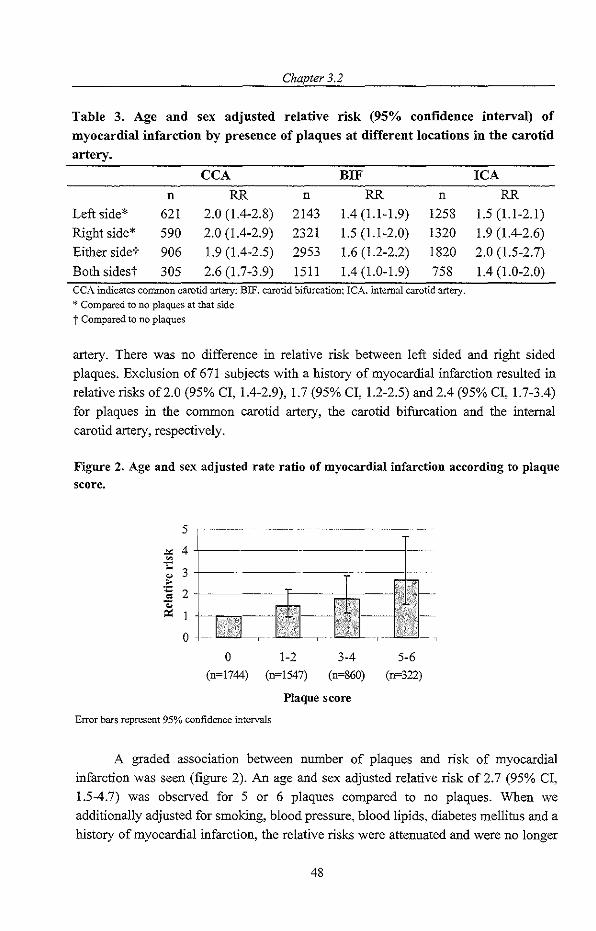
Chapter 3.2
Table 3. Age and sex adjusted relative risk (95% confidence interval) of myocardial infarction by presence of plaques at different locations in the carotid artery.
CCA BIF
n RR n RR n Left side* 621 2.0 (1.4-2.8) 2143 1.4 (1.1-1.9) 1258 Right side* 590 2.0 (1.4-2.9) 2321 1.5 (1.1-2.0) 1320 Either side-r 906 1.9 (1.4-2.5) 2953 1.6 (1.2-2.2) 1820
Both sides-r 305 2.6 (1.7-3.9) 1511 1.4 (1.0-1.9) 758 CCA indicates common carotid artery; BIP, carotid bifurcation: leA. internal carotid artery. <I< Compared to no plaques at that side
t Compared to no plaques
ICA
RR
1.5 (1.1-2.1) 1.9 (1.4-2.6) 2.0 (1.5-2.7)
1.4 (1.0-2.0)
artery. There was no difference in relative risk between left sided and right sided plaques. Exclusion of 671 subjects with a history of myocardial infarction resulted in relative risks of2.0 (95% cr, 1.4-2.9), 1.7 (95% cr, 1.2-2.5) and 2.4 (95% cr, 1.7-3.4) for plaques in the common carotid artery, the carotid bifurcation and the internal carotid artery, respectively.
Figure 2. Age and sex adjusted rate ratio of myocardial infarction according to plaque score.
5
-'" 4 .'" '" 3 " '" ~ 2
" 0::
0
o 1-2 3-4 5-6 (n~1744) (n~1547) (n~860) (n~322)
Plaque score
Error bars represent 95% confidence intervals
A graded association between number of plaques and risk of myocardial infarction was seen (figure 2). An age and sex adjusted relative risk of 2.7 (95% cr, 1.5-4.7) was observed for 5 or 6 plaques compared to no plaques. When we additionally adjusted for smoking, blood pressure, blood lipids, diabetes mellitus and a history of myocardial infarction, the relative risks were attenuated and were no longer
48

Plaques in the carotid artery and risk of myocardial infarction
significant. The relative risks associated with the number of calcified plaques and noncalcified plaques were highly comparable (table 4). The relative risk of myocardial
infarction for 4 to 6 calcified plaques compared to no calcified plaques was 2.0 (95%
CI, 1.0-4.2), while 4 to 6 non-calcified plaques compared to no non-calcified plaques
resulted in a relative risk of 2.1 (95% CI, 0.8-6.0). When the number of calcified
plaques and the plaque score were simultaneously added to the model to obtain the
independent risk estimate of calcification of plaques, the relative risk of 4-6 calcified
plaques compared to no calcified plaques was attenuated and not significant, 1.3 (95% CI,0.5-3.2).
Table 4. Age and sex adjusted relative risk (95% confidence interval) of myocardial iufarction by calcification ofthe plaque.
Number of plaques
0* 1-3 4-6
* Reference category.
Discussion
Calcified plaques
1.0 1.5 (1.0-2.4)
2.0 (1.0-4.2)
N on-calcified plaques
1.0 l.l (0.7-1.7)
2.1 (0.8-6.0)
We found a graded association between the number of plaques iu the carotid artery and risk of myocardial infarction. The risk of myocardial infarction was 2.7 times higher in
subjects with 5 or 6 plaques in the carotid artery compared to subjects without plaques
in the carotid artery. The presence of plaques in the carotid artery was associated with future myocardial infarction, irrespective of side and location of the plaque. The relationships with incident myocardial infarction were of equal strength for calcified
plaques and non-calcified plaques. Most studies that investigated the association between carotid plaques and
cardiovascular disease focused on cerebrovascular disease. In the Cardiovascular
Health Study (CHS), subjects with calcified plaques had a nearly two-fold increased
prevalence of cerebral infarcts on MRI scan compared to subjects with isodense
plaque.' In the British Regional Heart Study carotid plaques were cross-sectionally
associated with chest pain, intermittent claudication and prevalent ischemic heart disease. lo Only one study examined the association with the risk of coronary heart
disease prospectively. Salonen et al. found that small plaques anywhere in the carotid artery were associated with a relative risk of acute coronary events of 4.2 (1.5-11.5)
and large (stenotic) plaques with a relative risk of 6.7 (1.3-33.9), but analyses were conducted using a limited number of events (n=24).14
49

Chapter 3.2
We found no clear difference in risk for calcified and non-calcified plaques. Recent data on coronary artery calcifications have suggested that calcification is an active process that stabilizes vulnerable plaques and protects against rupture.17
However, whether subjects with predominantly calcified plaques in the carotid artery also have predominantly calcified plaques in the coronary arteries is not known. Recent evidence suggests that an other plaque characteristic, plaque-surface morphology (smooth versus irregular) is a systemic measure of plaque stability, and is related to future cardiac events18
In this older population, the prevalence of plaques was relatively high, especially in the carotid bifurcation and internal carotid artery. The predominance of plaques at these sites may be due to more turbulence of blood flow in the carotid bifurcation and the internal carotid artery.'·lo In our study, most established cardiovascular risk factors were associated with plaques in the carotid artery, except BM!. Similar results were found in other studies. Bonithon-Kopp et al. found in the Vascular Aging (EVA) Study that the presence of carotid plaques was strongly related to age, hypertension and hypercholesterolemia and weakly with ever smokingll As in our study, BMI showed no association with the presence of plaques. Ebrahim et al. found in The British Regional Heart Study that BDL-cholesterol, total cholesterol and systolic blood pressure were associated with plaques in the carotid artery.lo
Some limitations of the study need to be addressed. Missing data on one or more sites was present for part of the study subjects because of logistic reasons and poor visualization. Poor visualization may be due to overweight or tortuous vessels. We found that BMI was not associated with plaques in the carotid artery. If tortuous vessels would be associated with more extensive atherosclerosis, most severe cases of atherosclerosis would have missing data on carotid plaques, which would have led to an underestimation of the true association.
We computed a plaque score based on the number of sites with plaque. In the CHS as well as in the EVA Study siguificant associations between intima-media thickness and number of plaques were found. Several other cross-sectional studies have shown that carotid plaques preferentially occurred in older people with intimamedia thickening.5
.6.!' The British Regional Heart Study reported that carotid intima
media thickness and carotid plaque were correlated with each other but showed different patterns of association with risk factors and prevalent disease. lo
This is the first population-based study showing a graded association between plaques in the carotid artery and risk of myocardial infarction. The associations were present irrespective of side, location and calcification of the plaque. The results indicate that measurement of carotid plaques may play an important role in cardiovascular disease risk assessment.
50

Plaques in the carotid artery and risk qf myocardial infarction
References
I. Heiss G, Sharrett AR, Barnes R, Chambless LE, Szklo M, Alzola C. Carotid atherosclerosis measured by B-mode ultrasound in popUlations: associations with eardiovaseular risk factors in the ARIC study. Am J Epidemio!. 1991;134:250-6.
2. Wendelhag I, Wiklund 0, Wikstrand J. Arterial wall thicmess in familial hypercholesterolemia. Ultrasound measurement of intima-media thiclmess in the connnon carotid artery. Arterioscler Thromb. 1992;12:70-7.
3. Bots ML, Hofman A, de Bruyn AM, de Jong PT, Grobbee DE. Isolated systolic hypertension and vessel wall thicmess of the carotid artery. The Rotterdam Elderly Study. Arterioscler Thromb. 1993;13:64-9.
4. Gronholdt ML. Ultrasound and lipoproteins as predictors of lipid-rich, rupture-prone plaques in the carotid artery. Arterioscler Thromb Vase Bioi. 1999;19:2-13.
5. Prati P, Vanuzzo D, Casaroli M, Di Chiara A, De Biasi F, Feruglio GA, Touboul PJ. Prevalence and determinants of carotid atherosclerosis in a general population. Stroke. 1992;23: 1705-11.
6. Salonen R, Seppanen K., Rauramaa R, Salonen IT. Prevalence of carotid atherosclerosis and serum cholesterol levels in eastern Finland. Arteriosclerosis. 1988;8:788-92.
7. Tonstad S, Joakimsen 0, Stensland-Bugge E, Leren TP, Ose L, Russell D, Bonaa KH. Risk factors related to carotid intima-media thiclmess and plaque in children vvith familial hypercholesterolemia and control subjects. Arterioscler Thromb Vasc Bio!.
1996; 16:984-91.
8. Salonen R, Tervahauta M, Salonen IT, Pekkanen J, Nissinen A, Karvonen MJ. rntrasonographic manifestations of common carotid atherosclerosis in elderly eastern Finnish men. Prevalence and associations with cardiovascular diseases and risk
factors. Arterioscler Thromb. 1994; 14: 1631-40.
9. Manolio TA, Burke GL, O'Leary DH, Evans G, Beauchamp N, Knepper L, Ward B. Relationships of cerebral MR1 findings to ultrasonographic carotid atherosclerosis in older adults: the Cardiovascular Health Study. CHS Collaborative Research Group. Arterioscler Thromb Vase Bio!. 1999;19:356-65.
10. Ebrahim S, Papacosta 0, Whincup P, Warmamethee G, Walker M, Nicolaides AN, Dhanjil S, Griffin M, Belcaro G, Rumley A, Lowe GD. Carotid plaque, intima media thiclmess, cardiovascular risk factors, and prevalent cardiovascular disease in men and
women: the British Regional Heart Study. Stroke. 1999;30:841-50.
11. Bonithon-Kopp C, Touboul PJ, Berr C, Leroux C, Mainard F, Courbon D, Ducimetiere P. Relation of intima-media thickness to atherosclerotic plaques in carotid
arteries. The Vascular Aging (EVA) Study. Arterioscler Thromb Vase Bio!.
1996;16:310-6.
12. Polak JF, Shemanski L, O'Leary DH, Lefkowitz D, Price TR, Savage PJ, Brant WE, Reid C. Hypoechoic plaque at US of the carotid artery: an independent risk factor for incident stroke in adults aged 65 years or older. Cardiovascular Health Study.
Radiology. 1998;208:649-54.
51

Chapter 3.2
13. Belcaro G, Nicolaides AN, Laurora G, Cesarone MR De Sanctis M, Incandela L, Barsotti A. Ultrasound morphology classification of the arterial wall and cardiovascular events in a 6-year follow-up study. Arterioscler Thromb Vase BioI.
1996;16:851-6. 14. Salonen JT, Salonen R. Ultrasonographically assessed carotid morphology and the risk
of coronary heart disease. Arterioscler Thromb. 1991 ;11: 1245-9. 15. Hofman A, Grobbee DE, de Jong PT, van den Ouweland FA. Determinants of disease
and disability in the elderly: the Rotterdam Elderly Study. Eur J Epidemiol.
1991;7:403-22.
16. Lamberts H, Wood M, Hofmans-Okkes I. The International Classification o/Primary
Care in the European Community. London, UK: Oxford University Press; 1991. 17. Doherty TM, Detrano RC, Mautner SL, Mautner GC, Shavelle RM. Coronary
calcium: the good, the bad, and the uncertain. Am HeartJ. 1999;137:806-14.
18. Rothwell PM, Villagra R, Gibson R, Donders RC, Warlow CPo Evidence ofa chronic systemic cause of instability of atherosclerotic plaques. Lancet. 2000;355: 19-24.
19. Bonithon-Kopp C, Scarabin PY, Taquet A, Touboul PJ, Malmejac A, Guize L. Risk factors for early carotid atherosclerosis in middle-aged French women. Arterioscler Thromb. 1991; 11 :966-72.
52

Abstract
3.3
Carotid plaques and risk of stroke and subtypes of cerebral infarction
Background: Carotid plaques are considered to increase the risk of stroke. Few
studies have quantified this relationship in asymptomatic subjects and limited data are
available on the importance of location of plaques, or the effect on subtypes of
cerebral infarction. It is still controversial whether the plaques merely reflect
generalized atherosclerosis or are directly causally related to subsequent stroke. We
investigated the relationship between carotid plaques, measured at different locations,
and risk of stroke and subtypes of cerebral infarction in a population based study.
Methods: The study was based on the Rotterdam Study and included 4336 subjects
aged 55 years or over who were free from stroke. Presence of carotid plaques in left
and right sided common carotid artery, bifurcation and internal carotid artery was
assessed at baseline (1990-1993). Severe plaques were defined as presence of 5 to 6
plaques. After a mean follow up of 5.2 years 172 strokes had occurred. The
association between plaques and risk of stroke was analyzed using a Cox proportional
hazards regression model.
Results: Plaques in the common carotid artery, bifurcation and internal carotid artery
increased the risk of stroke and cerebral infarction approximately 1.5-fold. Severe
carotid plaques increased risk of non-lacunar infarction in the anterior (RR 3.6 (95%
CI 1.5-9.0)), but not in the posterior circulation (RR 0.5, 95% CI 0.1-3.8). A strong
and graded association with lacunar infarction was observed. with an almost 10-fold
increased risk in subjects with severe plaques (RR 9.8, 95% CI 2.0-46. 7).
Conclusion: Carotid plaques increase the risk of stroke and cerebral infarction,
irrespective of location in the carotid artery. A stronger association with infarctions in
the anterior than in the posterior circulaton was found. No clear difference was seen
in risk estimates for ips lateral and contralateral infarctions.
53

Chapter 3.3
Introduction Carotid plaques are frequently fouod in subjects who suffered from a stroke1
-3 Few
studies have quantified the association between carotid plaques and risk of subsequent
stroke in asymptomatic subjects4 Also, limited information is available on the
relationship with subtypes of cerebral infarction4•s, as well as about the impact of
location of the carotid plaque in relation to the risk of stroke. One could hypothesize
that because of more turbulence of bloodflow, bifurcation and internal carotid artery
plaques carry a higher risk than plaques in the common carotid artery.5.6 It is also still
controversial whether carotid plaques merely reflect generalized atherosclerosis or are
directly causally related to subsequent stroke by release of thrombo emboli.s"" We
investigated the association between asymptomatic plaques, measured at six locations
in the carotid arteries and the risk of stroke and subtypes of cerebral infarction in a
population-based cohort of elderly persons.
Methods Population
The study is part of the Rotterdam Study, a population-based cohort study on chronic
and disabling diseases in the elderly. All inhabitants of Ommoord, a suburb of
Rotterdam, aged 55 years or more were invited. People living in homes for the elderly
were included. Participation rate of those invited for the study was 78% and in total
7983 subjects participated.' The Medical Ethics Committee of Erasmus University
Rotterdam approved the study. Written informed consent to retrieve information from
treating physicians was obtained from all participants. Baseline measurements were
obtained from 1990 to 1993 and consisted of an interview at horne and two visits to the
research center for physical examination. From the 7129 subjects who visited the
research center, 215 experienced a previous stroke. Participants with a previous stroke
were excluded from the present study.
Assessment of carotid plaques
At baseline 5679 participants who were free from previous stroke uoderwent B-mode
ultrasonography of both carotid arteries with a 7.5-MHZ linear array transducer (ATL
Ultra-Mark IV to assess intima media thickness and presence of plaques.1O Missing
ultrasouod data were mainly due to restricted availability of ultrasonographers. The
left and right common carotid artery, bifurcation and internal carotid artery were
visualized and examined for the presence of plaques which were defined as focal
widenings relative to adjacent segments, with protrusion into the lumen, composed of
calcified or non-calcified components. Plaques were assessed in 5444 randomly
selected participants. Ultrasonographers and readers who assessed the presence of
plaques were blinded for all clinical information. The total plaque score reflected the
54

Carotid plaques and risk of stroke and subtypes of cerebral infarction
total number of sites witb plaques and ranged from 0 to 6 (left and right-sided common carotid artery, bifurcation and internal carotid artery). A total of 1108 participants had a missing plaque score at one or more locations, leaving 4336 participants of whom we had information on plaques at all locations in tbe carotid artery. The 1108 participants witb missing plaque score had on average slightly higher diastolic (P<O.OOI) and systolic blood pressure (P<0.05) levels and body mass index (P<O.OO I), compared with those without missings. The groups did not differ according to age, gender, diabetes, carotid IMT, and total cholesterol. A reproducibility study of tbe on line plaque assessment resulted in a kappa of 0.66 for the left, 0.68 for tbe right carotid artery and 0.67 for eitber side, indicating a moderate agreement. In a subgroup of 1775 participants from tbe Rotterdam study, assessment of stenosis in tbe right internal carotid artery was performed during tbe ultrasound examination. The prevalence of stenosis >80% was 0.3%.
Assessment of stroke and subtypes
During tbe baseline interview a previous stroke was assessed by asking "did you ever suffer from a stroke, diagnosed by a physician?" Medical records of subjects who answered 'yes' were checked in order to verify tbe diagnosis. 11 Once subjects enter tbe Rotterdam Study they are continuously monitored for major events tbrough automated linkage with tbe files from general practitioners. Information on vital status is obtained at regular intervals from tbe municipal authorities in Rotterdam. When an event or deatb has been reported, additional information is obtained by interviewing tbe general practitioner and scrutinizing information from hospital discharge records in case of admittance or referral. A neurologist (p.J.K.) reviewed information on all possible strokes. He classified tbe stroke as definite, probable or possible. 12 We only used definite and probable strokes in tbe analyses. If a CT or MRI, performed witbin 28 days, showed a hemorrhage or infarction tbe type of stroke was coded accordingly. We classified tbe stroke as cerebral infarction in case of no abnormality on CT or MRI. Strokes witbout neuro-imaging were classified as unspecified or as possible hemorrhagic or ischemic in case of tbe following symptoms. The stroke was considered to be hemorrhagic if tbere was sudden hemiplegia or otber focal signs witb permanent unconsciousness or death within hours. The stroke was classified as ischemic in case of limited impairment (isolated aphasia, isolated weakness of one limb, isolated facial weakness or isolated hemianopia), complete improvement within 72 hours or documented atrial fibrillation at time oftbe diagnosis. We further classified cerebral infarctions as lacunar or non-lacunar, based on clinical symptoms and/or neuro-imaging. Furtbermore, we subclassified tbem as located in tbe anterior or posterior circulation. For tbe present study, follow-up for stroke was complete in all participants untiIJanuary I, 1998.
55

Chapter 3.3
Medical history and riskJactors
Information on current health status and medical history at baseline was obtained using a computerized questionnaire. A neurologist verified all transient ischemic attacks reported by participants during the interview.13 Participants were classified as current, former or never smoker. Non-fasting blood samples were taken and serum total cholesterol and high-density lipoprotein (HDL) cholesterol was measured using an automated enzymatic procedure. Sitting blood pressure was measured twice on the right arm with a random-zero sphygmomanometer. In the analyses we used the
average of the two measurements. Hypertension was defmed as a systolic blood pressure of 160 mmHg or over, or a diastolic blood pressure of 95 mmHg or over, or current use of antihypertensive drugs for the indication of hypertension. I4 Diabetes mellitus was defined as use of oral blood glucose lowering drugs or insulin or random or post-load serum glucose level higher than 11.0 mmollL.
Data analysis
The relation between carotid plaques and the risk of stroke was assessed with a Cox proportional hazards regression model. We assessed the relation between total plaque score and risk of stroke and cerebral infarction. Participants without any plaques were taken as the reference. We also analyzed the presence of one or more plaques at different locations (left andlor right sided common carotid artery, bifurcation and
internal carotid artery) in relation to the risk of stroke and cerebral infarction. In order to distinguish between generalized atherosclerosis and thrombo-embolism we assessed whether the association between left and right-sided plaque score (0-3) and risk of infarction was different for ipsilateral and contralateral infarction. In these analyses subjects with zero plaques in left- and respectively right-sided carotid artery were taken as the reference. Further, we determined the relation between carotid plaques and subtypes of infarction, in particular non-lacunar infarction in the anterior and posterior circulation and lacunar infarction. All analyses were adjusted for age and gender and additionally for hypertension, diabetes mellitus, smoking, HDL- and totalcholesterol. We performed additional analyses excluding subjects with previous transient ischemic attack (n=168). Results are presented as rate ratios with corresponding 95% confidence intervals.
Results Table I shows the baseline characteristics of the study population. During 22674 person years of follow-up (mean follow-up 5.2 years) 172 definite or probable strokes occurred. A CT or MRI was performed in 65.2% of the cases. Subtyping revealed 106 cerebral infarctions, 14 intracerebral hemorrhages, I subarachnoid hemorrhage and 51 unspecified strokes. The infarction was lacunar in 21 cases (20%) of which 19 were
56

Carotid plaques and risk of stroke and subtypes of cerebral infarction
located in the anterior circulation. A total of 85 infarctions were non-lacunar 43 of
which were located in the anterior and 19 in the posterior circulation. In 23 infarctions
the location was unspecified. The group with stroke was on average older and
comprised a larger proportion of men than the cohort. We observed a higher
prevalence of carotid plaques at all locations in subjects with subsequent stroke. The
number of plaques in the left and right carotid artery were significantly correlated
(Spearman correlation coefficient 0.63).
Table 1. Baseline cbaracteristics ofthe study population.
Age
Gender (% female)
Systolic blood pressure (rrunHg)
Diastolic blood pressure (rrunHg)
Total cholesterol (mmoVL)
HDL cholesterol (mmoVL)
Diabetes (%)
Previous myocardial infarction (%)
Previous TlA (%)
Smoking (% current smokers)
Atrial fibrillation (%)
Any carotid plaques (%)
Plaque(s) in common carotid artery (%)
Plaque(s) in carotid bifurcation (%)
Plaque(s) in internal carotid artery (%)
Plaque(s) in left carotid artery (%)
Plaque(s) in right carotid artery (%)
Total plaque score (range 0-6) Valucs represent mcans (sd), or percentages.
Study population
(n=4336)
68.9 (8.8)
59.9
139.6 (225)
73.9 (I 1.8)
6.6 (1.2)
1.4 (0.4)
10.0
12.0
2.7
23.4
2.5
60.4
15.4
53.6
34.9
49.7
48.5
1.6
Carotid plaques and risk oj stroke and cerebral inJarction
Stroke
(n =172)
74.5 (9.0)
52.3
149.8 (23.8)
75.2 (13.3)
6.4 (1.2)
1.3 (0.4)
21.5
18.0
7.6
24.7
5.2
75.6
26.2
71.5
47.1
65.7
65.1
2.4
The risk of stroke and cerebral infarction gradually increased with increasing total
plaque score. The risk of stroke in subjects with severe plaques (score 5 to 6) was 2.7
fold increased (RR 2.7 (95% CI 1.6-4.5» and the risk of cerebral infarction more than
tripled (RR 3.12 (95% CI 1.62-6.02», compared to subjects without plaques (table 2).
Plaques in the common carotid artery increased the risk of stroke and cerebral
infarction 1.6 and respectively 1.8 times. In the bifurcation and internal carotid artery
plaques increased the risks approximately 1.5-fold. These associations did not
57

Chapter 3.3
materially change after adjustment for other factors and restriction to subjects without previous transient ischemic attack.
Table 2. Rate ratio of stroke and cerebral infarction in relation to carotid plaqnes, adjusted for age and gender.
Subjects Stroke Cerebral infarction at risk
No of Rate ratio No of Rate ratio events events
No plaque (ref.) 1715 42 1.00 29 1.00
Plaques at different locations :2:1 plaque in left or 669 45 1.75 (1.09-2.82) 25 1.55 (0.87-2.74)
right CCA :2:1 plaque in left or 2322 123 1.52 (1.04-2.22) 73 1.58 (0.99-2.53)
right BIF :2:1 plaque in left or 1514 81 1.39 (0.92-2.09) 49 1.53 (0.92-2.55)
rightICA
Total plaque score 1-2 1503 58 I.19 (0.78-1.81) 34 1.16 (0.68-1.97) 3-4 823 43 1.36 (0.85-2.16) 25 1.53 (0.86-2.74) 5-6 295 29 2.69 (1.61-4.52) 18 3.12 (1.62-6.02)
Any Elague 2621 130 1.48 (1.03-2.11) 77 1.44 (0.93-2.24) Groups with plaques at different locations are not mutually exclusive.
Carotid plaques in relation to inJarction in left and right hemisphere and subtypes oj infarction
A score of 3 plaques in the right carotid artery increased the risk of infarction in the ipsilateral rather than the contralateral hemisphere (RR 4.30 vs. 1.28), although the difference was not statisticaIly significant (table 3). For plaques in the left carotid artery the corresponding risks of ipsilateral and contralateral infarction were almost similar (RR 4.19 vs. 4.04). Presence of any plaque in the left or right carotid artery statistically significantly more than doubled the risk of infarction in the ipsilateral hemisphere, while no significantly increased risk of infarction in the contralateral hemisphere was found. Severe plaques (5-6 plaques) increased the risk of non-lacunar infarction in the anterior circulation 3.6 times (table 4). The risk of anterior circulation infarction was very similar for plaques at the different locations (common carotid artery, bifurcation or internal carotid artery). Neither total plaque score nor plaques at different segments of the carotid artery showed a significant relationship with the risk
58

Carotid plaques and risk of stroke and subtypes of cerebral infarction
of non-lacunar infarction in the posterior circulation. Increase in total plaque score
gradually increased risk of lacunar infarction. Participants with 3-4 plaques had a more
than five times increased risk and participants with 5-6 plaques had an almost ten-fold
increased risk of lacunar infarction. The results did not materially change after
exclusion of subjects with previous transient ischemic attack and after additional
adjustment for cardiovascular risk factors. The risk of lacunar infarction in participants
with plaques in the common carotid artery, bifurcation or internal carotid artery
significantly increased 5.7 to 8.1 times. The magnitude of these associations decreased
to respectively 3.5 and 5.3 after adjustment for cardiovascular risk factors, but did not
lose significance. Exclusion of subjects with previous transient ischemic attack did not
change the results.
Table 3. Rate ratio of ipsi- and contralateral infarction in relation to the side of
carotid plaqne, adjnsted for age and gender.
Infarction in right Infarction in left
hemisphere hemisphere
n=38 n=36
Severity of plaques No. of Rate ratio No. Of Rate ratio
left carotid artery* events events
0 II 1.00 10 1.00
8 1.16 (0.44-3.07) 13 1.91 (1.80-4.54)
2 11 2.12 (0.82-5.51) 10 2.95 (1.20-7.25)
3 8 4.04 (1.38-11.8) 3 4.19 (1.36-12.9)
Any plaque 27 1.79 (0.82-3.88) 26 2.45 (1.16-5.19)
Severity of plaques
right carotid artery*
0 10 1.00 13 1.00
8 1.75 (0.65-4.73) 14 1.92 (0.86-4.27)
2 14 3.36 (1.29-8.71) 7 2.07 (0.85-5.03)
3 6 4.30 (1.34-13.8) 2 1.28 (0.28-5.92)
Any plaque 28 2.58 (1.13-5.19) 23 1.90 (0.93-3.90) >I< Number of locations where plaques were observed (range Ow3).
59

Table 4. Rate ratio of subtypes of cerebml infarction in relation to carotid plaques, adjusted for age and gender. ~"= __ ~_~~~"~~=. __ "~~~~:~~_~~~ ~.,~~~~~~~==~_~~~_~~~~~~_~~~-=_,=,~_,"_~~~_~_~~_=="_~ ___ ~~=~~~~=~_x_~~~~:~~_~_-~~=~~_~_~~==~_~,~~~~~~,~~~~-~~~~~~~~:~~~~~~~~~~~
No plaque (ref.)
Plaques at different
locations
<: I plaque in left or
right CCA
<:1 plaque in left or
right BlF
<:1 plaque ill left or
right rCA
Total plaque score
Subjects Anterior circulation Posterior circulation Lacunal' infarction
at l'isl{ infarction infarction
No of
events
1715 II
669 II
2322 30
1514 23
Rate ratio
1.00
1.55 (0.60-4.08)
1.41 (0.66-3.02)
1.54 (0.68-3.45)
No of
events
10
2
9
6
Rate ratio
1.00
0.46 (0.09-2.25)
0.63 (0.24-1.64)
0.56 (0.19-1.63)
No of Rate ratio
events
2 1.00
6 8.08 (1.48-44.16)
19 6.49 (1.47-28.69)
10 5.68 (1.16-27.75)
1-2 1503 13 1.07 (0.47-2.42) 5 0.41 (0.13-1.32) 8 3.44 (0.92-12.84)
3-4 823 9 0.94 (0.36-2.50) 3 0.50 (0.13-1.87) 7 5.25 (1.31-21.07)
5-6 295 10 3.61 (1.45-9.01) 0.47 (0.06-3.80) 4 9.77 (2.04-46.68)
Any plaque 2621 32 1.29 (0.63-2.63) 9 0.44 (0.17-1.16) 19 4.39 (1.28-15.14) .~~"_~~~~==~~~:_-_~~~=~_, ___ "~~~~~_~. ~'"' __ ~"~~~ __ ~-~_~= __ ~"·~_~~~~~~~~~~.~·-~~~~_~~~~w ___ '~~~~~~p~~, __ ~~~~~~_-_~~~~__ ----~ __ ~~~~=~:~_~~~~~~~=

Carotid plaques and risk of stroke and subtypes ~f cerebral infarction
Discussiou The present study shows that carotid plaques increase the risk of stroke and cerebral infarction, irrespective of their location in the carotid artery. We found that carotid plaques increased the risk of infarction in the anterior but not in the posterior circulation. Finally, a strong association between carotid plaques and risk of lacunar infarction was found.
A few methodological issues need to be addressed. First, restriction to participants without a missing plaque score at any location could have led to a selection bias. We observed that participants with a missing score on average had slightly higher blood pressure levels and body mass index compared with those without missing values. However, no differences in other baseline variables where among carotid IMT was observed. Therefore, we think selection bias is unlikely. Misclassification of strokes could have occurred in our study, especially when information was limited. In the analyses of infarctions and their subtypes we used only infarctions in which a CT or MRI excluded a hemorrhage or infarctions with symptoms that were very suggestive of ischemic stroke. In most of these cases the stroke was diagnosed accurately. Classification of strokes was performed blinded for plaque score. Misclassification of stroke, if present, is therefore non-differential, again potentially resulting in an underestimation of the true effect The reproducibility of plaques showed a moderate agreement. However, since plaque assessment was performed blinded for case status, misclassification is likely to be non-differential, leading to an underestimation of the true associations. 15 The advantage of the present study is that it was done in asymptomatic participants in whom carotid stenosis was rare. From studies in symptomatic subjects we know that degree of carotid stenosis and echodensity of carotid plaques are associated with the risk of cerebral infarction. I
.3
The question remained what the role of carotid plaques is in relation to cerebral infarctions in asymptomatic subjects. The Cardiovascular Health Study is the only population-based cohort study that has investigated carotid plaques in relation to subsequent stroke in asymptomatic subjects! In that study, 4886 participants were followed for 3.3 years and 175 strokes occurred. The study focused on plaque
characteristics, not plaque localization or plaque load and concluded that hypoechogenic, but not hyperechogenic plaques were associated with increased risk of ischemic stroke. Plaques were scored as present in the bifurcation or internal carotid artery, without distinction between bifurcation and internal carotid artery. Plaques in the common carotid artery were not analyzed. Furthermore, not all subtypes of cerebral infarction were considered. The current study expands the findings of the Cardiovascular Health study in all these aspects.
The present study showed that the risk of stroke and cerebral infarction associated with carotid plaques does not depend on plaque location in the carotid
61
Chapter 3.3
artery. A considerable overlap of plaques at the different locations may explain the lack of difference in risk between location. Another explanation is that in an asymptomatic population carotid plaques primarily are indicators of generalized atherosclerosis and that location in the carotid artery does not matter.
We further aimed to clarity the underlying mechanism of the association between carotid plaques and cerebral infarction. The higher risk of cerebral infarction with increasing plaque score could indicate that a higher plaque score reflects more severe generalized atherosclerosis. On the other hand, if a participant has a high plaque score then the probability of having plaques that are vulnerable for thrombo-embolism is also higher. Thus, we can not distinguish on the basis of these results between generalized atheroslerosis and thrombo-embolism. For plaques in the right carotid artery we observed a higher risk of ipsilateral infarction compared to contralateral infarction whereas for plaques in the left carotid artery the risks of ipsilateral and contralateral infarction were similar. These results do not provide conclusive evidence that carotid plaques are sources of thrombo-emboli. Plaques in the left and right carotid artery were highly correlated and similarly distributed. Numbers were not high enough to examine the risks in subjects with plaques only at one side. We were not able to establish whether a ruptured plaque was present in the ipsilateral carotid artery when an ischemic stroke occurred. Two other studies also did not find differences in occurrence of carotid atherosclerosis and plaques between the ipsi- and contralateral carotid artery in symptomatic subjects7
.!6 An alternative approach to distinguish
between thrombo-embolism and generalized atherosclerosis was to compare associations between carotid plaques and risk of infarction in the anterior and posterior circulation. We observed that carotid plaques increased the risk of infarction in the anterior, but not in the posterior circulation, although a small number of posterior infarctions could be an alternative explanation for the absence of an association. These findings indicate that carotid plaques are sources of thrombo-emboli.
We found a strong and gradual association between carotid plaques and risk of lacunar infarction. Three possible mechanisms could underlie this association. First and most likely, carotid plaques are markers of intracranial atherosclerosis on small vessel level, which is believed to be an important causes of lacunar infarctions. Secondly, severe carotid plaques may develop into carotid stenosis and impairment of the bloodflow through the carotid arteries, leading to hypoperfusion of small cerebral vessels. This hypothesis is supported by reports on the association between carotid stenosis and lacunar infarction17
-19
, but is not very plausible, since carotid stenosis in our population was rare. A third explanation is that micro-emboli, originating from carotid plaques, occlude small perforating vessels and thereby cause a lacunar infarction. A recent study on patients with classical lacunar syndrome and diffusion
weighted imaging described a subset of patients with multiple lacunar infarctions
62

Carotid plaques and risk of stroke and subtypes of cerebral infarction
associated with an embolic cause from the heart and large arteries?O Nonetheless, it is
not likely that the majority of lacunar infarctions in the present study were caused by
thrombo-emboli.
This is the first prospective population-based study that investigated plaques at
all locations in the carotid artery in relation to stroke and subtypes of cerebral
infarction in asymptomatic and non-stenotic subjects. Increased total plaque score
increased the risk of stroke, infarction in the anterior circulation and lacunar infarction,
independent of plaque location.
References 1. Golledge J, Cuming R, Ellis M, Davies AH, Greenhalgh RM. Carotid plaque
characteristics and presenting symptom. Br J Surg. 1997;84:1697-701.
2. Gomez RG. Carotid plaque morphology and risk for stroke. Stroke. 1990;21: 148-151. 3. Sabetai MM, Tegos TJ, Nicolaides AN, EI-Atrozy TS, Dhanjil S, Griffin M, Belcaro
G, Geroulakos G. Hemispheric symptoms and carotid plaque echomorphology.J Vasc
Surg. 2000;31 :39-49. 4. Polak JF, Shemanski L, O'Leary DH, Lefkowitz D, Price TR, Savage PJ, Brant WE,
Reid C. Hypoechoic plaque at US of the carotid artery: an independent risk factor for incident stroke in adults aged 65 years or older. Cardiovascular Health Study. Radiology. 1998;208:649-54.
5. Rothwell PM. Carotid artery disease and the risk of ischaemic stroke and coronary vascular events. Cerebrovasc Dis. 2000; I 0:21-33.
6. Dempsey RJ, Diana AL, Moore RW. Thickness of carotid artery atherosclerotic plaque and ischemic risk. Neurosurgery. 1990;27:343-8.
7. Manolio TA, Burke GL, O'Leary DH, Evans G, Beauchamp N, Knepper L, Ward B. Relationships of cerebral MRI fmdings to ultrasonographic carotid atherosclerosis in older adults: the Cardiovascular Health Study. CHS Collaborative Research Group. Arterioscler Thromb Vasc BioI. 1999;19:356-65.
8. Handa N, Matsumoto M, Maeda H, Hougaku H, Kamada T. Ischemic stroke events and carotid atherosclerosis. Results of the Osaka Follow-up Study for illtrasonographic Assessment of Carotid Atherosclerosis (the OSACA Study). Stroke.
1995;26:1781-6. 9. Hofman A, Grobbee DE, de Jong PT, van den Ouweland FA. Determinants of disease
and disability in the elderly: the Rotterdam Elderly Study. Eur J Epidemiol.
1991;7:403-22. 10. Bots ML, Hofman A, De Jong PT, Grobbee DE. Common carotid intima-media
thiclmess as an indicator of atherosclerosis at other sites of the carotid artery. The Rotterdam Study. Ann Epidemiol. 1996;6:147-53.
II. Bots ML, Looman SJ, Koudstaal PJ, Hofman A, Hoes AW, Grobbee DE. Prevalence of stroke in the general population. The Rotterdam Study. Stroke. 1996;27:1499-501.
12. Bots ML, van der Wi!k EC, Koudstaal PJ, Hofman A, Grobbee DE. Transient
63

Chapter 3.3
neurological attacks in the general population. Prevalence, risk factors, and clinical
relevance. Stroke. 1997;28:768-73.
13. Bots ML, Hoes AW, Koudstaal PJ, Hofman A, Grobbee DE. Conuuon carotid intimamedia thickness and risk of stroke and myocardial infarction: the Rotterdam Study.
Circulation. 1997;96: 1432-7. 14. The fifth report of the Joint National Committee on Detection, Evaluation, and
Treatment of High Blood Pressure (JNC V). Arch Intern Med. 1993;153:154-83.
15. Rothman KJ, Greenland S. Modern Epidemiology. 1998:127-130. 16. Hallerstam S, Carlstrom C, Zetterling M, Konrad P, Rosfors S. Carotid atherosclerosis
in relation to symptoms from theTerritory supplied by the carotid artery. Eur J Vasc
Endovasc Surg. 2000;19:356-61.
17. Zhu CZ, Norris JW. Lacunar infarction and carotid stenosis (abstract). Ann Neurol.
1991 ;30:244.
18. Tegeler CH, Shi F, Morgan T. Carotid stenosis in lacunar stroke. Stroke.
1991;22:1124-8.
19. Bogousslavsky J. Carotid stenosis and lacunar stroke. Stroke. 1992;23:437-8.
20. Ay H, Oliveira-Filho J, Buonanno FS, Ezzeddine M, Schaefer PW, Rordorf G, Schwamm LH, Gonzalez RG, Koroshetz WJ. Diffusion-weighted imaging identifies a
subset oflacunar infarction associated with embolic source. Stroke. 1999;30:2644-50.
64

Chapter 4
Application in cardiovascular risk assessment


Abstract
4.1
Is carotid intima-media thickness useful in cardiovascular risk assessment?
Objective: We determined the contribution of common carotid IMT in the prediction
of fUture cardiovascular and cerebrovascular disease when added to established risk
factors.
Methods: We used data from a nested case-control study comprising 374 subjects with either an incident stroke or a myocardial infarction. and 1496 controls. All
subjects were aged 55 years and over and participated in the Rotterdam Study. Mean follow-up was 4.2 years (range 0.1-6.5). We evaluated which correlates of
cardiovascular and cerebrovascular disease contribute to the prediction of either a nevv incident myocardial infarction or a stroke. Logistic regression modeling and the
area under the Receiving Operating Characteristic curve (ROC area) were used to
quantify the predictive value of the established risk factors and the added value of
IMT.
Results: The ROC area of a model with age and sex only was 0.65 (95% confidence
interval. 0.62-0.69). Independent riskfactors were previous myocardial infarction and
stroke, diabetes mellitus, smo/':ing, systolic pressure, diastolic pressure, total and HDL cholesterol. These riskfactors increased the ROC area from 0.65 to 0.72 (0.69-0.75).
This model correctly predicted 17% of all subjects with cardiovascular and
cerebrovascular disease. When common carotid IMT was added to the previous model the ROC area increased to 0.75 (0.72-0.78). When only the IMT measurement was used, the ROC area was 0.71 (0.68-0.74), and 14% of all subjects were correctly
predicted. There was no difference in ROC area when using different measurement sites.
Conclusion: Adding intima-media thickness to a risk function for cardiovascular and
cerebrovascular disease does not result in a substantial increase in the predictive value when used as a screeningtool.
67

Chapter 4.1
Introduction Carotid intima-media thickness (IMT) measurements are widely being applied as a measure of atherosclerosis in studies on determinants of presence and progression of atherosclerosis and in studies on atherosclerosis as determinant of cardiovascular disease l
-4 Carotid IMT has been shown to be related to cardiovascular risk factors, prevalent cardiovascular disease and to atherosclerosis in the peripheral, coronary, and femoral arteries. Recently, evidence became available indicating that an increased carotid IMT is a strong predictor of coronary heart disease and stroke. 1
•4
-7 Therefore, it
has been suggested that measurements of carotid IMT may be used to identify highrisk subj ects. 8
The objective of the present study is to evaluate in elderly subjects of the general population which established risk factors, such as medical history, blood pressure and serum lipids, are independent predictors of cardiovascular and cerebrovascular disease and whether measurement of carotid IMT contributes to the prediction of cardiovascular and cerebrovascular disease when added to these risk factors. Eventually, we evaluated the predictive ability of the carotid IMT measurement alone, when used to replace established risk factors.
Methods Population
The Rotterdam Study is a single center prospective follow-up study on disease and disability in the elderly in 7,983 subjects, aged 55 years or over, living in the suburb of Ommoord in Rotterdam, The Netherlands, as detailed elsewhere: Baseline data were collected from March 1990 to July 1993 during a home interview and two visits at the research center. The overall participation rate of those invited for the study was 78%. The study has been approved by the Medical Ethics Committee of the Erasmus University and written informed consent was obtained from all participants.
Cardiovascular and cerebrovascular risk indicators
At baseline, information about the medical history of myocardial infarction and stroke, current medication, alcohol intake and smoking habits, was obtained by a trained research assistant. At the study center an extensive physical examination was performed, including height and weight measurement, two blood pressure measurements (taken with a random zero sphygmomanometer with the subject in sitting position, and averaged), a twelve lead ECG, serum total cholesterol and HDLcholesterol level and a non-fasting or postload glucose level.
Presence of hypertension was defined as a systolic pressure d 60 mmHg or a diastolic
pressure e:95mm Hg or current use of blood pressure lowering drugs for the indication of hypertension. Diabetes mellitus was considered to be present when subjects
68

Is carotid intima-media thickness useful in cardiovascular risk assessment?
currently used oral blood glucose-lowering drugs or insulin, or had a non-fasting or
postload glucose level above 11 mmol/L, assessed after a non-fasting venipuncture.
Incident cardiovascular and cerebrovascular disease
Information on incident fatal and non-fatal events is obtained from the general
practitioners (GPs) working in the district of Ommoord. The GPs report all possible
cases of myocardial infarction and stroke to the Rotterdam research center. Events are
coded according to the International Classification of Primary Care (ICPC) 10
Information on the vital status of the participants is obtained at regular intervals from
the municipal authorities in Rotterdam. When an event or death has been reported,
additional information is obtained by scrutinizing information from GP and hospital
discharge records in case of admittance or referral. Events are then confirmed by two
study physicians. Additionally, all data about new cases of myocardial infarction were
reviewed by a cardiologist and all data about stroke cases were reviewed by a
neurologist, both did not actually see the patient. In case of disagreement, consenSus is
reached in additional meetings. An incident myocardial infarction was considered to
have occurred when the event led to a hospitalization, and the hospital discharge
record comprised a diagnosis of a new myocardial infarction based on signs and
symptoms, ECG recordings, and repeated laboratory investigations during hospital
stay. All suspected cerebrovascular events reported by the GPs were submitted for
review to a neurologist (PJK). The neurologist classified the events as definite,
probable and possible stroke, based on all information, including symptoms and signs
obtained by interviewing the GP or, in case of hospital referral, hospital data. An
incident stroke was considered to have occurred when (1) the event had led to a
hospitalization and the hospital discharge record indicated a diagnosis of a new stroke.
The clinical diagnosis was based on signs, symptoms, and neuroimaging investigations during hospital stay; or (2) in case of no hospitalization, signs and symptoms
associated with the event obtained from the general practitioner were highly
suggestive of a stroke according to the neurologist (probable stroke); or (3) in case of
out hospital death, when the general practitioner reported that the cause of death was a
cerebrovascular accident and a cardiac cause was judged to be highly unlikely. For the
analyses, only definite and probable incident strokes were included.
Measurement oj intima-media thickness
To measure carotid intima-media thickness, ultrasonography of the common carotid
artery (CCA), carotid bifurcation (ElF), and internal carotid artery (ICA) of the left
and right carotid arteries was performed with a 7.5-MHz linear-array transducer (ATL
Ultra-Mark IV). On a longitudinal, two-dimensional ultrasound image of the carotid
artery, the anterior (near) and posterior (far) walls of the carotid artery are displayed as
69

Chapter 4.1
two bright white lines separated by a hypoechogenic space. The distance between the leading edge of the first bright line of the far wall and the leading edge of the second bright line indicates the intima-media thickness. For the near wall, the distance between the trailing edge of the fIrst bright line and the trailing edge of the second bright line at the near wall provides the best estimate of the near-wall intima-media thicknessll In accordance with the Rotterdam Study ultrasound protocol 12, a careful
search was performed to obtain the optimal representation of both the near and far walls of the distal common carotid artery, the carotid bifurcation and the internal
carotid artery. When an optimal longitudinal image was obtained, it was frozen on the R-wave of the ECG and stored on videotape. The actual measurements of intimamedia thickness were performed off-line. From videotape, the frozen images were digitized on the screen of a personal computer using additional dedicated software. This procedure has been described in detail previously 13.14 In short, with a cursor, or automatically by the computer, the interfaces of the common carotid artery were marked across a length of 10 mm. The computer then calculated the mean intimamedia thickness and the maximum intima-media thickness over the marked length for both near and far walL We used the average of the measurements of three frozen images of both the left and right side artery to obtain mean values of the mean and the maximum thickness for each subject. For the carotid bifurcation and the internal carotid artery the interfaces were marked across a variable length at the thickest part of the measurement site. Then the maximum intima-media thickness was calculated over the marked length. For the analyses, the maximum carotid intima-media thickness was determined as the mean of the maximum intima-media thickness of near- and far-wall measurements of both the left and right side arteries. A composite measure that combined the maximal common carotid intima-media thickness, the maximal bifurcation intima-media thickness and the maximal internal carotid artery intimamedia thickness when available was obtained by averaging the three measurements after standardization (subject maximum IMT minus cohort mean of the maximum IMT, divided by cohort standard deviation of the maximum IMT). The readers of the ultrasound images were unaware of the case status of the subject. Results from a reproducibility study of IMT measurements of the common carotid artery among 80 participants of the Rotterdam Study who underwent a second ultrasound of both carotid arteries within three months of the fIrst scan have been described elsewhere 15.
In short, mean differences (SD) in far-wall intima-media thickness of the common carotid artery between paired measurements of sonographers, readers, and visits were 0.005 mm (0.09), 0.060 mm (0.05), and 0.033 mm (0.12), respectively. Corresponding intraclass correlation coefficients were 0.63, 0.88 and 0.74, respectively.
70

Is carotid intima-media thickness useful in cardiovascular risk assessment?
Selection of cases and controls
Ultrasonography of the carotid arteries was perfonned in 5965 of the 7983 subjects in the Rotterdam Study. For subjects who had their baseline examination at the end of 1992 and in 1993, ultrasonography could not always be perfonned due to the restricted availability of ultrasonographers. As this may be considered as a random sample, for the present study the cases and controls were drawn from this cohort of 5965 subjects. For reasons of availability and completeness of infonnation on cardiovascular and cerebrovascular events, we restricted the present study to follow-up events registered by GPs, before May 1996. The mean duration of follow-up was 4.2 years (range 0.1-6.5 years, standard deviation 1.6). We selected 374 case subjects with incident cardiovascular and cerebrovascular disease of which 194 subjects had a myocardial infarction and 191 subjects a stroke (II subjects had both a myocardial infarction and a stroke, for which we used the event that occurred fIrst). For these subjects data on carotid IMT was obtained from the stored images on videotape. For each case subject, 4 control subjects were drawn. A subject was eligible as a control if he/she was free from myocardial infarction and stroke. The total number of control subjects was 1496, resulting in a total number of 1870 subjects.
Data analysis
First, data analysis was separately perfonned for the two outcomes (myocardial infarction and stroke) using the same control group. Only subjects with complete data on all risk factors and common carotid artery IMT measurement were included, resulting in a dataset of 1721 subjects: 328 cases (174 myocardial infarctions and 165 strokes, II had both) and 1393 controls. The association between each risk factor and myocardial infarction was quantifIed using logistic regression analyses, with adjustment for age and sex. The odds ratio (OR) and 95% confIdence interval (95% el) was used as measure of association. Variables associated with myocardial infarction (p-value <0.10) were then included in a multivariate logistic regression model to evaluate the independent contribution in the prediction of myocardial infarction. The fIrst (overall) model included all 'univariately' (i.e. age and sex
adjusted) significant variables from medical history and physical examination. Model reduction was perfonned by excluding variables that were not significantly related with myocardial infarction (odds ratio with p-value <0.10) from the overall modeL Subsequently, the reduced model was extended with carotid IMT measurements to evaluate their added value in the prediction of myocardial infarction. Differences in predictive value between all different prediction models (overall, reduced and extended) were estimated by comparison of the area under the receiver operating characteristic curve (ROC area) with standard errOL"" 7 A ROC curve of a multivariate logistic model plots the sensitivity and I-specificity at each consecutive threshold in
71

Chap/er4.1
the range of predicted probabilities of the model. The ROC area is a measure of the
discriminative or predictive ability of the model that can range from 0.5 (no
discrimination between subjects with and without myocardial infarction) to 1.0
(perfect discrimination). In all model comparisons, correlation between the models was taken into account because they were based on the same cases. IS
A ROC area reflects the overall added value of a model and does not directly indicate its clinical value.19
.20 Therefore, we additionally estimated for the final model
the absolute number of correctly predicted patients with and without myocardial
infarction.
A similar analytical approach was followed for stroke as outcome and for the
combined cardiovascular and cerebrovascular disease outcome (both myocardial infarction and stroke).
A different analysis was performed to evaluate any differences in the predictive
value for total cardiovascular and cerebrovascular disease (i.e. myocardial infarction or
stroke), between three IMT measurement sites and the combined IMT measure also
using logistic regression analyses in combination with ROC curves. This analysis was done on a restricted dataset with complete data for all three measurement sites,
resulting in a dataset of 512 subjects: 156 cases (74 myocardial infarctions and 74
strokes, 8 subjects had both) and 356 controls. Data on intima-media thickness at the carotid bifurcation was available in 64% of the 1870 subjects (74% in all myocardial infarction and stroke cases and in 61 % of the controls), of the internal carotid artery it
was available in 31% (47% in all cases and in 27% of the controls), and in 96% (92% in all cases and in 97% of the controls) it was available for the common carotid artery.
All analyses were performed by using SPSS software, version 9.0 (SPSS Inc., Chicago, Illinois).
Results The general characteristics are given in table I. The age and sex adjusted associations
for all evaluated risk indicators, including the carotid IMT measurements, with
myocardial infarction and stroke are given in table 2. Although the magnitude of the association of each variable with both cardiovascular and cerebrovascular outcomes
was different, the same predictors were of importance for both outcomes. Age, male gender, smoking, systolic blood pressure, hypertension, total cholesterol, HDL cholesterol, previous myocardial infarction, previous stroke, and the carotid IMT
measurements were highly associated with myocardial infarction. For stroke, also
diastolic blood pressure was a predictor, while total and HDL cholesterol and diabetes mellitus were much less important predictors.
72

Is carotid intima-media thickness usf4fu[ in cardiovascular risk assessment?
Table 1. Baseline Characteristics ofthe Stody Populatiou.
Characteristic Myocardial Stroke Coutrol
iufarctiou subjects
N 194 191 1496 Age (years) 72* 75* 70 Female (%) 39* 54* 62
Body mass index (kg/m2) 26.3 (3.4) 26.4 (3.9) 26.6 (3.8)
Current smoking (%) 30 28 22 Systolic blood pressure (rnmHg) 144 (22.5)* 149 (24.0)' 138 (21.3)
Diastolic blood pressure (rnmHg) 72 (12.3) 75 (13.4)* 72 (10.9)
Hypertension (%) 41* 52* 34
Total cholesterol (mmol/L) 6.9 (1.2)* 6.5 (1.2) 6.7 (1.3)
HDL cholesterol (mmol/L) 1.20 (0.28)* 1.31 (0.38) 1.34 (0.38)
Diabetes mellitus (%) 13* 14* 7 Previous MI (%) 31* 23* 12
Previous stroke (%) 6 11* 2 Max. common carotid IMT (mm) 1.17 (0.29)* 1.22 (0.35)* 1.02 (0.21)
Max. bifurcation IMT (mm) 1.76 (0.68)* 1.83 (0.73)' 1.44 (0.59)
Max. iuternal carotid IMT (mm) 1.42 (0.61)* 1.62 (0.81)* 1.15 (0.62) Values are unadjusted proportions or means with SD in parentheses.
MI indicates myocardial infarction: Max., maximal; IMT, intima-media thickness.
* p<O.05 compared with control subjects. adjusted for differences in age and sex.
Table 3 shows for both outcomes, myocardial infarction and stroke, the odds ratios of the independent predictors in two predictive models. Model I has the
independent predictors added, like previous cardiovascular and cerebrovascular
disease, diabetes mellitus, smoking, systolic and diastolic blood pressure and total and HDL-cholesterol. Model II was the same model extended with the common carotid intima-media thickness. For both outcomes almost the same independent predictors
were found, except for bloodlipids which were no independent predictors of stroke. In the prediction of myocardial infarction the ROC-area of model I was 0.75 (95% CI,
0.72-0.79) whereas for the prediction of stroke it was 0.73 (95% CI, 0.69-0.77).
Because for both outcomes the independent predictors were virtually the same and also the ROC-areas were similar, we decided to combine both myocardial infarction and
stroke as one combined cardiovascular and cerebrovascular disease outcome. For the
prediction of this combined outcome a model with age and sex only reached a ROC
area of 0.65 (95% CI, 0.62-0.69). The ROC-area of model I (table 3) increased
significantly (p-value = 0.01) from 0.65 to 0.72 (95% CI, 0.69-0.75). When common carotid intima-media thickness was added to model I (table 3) for the prediction of the
73

Chapter4.i
combined outcome (model II) there was a significant increase (p-value = 0.01) from
0.72 to 0.75 (95% CI, 0.72-0.78). When model III, a model with age, sex and carotid IMT only, was used instead of model II, the ROC area increased (p-value = 0.01) from 0.65 to 0.71 (0.68-0.74).
Table 2. Association of risk factors with myocardial infarction and stroke.
Risk factor Myocardial Stroke infarction
Characteristics from medical history and physical examination
Age (year)
Sex (female) Previous MI (yes/no)
Previous stroke (yes/no)
Smoking
Current smoking (yes/no) Former smoking (yes/no)
Body mass index (kg/m2)
Systolic blood pressure (10 mm Hg) Diastolic blood pressure (10 mm Hg)
Hypertension (yes/no) Total cholesterol (mmollL)
HDL cholesterol (mmollL)
Diabetes mellitus (yes/no)
Carotid IMT measurements
Max. common carotid IMT (per SD) Max. bifurcation IMT (per SD) Max. internal carotid IMT (per SD)
Combined IMT
n=194
LI9 (1.08-1.30) 0.37 (0.27-0.51)
2.64 (1.85-3.78)
2.18 (1.07-4.46)
1.80 (1.10-2.95) 1.44 (0.91-2.28)
1.00 (0.96-1.05)
1.10 (1.02-1.18) 1.00 (0.87-1.15) 1.11 (1.02-1.22)
1.31 (1.16-1.49)
0.38 (0.22-0.64)
1.23 (1.03-1.46)
1.51 (1.32-1.74)
1.44 (1.22-1.69) 1.27 (1.01-1.59) 1.71 (1.45-2.01)
n=191
1.45 (1.33-1.59)
0.64 (0.47-0.89)
1.61 (1.08-2.40)
4.20 (2.30-7.65)
1.90 (1.18-3.05) 1.26 (0.82-1.96)
0.99 (0.95-1.04) 1.18 (1.10-1.27) 1.31 (1.14-1.50)
1.17 (1.08-1.28)
0.98 (0.86-1.12)
0.91 (0.58-1.43)
1.13 (0.92-1.39)
1.56 (1.37-1.77) 1.51 (1.28-1.77)
1.56 (1.26-1.92) 1.68 (1.44-1.96)
Values arc odds ratios with 95% confidence intervals. adjusted for age and sex (except for age and sex).
MI indicates myocardial infarction: Max .• maximal: TIvlT. intima~media thickness: SD, standard deviation.
74

Table 3. Results of multivariate logistic regression analyses for stroke, myocardial infarction and cardiovascular and
cel'ebl'ovasculal' disease and the area under the receiver operator characteristic curve . • ~~'"~=.=.,~~~~~~ '_"C"·-=_~~~~~~~~~~~~~~.'W"~_~ ___ ~~~~_~"="=~~~~~~~ ,,~"~~~"~-~~~~~~=,,~~~~~~~~~~ ~~"~"~~~~ ~~~~~_:~~"_~~~,~~~_~ ____ ~_~~~""~_'~~:~~~===~~~~~
Myocardial Stroke Cardiovascular and cerebrovascular disease
Infarction n=194
Risk factor Model I
Age (per 5 yrs) 1.1 6 (1.03-1.30)
Sex (women) 0.48 (0.32-0.73)
Previous CVD 2.40 (1.65-3.49)
Diabetes mellitus 1.21 (1.00-1.48)
Smoking*
CUlTent 2.02 (1.18-3.45)
Fonner 1.44 (0.87-2.38)
Systolic pressure (per 10 I11mHg) 1.13 (1.02-1.25)
Diastolic pressure (per 10 I11mHg) 0.85 (0.70-1.03)
Total cholesterol (mllloVL) 1.34 (1.1 7-1.53)
HDL cholesterol (nlllloVL) 0.36 (0.20-0.64)
Max. comlllon carotid IMT (mm)
ROC area 0.75 (0.72-0.79)
n=191
Modell
1.42 (1.26-1.60)
0.91 (0.60-1.39)
1.94 (1.30-2.89)
1.15 (0.92-1.45)
2.11 (1.26-3.53)
1.35 (0.84-2.17)
1.14 (1.03-1.25)
1.16 (0.97-1.38)
0.99 (0.86-1.15)
0.84 (0.51-1.38)
0.73 (0.69-0.77)
(stroke and MI)
Model I
1.28 (1.18-1.40)
0.63 (0.46-0.85)
2.27 (1.70-3.04)
1.19 (1.00-1.41)
1.97 (1.34-2.89)
1.35 (0.94-1.93)
1.14 (1.06-1.23)
0.98 (0.86-1.13)
1.15 (1.04-1.27)
0.57 (0.39-0.85)
0.72 (0.69-0.75)
n=374
Model II
1.20 (1.1 0-1.32)
0.69 (0.50-0.94)
2.13 (1.58-2.86)
1.22 (1.03-1.45)
1.73 (1.17-2.56)
1.29 (0.90-1.85)
1.08 (1.00-1.16)
1.04 (0.90-1.20)
1.13 (1.02-1.25)
0.62 (0.41-1.92)
5.72 (3.40-12.6)
0.75 (0.72-0.78) Va~~cs arc odd;~~~ti~~~,:rt-h~95% ;;;~;fiJ;;;-;;;~i;t~~'afs:=~~~-~--~~---~~~---~~~"~~"~'_~M~~~~_~~~, __ ~c_~~ __ ~~~ __ ~~_~=_ ~'=~-~-~~~~~~~~~=~~~-~-~~.--~~~~~---
ROC area, area under the receiver operating characteristic curvc; n .... 1T, intima-mcdia thickness.
* Included as two indicator variables with no smoking as the reference category.

Chapter 4.1
In a subgroupanalysis on 512 subjects (156 cases and 356 controls), we then evaluated
the added contribution of each of the four IMT measurements in the prediction of the
combined outcome by separately adding them to model I (table 4). In this subset the
ROC-area of model I was 0.72 (95% CI, 0.67-0.77). This was increased (p-value =
0.07) to 0.74 (95% CI, 0.69-0.78) when common carotid IMT was added to model I, to
0.74 (95% Cl, 0.69-0.78) (p-value = 0.07) when bifurcation IMT was added, to 0.75
(95% CI, 0.70-0.79) (p-value = 0.01) when internal carotid IMT was added and to 0.75
(95% CI, 0.71-0.80) when the combined IMT measurement was added. Since the
increase in ROC-area for the different sites did not differ substantially, the COmmon
carotid IMT was used for the remainder of the analyses, also because it was available
for 95% of all 1870 subjects.
Table 4. Contribntion of different IMT measnres to model I
in the prediction of cardiovascnlar and cerebrovascnlar disease.
Model ROC area (95%CI)
Model I 0.72 (0.67-0.77)
Model I + max CCA IMT 0.74 (0.69-0.78)
Model I + max BIF IMT 0.74 (0.69-0.78)
Model I + max lCA lMT 0.75 (0.70-0.79)
Model I + combined IMT 0.75 (0.71-0.80) CCA indicates common carotid artery; BIF, carotid bifurcation;
leA internal carotid artery; IMT. intima-media-thickness;
ROC area. area under the receiver operating characteristic cunrc.
Model I contains age. sex, previous cardiovascular disease. diabetes mellitus. smoking. systolic and diastolic
blood pressure and total and HDL cholesteroL
To evaluate the difference in predictive value between the model with all
independent predictors (model I) and a model with only COmmon carotid IMT added to
age and sex (model III), we obtained an estimate of absolute incidences in the total
cohort of the combined outcome (cardiovascular and cerebrovascular disease) across
categories of the model's predicted probability. Initially, the absolute incidence is set
by the case:control ratio of I :4, giving 25%. Therefore, all subjects in the control
group were given a weight which was the inverse of the sampling fraction. The
sampling fraction was calculated by dividing 1496 controls by the cohort of 5965
subjects minus 374 cases, giving a sampling fraction of 0.27 and a weight of 3.74.
Hence, a new dataset was created which included all cases and the weighted control
group resembling the entire cohort. Table 5 shows the estimated distribution of
subjects with and without cardiovascular or cerebrovascular disease, across selected
probability categories of both model I and III. From this table one can directly obtain
the predictive value for presence or absence of cardiovascular and cerebrovascular
76

Table 5. Distribution of subjects with and without cardiovascular and cerebrovascular disease according to estimated
probability (risk) by diagnostic model I and III. _~~~C~"C"~_" __ ~_~~~~_~~~=~_"~~,,~,~ --- __ ~,_~~_~~~~'"~~,,_~,~'""=~. ~~~~~~~ __ ~~_~~~~~_~~~~~~ ___ •
Model I Model III Estimated CVD %2 N3 CVD+' CVD_S CVD o/;;r·---N'3---C-VD--+T,--C-VD-·_S~
probability' ------
,; 5 % 2.8 3048 (55) 84 (26) 2964 (57) 2.7 3081 (56) 82 (25) 2999 (58)
6-10 % 7.7 1688(31) 130 (40) 1558 (30) 8.9 1759 (32) 156 (48) 1603 (31)
11-15 % 12.4 491 (9) 61 (19) 430 (8) 9.1 464 (8) 42 (13) 422 (8)
16-20 % 14.8 189 (3) 28 (9) 161 (3) 21.1 128 (2) 27 (8) 101 (2)
2:21 % 21.0 119 (2) 25 (8) 94 (2) 20.4 103 (2) 21 (6) 82 (2)
Total 5535 328 5207 5535 328 5207 r~'v;Tt-;=;s ar~ ab;ohltc llulllbcr-of ~ub}ccts,,~th -p~;;;~7It~~f ih;~clt~;;'t;;t;n;;h\'een p;'cnthcsis, lmlCss· statcd"-oiile~vis~.--~~~~-~~~~~-~~~~
I Categories of estimated probability of risk, as estimated by the models;
2 Actual incidence of cardiovascular disease per probability category;
3 Estimated number (percentage, i.c. estimated incidence) of patients per probability category;
4 Estimated number (percentage) ofpeopJc with cardiovascular and cerebrovascular disease per probability category;
5 Estimated number (percentage) of people without cardiovascular and ccrcbrovascular disease per probability category.
Modell contains agc, scx, prcvious cardiovascular disease, diabetes mellitus, smoking, systolic and diastolic blood pressure and total and HDL cholesterol.
ModcllIl contains age, sex and common carotid intima-media thickness.

Chap/er4.1
disease per probability category (reading horizontally). For model I, for example, of all 3048 subjects with an estimated probability below 5%, 84 subjects had CVD, and 2964 did not,
yielding a predictive value of cardiovascular and cerebrovascular disease presence of
84/3048 = 2.8 % and of cardiovascular and cerebrovascular disease absence of 97.2%.
Of 119 subjects with an estimated probability of 21% and over, 25 subjects
experienced an event and 94 did not, a predictive value of cardiovascular and
cerebrovascular disease presence of 25/119 = 21 %. For model III of all 3081 subjects
with an estimated probability below 5%, 82 subjects experienced a cardiovascular and
cerebrovascular event, a predictive value for cardiovascular and cerebrovascular
disease presence of 2.4%. In the category of 21 % and over, the predictive value for
presence of cardiovascular and cerebrovascular disease was 20%. Table 5 also enables
to estimate the sensitivity and specificity at different probability thresholds (reading
vertically). Therefore, a threshold probability has to be used above which the
probability as estimated by the model is considered to be a "positive" testresult. For
example, when using model I at an arbitrary threshold probability of 15% it can be
seen that of all 308 (189+119) subjects with a 16% or higher risk of cardiovascular and
cerebrovascular disease, 53 (28+25) indeed had a myocardial infarction or stroke,
correctly predicting 17% (9%+8%) of all cardiovascular and cerebrovascular disease
patients (i.e. the sensitivity or true positive rate), while 255 (161+94) did not, so that
only 5% (3%+2%) of all subjects were without cardiovascular and cerebrovascular
disease (i.e. I-specificity or false positive rate), a specificity of 95%. Using the same
threshold of 15% when using carotid IMT (model III) showed that of all 231
(128+103) subjects with a 16% or higher risk, 48 (27+21) indeed experienced a
cardiovascular and cerebrovascular event (a sensitivity of 14%), while 183 (101+82)
did not, resulting in 4% false positives and a specificity of 96%. Similarly, sensitivity
and specificity Can be calculated for different threshold probabilities.
Discussion
Despite the observation of a significant association between carotid IMT an d risk of
future cardiovascular and cerebrovascular disease, the contribution of a single carotid
IMT measurement, which represents the average of the maximum IMT values of the
near and far walls of both left and right carotid artery, to estimate the risk of
cardiovascular and cerebrovascular disease on an individual level is small. Our study
showed that clinical cardiovascular and cerebrovascular risk factors, obtained with
medical history and physical examination like blood pressure and cholesterol
measurements can reasonably well predict the future occurrence of cardiovascular and
cerebrovascular disease. Using these parameters facilitates the early prediction of
about 17% of all future cardiovascular and cerebrovascular disease cases (sensitivity)
with only 5% false positive predictions (95% specificity). Using the carotid IMT
78

Is carotid intima-media thickness useful in cardiovascular risk assessment?
measurement was not substantially worse in tbe classification on risk of future cardiovascular and cerebrovascular disease, since sensitivity for carotid IMT alone was 14%, while tbe percentage of false positives was only 4% (96% specificity). For
tbese estimates we used an arbitrary threshold probability of 15% above which subjects were classified as future cases of cardiovascular and cerebrovascular disease. Using other risk tbresholds tban 15% as we used, did not yield otber results. Whetber using sequential measurements will be more useful still needs to be studied.
To appreciate tbe results of tbe present analysis, some aspects need to be discussed. First, it should be noted tbat tbe estimates of sensitivity, specificity and predictive values apply to a prognostic setting, witb a low baseline risk of tbe disease, and should not be confused with (tbe usually much higher) estimates obtained from a diagnostic setting, in which tbe a priori chance of having tbe disease under study is much higher. We showed that established risk factors correctly classified 17% of all subjects witb cardiovascular and cerebrovascular disease while tbe carotid IMT measurement correctly classified 14%. Thus, performing a carotid IMT measurement only, leads to a 3% reduction in correctly predicting the presence of cardiovascular and cerebrovascular disease. Altbough tbe value of carotid IMT as a proxy of atherosclerosis in epidemiologic studies is without debate, in daily practice carotid IMT measurement is still a time and money consuming investigation, which is not easily performable in primary care. Therefore its value as a screeningtool seems to be limited. Second, common carotid IMT was measured only once at baseline. Altbough studies indicate good reproducibility, automatic edge detection computer programs may furtber reduce tbe measurement error as well as duplicate measurements may. Third, tbe present analysis was restricted to those 95% of all subjects witb complete data on all risk indicators and carotid IMT measurements. It is not likely tbat the found associations would be different if all tbose witb missing data were not excluded, since tbere was no reason to believe tbat tbe risk indicators and ultrasonography were obtained from a selected sample of tbe study cohort. This is furtber examplified by tbe analyses on tbe restricted dataset in which tbe ROC-areas were virtually the same. Fourth, due to tbe case-control design of tbe present study the odds ratios (regression
coefficients) of tbe predictors are correctly estimated whereas a baseline risk of cardiovascular and cerebrovascular disease (i.e. tbe intercept or constant of a model) could not be directly estimated. Therefore, we applied a weighting procedure to the control group to obtain table 5. If one desires to estimate tbe absolute risk for subjects in a different population, one can directly use tbe (adjusted) regression coefficients tbough must first adjust tbe constant for tbe prevalence of cardiovascular and cerebrovascular disease in tbe population at hand.
In several studies a high carotid IMT was related to future cardiovascular and cerebrovascular events4
.5.' Despite tbe different ultrasound protocols used in tbese
79

Chapter 4.1
studies the results for the common carotid intima-media thickness are remarkably similar. Odds ratio per standard deviation increase in the ARIC study for coronary
heart disease was 1.92 (95% CI, 1.66-2.22) for women and 1.32 (95% CI, 1.13-1.54)
for men. In the Cardiovascular Health Study the relative risk for coronary heart disease
and stroke as a combined outcome was 1.35 (95% CI, 1.25-1.45). In the Rotterdam
Study the odds ratios were 1 Al (95% CI, 1.25-1.82) for stroke and 1.43 (95%CI, 1.16-
1.78) for myocardial infarction. Recently, Touboul et al. found in cross-sectional analyses on data from the GENIC study that an increased common carotid IMT was
associated with brain infarctions, both overall and in the main sUbtypes.21 In all
studies, including the present one, the association remained when cardiovascular and cerebrovascular risk factors were accounted for.
In conclusion, the present paper indicates that a single carotid lMT
measurement is of the same importance as commonly used risk factors in the prediction of cardiovascular and cerebrovascular disease. Relative to the other easy
obtainable and established risk factors, it does not add substantially when used as a screening tool to discriminate subjects with high and low risk of getting cardiovascular
and cerebrovascular disease.
References 1. Salonen JT, Salonen R. Ultrasound B-rnode imaging in observational studies of
atherosclerotic progression. Circulation. 1993;87:II56-65. 2. Chambless LE, Zhong MM, Arnett D, Folsom AR, Riley WA, Heiss G. Variability in
B-rnode ultrasound measurements in the atherosclerosis risk in communities (ARIe) study. Ultrasound Med BioI. 1996;22:545-54.
3. 0' Leary D, Polak JF, Kromnal RA, Savage PJ, Borhani NO, Kittner SJ, Tracy R, Gardin 1M, Price TR, Furberg CD. Thickening of the carotid wall. A marker for atherosclerosis in the elderly? Cardiovascular Health Study Collaborative Research Group. Stroke. 1996;27:224-31.
4. Bots ML, Hoes A W, Koudstaal PJ, Hofman A, Grobbee DE. Common carotid intimamedia thickness and risk of stroke and myocardial infarction: the Rotterdam Study. Circulation. 1997;96: 1432-7.
5. Chambless LE, Heiss G, Folsom AR, Rosamond W, Szklo M, Sharrett AR, Clegg LX. Association of coronary heart disease incidence with carotid arterial wall thickness
and major risk factors: the Atherosclerosis Risk in Communities (ARlC) Study, 1987-1993. Am J Epidemiol. 1997;146:483-94.
6. Hodis HN, Mack WJ, LaBree L, Selzer RH, Liu CR, Liu CH, Azen SP. The role of carotid arterial intima-media thickness in predicting clinical coronary events. Ann Intern Med. 1998;128:262-9.
7. O'Leary DH, Polak JF, Kromnal RA, Manolio TA, Burke GL, Wolfson SK, Jr. Carotid-artery intima and media thickness as a risk factor for myocardial infarction
80

Is carotid intima-media thicJ..:ness us~ful in cardiovascular risk assessment?
and stroke in older adults. Cardiovascular Health Study Collaborative Research
Group. N Engl J Med. 1999;340: 14-22.
8. Grobbee DE, Bots ML. Carotid artery intima-media thickness as an indicator of
generalized atherosclerosis. J Intern Med. 1994;236:567-73.
9. Hofman A, Grobbee DE, de long PT, van den Ouwe1and FA. Determinants of disease
and disability in the elderly: the Rotterdam Elderly Study. Eur J Epidemiol.
1991;7:403-22. 10. Lamberts H, Wood M, Hofmans-Okkes l. The International Classification of Primary
Care in the European Community. London, UK: Oxford University Press; 1991. 11. Wikstrand l, Wendelhag 1. Methodological considerations of ultrasound investigation
of intima-media thickness and lumen diameter. J Intern Med. 1994;236:555-9.
12. Bots ML, van Meurs lCH1v:1, Grobbee DE. Assessment of early atherosclerosis: a new perspective. J Drug Res. 1991;16:150-154.
13. Wendelhag I, Gustavsson T, Suurku1a M, Berglund G, Wikstrand l. Ultrasound measurement of wall thickness in the carotid artery: fundamental principles and
description ofa computerized analysing system. Clin Physiol. 1991;11:565-77.
14. Bots MI., Hofman A, Grobbee DE. Common carotid intima-media thickness and
lower extremity arterial atherosclerosis. The Rotterdam Study. Arterioscler Thromb.
1994;14:1885-91.
15. Bots ML, Mulder PG, Hofman A, van Es GA, Grobbee DE. Reproducibility of carotid
vessel wall thickness measurements. The Rotterdam Study. J Clin Epidemiol.
1994;47:921-30.
16. Hanley lA, McNeil BJ. The meaning and use of the area under a receiver operating characteristic (ROC) curve. Radiology. 1982;143:29-36.
17. DeLong ER, DeLong DM, Clarke-Pearson DL. Comparing the areas under two or more correlated receiver operating characteristic curves: a nonparametric approach. Biometrics. 1988;44:837-45.
18. Hanley lA, McNeil BJ. A method of comparing the areas under receiver operating
characteristic curves derived from the same cases. Radiology. 1983;148:839-43.
19. Moons KG, Stijnen T, Michel BC, Buller HR, Van Es GA, Grobbee DE, Habbema ID. Application of treatment thresholds to diagnostic-test evaluation: an alternative to
the comparison of areas under receiver operating characteristic curves. Med Decis
Maldng.1997;17:447-54.
20. Moons KG, van Es GA, Michel BC, Buller HR, Habbema JD, Grobbee DE.
Redundancy of single diagnostic test evaluation. Epidemiology. 1999;10:276-81.
21. Toubou1 Pl, E1baz A, Koller C, Lucas C, Adrai V, Chedru F, Amarenco P. Common
carotid artery intima-media thickness and brain infarction : the Etude du Profil Genetique de l'lnfarctus Cerebral (GENIC) case-control study. The GENIC
Investigators. Circulation. 2000; 102:313-8.
81


4.2
Carotid intima-media thickness and ankle-brachial index: Powerful predictors of myocardial infarction
Abstract Aim: The objective of this study was to compare the relative risks associated with myocardial infarction for nvo measurements of atherosclerosis, carotid intima-media
thickness (IMT) and ankle-brachial index (ABI). We additionally examined the synergy
between both measures. Methods: The study was conducted within the Rotterdam Study, a prospective follow
up study in 7983 subjects, aged 55 years and over. At baseline (1990-1993),
ultrasound images of the carotid arteries were made (n=5854) and ankle-brachial
index was measured (n=6450). During a mean follow-up of 5.4 years, 228 new
myocardial infarctions occurred. Cox regression analysis was used to calculate the risk of myocardial infarction using tertiles of IMT and ABl. When both were
combined, subjects with a low IMT and a high ABI were considered the reference group. The area under the receiver operating characteristic curve (ROC area) was then calculated for both measurements separately and for the two measurements
combined (n=5141). Results: The rate ratio (RR) of myocardial infarction for the highest tertile of IMT
compared to the lowest was 2.26 (95% CI, 1.52-3.36). For the lowest terti Ie of ABI
compared to the highest, the RR was 2,18 (1,50-3.15). The RRfor subjects with a high
carotid IMT and a low ABI compared to the reference group, was 3.04 (95% CL 1.66-5,57).
Couclusions: The associations of carotid intima-media thickness and the anklebrachial index with myocardial infarction are of equal strength, The risk ratio of
myocardial infarction was highest when both measures were in the tertile indicating
atherosclerosis.
83

Chapter 4.2
Introduction Carotid intima-media thickness (IMT) is widely applied as a measure of atherosclerosis in studies on determinants of presence of atherosclerosis and in studies on atherosclerosis as determinant of cardiovascular disease. l
-4 Carotid IMT has been shown to be related to cardiovascular risk factors and prevalent cardiovascular disease. Recently, evidence became available indicating that an increased carotid IMT is a strong predictor of coronary heart disease and stroke. I
.4-7 Ankle-brachial systolic blood pressure index (ABI) has also been found to be an independent predictor of future
cardiovascular events.S•11 Both carotid IMT and ABI are non-invasive measures of
generalized atherosclerosis and have been proposed to be useful in risk stratification. However, the possible superiority of one above the other or the added value of both measurements has never been evaluated. The objective of this study was to compare the relative risks associated with myocardial infarction in an older population. Furthermore, we evaluated the synergy between both measures of atherosclerosis.
Methods Population
The Rotterdam Study is a single center prospective follow-up study on disease and disability among 7,983 subjects, aged 55 years or over, living in the suburb of Ommoord in Rotterdam, The Netherlands. 12 Baseline data were collected from March 1990 to July 1993 during a home interview and two visits at the research center. The overall participation rate of those invited for the study was 78%. The study has been approved by the Medical Ethics Committee of Erasmus University and written
informed consent was obtained from all participants.
Cardiovascular Risk Indicators
At baseline, interview information, including current medication, alcohol intake and smoking habits, was obtained by a trained research assistant during a home interview. A medical history of myocardial infarction was assessed by asking the subject "Did you ever suffer from a myocardial infarction for which you were hospitalized?". Reported myocardial infarctions were verified by electrocardiograms and hospital discharge records. During the interview a previous stroke was assessed by asking, "did you ever suffer from a stroke, diagnosed by a physician?" Medical records of subjects who answered 'yes' were checked and a previous stroke was considered to have happened if it was confirmed by the medical records. Two blood pressure measurements were taken with a random zero sphygmomanometer with the subject in sitting position, and averaged. A twelve lead ECG was recorded and stored digitally.
Hypertension was defined as a systolic pressure <:160 mmHg or a diastolic pressure
<:95 mmHg or current use of blood pressure lowering drugs for the indication of
84

Carotid intima-media thickness and ankle-brachial index
hypertension. Diabetes mellitus was considered to be present when subjects currently used oral blood glucose-lowering drugs or insulin, or had a non-fasting or postload glucose level above II mmoVL. Serum total cholesterol and HDL-cholesterol values were assessed by an automated enzymatic procedure in a non-fasting blood sample.
Incident Cardiovascular Disease
Information on incident fatal and non-fatal events was obtained from the general practitioners (GPs) working in the district of Ommoord. The GPs involved reported all possible cases of myocardial infarction to the Rotterdam Study research center. Events were presented as coded information according to the International Classification of Primary Care (ICPC).13 Information on the vital status of the participants was obtained at regular intervals from the municipal authorities in Rotterdam. When an event or death had been reported, additional information was obtained by scrutinizing information from GP and hospital discharge records in case of admittance or referral. Events were then confirmed by two Rotterdam Study physicians. In case of disagreement, consensus was reached by discussion. A myocardial infarction was considered to have occurred when the event lead to a hospitalization, and the hospital discharge record comprised a diagnosis of a new myocardial infarction based on signs and symptoms, ECG recordings, and repeated laboratory investigations during hospital stay. The diagnosis is considered a probable myocardial infarction, when it was described as probable by a cardiologist. When the GP thought there was a certain new myocardial infarction, but he had no additional evidence, the myocardial infarction was also considered to be probable.
Measurement of intima-media thid:ness
To measure carotid intima-media thickness, ultrasonography of the left and right common carotid artery was performed with a 7.5-MHz linear-array transducer (ATL Ultra-Mark IV). On a longitudinal, two-dimensional ultrasound image of the carotid artery, the anterior (near) and posterior (far) walls of the carotid artery are displayed as two bright white lines separated by a hypoechogenic space. The distance between the leading edge of the first bright line of the far wall and the leading edge of the second bright line indicates the intima-media thickness. For the near wall, the distance between the trailing edge of the first bright line and the trailing edge of the second bright line at the near wall provides the best estimate of the near-wall intima-media thickness. In accordance with the Rotterdam Study ultrasound protocol, a careful search was performed for all interfaces of the near and far walls of the distal common carotid artery. When an optimal longitudinal image was obtained, it was frozen on the R-wave of the ECG and stored on videotape. The actual measurements of intimamedia thickness were performed off-line. From videotape, the frozen images were
85

Chapter 4.2
digitized on the screen of a personal computer using additional dedicated software. This procedure has been described in detail previously.'4.!5 In short, with a cursor, or
automatically by the computer, the interfaces of the common carotid artery were
marked across a length of 10 mm. We then calculated the point-maximum intimamedia thickness over the marked length for both near and far wall of the common
carotid artery. The average of multiple frozen images of each arterial segment was used for this. For the analyses, the maximum carotid intima-media thickness was
determined as the mean of the maximum intima-media thickness of near- and far-wall
measurements of both the left and right side arteries for the common carotid artery.
The readers of the ultrasound images were unaware of the case status of the subject.
Results from a reproducibility study of IMT measurements of the common carotid
artery among 80 participants of the Rotterdam Study who underwent a second ultrasound of both carotid arteries within three months of the first scan have been
described elsewhere16
Measurement of ankle-brachial index
Blood pressure was calculated as the mean of two consecutive measurements with a random-zero sphygmomanometer at the right brachial artery in sitting position. The
systolic blood pressure level of the posterior tibial artery at both the left and right leg was measured using an 8 MHz continuous wave Doppler probe (Huntleigh
Technology, Bedfordshire, UK) and a random-zero sphygmomanometer. For each leg a single blood pressure reading was taken with the subject in supine position. The ratio
of the systolic pressure at the ankle to the systolic blood pressure at the arm (ankle
brachial index) was calculated for each leg. The lowest ankle-brachial index in either
leg was used in the analyses. In subjects in which it was not possible to measure the
blood pressure at the posterior tibial artery due to occlusion of the artery, the ankle
brachial index was set at O.
Selection of Case Subjects
For reasons of availability and completeness of information on cardiovascular events,
we restricted the present study to follow-up events registered by GPs with computerized files, before January 1, 1998. The mean duration of follow-up was 5.4 years (range 0.02-8.32). After review of all available information, 308 myocardial
infarctions were classified, 180 in men and 128 in women. Ultrasonography of the
carotid arteries was performed in 5854 (including 228 cases) of the 7129 subjects who
visited the research center. For subjects who had their baseline Rotterdam Study
examination at the end of 1992 or in 1993, ultrasonography was not always performed
due to the restricted availability of ultrasonographers. In 96.5 % of the 5854 subjects
we were able to assess the common carotid intima-media thickness from the
86

Carotid intima-media thickness and ankle-brachial index
videotapes. The ankle-brachial index was available in 6450 (including 233 cases) of the 7983 subjects. The measurement was missing in 1533 subjects; 854 subjects did not visit the research center, 4 subjects died before their visit to the research center and in 675 subjects the systolic arm blood pressure, the systolic ankle blood pressure or both were not measured. For 5141 of the 7129 subjects both carotid intima-media thickness and ankle-brachial index were available, including 191 cases.
Data analyses Means and proportions of risk factors for myocardial infarction at baseline were calculated for all subjects in the total population with data on both measurements and for subjects with a myocardial infarction during follOW-Up. Tertiles of carotid IMT and ABI were calculated. Cox regression analysis was then used to calculate the risk of myocardial infarction for the highest compared to the lowest tertile of IMT and for the lowest compared to the highest tertile of ABI. These analyses were also performed with exclusion of subjects with a history of myocardial infarction or stroke. To investigate the implications for clinical practice, we also dichotomized IMT and ABI. In agreement with other studies, a threshold value of <0.90 was used for ABI. Since about 20% of all subjects was found to have an ABI <0.90, for the threshold value of carotid IMT we used the value at the upper 20 percent of the distribution, a value of 1.1675 mm. We used Cox regression to calculate the risk of myocardial infarction for values of carotid IMT above the threshold value compared to values under the threshold value and for values of ABI under the threshold value compared to values above the threshold value. Next, we divided all subjects into nine groups according to tertiles of carotid IMT and ABL Cox regression analyses was used to evaluate the risk of incident myocardial infarction in each of the nine groups. Subjects in the lowest tertile of IMT and in the highest tertile of ABI were considered the reference group. Subsequently, the risk of myocardial infarction was calculated for tertiles of IMT in strata of ABI and for tertiles of ABI in strata of IMT. As a measure of interaction we calculated the synergy index, defined as the ratio of the relative risk of both measurements indicating severe atherosclerosis minus 1 divided by the risk of a high IMT plus the risk of a low ABI minus 2. A synergy index of 1 indicates no synergy. Finally, as a measure of predictive ability of carotid IMT and ABI, the area under the receiver operating characteristic curve (ROC area) was calculated for both measurements separately and for the two measurements combined. For better comparison, only subjects with data on both measurements were used for the ROC analyses (n=5141).
87

Chapter 4.2
Table L Baseline cbaracteristics ofthe stody population.
Characteristic
N Age (years)
Female (%) Body mass index (kg/m')
Systolic blood pressure (mmHg)
Diastolic blood pressure (mmHg)
Hypertension (%)
Total cholesterol (mmoVL)
HDL cholesterol (mmol/L)
Current smoking (%)
Diabetes mellitos (%)
History of stroke (%)
History of myocardial infarction (%)
Mean common carotid IMT (mm)
Max. common carotid IMT (mm)
Ankle-brachial index
Myocardial infarction
308
72.4 (8.6)
42
26.4 (3.5)
145 (22.1)
74 (12.0)
40
6.92 (1.14)
1.20 (0.29)
29
12
6
25
0.86 (0.19)
1.15 (0.27)
0.99 (0.25) Values arc unadjusted proportions or means with SD in parentheses. Max. indicates maximum~ IMT. intima-media thickness.
Resnlts
Total popnlation
7983 70.6 (9.8)
61
26.3 (4.0)
139 (22.4)
74 (11.7)
32
6.60 (1.23)
1.35 (0.37)
23
7
3 11
0.80 (0.16)
1.03 (0.22)
1.05 (0.24)
Table 1 describes the baseline characteristics of the stody population. Subjects who
had a myocardial infarction were more likely to smoke. have hypertension, diabetes or
a history of myocardial infarction. Levels of total and HDL cholesterol were also
higher in subjects with incident myocardial infarction. Analyses in tertiles of IMT
showed an age and sex adjusted rate ratio of 2.26 (95% CI, 1.52-3.36) for the highest
tertile compared to the lowest tertile (table 2). For the lowest compared to the highest
tertile of AEI the rate ratio was 2.18 (95% CI, 1.50-3.15). Additional adjustment for
other risk factors like body mass index, smoking, diabetes mellitus, systolic and
diastolic blood pressure and total and HDL cholesterol attenuated the rate ratios but
they remained statistically significant. When 852 subjects with a history of myocardial
infarction or stroke were excluded from the analyses, the highest tertile of IMT
compared to the lowest tertile showed an age and sex adjusted rate ratio of 2.29 (95%
CI, 1.44-3.65), while the lowest compared to the highest tertile of AEI showed a rate
ratio of 2.07 (95% CI, 1.34-3.20). When both measurements were analyzed
dichotomously, using a cut-off value ofIMT (0.1675 mm) at the same percentile as the
well known cut-off value of 0.9 for AEI, an age and sex adjusted rate ratio of 1.89
88

Carotid intima-media thickness and ankle-brachial index
(95% CI, 1.40-2.55) was found for a high IMT as compared to a low IMT. For an ABI
<0.9 as compared to 2:0.9 the rate ratio was 1.66 (95% CI, 1.20-2.29).
Figure 1. Association (rate ratios) of tertiles of intima-media thickness and ankle
brachial index with myocardial infarction, adjusted for age and sex.
Risk ratio
3,5
3
2,5
2
1,5
I
0,5
o
T ertiles of ABI
* Reference category
high
intermed. Tertiles of
IMT
Figure I shows the rate ratios of myocardial infarction in analyses in which
subjects were stratified into nine groups according to IMT as well as ABI. The risk of
myocardial infarction was highest among subjects with a high IMT and a low ABI
(rate ratio 3.04, 95% CI, 1.66-5.57). In each stratum of ABI, the risk ratio increased
with increasing IMT. Similarly, in each stratum of IMT, the risk ratio increased with
decreasing ABI, except in the stratum oflow IMT. The synergy index was calculated
to be 7.3, indicating that the excess risk of having both IMT in the highest tertile and
ABI in the lowest tertile, is a factor 7 higher than the sum of the excess risk ratios of
both individual measurements. When we assumed the risk ratio oflow IMT and a low
ABI compared to the reference group to be I, instead of 0.75, the synergy index was
3.8.
89

Chapter 4.2
Table 2. Age and sex adjusted rate ratios (95% confidence interval) oftertiles of
IMT and ABI for incident myocardial infarction with corresponding ROC areas.
PerSD
1" quartile
2nd quartile 3,d quartile
ROC area (95% CIl
IMT
1.38 (1.26-1.52)
1.00'
1.39 (0.92-2.10)
2.26 (1.52-3.36)
0.69 (0.65-0.73)
ABI
0.76 (0.68-0.86)
2.18 (1.50-3.15)
1.60 (1.10-2.35)
1.00*
0.68 (0.63-0.72) IMT indicates carotid intima-media thickness: AB!. ankle-brachial index: SD, standard deviation: ROC area.,
area under the Receiver Operating Characteristic curve
* Reference category.
Table 2 sbows tbe contribution ofbotb common carotid artery IMT and ABI in
the prediction of incident myocardial infarction. We first computed standard
deviations of the measurements for tbe study population. The rate ratio per SD
increase of maximal common carotid artery IMT was 1.38 (95% CI, 1.26-1.52). The
age and sex adjusted area under tbe ROC curve for tbis measurement was 0.69 (95%
CI, 0.65-0.73). For each increase in SD of ankle-brachial index tbe rate ratio was 0.76
(95% CI, 0.68-0.86), witb a ROC area of 0.68 (95% CI, 0.63-0.72). The ROC area was
not increased when carotid IMT and ABI were simultaneously added to tbe model,
0.69 (95% CI, 0.65-0.73).
Discussion The present study shows tbat the associations of common carotid artery intima-media
tbickness and ankle-brachial index witb myocardial infarction are of equal strengtb.
The risk of myocardial infarction was highest when botb IMT was in tbe highest tertile
and ABI was in tbe lowest tertile.
Several metbodological issues need to be addressed. A single measurement of
tbe botb IMT and ABI was used. Since taking tbe mean of consecutive measurements
would reduce tbe difference between tbe measured value and tbe true value, our
measurement may have lead to an underestimation of tbe true rate ratio. Missing
values of intima-media tbickness were present for a part of the subjects because of
logistic reasons and because of technical difficulties in visualization of the carotid
artery. Missing data because of technical difficulties witb visualization are usually due
to the presence of tortured vessels. If tbis would be associated witb more extensive
atberosclerosis, severe cases of atberosclerosis would have missing data. If present,
this may have lead to an underestimation of tbe true association. Values of ABI were
missing in 1533 subjects, mainly due to non-response or logistic reasons leading to not
measuring blood pressure. The characteristics of tbese subjects did not differ
90

Carotid intima-media thicJ..-ness and ankle-brachial index
substantially from the subjects in which ABI was detennined. When comparing the ROC areas we assured that all subjects with any missing data on one of the measurement sites were excluded from the analyses.
Some other studies have addressed the association of either IMT or ABI with myocardial infarction, showing that both non-invasive measures of atherosclerosis are predictors of future cardiovascular disease, independent of other risk factors. In the Kuopio Ischemic Heart Disease Risk Factor Study among 1257 middle-aged Finish men, an increase of 0.1 mm of maximum common carotid intima-media thickness was associated with an 11% (95% CI, 6% to 16%) increase in the risk of myocardial infarction. I
•17 In the Atherosclerosis Risk in Communities Study (ARIC), a study
among 15792 men and women, aged 45-64 years, relative risks were 1.92 (95% CI, 1.66-2.22) for men and l.32 (95% CI, 1.13-1.54) for women per SD increase of common carotid intima-media thickness.' In the Cardiovascular Health Study, a prospective study among 5201 men and women of 65 years and older, a relative risk of myocardial infarction of 1.33 (95% CI 1.21-1.48) per SD increase of maximal common carotid artery intima-media thickness was reported7 For ABI, Komitzer et al. found in a study among 2023 middle aged Belgian men a relative risk for an ABI
<0.90 versus :2:0.90 to be 4.97 (p = .006) for coronary mortality. IS In the Edinburgh artery study, a study among 1592 men and women aged 55-74 years, a relative risk of l.39 (95% CI, 0.90-2.16) for non-fatal myocardial infarction and a relative risk of2.21 (95% cr, 1.25-3.90) for fatal myocardial infarction was found.1O In the Cardiovascular Health Study, Newman et al. reported a relative risk of myocardial infarction of 1.61
(95% CI, 1.18-2.19) for an ABI <0.90 as compared to an ABI :2:0.909
To our knowledge, no other study compared the association of the two measurements with myocardial infarction. In the present study, we found no difference in relative risks of myocardial infarction associated with these two measurements. The risk of myocardial infarction was clearly highest when both measurements indicated presence of atherosclerosis in the highest tertile. When the two measurements were combined, the ROC area did not increase, indicating that overall the combination of the two measurements was not better than the use of one single measurement. The apparent discrepancy between these results may be due to the fact that the ROC analyses examines the additive predictive value over the whole range of the other atherosclerotic measurement, while for example, the ABI was only associated with risk of myocardial infarction among subjects with an intennediate or high IMT.
In conclusion, the associations of common carotid artery intima-media thickness and the ankle-brachial index with myocardial infarction are of equal strength. The risk of myocardial infarction was highest when both intima-media thickness was in the highest tertile and the ankle-brachial index was in the lowest tertile.
91

Chapter 4.2
References 1. Salonen JT, Salonen R. U1trasound B-mode imaging in observational studies of
atherosclerotic progression. Circulation. 1993;87:1156-65.
2. Chambless LE, Zhong MM, Arnett 0, Folsom AR, Riley WA, Heiss G. Variability in B-rnode ultrasound measurements in the atherosclerosis risk in comm1lllities (ARIC) study. Ultrasound Med BioI. 1996;22:545-54.
3. 0' Leary 0, Polak JF, Kronmal RA, Savage PJ, Borhani NO, Kittner SJ, Tracy R, Gardin JM, Price TR, Furberg CD. Thickening of the carotid wall. A marker for atherosclerosis in the elderly? Cardiovascular Health Study Collaborative Research
Group. Stroke. 1996;27:224-31.
4. Bots ML, Hoes A W, Koudstaal PJ, Hofman A, Grobbee DE. Common carotid intimamedia thickness and risk of stroke and myocardial infarction: the Rotterdam Study. Circulation. 1997;96:1432-7.
5. Chambless LE, Heiss G, Folsom AR, Rosamond W, Szklo M, Sharrett AR, Clegg LX. Association of coronary heart disease incidence with carotid arterial wall thickness and major risk factors: the Atherosclerosis Risk in Communities (ARIC) Study, 1987-
1993. Am J Epidemiol. 1997;146:483-94.
6. Hodis HN, Mack WJ, LaBree L, Selzer RH, Liu CR, Liu CH, Azen SP. The role of carotid arterial intima-media thickness in predicting clinical coronary events. Ann Intern Med. 1998;128:262-9.
7. O'Leary DH, Polak JF, Kronmal RA, Manolio TA, Burke GL, Wolfson SIC, Jr. Carotid-artery intima and media thickness as a risk factor for myocardial infarction and stroke in older adults. Cardiovascular Health Study Collaborative Research Group. N EnglJ Med. 1999;340:14-22.
8. Vogt MT, McKenna M, Anderson SJ, Wolfson SIC, Kuller LH. The relationship between ankle-ann index and mortality in older men and women. JAm Geriatr Soc. 1993;41:523-30.
9. Newman AB, Shemanski L, Manolio TA, Cushman M, Mittelmark M, Polak JF, Powe NR, Siscovick D. Ankle-arm index as a predictor of cardiovascular disease and mortality in the Cardiovascular Health Study. The Cardiovascular Health Study Group. Arterioscler Thromb Vase BioI. 1999; 19:538-45.
10. Leng GC, Fowkes FG, Lee AJ, Dunbar J, Housley E, Ruckley CV. Use of ankle brachial pressure index to predict cardiovascular events and death: a cohort study.
Bmj. 1996;313:1440-4.
II. Newman AB, Sutton-Tyrrell K, Vogt MT, Kuller LH. Morbidity and mortality in hypertensive adults with a low ankle/arm blood pressure index. Jama. 1993;270:487-9.
12. Hofman A, Grobbee DE, de Jong PT, van den Ouweland FA. Determinants of disease and disability in the elderly: the Rotterdam Elderly Study. Eur J Epidemiol. 1991;7:403-22.
13. Lamberts H, Wood M, Hofmans-Okkes I. The International Classification of Primary
Care in the European Community. London, UK: Oxford University Press; 1991.
92

Carotid intima-media thickness and ankle-brachial index
14. Wendelhag I. Gustavsson T. Suurkula M. Berglund G, Wikstrand J. Ultrasound measurement of wall thickness in the carotid artery: fundamental principles and description ofa computerized analysing system. Clin Physiol. 1991;11:565-77.
15. Bots ML, Hofman A, Grobbee DE. Common carotid intima-media thickness and lower extremity arterial atherosclerosis. The Rotterdam Study. Arterioscler Thromb.
1994; 14: 1885-91. 16. Bots ML, Mulder PG, Hofman A, van Es GA, Grobbee DE. Reproducibility of carotid
vessel wall thickness measurements. The Rotterdam Study. J Clin Epidemiol.
1994;47:921-30. 17. Salonen JT, Salonen R. Ultrasonographically assessed carotid morphology and the risk
of coronary heart disease. Arterioscler Thromb. 1991; 11: 1245-9. 18. Kornitzer M, Dramaix M, Sobolski J, Degre S, De Backer G. Ankle/arm pressure
index in asymptomatic middle-aged males: an independent predictor of ten-year coronary heart disease mortality. Angiology. 1995;46:211-9.
93


Chapter 5
Progression of atherosclerosis in the carotid artery


Progression of atherosclerosis in the carotid artery
Abstract Aim: We investigated the relationship between cardiovascular risk Jactors and progression oj carotid atherosclerotic plaques in an older population.
Methods and results: The study was conducted within the Rotterdam Study. a population-based study among 7983 subjects oj 55 years and older. At baseline (1990-
1993) and during the third examination phase (1997-1999), ultrasound images oJboth
carotid arteries were made and presence oj plaques in the common carotid artery, the
carotid biforcation and internal carotid artery was assessed. Mean Jollow-up time
between examination phases was 6.5 years. Progression oJplaques was calculated by
subtracting the number oj sites with plaque at the first examination phase Jrom the number oj sites with plaque at the third examination phase. Severity oj plaque
progression was categorized as mild (J or 2 new plaques) or severe (more than 2 new plaques). Logistic regression analyses with adjusttnentJor age, sex andJollow-up time
was used to calculate odds ratios (OR) oj baseline risk Jactors Jor progression oj plaques. Systolic blood pressure (OR 1.08, 1.04-1.12, per 10 mm Hg) and total cholesterol (OR 1.15, 1.08-1.23) were associated with progression oJplaques in the carotid artery. Current smokers have an OR oj 1.58 (95% CI, 1.28-1.95), compared to
non~smokers for progression of carotid plaques. Associations were less strong for
body mass index, diastolic blood pressure and HDL cholesterol. The estimates were
generally higher Jor severe progression than Jor mild progression. Analyses Jor
categories oj riskJactors showed OR's Jor severe progression oJ2.49 (1.69-3.67), 1.44 (1.02-2.05), 3.16 (2.12-4.70) and 0.77 (0.52-1.13) Jor the highest compared to the
lowest quartile oj systolic blood pressure, diastolic blood pressure, total and HDL
cholesterol, respectively. Conclusions: In this population-based study we Jound that the most important risk
Jactors Jor progression oj carotid atherosclerosis were age, sex, systolic blood pressure, total cholesterol and smof..:ing.
97

Chapter 5
Introduction
Carotid intima-media thickness and plaques have been found to be associated with cardiovascular risk factors l
-? So far, few studies have examined the association between classical cardiovascular risk factors and progression of carotid intima-media thickness8
-13 In general, level ofLDL cholesterol, systolic blood pressure and smoking
were associated with progression of carotid intima-media thickness. The Atherosclerosis Risk in Communities (ARIC) Study found that smoking was the most important risk factor for progression of intima-media thickness. 14 Presence of plaques
at different sites in the carotid artery is an alternative measure of carotid atherosclerosis and can be obtained relatively easily. No study has yet examined determinants of progression of carotid plaques.
In the present population-based study we investigated the relationship between classical cardiovascular risk factors and progression of carotid atherosclerotic plaques in an older population.
Methods
Study population
The Rotterdam study is a population-based prospective follow-up study aiming at the assessment of determinants and occurrence of chronic diseases in the elderly. The study focuses on cardiovascular, neurogeriatric, ophtalmologic and locomotor diseases, and has been described in more detail elsewhere. IS In brief, all residents aged 55 and over of a defined area in Rotterdam were invited to participate. A total of 7983 men and women (78 percent of those eligible) entered the study. During the first examination phase (1990-1993), all participants were interviewed at home and visited the study center twice. Between the first and third examination phase, 1992 persons had died, 35 were lost to follow-up, and 55 were not invited for the third examination phase because they were living outside the area. For the third examination phase (1997-1999) 5901 subjects were invited. Of the invited subjects, 4730 (80%) participated, of which 4023 completed both visits of the third examination. The study was approved by the Medical Ethics Committee of the Erasmus University, and written informed consent was obtained from all participants.
Baseline cardiovascular risk Jactors
Baseline interview information included current medication and smoking habits. For smoking, the number of packyears was calculated by multiplying the number of cigarette packs per day with the total number of years of smoking for both current and former smokers. Height and weight were measured at the study center. Two blood pressure measurements were taken with a random zero sphygmomanometer with the subject in sitting position, and averaged. Diabetes mellitus was considered to be present
98

Progression of atherosclerosis in the carotid artery
when subjects currently used oral blood glucose-lowering drugs or insulin, or had a non-fasting or postload glucose level of ILl mmoVL or over. Serum total cholesterol and HDL-cholesterol were measured by an automated enzymatic procedure. A history of myocardial infarction was assessed by asking the subject "Did you ever suffer from a myocardial infarction for which you were hospitalized?". Reported myocardial infarctions were verified by electrocardiograms and hospital discharge records. During the interview a previous stroke was assessed by asking, "did you ever suffer from a stroke, diagnosed by a physician?" Reported strokes were checked and a previous stroke was considered to have occurred if it was confirmed by medical records.
Assessment of carotid plaques
Of the 7983 subjects who participated in the Rotterdam Study, 7129 visited the research center for their first examination phase. Ultrasonography of the carotid artery was performed with a 7.5 MHz linear-array transducer (ATL Ultra-Mark IV) in 5854 subjects. Ultrasound data were mainly missing due to logistic reasons. In particular, for subjects who had their baseline examination at the end of 1992 or in 1993, ultrasonography could not always be performed due to the restricted availability of ultrasonographers. Data on plaques were available for 5611 subjects. From October 1991 onwards plaque assessment was performed on-line (n= 3989). For 1622 subjects with a missing on-line plaque assessment the off-line assessment was used. Of the 4023 subjects who visited the research center during the third examination phase, ultrasonography of the carotid artery was performed in 3860 subjects. For 3841 of these subjects on-line plaque assessment was performed. For 19 subjects plaque assessment was not possible due to technical difficulties in visualization of the carotid artery. For both the first and the third examination phase, plaques were defined as focal widenings relative to adjacent segments, with protrusion into the lumen and composed of calcified and/or non-calcified components, using identical protocols.
For both the first and the third examination phase, a plaque score was derived by counting the number of sites with a plaque, leading to a maximum score of 6 (left and right common carotid artery, left and right carotid bifurcation, left and right
internal carotid artery). When data on one or more sites were missing, a weighted plaque score was computed based on the available number of sites. For these subjects we divided the number of sites with a plaque by the number of sites with available plaque assessment and multiplied by 6. Change in the number of plaques was calculated for all subjects by subtracting the plaque score at the fITst examination phase from the plaque score at the third examination phase. Since not all subjects from
the third examination phase with a plaque assessment also had a plaque assessment in the fITst examination phase, progression could be computed for a total of 3118 subjects. Subjects with a decrease in the number of plaques (508 subjects, 16%) were
99

Chapter 5
considered to have no progression. Mean follow-up time between examination phases was 6.5 years (range 5.2-9.4 years; standard deviation 0.4).
Data analyses First, levels of baseline risk factors were computed for both subjects with and without progression of atherosclerosis. Logistic regression analyses were then used to calculate the associations (odds ratios with 95% confidence intervals) between baseline risk factors and progression of plaques, initially with adjustment for age, sex and follow-up time, secondly with additional adjustment for other cardiovascular risk factors.
Next, progression was categorized into two categories. Mild progression referred to the occurrence of one or two new plaques. Severe progression referred to the occurrence of more than two new plaques. Polytomous logistic regression analyses were then used to calculate the associations (odds ratios) between baseline risk factors and severity of progression of plaques. Finally, associations between quartiles of continuous baseline risk factors and progression of carotid plaques were calculated in which the lowest quartile was considered the reference category.
The associations between risk factors and progression of plaques were additionally performed separately for men and women and for younger (:::65 years) and older (>65 years) subjects. Interactions between age and other risk factors and between sex and other risk factors were examined by adding interaction terms to the logistic regression models.
Figure 1. Progression of carotid plaqnes for men and women.
~ 60 ~ 50 0 ~
::: 40 0 .~ 30 -;..; 0 20 p.. 0 10 ! ... ~ 0
0 1 2 3 4 5 6
Increase in number of plaques
100
liMen
raWomen

Progression of atherosclerosis in the carotid artery
Table 1. General characteristics of the study popnlation at baseline.
Follow-up time, years
Age, years
Female, %
Body mass index, kg/m2
Systolic blood pressure, mmHg
Diastolic blood pressure, mmHg
Total cholesterol, mmol/L
HDL cholesterol, mmol/L
Current smoking, %
Fonner smoking, % Diabetes mellitus, %
History of myocardial infarction, %
History of stroke, %
No progression of carotid
atherosclerosis
(N= 1721)
6.5 (0.4)
65.0 (6.5)
62
26.3 (4.5)
134 (20.5)
73 (11.0)
6.6 (1.2)
1.38 (0.38)
19
44
6
9
2 Values are unadjusted proportions or means with SD in parentheses.
Resnlts
Progression of carotid
atherosclerosis
(N = 1397)
6.6 (0.4)
66.4 (6.9)
55
26.3 (3.5)
138 (20.6)
74 (10.8)
6.8 (1.2)
1.34 (0.34)
24
46
7
10
I
Table I gives the baseline characteristics of the study population. Subjects with
progression of plaques were slightly older and more likely to be male. Of all 3118
subjects, 1397 subjects showed progression of the number of plaques. In 48% the
number of plaques increased with one, in 29% with two and in the remaining with
more than two (figure I).
Odds ratios of risk factors for progression of plaques in the carotid artery are
given in table 2. Strong associations were found for male sex, smoking, systolic blood
pressure and total cholesterol. Current smokers had an age, sex and follow-up time
adjusted odds ratio of 1.58 (95% cr, 1.28-1.95), compared to non-smokers for
progression of carotid plaques. For systolic blood pressure an odds ratio of 1.08 (95%
cr, 1.04-1.12) for progression of plaques was found and for total cholesterol an odds
ratio of 1.15 (95% cr, 1.08-1.23). When additionally adjusted for other risk factors
(model II) the odds ratios did not materially change. Similarly, exclusion of 421
subjects who initiated lipid lowering or blood pressure lowering medication during
follOW-Up, did not change the results.
101

Chapter 5
Table 2. Odds ratios (95% CI) of progression of atherosclerosis for baseline risk
factors.
Age (per year)
Sex (women)
Body mass index (kg/m')
Systolic blood pressure (per 10 mm Hg)
Diastolic blood pressure (per 10 mm Hg)
Total cholesterol (mmolJL)
HDL cholesterol (mmolJL)
Smoking
Diabetes mellitus
Current smokers
Past smokers
Number of cigarettes
Pack years of smoking
Previous myocardial infarction
Previous stroke
Modell
1.03 (1.02-1.04)
0.74 (0.63-0.84)
1.00 (0.98-1.02)
1.08 (1.04-1.12)
1.05 (0.98-1.12)
1.15 (1.08-1.23)
0.82 (0.67-1.02)
1.58 (1.28-1.95)
1.21 (1.00-1.45)
1.02 (1.01-1.03)
1.01 (1.00-1.01)
0.95 (0.90-0.99)
0.95 (0.74-1.22)
0.79 (0.43-1.43) Model I: adjusted for age. sex and follow~up time (where appropriate).
Model II
1.03 (1.02-1.04)
0.79 (0.65-0.95)
1.00 (0.98-1.01)
1.11 (1.06-1.17)
0.93 (0.85-1.01)
1.15 (1.08-1.23)
0.82 (0.66-1.03)
1.64 (1.19-2.26)
1.16 (0.93-1.43)
0.99 (0.97-1.01)
1.00 (1.00-1.01)
0.94 (0.90-0.99)
0.94 (0.73-1.22)
0.67 (0.36-1.25)
Model II: adjusted for age, sex, follow-up time, body mass index. smoking, systolic blood pressure, total and
HDL cholesteroL diabetes mellitus and history of myocardial infarction and stroke.
Progression of plaques was then further classified into mild progression (1-2
new plaques) and severe progression (>2 new plaques). Table 3 shows the odds ratios
of the same risk factors for mild and severe progression. The estimates were generally
higher for severe progression than for mild progression. In table 4, odds ratios for
progression of plaques are shown for categories of continuous baseline risk factors.
For severe progression, odds ratios for the highest compared to the lowest quartile of
systolic blood pressure and total cholesterol the odds ratios were 2.49 (95% cr, 1.69-
3.67) and 3.16 (95% cr, 2.12-4.70), respectively. For body mass index, diastolic blood
pressure and HDL cholesterol the associations were less strong, odds ratios for the
highest quartile compared to the lowest quartile were 1.46 (95% cr, 1.02-2.08), 1.44
(95% cr, 1.02-2.05) and 0.77 (95% cr, 0.52-1.13), respectively.
Analyses in strata of sex showed similar magnitudes of the associations
between risk factors and plaque progression for men and women. Only the association
between former smoking and progression of atherosclerosis was different for men and
women (p for interaction = 0.04). No association was found between former smoking
and progression of atherosclerosis among women (odds ratio 1.09, 95% cr, 0.87-1.36),
while men had an increased risk compared to non-smokers (odds ratio 1.78, 95% cr,
102

Progression of atherosclerosis in the carotid artery
1.18-2.68). When we examined associations among younger (:565 years) and older
(>65 years) subjects, the association between sex and progression of plaques was
stronger in younger (odds ratio 0.65, 95% cr, 0.53-0.80), than in older (odds ratio
0.82, 95% cr, 0.67-1.00) subjects (p for interaction = 0.03). Similarly, smoking and
HDL cholesterol were only significant risk factors for progression among younger
subjects (P's for interaction were <0.001). No significant interactions with age were
found for systolic blood pressure, diastolic blood pressure and total cholesterol.
Table 3. Odds ratios (95% CI) of mild and severe progression of atherosclerosis
for baseline risk factors.
Age (per year)
Sex (women)
Body mass index (kg/m2)
Systolic blood pressure (per 10 mm Hg)
Diastolic blood pressure (per 10 mm Hg)
Total cholesterol (mmollL)
HDL cholesterol (mmollL)
Smoking
Current smokers
Past smokers
Number of cigarettes
Pack years of smoking (per 5)
Diabetes mellitus
Previous myocardial infarction
Previous stroke
Mild progression
(1-2 new plaqnes)
(N = 1076)
1.03 (1.02-1.04)
0.78 (0.67-0.91)
1.00 (0.98-1.01)
1.07 (1.03-1.11)
1.03 (0.96-1.1 0)
1.09 (1.02-1.16)
0.88 (0.70-1.10)
1.42 (1.13-1.78)
1.13 (0.93-1.38)
1.01 (1.00-1.02)
1.02 (1.00-1.04)
0.97 (0.92-1.01)
0.86 (0.66-1.13)
0.79 (0.41-1.51) Odds ratios arc adjusted for age, sex and follow~up time.
103
Severe progression (>2 new plaques)
(N = 321)
1.04 (1.02-1.06)
0.60 (0.47-0.77)
1.01 (0.98-1.04)
1.15 (1.08-1.22)
1.13 (1.01-1.26)
1.26 (1.14-1.40)
0.70 (0.48-1.03)
2.17 (1.50-3.13)
1.42 (1.01-1.99)
1.03 (1.01-1.04)
1.04 (1.01-1.07)
0.87 (0.77-0.98)
1.18 (0.79-1.73)
0.93 (0.35-2.47)

Chapter 5
Table 4. Odds ratios (95% eI) of mild and severe progression of atherosclerosis
for categories of continuous baseline risk factors.
Total progression Mild progression Severe progression (1-2 new plaques) (>2 new plaques)
N 1397 1076 321
Body mass index (kg/m2)
1" quartile 1.00* 1.00* 1.00'
2nd quartile 1.08 (0.88-1.33) 1.07 (0.86-1.33) 1.14 (0.79-1.64)
3rd quartile 1.23 (1.00-1.5 I) 1.19 (0.96-1.47) 1.18 (0.82-1.70) 4th quartile 1.16 (0.94-1.42) 1.04 (0.84-1.30) 1.46 (1.02-2.08)
Systolic blood pressure (mmHg)
I" quartile 1.00' 1.00* 1.00'
2nd quartile 1.25 (1.01-1.53) 1.15 (0.92-1.43) 1.71 (1.15-2.54)
3rd quartile 1.36 (1.11-1.67) 1.21 (0.97-1.51) 2.08 (1.41-3.06) 4th quartile 1.70 (1.38-2.09) 1.53 (1.23-1.91) 2.49 (1.69-3.67)
Diastolic blood pressure (mmHg)
I" quartile 1.00* 1.00' 1.00'
2nd quartile 1.00 (0.82-1.22) 1.05 (0.85-1.30) 0.96 (0.67-1.38)
3rd quartile 1.02 (0.83-1.25) 0.98 (0.78-1.22) 1.14 (0.79-1.63) 4th quartile 1.19 (0.97-1.46) 1.15 (0.92-1.43) 1.44 (1.02-2.05)
Total cholesterol (mmolJL)
1" quartile 1.00* 1.00' 1.00*
2nd quartile 1.57 (1.28-1.92) 1.35 (1.09-1.68) 2.67 (1.80-3.97)
3rd quartile 1.59 (1.28-1.98) 1.42 (1.13-1.79) 2.32 (1.51-3.55) 4th quartile 1.65 (1.34-2.03) 1.30 (1.04-1.62) 3.16 (2.12-4.70)
HDL cholesterol (mmolJL)
I" quartile 1.00' 1.00' 1.00'
2nd quartile 1.12 (0.91-1.37) 1.12 (0.90-1.40) 0.94 (0.67-1.32)
3rd quartile 0.92 (0.73-1.16) 0.98 (0.76-1.26) 0.75 (0.50-1.12) 4th quartile 0.92 (0.73-1.16) 0.96 (0.75-1.22) 0.77 (0.52-1.13)
Smoking (packyears)
Never 1.00' 1.00' 1.00*
1-15 1.42 (1.08-1.86) 1.11 (0.89-1.38) 1.44 (0.99-2.09)
16-30 1.53 (1.17-1.99) 1.26 (0.99-1.60) 1.75 (1.17-2.60) >30 1.28 (0.87-1.87) 1.38 (1.10-1.74) 1.98 (1.36-2.88)
* Reference category. Odds ratios are adjusted for age, sex and follow~up time.
104

Progression of atherosclerosis in the carotid artery
Discussion
We found that the most important risk factors for progression of the number of plaques in the carotid artery during 6.5 years of follow-up were age, sex, systolic blood pressure, total cholesterol and smoking. The highest risk was found for the highest level of risk factors and the associations were stronger for severe progression than for mild progression.
Some methodological issues need to be discussed. Data on carotid plaques was missing for part of the study subjects on one or both examination phases because of logistic reasons, which can be considered random, and because of poor visualization.
Poor visualization may be due to overweight or tortuous vessels. We found that BMI was not associated with presence and progression of plaques in the carotid artery. If tortuous vessels would be associated with more extensive atherosclerosis, most severe
cases of atherosclerosis or progression of atherosclerosis would have missing data on carotid plaques, which would have led to an underestimation of the true association. Selection bias due to subjects lost to follow-up may have occurred. This might have led to an underestimation of the risk estimates, since subjects with more severe progression of atherosclerosis are less likely to survive.
We calculated the progression of plaques as the difference between the number of plaques at the end of follow up and the number of plaques at baseline. About 16% of all subjects were found to have had a decrease in the number of plaques. Since we consider this to be mainly measurement error, we added these subjects to the group without progression. A drawback of the scoring method is that progression of existing plaques at baseline, reSUlting in diameter change, cannot be measured and therefore some misclassification may have occurred. Furthermore, subjects with the maximum number of plaques at baseline (i.e. 6 plaques) cannot have had plaque progression. When we excluded these subjects (n=68) from the analyses, the risk estimates did not materially change. Although our approach to measuring plaque progression is rather crude, the method is easily applicable in practice and can be completed in a short period of time.
Few studies have examined the associations between risk factors and progression of carotid atherosclerosis, but in most studies progression of intima-media thickness was investigated. 14.16,17 Howard and co-workers examined in the
Atherosclerosis Risk in Communities (ARIC) study the association between cigarette smoking and progression of atherosclerosis as measured by an increase in intima
media thickness. 14 They concluded that both active smoking and environmental tobacco smoke are associated with progression of atherosclerosis. Their findings suggest that the impact of smoking was larger among subjects with diabetes mellitus and subjects with hypertension. Lakka and colleagues showed that elevated systolic blood pressure and pulse pressure were important risk factors for progression of
105

Chapter 5
preclinical atherosclerosis.1 7 One trial investigated classical risk factors for progression of plaques in the carotid artery. IS Delcker and colleagues showed in a study among 59
patients with known cardiovascular risk factors or neurological symptoms, that
diastolic blood pressure was the strongest predictor of plaque progression as measured
by an increase in plaque volume, followed by diabetes mellitus.
We found less strong associations with progression of the number of carotid
plaques for body mass index, diastolic blood pressure and HDL cholesterol. Cross
sectionally, it has been previously shown that HDL-cholesterol, total cholesterol and
systolic blood pressure are significantly associated with presence of plaques in the carotid artery19.20 Also in cross-sectional studies, body mass index has not been found
to be strongly associated with atherosclerosis. The effect Df medication, started after the baseline measurements might have diluted the associations of both total cholesterol
and HDL cholesterol with plaque progression. However, when we excluded all subjects who used lipid lowering or blood pressure lowering medication at baseline or
initiated this during follow-up, the results did not materially change. The association
between HDL cholesterol and progression of plaques was present in subjects below 65 years of age. Other studies have found that HDL cholesterol was only weakly
associated with coronary heart disease at older age.21 Diastolic blood pressure tends to
decrease in elderly subjects. A J-shaped relation has been found for the association
between diastolic blood pressure and progression of aortic atherosclerosis22 This may
partly explain the weak association we found with progression of carotid atherosclerosis, although we did not find a clear J-shape.
We found the association between cardiovascular risk factors and progression of carotid plaques to be stronger for subjects with severe progression than for subjects
with mild progression. This can probably be at least partially explained by less misclassification of the presence of progression among subjects with severe
progression. Analyses of interaction between risk factors showed that the associations between sex and current smoking with progression of carotid plaques were found to be
stronger in younger subjects. In conclusion, in this population-based study we examined the association
between classical risk factors and progression plaques in the carotid artery, during 6.5
years of follow-up. Most important risk factors were age, sex, systolic blood pressure, total cholesterol and smoking.
References I. Bots ML, Witteman Je, Hofman A, de Jong PT, Grobbee DE. Low diastolic blood
pressure and atherosclerosis in elderly subjects. The Rotterdam study. Arch Intern
Med. 1996;156:843-8.
106

Progression of atherosclerosis in the carotid artery
2. Bots ML, Hofman A, de Bruyn AM, de Jong PT, Grobbee DE. Isolated systolic hypertension and vessel wall thickness of the carotid artery. The Rotterdam Elderly Study. Arterioscler Thromb. 1993;13:64-9.
3. Wendelhag I, Wiklund 0, Wikstrand J. Arterial wall thickness in familial hypercholesterolemia. Ultrasound measurement of intima-media thickness in the common carotid artery. Arterioscler Thromb. 1992;12:70-7.
4. Salonen JT, Nyyssonen K, Tuomainen TP, Maenpaa PH, Korpela H, Kaplan GA, Lynch J, Helmrich SP, Salonen R. Increased risk of non-insulin dependent diabetes mellitus at low plasma vitamin E concentrations: a four year follow up study in men. Bmj.1995;311:1124-7.
5. Bonithon-Kopp C, Scarabin PY, Taquet A, Touboul PJ, Malmejac A, Guize L. Risk factors for early carotid atherosclerosis in middle-aged French women. Arterioscler
Thromb.1991;11:966-72.
6. Salonen R, Seppanen K, Raurarnaa R, Salonen JT. Prevalence of carotid atherosclerosis and serum cholesterol levels in eastern Finland. Arteriosclerosis.
1988;8:788-92.
7. Salonen JT, Salonen R. Association of serum low density lipoprotein cholesterol, smoking and hypertension with different manifestations of atherosclerosis. Int J
Epidemiol.1990;19:911-7.
8. Belcaro G, Laurora G, Cesarone MR, De Sanctis MT, Incandela L, Barsotti A. Progression of subclinical atherosclerosis in 6 years. Ultrasound evaluation of the average, combined femoral and carotid bifurcation intima-media thickness. Vasa. 1995;24:227-32.
9. Bonithon-Kopp C, Jouven X, Taquet A, Touboul PJ, Guize L, Scarabin PY. Early carotid atherosclerosis in healthy middle-aged women. A follow-up study. Stroke.
1993;24:1837-43.
10. Hodis HN, Mack WJ, LaBree L, Selzer RH, Liu CR, Liu CH, Azen SP. The role of carotid arterial intima-media thickness in predicting clinical coronary events. Ann
Intern Med. 1998; 128:262-9.
11. Salonen JT, Salonen R. Ultrasound B-mode imaging in observational studies of atherosclerotic progression. Circulation. 1993;87:1156-65.
12. Salonen R, Nyyssonen K, Porkkala E, Rununukainen J, Belder R, Park JS, Salonen JT. Kuopio Atherosclerosis Prevention Study (KAPS). A population-based primary
preventive trial of the effect ofLDL lowering on atherosclerotic progression in carotid and femoral arteries. Circulation. 1995;92: 1758-64.
13. Salonen R, Salonen JT. Progression of carotid atherosclerosis and its detenninants: a population-based uJtrasonography study. Atherosclerosis. 1990;81 :33-40.
14. Howard G, Wagenknecht LE, Burke GL, Diez-Roux A, Evans GW, McGovern P, Nieto FJ, Tell GS. Cigarette smoking and progression of atherosclerosis: The Atherosclerosis Risk in Communities (ARIC) Study [see comments]. Jama.
1998;279:119-24.
107

Chapter 5
15. Hofman A, Grobbee DE, de long PT, van den Ouweland FA. Determinants of disease and disability in the elderly: the Rotterdam Elderly Study. Eur J Epidemio!.
1991;7:403-22. 16. Espeland MA, Applegate W, Furberg CD, Lefkowitz D, Rice L, Hunningbake D.
Estrogen replacement therapy and progression of intimal-medial thickness in the carotid arteries of postmenopausal women. ACAPS Investigators. Asymptomatic
Carotid Atherosclerosis Progression Study. Am J Epidemio!. 1995;142:1011-9.
17. Lakka TA, Salonen R, Kaplan GA, Salonen JT. Blood pressure and the progression of carotid atherosclerosis in middle- aged men. Hypertension. 1999;34:51-6.
18. Delcker A, Diener HC, Wilhelm H. Influence of vascular risk factors for atherosclerotic carotid artery plaque progression. Stroke. 1995;26:2016-22.
19. Ebrahim S, Papacosta 0, Wbincup P, Wannarnethee G, Walker M, Nicolaides AN, Dhanjil S, Griffin M, Be1caro G, Rumley A, Lowe GD. Carotid plaque, intima media thickness, cardiovascular risk factors, and prevalent cardiovascular disease in men and women: the British Regional Heart Study. Stroke. 1999;30:841-50.
20. Bonithon-Kopp C, Touboul Pl, Berr C, Leroux C, Mainard F, Courbon D, Ducimetiere P. Relation of intima-media thickness to atherosclerotic plaques in carotid arteries. The Vascular Aging (EVA) Study. Arterioscler Thromb Vase Bioi.
1996;16:310-6. 21. Kromnal RA, Cain KC, Ye Z, Omenn GS. Total serum cholesterol levels and
mortality risk as a function of age. A report based on the Framingham data. Arch Intern Med. 1993;153:1065-73.
22. Witteman lC, Grobbee DE, Valkenburg HA, van Hemert AM, Stijnen T, Burger H, Hofman A. J-shaped relation between change in diastolic blood pressure and progression of aortic atherosclerosis. Lancet. 1994;343:504-7.
108

Chapter 6
General discussion


General discussion
The aim of this thesis is to advance knowledge on carotid intima-media thickness and plaques in relation to occurrence of atherosclerosis and cardiovascular disease. This final chapter will discuss the general applicability of carotid ultrasonography and position of measurements of carotid atherosclerosis as an indicator of cardiovascular risk. Preceding this discussion, some background on the studies is given and the main results are summarized. Methodological aspects and clinical implications are reviewed and finally suggestions for future research on carotid ultrasonographic measures of atherosclerosis will be given.
Background
Atherosclerosis is the most important underlying cause of cardiovascular diseases and interest has increased over the last two decades to assess atherosclerosis noninvasively. Carotid ultrasonography is a powerful tool to study subclinical atherosclerosis in a non-invasive manner in populations at large in particular by measuring intima-media thickness and carotid plaques. This technique facilitates to study atherosclerosis, its determinants and its consequences. Several population-based and hospital-based observational studies have implemented carotid intima-media thickness measurements as a non-invasive measure of atherosclerosis. In several intervention studies carotid intima-media thickness is used as proxy endpoint. At the start of this thesis ample evidence was available on the cross-sectional relationship of risk factors with carotid intima-media thickness and plaques,!-6 and the relation of subclinical atherosclerosis with cardiovascular disease4 .'.!O Although the value of non
invasive assessment of intima-media thickness as a measure of generalized atherosclerosis is now widely accepted, the value of the use of these measurements in medical practice has scarcely been addressed. The contribution of the current thesis is both to examine intima-media thickness and carotid plaques in relation to cardiovascular risk factors and risk of myocardial infarction and stroke and to provide more information on the applicability of ultrasonographic measurements of atherosclerosis in the prediction of future cardiovascular events. Also, the usefulness of intima-media thickness combined and compared with another established measure of atherosclerosis, the ankle-brachial blood pressure index, was examined.
The studies described in this thesis were performed as part of the Rotterdam Studyll This is a population-based prospective cohort study among 7983 subjects, aged 55 years and over at baseline. During the first examination phase (1990-1993), all participants were interviewed at home and visited the study center twice. Between the first and third examination phase, 1992 persons had died, 35 were lost to follow-up, and 55 were not invited for the third examination phase because they were living outside the area. For the third examination phase (1997-1999) 5901 subjects were invited. Of the invited subjects, 4730 (80%) participated, of which 4023 completed
III

Chapter 6
both visits of the third examination. In both the fIrst (n=5854) and the third (n=3860) examination round ultrasonography was performed and intima-media thickness was measured and carotid plaques were assessed.
Main Findings Intima-media thickness
Several population-based studies have addressed the association between intima-media thickness and incident cardiovascular disease7-9.12.13 In these studies, intima-media
thickness of the common carotid artery was used as a measure of atherosclerosis. Since data were lacking on which carotid intima-media thickness measure (i.e. common, bifurcation, internal or combined) best predicts cardiovascular events, we examined the association between carotid intima-media thickness at three sites of the carotid artery and risk of incident myocardial infarction. The risk of myocardial infarction gradually increased with an increase in carotid intima-media thickness, indicating that carotid intima-media thickness is a predictor of future myocardial infarction. Intimamedia thickness measurements of the common carotid artery, the carotid bifurcation and the internal carotid artery as well as a combined measure show the same magnitude of risk.
Carotid plaques
Until now, only a few studies have used non-stenotic carotid plaques as an indicator of atherosclerosis. These studies mainly investigated risk of cerebrovascular disease associated with carotid plaques. l3
-IS In this thesis, we examined the risk of stroke
associated with carotid plaques and additionally focused on different subtypes of cerebral infarction. Presence of carotid plaques was found to be related to risk of stroke and cerebral infarction, irrespective of their location in the carotid artery. We found no clear indication that carotid plaques were more strongly associated with ipsilateral than with contralateral cerebral infarctions. Furthermore, a strong relation was found with lacunar infarction and infarction in the anterior circulation. No increased risk of infarction in the posterior circulation was found.
To further investigate the use of carotid plaques as a measure of advanced atherosclerosis we examined the association between carotid plaques at different locations of the carotid artery and risk of myocardial infarction. The results showed a graded association between the number of plaques in the carotid artery and risk of myocardial infarction. There was an almost three-fold increased risk of myocardial infarction for subjects with 5 or 6 plaques in the carotid artery compared to subjects without plaques in the carotid artery. The relationships with incident myocardial infarction were found to be of equal strength for calcifIed and non-calcifIed plaques. The risk of myocardial infarction associated with the presence of 5 or 6 plaques is
112

General discussion
comparable to tbat associated witb tbe highest quartile compared to tbe lowest of intima-media tbickness (relative risk 3.2).
Cardiovascular risk assessment
Carotid intima-media tbickness has been shown to be related to cardiovascular risk factors, prevalent cardiovascular disease and to atberosclerosis in the peripheral, coronary, and femoral arteries. Furtbermore, an increased carotid intima-media tbickness is a strong predictor of coronary heart disease and stroke. Therefore, it has been suggested that measurements of carotid intima-media thickness and carotid plaques may be used to identifY high-risk subjects and serve as a tool for risk stratification. Data on the clinical application of carotid ultrasonography have not been published so far. We examined whetber measurement of carotid intima-media tbickness contributes to tbe prediction of cardiovascular disease when added to established risk factors, such as medical history, blood pressure and serum lipids. The study showed tbat despite the observation of a significant association between carotid intima-media tbickness and risk of future coronary heart disease and cerebrovascular disease, tbe contribution of a single carotid intima-media tbickness measurement to estimate tbe risk of cardiovascular disease on an individual level is small, although tbe intima-media thickness measurement predicted future myocardial infarction and stroke as well as "classical" risk factors. Relative to tbe otber easy obtainable and established cardiovascular risk factors, it does not add substantially when used as a screening tool to discriminate subjects in tbe general population witb high and low risk of getting cardiovascular disease.
Change in atherosclerosis
Follow-up measurements in population-based studies have made it possible to examine changes in carotid atberosclerosis over time.19
.24 The Atherosclerosis Risk in
Communities (ARIC) Study has shown tbat botb active smoking and environmental tobacco smoke are associated with progression of atberosclerosis as measured by the carotid intima-media tbickness?' In tbe Asymptomatic Carotid Atherosclerotic
Progression Study (ACAPS) estrogen replacement therapy was shown to reduce progression of atherosclerosis in women not receiving lipid-lowering medication?6 At present, data on change in intima-media tbickness from the Rotterdam Study are not yet available for analyses.
In tbis tbesis we investigated tbe relation between classical cardiovascular risk factors and progression of tbe number of plaques in the carotid artery. We found tbat tbe most important risk factors for progression of tbe number of plaques in the carotid artery duriug 6.5 years of follow-up were age, sex, systolic blood pressure, total
113

Chapter 6
cholesterol and smoking. The highest risk was found for the highest level of these risk
factors, both for moderate and for severe progression.
Study design Most of the studies described in this thesis were analyzed as cohort studies. Cox
proportional hazards models were used in which the time to incidence of an event is
modeled to compute the risk estimates.27 Also, both the nested case-control approach and the case-cohort approach have been used in this thesis. In a case-cohort approach,
a combination of a cohort design and a case-control design is made.28.29 We used this
approach mainly for efficiency reasons. Since intima-media thickness had been
quantified for a random sample of 1958 of the 5854 subjects who underwent a carotid ultrasonography and for all cases, this enabled us to evaluate the association between
intima-media thickness and incident myocardial infarction by using a standard Cox proportional hazards model. Although the selection of controls in a nested case-control
is important to obtain unbiased risk estimates, a nested case-control study was used to estimate the absolute number of correctly predicted patients with and without cardiovascular disease in chapter 4.1?7
Carotid ultrasouographic measures of atherosclerosis
Intima-media thickness as measure of atherosclerosis
In our studies, carotid intima-media thickness is used as a measure of generalized
atherosclerosis. Some have argued that carotid intima-media thickness may not reflect
atherosclerosis but rather is an adaptive response of the vessel wall to changes in shear stress and tensile stress. However, it has been shown that carotid intima-media
thickness, even at lower levels, may validly indicate the presence of atherosclerosis elsewhere in the arterial system.30
*32 Furtheffi1ore, for intima-media thickness ranging from 0.60 to 0.90 mm, graded associations were found with cardiovascular risk factors
and prevalent cardiovascular disease.33 Also, the risk of future cardiovascular and cerebrovascular disease increases gradually with increasing common carotid intimamedia thickness.7.s.B.34 Therefore, in our view, intima-media thickness of the carotid
artery can validly and effectively be used as a measure of generalized atherosclerosis.
Measures of progression
Several cross-sectional studies, including studies from the Rotterdam Study, showed
that several cardiovascular risk factors are associated with intima-media thickening.1.4.5.22.35 Intervention studies have illustrated the usefulness of carotid
ultrasonography to monitor changes of intima-media thickness over time.,,·36-39
Observational studies on progression of intima-media thickness are scarce.2l·25 The
usefulness of a measure of progression of intima-media thickness will depend on the
114

General discussion
amount of measurement error in the measurements and the follow-up time between measurements. Whether the availability of more than two measurement points in time with regression through these points offers a better measurement of progression of intima-media thickness than subtraction of the baseline measurement from the followup measurement is not known yet.
Carotid plaques as measure of atherosclerosis
Most studies using carotid ultrasonography have measured intima-media thickness. This is a measure of the combined thickness of the intima and the media of the vessel wall and thus does not directly measure atherosclerosis, which developes from the intima. Assessment of plaques in the carotid arteryprovides a direct measure of the extend of atherosclerosis. Carotid plaques in the carotid bifurcation and the internal carotid artery have been significantly associated with intima-media thickness.31
.40.41 In chapter 3.2 we showed that established cardiovascular risk factors were significantly and positively related to plaques in the carotid artery and a graded association between the number of carotid plaques and risk of myocardial infarction was found. Therefore, although the carotid plaque assessment used in our studies is a rather crude measure, our findings show that the number of carotid plaques represents the degree of atherosclerosis. The risk of myocardial infarction associated with the presence of 5 or 6 plaques, relative risk 2.7, is comparable to that associated with the highest quartile compared to the lowest of intima-media thickness (relative risk 3.2). Carotid plaque assessment as used in this thesis can be easily performed in a short period of time without the use of expensive computer software, which is necessary for measurement of intima-media thickness. More research is needed to assess whether additional information about the type of carotid plaques is useful. Recent studies have argued that plaque characteristics like plaque density and calcification may have additional value in establishing the relationships with future cardiovascular events. 14
•15
.42 Another plaque characteristic, plaque-surface morphology (smooth versus irregular), as can be obtained by carotid angiograms, is a systemic measure of plaque stability and has been found to be related to future cardiac events.43 We found no difference in risk for
calcified and non-calcified carotid plaques in the Rotterdam Study. Other data about plaque characteristics are not available. Measurement of plaque characteristics is subjective and difficult to standardize, and the usefulness in classifYing type of plaque by ultrasonography has to be investigated.
Clinical implications
Current guidelines for treatment of risk factors are based on absolute risk of cardiovascular disease for the individual patient.4448 It is now well known that combined measurement of several cardiovascular risk factors can not only give the
115

Chapter 6
general practitioner insight in the cardiovascular risk profile of his or her patient, but it can also be a basis for more tailored cardiovascular prevention. Carotid ultrasonography has the advantage of being non-invasive and safe and may help the physician to estimate the risk of cardiovascular disease for the individual patient. This has led to the question whether carotid ultrasonography could contribute to the assessment of the cardiovascular risk profile of the patient. In chapter 4.1 we showed that cardiovascular risk assessment using classical cardiovascular risk factors like medical history, blood pressure and serum lipids reasonably predicts the occurrence of
future cardiovascular events. Carotid intima-media thickness measurement was ahnost as good as a predictor as all classical risk factors combined. Adding intima-media thickness to commonly used risk factors to assess cardiovascular risk, however, did not contribute very much. We concluded that intima-media thickness does not add substantially when this measurement is used as a screeningtool in a setting where information on classical risk factors is available. Although the use of carotid intimamedia thickness as a measure of atherosclerosis in etiologic research is beyond dispute, for clinical practice the carotid intima-media thickness measurement is still a time and money consuming investigation, which is not easily performable in primary care without extra training.
For clinical practice, the use of other non-invasive measures of atherosclerosis, like the ankle-brachial index could be promising. This blood pressure index is rather easy to obtain, inexpensive and can be implemented without extra training. In chapter 4.2 we showed that the ankle-brachial index is strongly associated with future myocardial infarction. Our study indicated that the ankle-brachial index predicted future myocardial infarction equally well as carotid intima-media thickness. Risk of myocardial infarction was highest when both intima-media thickness was in the highest tertile and the ankle-brachial index was in the lowest tertile. Usefulness as a predictor of myocardial infarction in clinical practice needs to be further investigated. Recent results indicate that coronary calcium as measured by electron beam computer tomography might be useful in the prediction of future myocardial infarction.49
•51
Coronary calcium is invariably associated with significant atherosclerosis5! Since computer tomography can visualize local calcium deposits in the coronary arteries, its predictive value for coronary events may be better than that for carotid ultrasound.50
.52
At present, only data from small studies are available. Prospective data from large population-based studies that examined the predictive value of this measurement for cardiovascular disease in clinical practice are awaited.
Fnture research on atherosclerosis
The objectives of the studies described in this thesis were to examine ultrasonographic measures of atherosclerosis in relation to determinants of atherosclerosis and
116

General discussion
cardiovascular disease outcomes and the use of these measurements in the prediction of future cardiovascular disease in clinical practice.
The studies presented in this thesis support the view that carotid ultrasonographic measures of atherosclerosis can be used as valid subclinical measures of atherosclerosis in etiologic research. Non-invasive measurement of intima-media thickness enables us to examine determinants of cardiovascular disease in subjects of young age in which waiting for cardiovascular disease outcomes would need long periods of follow-up. More research is needed to obtain more information about the value of measurement of plaque characteristics, like surface regularity, density, homogeneity and calcification, in etiologic research. However, the validity of the assessment of plaque characteristics by carotid ultrasonography is not known yet. Knowledge about plaque characteristics could learn us more about etiologic factors in the process of atherosclerosis and the role of atherosclerosis in the development of cardiovascular disease. Little is known about the determinants of progression of carotid atherosclerosis and the relationship between progression of carotid intimamedia thickness or carotid plaques and incident cardiovascular disease and mortality in longitudinal studies. Longer follow-up periods are needed to obtain sufficient numbers of cases to examine the association between progression and incident cardiovascular disease.
Although our results suggest a limited value of carotid ultrasonography for risk assessment at an individual level in general practice, they await confirmation by other studies. It is still unclear whether intima-media thickness and plaque assessment can be useful in targeting therapy in high-risk patients. Recent evidence suggests the limited usefulness of carotid intima-media thickness in the prediction of cardiovascular events in patients with stable angina, whereas carotid plaques provided better prediction.53 In contrast, intima-media thickness measurements in high-risk patients particpating in the SMART study showed a strong relation to (re-levents, with a better predictive value than functional measurements like distensibility.lO Several other measures have been proposed to be of possible use in general practice. The ankIebrachial blood pressure index is a non-invasive and easy obtainable measure of atherosclerosis. Although we found the risk estimates of myocardial infarction for the ankle-brachial index to be equal to intima-media thickness, the value of this measurement in cardiovascular risk assessment should be further examined. Possibly, the more recently developed imaging tecbniques like electron beam computed tomography can prove to be a good tool for prediction of myocardial infarction, especially because this can directly visualize calcifications present in the coronary arteries. Prospective data on this measurement are needed.
117

Chapter 6
References 1. Bots ML, Hofman A, de Bruyn AM, de long PT, Grobbee DE. Isolated systolic
hypertension and vessel wall thickness of the carotid artery. The Rotterdam Elderly Study. Arterioscler Thromb. 1993;13:64-9.
2. Bots ML, Witteman lC, Hofman A, de long PT, Grobbee DE. Low diastolic blood pressure and atherosclerosis in elderly subjects. The Rotterdam study. Arch Intern
Med.1996;156:843-8.
3. Bots ML, Launer Ll, Lindemans l, Hofman A, Grobbee DE. Homocysteine, atherosclerosis and prevalent cardiovascular disease in the elderly: The Rotterdam Study. J Intern Med. 1997;242:339-47.
4. Heiss G, Sharrett AR, Barnes R, Chambless LE, Szklo M, Alzola C. Carotid atherosclerosis measured by B-mode ultrasound in populations: associations with
cardiovascular risk factors in the ARIC study. Am J Epidemio!. 1991;134:250,6.
5. WendeJhag I, Wiklund 0, Wikstrand l. Arterial wall thickness in familial hypercholesterolemia. Ultrasound measurement of intima-media thickness in the
common carotid artery. Arterioscler Thromb. 1992;12:70-7.
6. Wendelhag I, Wiklund 0, Wikstrand l. Intima,media thickness after cholesterol lowering in familial hypercholesterolemia. A three-year ultrasound study of common carotid and femoral arteries. Atherosclerosis. 1995;117:225,36.
7. Bots ML, Hoes AW, Koudstaal Pl, Hofman A, Grobbee DE. Common carotid intima, media thickness and risk of stroke and myocardial infarction: the Rotterdam Study. Circulation. 1997;96:1432,7.
8. Chambless LE, Folsom AR, Clegg LX, Sharrett AR, Shahar E, Nieto Fl, Rosamond WD, Evans G. Carotid wall thickness is predictive of incident clinical stroke: the Atherosclerosis Risk in Communities (ARIC) study. Am J Epidemio!. 2000;151:478-
87. 9. O'Leary DH, Polak IF, Kronmal RA, Manolio TA, Burke GL, Wolfson SK, lr.
Carotid-artery intima and media thickness as a risk factor for myocardial infarction and stroke in older adults. Cardiovascular Health Study Collaborative Research Group. N Engl J Med. 1999;340:14-22.
10. Simons PC, Algra A, Bots ML, Grobbee DE, van der Graaf Y. Common carotid intima-media thicmess and arterial stiffuess: indicators of cardiovascular risk in highrisk patients. The SMART Study (Second Manifestations of ARTerial disease). Circulation. 1999;100:951,7.
11. Hofman A, Grobbee DE, de long PT, van den Ouweland FA. Determinants of disease and disability in the elderly: the Rotterdam Elderly Study. Eur J Epidemio!.
1991;7:403,22.
12. Chambless LE, Heiss G, Folsom AR, Rosamond W, Szklo M, Sharrett AR, Clegg LX. Association of coronary heart disease incidence with carotid arterial wall thickness and major risk factors: the Atherosclerosis Risk in Communities (ARlC) Study, 1987, 1993. Am J Epidemio!. 1997;146:483,94.
118

General discussion
13. Salonen JT, Salonen R. Ultrasonographically assessed carotid morphology and the risk of coronary heart disease. Arterioscler Thromb. 1991; II: 1245-9.
14. Manolio TA, Burke GL, O'Leary DH, Evans G, Beauchamp N, Knepper L, Ward B.
Relationships of cerebral MRI findings to ultrasonographic carotid atherosclerosis in older adults : the Cardiovascular Health Study. CHS Collaborative Research Group. Arterioseler Thromb Vase BioI. 1999;19:356-65.
15. Ebrahim S, Papacosta 0, Whincup P, Wannarnethee G, Walker M, Nicolaides AN, Dhanjil S, Griffin M, Belcaro G, Rumley A, Lowe GD. Carotid plaque, intima media thickness, cardiovascular risk factors, and prevalent cardiovascular disease in men and women: the British Regional Heart Study. Stroke. 1999;30:841-50.
16. Bonithon-Kopp C, Touboul Pl, Berr C, Leroux C, Mainard F, Courbon D, Ducimetiere P. Relation of intima-media thickness to atherosclerotic plaques in carotid
arteries. The Vascular Aging (EVA) Study. Arterioscler Thromb Vase Bioi.
1996;16:310-6.
17. Polak JF, Shemanski L, O'Leary DH, Lefkowitz D, Price TR, Savage PJ, Brant WE, Reid C. Hypoechoic plaque at US of the carotid artery: an independent risk factor for incident stroke in adults aged 65 years or older. Cardiovascular Health Study.
Radiology. 1998;208:649-54.
18. Belcaro G, Nicolaides AN, Laurora G, Cesarone MR, De Sanctis M, Incandela L, Barsotti A. Ultrasound morphology classification of the arterial wall and cardiovascular events in a 6-year follow-up study. Arterioscler Thromb Vasc BioI. 1996;16:851-6.
19. Be1caro G, Laurora G, Cesarone MR, De Sanctis MT, Incandela L, Barsotti A. Progression of subclinical atherosclerosis in 6 years. Ultrasound evaluation of the
average, combined femoral and carotid bifurcation intima-media thickness. Vasa. 1995;24:227-32.
20. Bonithon-Kopp C, Jouven X, Taquet A, Touboul Pl, Guize L, Scarabin PY. Early carotid atherosclerosis in healthy ntiddle-aged women. A follow-up study. Stroke.
1993;24:1837-43.
21. Hodis HN, Mack WJ, LaBree L, Selzer RH, Liu CR, Liu CH, Azen SP. The role of carotid arterial intima-media thickness in predicting clinical coronary events. Ann Intern Med. 1998;128:262-9.
22. Salonen JT, Salonen R. Ultrasound B-mode imaging in observational studies of
atherosclerotic progression. Circulation. 1993;87:1I56-65.
23. Salonen R, Nyyssonen K, Porkkala E, Rummukainen J, Belder R, Park JS, Salonen IT. Kuopio Atherosclerosis Prevention Study (KAPS). A population-based primary preventive trial of the effect ofLDL lowering on atherosclerotic progression in carotid and femoral arteries. Circulation. 1995;92: 1758-64.
24. Salonen R, Salonen JT. Progression of carotid atherosclerosis and its determinants: a
population-based ultrasonography study. Atherosclerosis. 1990;81:33-40.
25. Howard G, Wagenknecht LE, Burke GL, Diez-Roux A, Evans GW, McGovern P, Nieto FJ, Tell GS. Cigarette smoking and progression of atherosclerosis: The
119

Chapter 6
Atherosclerosis Risk in Communities (ARIe) Study [see comments]. Jama.
1998;279: 119-24. 26. Espeland MA, Applegate W, Furberg CD, Lefkowitz D, Rice L, Hunningbake D.
Estrogen replacement therapy and progression of intimal-medial thickness in the carotid arteries of postmenopausal women. ACAPS Investigators. Asymptomatic
Carotid Atherosclerosis Progression Study. Am J Epidemiol. 1995;142:1011-9.
27. Rothman KJ, Greenland S. Modern Epidemiology. 2 ed: Lippincott-Raven; 1998. 28. Barlow WE. Robust variance estimation for the casewcohort design. Biometrics.
1994;50:1064-72.
29. Barlow WE, Ichikawa L, Rosner D, Izumi S. Analysis of case-cohort designs. J Clin
Epidemio!. 1999;52:1165-72.
30. Bots ML, Hofinan A, Grobbee DE. Common carotid intima-media thickness and lower extremity arterial atherosclerosis. The Rotterdam Study. Arterioscler Thromb. 1994;14:1885-91.
31. Bots ML, Hofinan A, De Jong PT, Grobbee DE. Common carotid intima-media thickness as an indicator of atherosclerosis at other sites of the carotid artery. The Rotterdam Study. Ann Epidemio!. 1996;6:147-53.
32. Wendelhag I, Gustavsson T, Suurknla M, Berglund G, Wikstrand J. U1ttasound measurement of wall thickness in the carotid artery: fundamental principles and description of a computerized analysing system. Clin Physiol. 1991; II :565-77.
33. Grobbee DE, Bots ML. Carotid artery intima-media thickness as an indicator of generalized atherosclerosis. J Intern Med. 1994;236:567-73.
34. Bots ML, Hofinan A, Grobbee DE. Increased COmmon carotid intima-media thickness. Adaptive response or a reflection of atherosclerosis? Findings from the Rotterdam Study. Stroke. 1997;28:2442-7.
35. Bots ML, Witteman JC, Grobbee DE. Carotid intima-media wall thickness in elderly women with and without atherosclerosis of the abdominal aorta. Atherosclerosis. 1993;102:99-105.
36. Lonn E, Yusuf S, Dzavik V, Doris C, Yi Q, Smith S, Moore-Cox A, Bosch J, Riley W, Teo K. Effects of ramipril and vitamin E on atherosclerosis: the study to evaluate carotid uIttasound changes in patients treated with ramipril and vitamin E (SECURE). Circulation. 2001; I 03 :919-25.
37. Salonen JT, Nyyssonen K, Salonen R, Lakka HM, Kaikkonen J, Porkkala-Sarataho E, Voutilainen S, Lakka TA, Rissanen T, Leskinen L, Tuomainen TP, Valkonen VP, Ristonmaa U, Poulsen HE. Antioxidant Supplementation in Atherosclerosis Prevention (ASAP) study: a randomized trial of the effect of vitamins E and C on 3-year progression of carotid atherosclerosis. J Intern Med. 2000;248:377-86.
38. Hodis HN, Mack WJ, LaBree L, Selzer RH, Liu C, AJaupovic P, Kwong-Fu H, Azen SP. Reduction in carotid arterial wall thickness using lovastatin and dietary therapy: a randomized controlled clinical trial. Ann Intern Med. 1996; 124:548-56.
39. Mercuri M, Bond MG, Sirtori CR, Veglia F, Crepaldi G, Feruglio FS, Descovich G, Ricci G, Rubba P, Mancini M, Gallus G, Bianchi G, D'Alo G, Ventura A. Pravastatin
120

General discussion
reduces carotid intima-media thickness progression in an asymptomatic hypercholesterolemic mediterranean population: the Carotid Atherosclerosis Italian
Ultrasound Study. AmJ Med. 1996;101:627-34.
40. Polak JF. O'Leary DH, Kronma1 RA, Wolfson SK, Bond MG, Tracy RP, Gardio 1M, Kittner SJ, Price TR, Savage PJ. Sonographic evaluation of carotid artery
atherosclerosis in the elderly: relationship of disease severity to stroke and transient ischemic attack. Radiology. 1993;188:363-70.
41. Howard G, Burke GL, Evans GW, Crouse JR, 3rd, fu1ey W, Arnett D, de Lacy R,
Heiss G. Relations of intimal-medial $ickness among sites within the carotid artery as evaluated by B-mode ultrasound. ARlC Investigators. Atherosclerosis Risk in Communities. Stroke. 1994;25:1581-7.
42. Mathiesen EB, Bonaa KH, Joakimsen O. Echo1ucent plaques are associated with high
risk of ischemic cerebrovascular events in carotid stenosis : the tromso study. Circulation. 2001; 1 03 :2171-5.
43. Rothwell PM, Villagra R, Gibson R, Donders RC, War10w CPo Evidence of a chronic
systemic cause of instability of atherosclerotic plaques. Lancet. 2000;355:19-24.
44. Chalmers J, MacMahon S, Mancia G, Wbitworth J, Beilin L, Hansson L, Neal B,
Rodgers A, Ni Mhurchu C, Clark T. 1999 World Health Organization-International
Society of Hypertension Guidelines for the management of hypertension. Guidelines sub-committee of the World Health Organization. Clin Exp Hypertens. 1999;21: 1 009-
60.
45. Simoons ML, Casparie AF. [Therapy and prevention of coronary heart diseases
through lowering of the serum cholesterol levels; third consensus 'Cholesterol'. Consensus Working Group, CBO]
Behandeling en preventie van coronaire hartziekten door verlaging van de serumcholesterolconcentratie; derde consensus 'Cholesterol'. Ned Tijdschr Geneeskd. 1998; 142:2096-1 01.
46. Pyora1a K, Wood D. Prevention of coronary heart disease in clinical practice.
European recommendations revised and reinforced. Eur Heart J. 1998;19:1413-5.
47. Sever P, Beevers G, Bulpitt C, Lever A, Ramsay L, Reid J, Swales J. Management
guidelines in essential hypertension: report of the second working party of the British Hypertension Society. Bmj. 1993;306:983-7.
48. Smith GD, Song F, Sheldon TA. Cho1estero11owering and mortality: the importance
of considering initia11eve1 of risk. Bmj. 1993 ;306: 1367-73.
49. Janowitz \VR.. CT imaging of coronary artery calcium as an indicator of atherosclerotic disease: an overview. J Thorac Imaging. 2001; 16:2-7.
50. Janowitz WR, Agatston AS, Kaplan G, Viamonte M, Jr. Differences in prevalence and extent of coronary artery calcium detected by ultrafast computed tomography in
asymptomatic men and women. Am J Cardiol. 1993;72:247-54.
51. O'Malley PG, Taylor AJ, Jackson JL, Doherty TM, Detrano RC. Prognostic value of
coronary electron-beam computed tomography for coronary heart disease events in asymptomatic populations. Am J Cardiol. 2000;85:945-8.
121

Chapter 6
52. Detrano RC, Wong ND, Doherty TM, Shavelle RM, Tang W, Ginzton LE, BudoffMJ, Narahara KA. Coronary calcium does not accurately predict near-term future coronary
events in high-risk adults. Circulation. 1999;99:2633-8.
53. Held C, Hjemdahl P, Eriksson SV, Bjorkander II, Forslund L, Rehnqvist N. Prognostic implications of intima-media thickness and plaques in the carotid and femoral arteries in parients with stable angina pectoris. Eur Heart J. 2001;22:62-72.
122

Chapter 7
Summary


Summary
This thesis describes our research on carotid intima-media thickness and plaques in relation to cardiovascular risk factors and cardiovascular disease. All studies were performed within the Rotterdam Study, a population-based prospective cohort study on frequency and determinants of disease in the elderly. At baseline (1990-1993), 7983 inhabitants of a suburb of Rotterdam, aged 55 years or older, participated (response 78%). Follow-up examinations on carotid atherosclerosis were performed during the third examination phase (1997-1999). All study participants are continuously being followed for the development of cardiovascular diseases using medical records from the general practitioners.
Chapter 1 provides a general introduction to this thesis. Two chapters in this thesis address the association between intima-media thickness and incident cardiovascular disease. The fIrst one (chapter 2) is an overview of the use of intimamedia thickness measurements, addresses validity and reproducibility of the measurement and summarizes the cross-sectional and sparse longitudinal fmdings so far. The second paper, presented in chapter 3.1 describes the association between carotid intima-media thickness at three sites of the carotid artery and risk of incident myocardial infarction. The age and sex adjusted risk ratio for myocardial infarction per SO increase of common carotid intima-media thickness was 1.44 (95% confIdence interval, 1.28-1.62). Per SO increase of carotid bifurcation intima-media thickness, internal carotid artery intima-media thickness and the combined measure, the risk ratios were 1.34 (95% cr, 1.17-1.53), 1.12 (95% cr, 0.94-1.33) and 1.47 (95% cr, 1.31-1.65), respectively. Furthermore, risk of myocardial infarction gradually increased with an increase in carotid intima-media thickness. Therefore, we concluded that an increased carotid intima-media thickness is a strong predictor of future myocardial infarction and all measurement sites have the same ability to predict future myocardial infarction.
rn chapter 3.2 we examined the association between carotid plaques at different locations in the carotid artery and risk of myocardial infarction. This study showed a graded association between the number of plaques in the carotid artery and risk of myocardial infarction. The risk ratio of myocardial infarction was 2.7 (95% cr,
1.5-4.7) for subjects with 5 or 6 plaques in the carotid artery compared to subjects without plaques in the carotid artery. The relationships with incident myocardial infarction were found to be of equal strength for calcifIed and non-calcifIed plaques. We examined the risk of stroke and focused on different subtypes of cerebral infarction in chapter 3.3. The study shows that carotid plaques increase the risk of stroke and cerebral infarction, irrespective of their location in the carotid artery. There was no evidence that carotid plaques are more strongly related to ipsilateral than to contralateral infarctions. Plaques in the carotid artery were found to increase the risk of infarction in the anterior circulation, but not in the posterior circulation. There was an
125

Chapter 7
almost 10-fold increased risk of lacunar infarction when 5 or 6 plaques were present when compared to no plaques in the carotid artery.
In chapter 4.1 we studied the application of intima-media thickness in cardiovascular risk assessment. Our study showed that classical cardiovascular risk factors have a area under the Receiver Operating Characteristic curve (ROC area) for prediction of cardiovascular disease of 0.72 (95% CI, 0.69-0.75). When common carotid intima-media thickness was added to these risk factors the ROC area increased marginally to 0.75 (95% CI, 0.72-0.78). When common carotid intima-media thickness alone was used, the ROC area was almost similar to the ROC area for classical risk factors, 0.71 (95% cr, 0.68-0.74). These results indicate that a carotid intima-media thickness measurement can predict equally well compared to the commonly used risk factors combined. However, given the other risk factors, intimamedia thickness only marginally increases the predictive value of the model. In chapter 4.2 we evaluated which of the non-invasive measurements of generalized atherosclerosis, carotid intima-media thickness and the ankle-brachial blood pressure index, is most strongly associated with future myocardial infarction and whether combination of the two measurements has additive value. The study shows that the relative risks for myocardial infarction of both measurements are equally high. Risk of myocardial infarction was highest when both the intima-media thickness was in the highest tertile and the ankle-brachial index was in the lowest tertile.
Chapter 5 describes a study in which we investigated the relationship between classical cardiovascular risk factors and progression of carotid atherosclerotic plaques during 6.5 years of follow-up. The most important risk factors for progression of atherosclerosis in the carotid artery were age, sex, systolic blood pressure, total cholesterol and smoking. The estimates were generally higher for severe progression than for mild progression.
In chapter 6 we discuss the results of the studies presented in this thesis. Our results are in agreement with the view that both carotid intima-media thickness and carotid plaqnes are measures of generalized atherosclerosis. These measures can be used as intermediate endpoints in observational and intervention research. Both measures of atherosclerosis show graded associations with incident myocardial infarction and stroke. However, the contribution of carotid intima-media thickness in cardiovascular risk assessment is limited, although these results should be confirmed by other studies. We showed that most classical risk factors are associated with progression of carotid plaques. The value of plaque characteristics, like plaque density, calcification and surface morphology, both in etiologic research and in prediction of cardiovascular disease should be further evaluated. Longer follow-up periods in large population-based studies are needed to obtain knowledge about the relationship between progression of carotid atherosclerosis and incident cardiovascular disease and
126

Summary
mortality. Finally, the role of carotid ultrasonographic measures of atherosclerosis in etiologic and prediction research should be further compared to that of other measures of atherosclerosis, like the ankle-brachial index and coronary calcifications as measured by electron beam computed tomography.
127


Chapter 8
Samenvatting


Samenvatting
Dit proefschrift beschrijft het onderzoek naar de intima-media dikte metingen en de plaque beoordeling van de halsslagader in relatie tot cardiovasculaire risicofactoren en cardiovasculaire ziekten. AIle beschreven onderzoeken werden uitgevoerd binnen het Erasmus Gezondheid en Ouderen (ERGO) onderzoek, een populatie-studie over frequentie en oorzaken van ziekten bij ouderen. Het onderzoek begon in 1990-1993 onder 7983 inwoners van Ommoord, een wijk van Rotterdam, die op dat moment 55 jaar of ouder waren. In 1997-1999 werd iedereen voor de derde keer onderzocht op atheroscIerose van de arteria carotis. Vanaf het begin van de studie worden aIle deelnemers gevolgd v~~r het optreden van cardiovasculaire ziekten door koppeling met huisartsenbestanden.
Hoofdstnk 1 is een algemene inleiding van dit proefschrift. Twee hoofdstukken in dit proefschrift behandelen de relatie tussen intima-media dikte en het optreden van cardiovasculaire ziekten. Het eerste (hoofdstnk 2) biedt een overzicht van het gebruik van deze vaatwanddikte meting, behandelt validiteit en reproduceerbaarheid van de meting en vat de bevindingen samen uit de cross-sectionele onderzoeken en schaarse prospectieve onderzoeken die tot nu toe verricht zijn. De tweede studie, beschreven in hoofdstnk 3.1 beschrijft de relatie tussen intima-media dikte op drie verschillende locaties in de halsslagader en de kans op het krijgen van een myocardinfarct. Het v~~r leeftijd en geslacht gecorrigeerde relatieve risico van het krijgen van een myocardinfarct per standaard deviatie toename van de wanddikte van de arteria carotis communis was 1.44 (95% betrouwbaarheidsinterval, 1.28-1.62). De reIatieve risico's per standaard deviatie toename van de wanddikte van de arteria carotis bifurcatie, de arteria carotis interna en een gecombineerde maat waren respectievelijk 1.34 (95% BI, 1.17-1.53), 1.12 (95% BI, 0.94-1.33) en 1.47 (95% BI, 1.31-1.65). Bovendien nam de kans op een myocardinfarct geleidelijk toe met het toenemen van de vaatwanddikte. Wij concIudeerden hieruit dat een toegenomen intima-media dikte van de halsslagader een sterke voorspeller is van het optreden van een myocardinfarct en dat de metingen van aIle meetlocaties dezelfde mogelijkheden hebben om een toekomstig myocardinfarct te voorspellen.
In hoofdstnk 3.2 onderzochten we de relatie tussen plaques op verschillende
locaties in de halsslagader en het risico van een myocardinfarct. Dit onderzoek toont een gradueel verband tussen het aantal plaques in de halslagader en de kans op het krijgen van een myocardinfarct. Het relatieve risico van het krijgen van een myocardinfarct was 2.7 (95% BI, 1.5-4.7) keer zo groot v~~r mensen met 5 of 6 plaques in de halsslagader in vergelijking met mensen zonder plaques. De relaties met het optreden van een myocardinfarct waren van gelijke grootte voor gecalcificeerde
plaques en niet-gecalcificeerde plaques. Ook onderzochten we het risico van een beroerte en keken daarbij naar verschillende subtypen van beroerte in hoofdstnk 3.3. Dit onderzoek toont aan dat aanwezigheid van plaques in de halssIagader de kans op
131

ChapterS
een beroerte verhoogt, onafhankelijk van de locatie van de plaque in de arterie. Br waren geen aanwijzingen dat plaques sterker zijn gerelateerd aan ipsilaterale infarcten dan aan contralaterale herseninfarcten. Plaques in de halsslagader verhogen de kans op een herseninfarct in de voorste cerebrale circulatie, maar niet in de achterste circulatie. Br was een bijna 10-voudig verhoogde kans op een lacunair infarct bij mensen met 5 of 6 plaques in de halsslagader, vergeleken met mensen zonder plaques.
In hoofdstuk 4.1 onderzochten we het gebruik van de intima-media dikte meting bij de cardiovasculaire risico schatting. Ons onderzoek toont aan dat klassieke
risicofactoren een oppervlakte onder de Receiver Operating Characteristic (ROC) curve voor het voorspellen van cardiovasculaire ziekte hebben, van 0.72 (95% BI, 0.69-0.75). Warmeer de intima-media dikte van de arteria carotis communis werd toegevoegd aan deze risicofactoren, steeg de ROC oppervlakte naar 0.75 (95% BI, 0.72-0.78). Warmeer deze intima-media dikte meting alleen werd gebruikt, was de ROC oppervlakte bijna gelijk aan die van de klassieke risicofactoren, 0.71 (95% BI, 0.68-0.74). Deze resultaten wijzen er op dat een intima-media dikte meting net zo goed is als klassieke cardiovasculaire risicofactoren in het voorspellen van cardiovasculaire ziekte. Bchter, gegeven deze risicofactoren, verhoogt de intima-media dikte slechts marginaal de predictieve waarde van het mode!. In hoofdstuk 4.2 onderzochten we welke van de niet-invasieve metingen van gegeneraliseerde atherosclerose, intimamedia dikte van de halsslagader en de enkel-arm bloeddruk index, het sterkst gerelateerd is aan het optreden van een myocardinfarct en of een combinatie van de twee metingen aanvullende waarde heeft. Dit onderzoek toont aan dat het relatieve risico op een myocardinfarct voor beide metingen even hoog is. De kans op een myocardinfarct was het hoogst warmeer zowel de waarde van de intima-media dikte in het hoogste tertiel was als de waarde van de enkel-arm index in het laagste tertie!.
Hoofdstuk 5 beschrijft een onderzoek waarin we hebben gekeken naar de relatie tussen klassieke cardiovasculaire risicofactoren en progressie van plaques in de halsslagader tijdens een follow-up peri ode van 6.5 jaar. De meest belangrijke risicofactoren voor progressie van atherosclerose in de halsslagader waren leeftijd, geslacht, systolische bloeddruk, serum totaal cholesterol en roken. De risicoschattingen waren in het algemeen hoger voor ernstige progressie dan voor milde progressie.
In hoofdstuk 6 bespreken we de in dit proefschrift gepresenteerde resultaten. Onze resultaten zijn in overeenstemming met de huidige gedachte dat zowel intima
media dikte als plaques in de halsslagader, maten van gegeneraliseerde atherosclerose zijn. Deze maten kunnen gebruikt worden als intermediaire eindpunten in observationele studies en klinische trials. Beide maten van atherosclerose tonen graduele verbanden met het optreden van een myocardinfarct en beroerte. Bchter, de bijdrage van de intima-media dikte meting in de cardiovasculaire risicoschatting is
132

Samenvatting
beperkt, alhoewel deze resultaten nog bevestigd dienen te worden door andere
onderzoeken. Wij hebben aangetoond dat de meeste klassieke risicofactoren zijn gerelateerd aan progressie van plaques in de halsslagader. De waarde van plaque
karakteristieken zoals echodensiteit, calcificatie en oppervlakte, voor zowel etiologisch
onderzoek als voor het voorspellen van cardiovasculaire ziekte moet verder onderzocht worden. Langere follow-up tijden in grote populatie-studies zijn nodig om meer
inzicht te verkrijgen in de relatie tussen progressie van atherosclerose in de arteria carotis en het optreden van cardiovasculaire ziekte en mortaliteit. Tenslotte zal de rol
van echografie van de halsslagader als maat van atherosclerose in etioiogisch onderzoek en onderzoek naar voorspelling van ziekte moeten worden vergeleken met andere maten van atherosclerose, zoals de enkel-arm index en de coronaire
calcificaties zoals gemeten met de electron beam computed tomography (EBCT) scan.
133


Dankwoord
Dit proefschrift zou zeker niet tot stand gekomen zijn zonder de hulp van veel andere mensen waarvan ik er hier toch een aantal wil bedanken.
Daarbij zou ik willen beginnen met mijn copromotor Jacqueline Witteman. Jacqueline, uiteindelijk heb je van mij met veel moeite toch een onderzoeker kunnen maken en heb je me meer epidemiologisch leren denken. Ik wil je bedanken voor de vele leermomenten en hoop je ondanks mijn meer klinische toekomst toch nog tegen te blijven komen. Als tweede copromotor wi! ik graag Michiel Bots bedanken. Beste Michiel, ik ben je op veel momenten bijzonder dankbaar geweest voor je soms heerlijke pragmatische aanpak. Als ik weer eens vast zat wist je me door middel van een heldere blik op de zaak weer te motiveren en je was echt altijd beschikbaar voor vragen en het doorspreken van stnkken. Ook bij j ou hoop ik dat we in de toekomst nog samen wat dingen kunnen afronden.
Vervolgens wi! ik mijn beide promotoren bedanken, professor Grobbee en professor Hofinan. Beste Rick, ondanks de afstand hield je je toch altijd op de hoogte omtrent de vorderingen van mijn onderzoek. Mijn rites naar Utrecht waren de moeite altijd waard, jij wist de analyses en stnkken in een praktisch kader te plaatsen zodat de toepasbaarheid van mijn onderzoek gewaarborgd bleef. Verder wil ikje bedanken v~~r het feit datje altijd knopen durfde door te hakken. Beste Bert, ik wilje bedanken voor het kritisch doorlezen van mijn manuscripten en de vrijheid die ik had, terwijl je er altijd was als ik je echt eens nodig had.
Carl Moons mag niet onvermeld blijven. Carl, als ik weer eens hulp nodig had bij het maken van predictiemodellen, was je altijd beschikbaar. Ik heb je snelle en directe aanpak erg gewaardeerd!
De ruim drie en half jaar die ik op de afdeling epidemiologie heb rondgelopen werd een stnk aangenamer door mijn collegae, natuurlijk in de eerste plaats door mijn
kamergenoten Henning Tiemeier en Monika Hollander. Henning, ik zal onze vele vruchtbare discussies die we hebben gevoerd over uiteenlopende onderwerpen, van epidemiologie tot politiek, missen. Monika, als medepromovendus op hetzelfde project hebben we vaak gesproken over onze wederzijdse bevindingen. Ik heb door jou nog veel geleerd van de neurologie! Ik heb het altijd heel prettig gevonden dat alle zaken open en eerlijk doorgesproken konden worden. Alle hart- en vaatziekten onderzoekers, Liesbeth, Irene, Hok-Hay, Margo, Rogier, Rozemarijn, Wim en Jan wi! ik bedanken voor het feit dat ik altijd binnen kon lopen voor allerhande vragen en het uiten van
135

mijn onvrede als weer eens iets niet lukte. De leden van het "vrijdagmiddagborrelclubje··, AIjan, Marjolein, Sarah, Niels, Ewoud, Kristel en Anna, wil ik bedanken voor de vele goede weekafsluitingen in WP, misschien moeten we het tijdstip of de locatie verplaatsen zodat ik mee kan blijven doen?
De talrijke echo's van de carotiden en de enorme hoeveelheid vaatwandmetingen waren nooit verricht zonder het niet aflatende enthousiasme van lnge Haumersen, Toos Stehman, Pauli van Eldik en Louise van Kleeff. Meiden, jullie inzet is echt enorm geweest! lk zal de gezellige besprekingen op de
donderdagmiddagen gaan missen, maar ik hoop dat jullie de traditie zullen voortzetten. Aile andere medewerkers van het ERGO-centrum wi! ik bedanken voor het verzamelen van de andere data en de gezellige uren op het centrum. Uiteraard geeft mij dit meteen de gelegenheid aile deelnemers aan het ERGO-onderzoek te bedanken voor hun belangenloze bijdrage aan het onderzoek.
Professor Theo Stijnen wil ik bedanken voor de geweldige manier waarop hij nog steeds bereikbaar is voor statistische problemen. Theo, met de manier waarop jij voor mij soms onbegrijpelijke statistiek duidelijk wist te maken was ik echt heel blij. Hierbij moet ik natuurlijk ook Bettina, Maria en Lydia bedanken voor het feit dat ze altijd bereid waren een antwoord te verzinnen als ik weer eens zo maar binnenliep. Eric, Nano en Rene Molhoek wil ik bedanken voor aile IT hulp. Jullie kunnen mij nog altijd bellen als jullie weer eens hulp nodig hebben! Natuurlijk ook dank aan Frank voor de datastroom richting mijn computer. Een aparte vermelding dient Marga van den Bergh te krijgen. Marga, het beheren van een agenda is op de afdeling een hele klus en ik bewonder de manier waarop je altijd vrolijk mijn afspraken verzette.
Uiteraard mag mijn familie niet ontbreken in het dankwoord van mijn proefschrift. Lieve pa en ma, ik waardeer het enorme vanzelfsprekende vertrouwen dat jullie altijd in mij hebben en het feit dat jullie me altijd steunen. lk wi! jullie bedanken dat jullie me altijd hebben gestimuleerd om te studeren. Afre, heel erg bedankt voor bet altijd maar aanhoren van mijn promotieperikelen, ik zal de afgelopen hectische periode proberen goed te maken! lk ben er trots op dat je m'n boekje een prachtig "gezicht" hebt gegeven.
Als allerlaatste wi! ik natuurlijk nog mijn beide paranimfen bedanken. Monika, na aI die tijd naast me te hebben gezeten, past het goed dat je tijdens mijn promotie ook naast me staat. lk hoop jouw neurologische en epidemiologische kennis niet te veer te hoeven aanspreken. Michel, ik ben er trots op en het stelt me gerust te weten dat jij aan mijn zijde staat. Je bent mijn favoriete broertje en ik stel je nuchtere intelligentie altijd zeer op prijs.
136
About the author
Antonio Iglesias del Sol was born on May 17"', 1970 in Amsterdam, The Netherlands. He attended secondary school at the Rijksscholengemeenschap in Purmerend. In 1990
he started his medical study at the University of Amsterdam. During this period he
contributed in a research project at the Nephrology Unit at the department ofInternal
Medicine of the Academic Medical Center in Amsterdam on the effectivity of furosemide. He graduated medical school in March, 1997 after which he worked as a
resident in Internal Medicine and after that as a resident in Cardiology in the Academic
Medical Center in Amsterdam. In February 1998 he started the work described in this
thesis at the department of Epidemiology & Biostatistics of the Erasmus Medical Centre Rotterdam (head: Prof. Dr. A Hofman), in close collaboration with the Julius
Centre for Patient Oriented Research and general Practice of the University Medical Centre Utrecht in Utrecht (head: Prof. Dr. D.E. Grobbee). During this period he obtained a Master of Science degree in Clinical Epidemology at the Netherlands
Institute of Health Sciences in Rotterdam. In September 200 I he started his training as an internist at the department of Internal Medicine in the Free University Medical
Center in Amsterdam (head: Prof. Dr. S.A. Darmer).
137


