cardiopulmonary resuscitation artefact suppression using a kalman filter and the frequency of chest...
TRANSCRIPT

C
Cf
Ja
b
a
ARR2A
KCASCK
1
iemciddocrpiEsb
i
0d
Resuscitation 81 (2010) 1087–1094
Contents lists available at ScienceDirect
Resuscitation
journa l homepage: www.e lsev ier .com/ locate / resusc i ta t ion
linical paper
ardiopulmonary resuscitation artefact suppression using a Kalman filter and therequency of chest compressions as the reference signal�
esus Ruiza, Unai Irustaa, Sofia Ruiz de Gaunaa,∗, Trygve Eftestølb
Department of Electronics and Telecommunications, University of the Basque Country, Alameda de Urquijo s/n, 48013 Bilbao, Vizcaya, SpainDepartment of Electrical Engineering and Computer Science, University of Stavanger, 4036 Stavanger, Norway
r t i c l e i n f o
rticle history:eceived 5 October 2009eceived in revised form4 December 2009ccepted 22 February 2010
eywords:ardiopulmonary resuscitation (CPR)utomated external defibrillator (AED)
a b s t r a c t
Aim: To develop a new method to suppress the artefact generated by chest compressions during cardiopul-monary resuscitation (CPR) using only the frequency of the compressions as additional information.Materials and methods: The CPR artefact suppression method was developed and tested using a databaseof 381 ECG records (89 shockable and 292 non-shockable) from 299 patients. All records were extractedfrom real out-of-hospital cardiac arrest episodes. The suppression method consists of a Kalman filter thatuses the frequency of the measured compressions to estimate the artefact and to remove it from the ECG.The performance of the filter was evaluated by comparing the sensitivity and specificity of an automatedexternal defibrillator before and after the artefact suppression.
udden cardiac arrest (SCA)PR artefact suppressionalman filter
Results: For the test database, the sensitivity improved from 57.8% (95% confidence interval, 43.3–71.0%)to 93.3% (81.5–98.4%) and the specificity decreased from 92.5% (87.0–95.9%) to 89.1% (83.0–93.3%).Conclusion: For a similar sensitivity, we obtained better specificity than that reported for other methods,although still short of the values recommended by the American Heart Association. The results suggestthat the CPR artefact can be accurately modelled using only the frequency of the compressions. Thisinformation could be easily acquired through the defibrillator’s CPR help pads, with minimal hardware
modifications.. Introduction
The treatment for out-of-hospital sudden cardiac arrest (SCA)s immediate bystander cardiopulmonary resuscitation (CPR) andarly electrical defibrillation if a malignant ventricular arrhyth-ia is observed.1,2 However, chest compressions during CPR may
ause rhythmic artefacts in the ECG, compromising the reliabil-ty of the shock/no-shock decision.3 In current automated externalefibrillator (AED) operation, CPR must therefore be discontinueduring the rhythm analysis interval. Unfortunately, these hands-ff intervals reduce the probability of restoration of spontaneousirculation (ROSC) after the delivery of the shock.4–6 Some recentlyeleased defibrillators (ZOLL R series ALS and Plus) already incor-orate CPR artefact filtering thus minimizing the hands-off interval
n manual operation. However, the diagnosis based on the filteredCG is not reliable enough for its use in automatic operation. Theuppression of the CPR artefact would allow a reliable diagnosisy the AED during chest compressions, therefore, eliminating the
� A Spanish translated version of the abstract of this article appears as Appendixn the final online version at doi:10.1016/j.resuscitation.2010.02.031.∗ Corresponding author. Tel.: +34 94 601 7341; fax: +34 94 601 4259.
E-mail address: [email protected] (S. Ruiz de Gauna).
300-9572/$ – see front matter © 2010 Elsevier Ireland Ltd. All rights reserved.oi:10.1016/j.resuscitation.2010.02.031
© 2010 Elsevier Ireland Ltd. All rights reserved.
hands-off intervals and increasing the probability of resuscitationsuccess.
Strohmenger et al. first reported the spectral overlap betweenthe CPR artefact and the dominant frequencies of human ventric-ular fibrillation (VF).7 This overlap depends mainly on the rateand depth of the chest compressions, which may vary through-out the resuscitation intervention. Consequently, adaptive filtersare more efficient than fixed coefficient filters in suppressing theCPR artefact.7–9 The first adaptive filters were tested using artifi-cial mixtures of human VF and CPR artefact samples collected frompigs in asystole.8,10–12 The artefact was added to the ECG at severalsignal-to-noise ratios (SNRs) to assess the performance of the fil-ter under different levels of corruption. The authors reported goodtheoretical results in terms of the improvement in SNR after thefiltering process.
In 2004, Eilevstjønn et al. tested for the first time a CPR sup-pression method using real out-of-hospital SCA episodes, reportingresults for the sensitivity (the ability to detect shockable rhythms)and the specificity (the ability to detect non-shockable rhythms).13
Apart from the ECG registered from the defibrillation pads,Eilevstjønn et al. used up to four additional reference signals toestimate the CPR artefact. However, the acquisition of any referencesignal implies profound hardware modifications for current AEDs.Therefore, efforts were focused on the simplification of the sup-

1 ation 8
pfiaocscicAsfid
mbtKes
2
2
ISAbouc
avsic
8Adg
r
Fi
088 J. Ruiz et al. / Resuscit
ression methods. Recently, two contributions presented adaptivelters based solely on the analysis of the surface ECG.9,14 Sensitivitynd specificity results were not as good, revealing the limitationsf the method if the CPR artefact is directly estimated from theorrupted ECG. In 2009, Irusta et al. presented an intermediateolution, that only requires recording the instants when the chestompressions are given (e.g., through the CPR aid pads).15 Thesenstants were used to calculate the instantaneous frequency of theompressions, which served to accurately model the CPR artefact.least mean square (LMS) filter estimated the artefact and then
ubtracted it from the corrupted ECG. The sensitivity and the speci-city were comparable to those presented by Eilevstjønn et al., thusemonstrating the validity of the approach.
In the current paper, we describe a new CPR artefact suppressionethod based on the same hypothesis; that is, the CPR artefact can
e accurately estimated and suppressed using the instants whenhe chest compressions are delivered. In this paper we propose aalman filter to estimate the CPR artefact using only this refer-nce signal. The Kalman filter was adjusted and tested using theensitivity and specificity of a commercial AED algorithm.
. Materials and methods
.1. Data collection
Irusta et al. fully describe the ECG database used in this study.15
t consists of ECG records extracted from real out-of-hospitalCA interventions registered using the Laerdal’s HeartStart 4000ED, which was modified to acquire additional reference channelsesides the surface ECG through an additional chest pad.16,17 Onef these reference signals was the compression depth which wassed in our study to estimate the instantaneous frequency of thehest compressions.
The records were classified into five rhythm classes and labelleds shock or no-shock by expert reviewers: VF and pulseless fastentricular tachycardia (VT, rate > 150 beats per minute) in thehockable category, and asystole (ASY), pulseless electrical activ-ty (PEA), and pulse-generating rhythm (PR) in the non-shockableategory.
The dataset is composed of 381 ECG records from 299 patients:9 shockable (84 VF and 5 VT) and 292 non-shockable records (88
SY, 166 PEA, and 38 PR). Each record in a class corresponded to aifferent patient. For training and testing purposes, we randomlyenerated two equal sized groups.The records were 31 s long: in the first 15.5 s a CPR artefact cor-upted the ECG, whereas in the last 15.5 s, CPR was stopped, so the
ig. 1. Average power spectral density (PSD) of the signals under study: the power distrisolated CPR artefact distribution. .
1 (2010) 1087–1094
ECG was free-of-artefacts (see Figs. 4 and 5). We could compare theAED diagnosis in the two intervals and evaluate the influence of theCPR artefact in the shock/no-shock decision because the underlyingrhythm annotated by the reviewers was the same in both intervals.
The records were down-sampled from 500 to 250 Hz and storedwith a resolution of 1.031 �V per least significant beat. They werepre-processed with an order-four Butterworth band pass filter(0.7–30 Hz).
2.2. Spectral analysis
The spectral analysis of the ECG records has been used as a pre-liminary tool for the design of efficient artefact filtering methods.We defined three types of data for the study: free-of-artefact shock-able segments (last 15.5 s of the VF/VT records), free-of-artefactnon-shockable segments (last 15.5 s of the PEA/PR records) and iso-lated CPR artefact segments (first 15.5 s of the ASY records). Weestimated the power spectral density (PSD) of each segment usingthe Welch method with a Hanning window of 4.8 s.
First we analysed the mean compression rate using the isolatedCPR artefact segments. The mean frequency was 121 cpm althoughthe variability between records was very high with compressionrates ranging from 73 to 170 cpm.
Fig. 1 plots the normalized average PSD of each type of data. Thewell-known overlap between the CPR artefact and the shockablerecords is clearly observable in Fig. 1a, and is reflected by the prox-imity of the dominant frequencies of the shockable segments to theaverage fundamental frequency of the CPR artefact. Furthermore,the overlap is higher with the PEA/PR segments, whose dominantfrequencies are closer to the CPR artefact (Fig. 1b). As expected, thespectral analysis anticipates the challenge of artefact removal fromcorrupted non-shockable records.
2.3. CPR artefact suppression filter
The corrupted ECG acquired through the defibrillation padscan be considered as the sum of the underlying ECG and the CPRartefact:8–15
sin[n] = sECG[n] + sCPR[n]
We define a signal model of the artefact based on the instantaneousfrequency of the compressions. The Kalman filter obtains an esti-mate of the artefact at each instant sCPR[n], using the corrupted ECGsin[n] and the instantaneous frequency of the compressions. Thus,
butions of the shockable records (a) and the non-shockable records (b) overlap the

J. Ruiz et al. / Resuscitation 81 (2010) 1087–1094 1089
F the ii panelT the c
t
s
Iit
2
fcdo
psaF
ig. 2. Example of the extraction of the components of the CPR artefact model fromnstants of the compressions are marked on the compression depth signal (secondhe amplitude factor A[n], shown in the last panel, permits the distinction between
he underlying ECG in sin[n] is finally estimated as:
ECG[n] = sin[n] − sCPR[n]
n the following sections, we describe the process to derive thenstantaneous frequency of the chest compressions, the model ofhe CPR artefact, and the formulation of the Kalman filter.
.4. The reference signal
The time-varying frequency of the compressions is calculatedrom the markers corresponding to the instants when the chestompressions are at their maximum. Since current AEDs are notesigned for the acquisition of these instants, they were markedn the compression depth signal associated with each ECG record.
Fig. 2 shows an example of this methodology. The two upperanels show an ECG record sin[n] and the corresponding compres-ion depth signal. Fig. 2 depicts the instants when the depression ist its maximum (negative peaks of the compression depth signal).or a given series of maximum depression instants ti, the frequency
nstants of the compressions. The upper panel shows the corrupted ECG sin[n]. The). The third and fourth panels display the phase factor �[n] and its cosine function.ompression intervals and the pauses.
between two consecutive compressions is calculated as:
fi = 1ti+1 − ti
We assume that the frequency of the compression is constantbetween two markers, but may vary along the chest compressionseries.
2.5. A signal model of the CPR artefact
Irusta et al. proposed a complete model for the CPR artefactderived from the analysis of asystole corrupted by CPR.15 During thechest compressions, the artefact presents an almost periodic wave-form with a fundamental frequency that follows the frequency
of the compressions. This periodicity is observed in the spectraldomain as the power distribution is grouped around the fundamen-tal frequency and its harmonics. However, CPR artefacts can varyfrom a quasi-sinusoidal waveform, in which most of the power isconcentrated around the fundamental frequency, to instances pre-
1 ation 8
sIC
s
wtvecw
�
Tclf
satiftiiasa
2
dra
Fc
090 J. Ruiz et al. / Resuscit
enting up to six significant harmonics. From these observations,rusta et al. established the following mathematical model for thePR artefact:
CPR[n] = A[n]N∑
k=1
Ak[n] · cos(k�[n] + �k[n]), (1)
here A[n]: the amplitude factor. It forces sCPR[n] to zero duringhe intervals without chest compressions. Ak[n] and �k[n]: the time-arying amplitudes and phases of the harmonic k (1 to N) adaptivelystimated by the Kalman filter. �[n]: the instantaneous phase of theompressions obtained by sampling the continuous phase 2�fit,ith a sampling frequency fs:
[n] = 2�fit∣∣t=n/fs
= 2�fifs
n (2)
he third panel of Fig. 2 displays the instantaneous phase of theompressions �[n], calculated from Eq. (2); its cosine function fol-ows the frequency variations of the chest compressions (Fig. 2,ourth panel).
In addition, we must consider the intervals without compres-ions, that is, during the mandatory hands-off time for rhythmnalysis or during the ventilation pauses. During these intervals,he estimated artefact sCPR[n] must be zero because no artefact isnduced on the ECG. To reflect the different intervals, the amplitudeactor is set to A[n] = 1 during the chest compressions intervals ando A[n] = 0 when compressions are stopped. To avoid abrupt changesn the amplitude, two transition periods are defined: one for thenitiation of the chest compressions and one for the initiation ofpause. A pause is detected if an interval between two compres-
ions (ti + 1 − ti) is above 1 s. The lower panel of Fig. 2 depicts themplitude factor A[n].
.6. The Kalman filter
The formulation of the Kalman filter is based on the approachescribed by Ruiz de Gauna et al., where it provided an efficientecursive algorithm for the estimation of the amplitudes Ak[n],nd the phases �k[n] of sCPR[n], restricted to two harmonics.14 We
ig. 3. Sensitivity (�) and specificity (�) of the AED shock advice algorithm after filteringonsidered in the model of the artefact (N). The horizontal axis is in logarithmic scale.
1 (2010) 1087–1094
adapted the model to generalize the number of harmonics to N.Thus, the Kalman filter estimates Ak[n] and �k[n] at each instant nfor k = 1 . . . N.
The filter requires the adjustment of the variance of the noiseprocess (qk), which controls how fast the harmonic componentk of the CPR artefact model can vary.14 To reduce the adjustableparameters, we fixed qk as:
qk = 1k
q, k = 1, . . . , N (3)
In this way, we assign a higher potential variability to the moresignificant harmonics and reduce the adjustable parameters of theKalman filter to N and q.
2.7. Evaluation of the filter performance
The final purpose of removing the artefact during the chest com-pressions intervals is to ensure a reliable AED diagnosis during CPR.Therefore, the filtering method was evaluated by measuring thesensitivity and the specificity of an offline version of the shockadvice algorithm of the Reanibex 200 AED (Osatu S. Coop., Ermua,Spain). The algorithm processes three consecutive 4.8 s signal inter-vals and advises a shock if at least two are classified as shockable.Therefore, the 15.5 s intervals defined for the ECG records are suffi-cient for a diagnosis by the AED algorithm. The optimum operatingpoint of the filter was adjusted with the training set and its perfor-mance validated with the test set. The 95% confidence intervals (CI)were calculated using the adjusted Wald interval.18
3. Results
3.1. Parameter adjustment and training results
The Kalman filter was tested considering up to six harmonicsin the CPR artefact model (N = 6) and varying the values of q in the0–2.5 × 10⁻³ range. Fig. 3 shows the sensitivity and the specificitycalculated for the first 15.5 s interval of the filtered records in thetraining database.
as a function of the variance of the noise process (q) and the number of harmonics
J. Ruiz et al. / Resuscitation 8
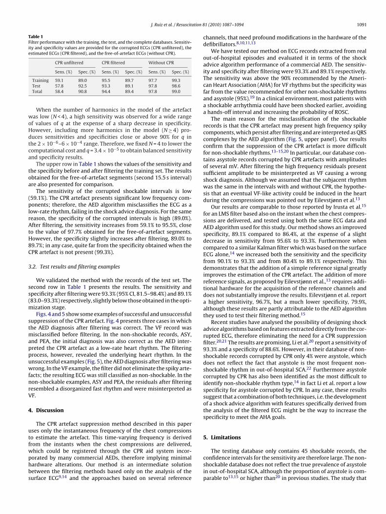
Table 1Filter performance with the training, the test, and the complete databases. Sensitiv-ity and specificity values are provided for the corrupted ECGs (CPR unfiltered), theestimated ECGs (CPR filtered), and the free-of-artefact ECGs (without CPR).
CPR unfiltered CPR filtered Without CPR
Sens. (%) Spec. (%) Sens. (%) Spec. (%) Sens. (%) Spec. (%)
woHdtca
toa
(plrAtH8C
3
ss(m
stmappuwfnrV
4
utfwphbs
Training 59.1 89.0 95.5 89.7 97.7 99.3Test 57.8 92.5 93.3 89.1 97.8 98.6Total 58.4 90.8 94.4 89.4 97.8 99.0
When the number of harmonics in the model of the artefactas low (N < 4), a high sensitivity was observed for a wide range
f values of q at the expense of a sharp decrease in specificity.owever, including more harmonics in the model (N ≥ 4) pro-uces sensitivities and specificities close or above 90% for q inhe 2 × 10⁻⁶–6 × 10⁻⁴ range. Therefore, we fixed N = 4 to lower theomputational cost and q = 3.4 × 10⁻⁵ to obtain balanced sensitivitynd specificity results.
The upper row in Table 1 shows the values of the sensitivity andhe specificity before and after filtering the training set. The resultsbtained for the free-of-artefact segments (second 15.5 s interval)re also presented for comparison.
The sensitivity of the corrupted shockable intervals is low59.1%). The CPR artefact presents significant low frequency com-onents; therefore, the AED algorithm misclassifies the ECG as a
ow-rate rhythm, failing in the shock advice diagnosis. For the sameeason, the specificity of the corrupted intervals is high (89.0%).fter filtering, the sensitivity increases from 59.1% to 95.5%, close
o the value of 97.7% obtained for the free-of-artefact segments.owever, the specificity slightly increases after filtering, 89.0% to9.7%; in any case, quite far from the specificity obtained when thePR artefact is not present (99.3%).
.2. Test results and filtering examples
We validated the method with the records of the test set. Theecond row in Table 1 presents the results. The sensitivity andpecificity after filtering were 93.3% (95% CI, 81.5–98.4%) and 89.1%83.0–93.3%) respectively, slightly below those obtained in the opti-
ization stage.Figs. 4 and 5 show some examples of successful and unsuccessful
uppression of the CPR artefact. Fig. 4 presents three cases in whichhe AED diagnosis after filtering was correct. The VF record was
isclassified before filtering. In the non-shockable records, ASY,nd PEA, the initial diagnosis was also correct as the AED inter-reted the CPR artefact as a low-rate heart rhythm. The filteringrocess, however, revealed the underlying heart rhythm. In thensuccessful examples (Fig. 5), the AED diagnosis after filtering wasrong. In the VF example, the filter did not eliminate the spiky arte-
acts; the resulting ECG was still classified as non-shockable. In theon-shockable examples, ASY and PEA, the residuals after filteringesembled a disorganized fast rhythm and were misinterpreted asF.
. Discussion
The CPR artefact suppression method described in this paperses only the instantaneous frequency of the chest compressionso estimate the artefact. This time-varying frequency is derivedrom the instants when the chest compressions are delivered,
hich could be registered through the CPR aid system incor-orated by many commercial AEDs, therefore implying minimalardware alterations. Our method is an intermediate solutionetween the filtering methods based only on the analysis of theurface ECG9,14 and the approaches based on several reference1 (2010) 1087–1094 1091
channels, that need profound modifications in the hardware of thedefibrillators.8,10,11,13
We have tested our method on ECG records extracted from realout-of-hospital episodes and evaluated it in terms of the shockadvice algorithm performance of a commercial AED. The sensitiv-ity and specificity after filtering were 93.3% and 89.1% respectively.The sensitivity was above the 90% recommended by the Ameri-can Heart Association (AHA) for VF rhythms but the specificity wasfar from the value recommended for other non-shockable rhythmsand asystole (95%).19 In a clinical environment, most patients witha shockable arrhythmia could have been shocked earlier, avoidinga hands-off interval and increasing the probability of ROSC.
The main reason for the misclassification of the shockablerecords is that the CPR artefact may present high frequency spikycomponents, which persist after filtering and are interpreted as QRScomplexes by the AED algorithm (Fig. 5, upper panel). Our resultsconfirm that the suppression of the CPR artefact is more difficultfor non-shockable rhythms.13–15,20 In particular, our database con-tains asystole records corrupted by CPR artefacts with amplitudesof several mV. After filtering the high frequency residuals presentsufficient amplitude to be misinterpreted as VF causing a wrongshock diagnosis. Although we assumed that the subjacent rhythmwas the same in the intervals with and without CPR, the hypothe-sis that an eventual VF-like activity could be induced in the heartduring the compressions was pointed out by Eilevstjønn et al.13
Our results are comparable to those reported by Irusta et al.15
for an LMS filter based also on the instant when the chest compres-sions are delivered, and tested using both the same ECG data andAED algorithm used for this study. Our method shows an improvedspecificity, 89.1% compared to 86.4%, at the expense of a slightdecrease in sensitivity from 95.6% to 93.3%. Furthermore whencompared to a similar Kalman filter which was based on the surfaceECG alone,14 we increased both the sensitivity and the specificityfrom 90.1% to 93.3% and from 80.4% to 89.1% respectively. Thisdemonstrates that the addition of a simple reference signal greatlyimproves the estimation of the CPR artefact. The addition of morereference signals, as proposed by Eilevstjønn et al.,13 requires addi-tional hardware for the acquisition of the reference channels anddoes not substantially improve the results. Eilevstjønn et al. reporta higher sensitivity, 96.7%, but a much lower specificity, 79.9%,although these results are partly attributable to the AED algorithmthey used to test their filtering method.15
Recent studies have analysed the possibility of designing shockadvice algorithms based on features extracted directly from the cor-rupted ECG, therefore eliminating the need for a CPR suppressionfilter.20,21 The results are promising, Li et al.20 report a sensitivity of93.3% and a specificity of 88.6%. However, in their database of non-shockable records corrupted by CPR only 4% were asystole, whichdoes not reflect the fact that asystole is the most frequent non-shockable rhythm in out-of-hospital SCA.22 Furthermore asystolecorrupted by CPR has also been identified as the most difficult toidentify non-shockable rhythm type,14 in fact Li et al. report a lowspecificity for asystole corrupted by CPR. In any case, these resultssuggest that a combination of both techniques, i.e. the developmentof a shock advice algorithm with features specifically derived fromthe analysis of the filtered ECG might be the way to increase thespecificity to meet the AHA goals.
5. Limitations
The testing database only contains 45 shockable records, theconfidence intervals for the sensitivity are therefore large. The non-shockable database does not reflect the true prevalence of asystolein out-of-hospital SCA, although the proportion of asystole is com-parable to13,15 or higher than20 in previous studies. The study that

1092 J. Ruiz et al. / Resuscitation 81 (2010) 1087–1094
Fig. 4. Examples of successful filtering for a VF, an ASY, and a PEA record. The original corrupted signal, the filtered signal, and the estimated CPR artefact are shown for allcases. The shock/no-shock decision of the AED algorithm is shown for the original and filtered signals.

J. Ruiz et al. / Resuscitation 81 (2010) 1087–1094 1093
Fig. 5. Examples of unsuccessful filtering for a VF, an ASY, and a PEA record. The original corrupted signal, the filtered signal, and the estimated CPR artefact are shown forall cases. The shock/no-shock decision of the AED algorithm is shown for the original and filtered signals.

1 ation 8
oiittntcrioLfd
wrn
6
oifsoaifA
C
rA
et
A
CPcdp
R
1
1
1
1
1
1
1
1
1
1
2
21. Neurauter A, Eftestøl T, Kramer-Johansen J, et al. Improving countershock
094 J. Ruiz et al. / Resuscit
riginated the ECG database16,17 was aimed at measuring CPR qual-ty according to the guidelines 2000. These guidelines were updatedn 2005, the compression:ventilation ratio was changed from 15:2o 30:2. We think that changes in this ratio would not affect the fil-er accuracy. The instantaneous frequency of the compressions wasot acquired online using a dedicated HW, but rather derived fromhe compression depth signal. In a practical setting this informationan be easily derived from the acceleration, force or pressure signalecorded by the CPR aid pads. In the ECG database CPR was admin-stered manually. We have therefore not analysed the implicationsf using mechanical chest compression systems, such as the JolifeUCAS or the Zoll AutoPulse devices. However, modelling the arte-act in such systems should be easier, because the frequency andepth of the compressions are constant.
The performance of the filter was optimized in combinationith a particular AED algorithm; when used on a different AED the
esults for sensitivity and specificity will vary and the filter wouldeed to be optimized again for that particular algorithm.
. Conclusions
We removed the CPR artefact from human ECG records of out-f-hospital episodes using a Kalman filter that needs only thenstantaneous frequency of the compressions to estimate the arte-act. The method was evaluated in terms of the sensitivity and thepecificity of the shock advice algorithm of a commercial AED. Webtained a satisfactory sensitivity of 93.3%. The specificity of 89.1%,lthough significantly superior to the values reported in other stud-es, is still far from the recommendations. Future research shouldocus on the design of more robust combinations of filtering andED shock advice algorithms.
onflict of interest statement
Authors, J. Ruiz, U. Irusta and S. Ruiz de Gauna have receivedesearch support from Osatu S. Coop. (Ermua, Spain) for studies onED shock advice algorithms.
Trygve Eftestøl was, in the period 1995–2000, a part-timemployee of Laerdal Medical (Stavanger, Norway), which providedhe modified defibrillator used in the study.
cknowledgements
This work received financial support from the Ministerio deiencia e Innovacion of Spain through the project TEC2006-11978.rof. Andreas Steen and Dr. Lars Wik, principal investigators of thelinical study, kindly provided the data from out-of-hospital car-iac arrest episodes for which Laerdal Medical (Stavanger, Norway)rovided invaluable technical and administrative support.
eferences
1. Valenzuela TD, Roe DJ, Nichol G, Clark LL, Spaite DW, Hardman RG. Outcomesof rapid defibrillation by security officers after cardiac arrest in casinos. N EnglJ Med 2000;343:1206–9.
2
1 (2010) 1087–1094
2. Handley AJ, Koster R, Monsieurs K, Perkins GD, Davies S, Bossaert L. Euro-pean Resuscitation Council Guidelines for Resuscitation 2005—Section 2. Adultbasic life support and use of automated external defibrillators. Resuscitation2005;67:S7–23.
3. Snyder D, Morgan C. Wide variation in cardiopulmonary resuscitationinterruption intervals among commercially available automated external defib-rillators may affect survival despite high defibrillation efficacy. Crit Care Med2004;32:S421–4.
4. Eftestøl T, Sunde K, Steen PA. Effects of interrupting precordial compressions onthe calculated probability of defibrillation success during out-of-hospital cardiacarrest. Circulation 2002;105:2270–3.
5. Yu T, Weil MH, Tang WC, et al. Adverse outcomes of interrupted precor-dial compression during automated defibrillation. Circulation 2002;106:368–72.
6. Edelson DP, Abella BS, Kramer-Johansen J, et al. Effects of compression depthand pre-shock pauses predict defibrillation failure during cardiac arrest. Resus-citation 2006;71:137–45.
7. Strohmenger HU, Lindner KH, Brown CG. Analysis of the ventricular fibrillationECG signal amplitude and frequency parameters as predictors of countershocksuccess in humans. Chest 1997;111:584–9.
8. Langhelle A, Eftestøl T, Myklebust H, Eriksen M, Holten BT, Steen PA. Reduc-ing CPR artefacts in ventricular fibrillation in vitro. Resuscitation 2001;48:279–91.
9. Aramendi E, de Gauna SR, Irusta U, Ruiz J, Arcocha MF, Ormaetxe JM. Detec-tion of ventricular fibrillation in the presence of cardiopulmonary resuscitationartefacts. Resuscitation 2007;72:115–23.
0. Aase SO, Eftestøl T, Husøy JH, Sunde K, Steen PA. CPR artifact removal fromhuman ECG using optimal multichannel filtering. IEEE Trans Biomed Eng2000;47:1440–9.
1. Husøy JH, Eilevstjønn J, Eftestøl T, Aase SO, Myklebust H, Steen PA. Removalof cardiopulmonary resuscitation artifacts from human ECG using an effi-cient matching pursuit-like algorithm. IEEE Trans Biomed Eng 2002;49:1287–98.
2. Rheinberger K, Steinberger T, Unterkofler K, Baubin M, Klotz A, Amann A.Removal of CPR artifacts from the ventricular fibrillation ECG by adaptiveregression on lagged reference signals. IEEE Trans Biomed Eng 2008;55:130–7.
3. Eilevstjønn J, Eftestøl T, Aase SO, Myklebust H, Husøy JH, Steen PA. Feasibilityof shock advice analysis during CPR through removal of CPR artefacts from thehuman ECG. Resuscitation 2004;61:131–41.
4. Ruiz de Gauna S, Ruiz J, Irusta U, Aramendi E, Eftestøl T, Kramer-Johansen J. Amethod to remove CPR artefacts from human ECG using only the recorded ECG.Resuscitation 2008;76:271–8.
5. Irusta U, Ruiz J, Ruiz de Gauna S, Aramendi E, Eftestøl T, Kramer-Johansen J. Aleast mean square filter for the estimation of the cardiopulmonary resuscitationartifact based on the frequency of the compressions. IEEE Trans Biomed Eng2009;56:1052–62.
6. Wik L, Kramer-Johansen J, Myklebust H, et al. Quality of cardiopulmonaryresuscitation during out-of-hospital cardiac arrest. JAMA 2005;293:299–304.
7. Kramer-Johansen J, Myklebust H, Wik L, et al. Quality of out-of-hospital car-diopulmonary resuscitation with real time automated feedback: a prospectiveinterventional study. Resuscitation 2006;71:283–92.
8. Agresti A, Coull BA. Approximate is better than “exact” for interval estimationof binomial proportions. Am Statist 1998;52:119–26.
9. Kerber RE, Becker LB, Bourland JD, et al. Automatic external defibrillators forpublic access defibrillation: recommendations for specifying and reportingarrhythmia analysis algorithm performance, incorporating new waveforms andenhancing safety. A statement for health professionals from the American HeartAssociation Task Force on automatic external defibrillation subcommittee onAED safety and efficacy. Circulation 1997;95:1677–82.
0. Li Y, Bisera J, Geheb F, Tang V, Weil MH. Identifying potentially shockablerhythms without interrupting cardiopulmonary resuscitation. Crit Care Med2008;36:198–203.
success prediction during cardiopulmonary resuscitation using ventricu-lar fibrillation features from higher ECG frequency bands. Resuscitation2008;79:453–9.
2. Cobb LA, Fahrenbruch CE, Olsufka M, Copass MK. Changing incidence of out-of-hospital ventricular fibrillation 1980–2000. JAMA 2002;288:3008–13.